A mouse model reproducing the pathophysiology
of neonatal group B streptococcal infection
Elva Bonifácio Andrade
1,2,3,4
, Ana Magalhães
2,3
, Ana Puga
1
, Madalena Costa
1,4,5
, Joana Bravo
1,2,3
,
Camila Cabral Portugal
2,3
, Adília Ribeiro
1,2,3
, Margarida Correia-Neves
6,7,8
, Augusto Faustino
1
, Arnaud Firon
9
,
Patrick Trieu-Cuot
9
, Teresa Summavielle
2,3,4
& Paula Ferreira
1,2,3
Group B streptococcal (GBS) meningitis remains a devastating disease. The absence of an
animal model reproducing the natural infectious process has limited our understanding of
the disease and, consequently, delayed the development of effective treatments. We describe
here a mouse model in which bacteria are transmitted to the offspring from vaginally
colo-nised pregnant females, the natural route of infection. We show that GBS strain BM110,
belonging to the CC17 clonal complex, is more virulent in this vertical transmission model
than the isogenic mutant BM110
ΔcylE, which is deprived of hemolysin/cytolysin. Pups
exposed to the more virulent strain exhibit higher mortality rates and lung in
flammation than
those exposed to the attenuated strain. Moreover, pups that survive to BM110 infection
present neurological developmental disability, revealed by impaired learning performance
and memory in adulthood. The use of this new mouse model, that reproduces key steps
of GBS infection in newborns, will promote a better understanding of the physiopathology of
GBS-induced meningitis.
DOI: 10.1038/s41467-018-05492-y
OPEN
1ICBAS—Instituto de Ciências Biomédicas de Abel Salazar, Universidade do Porto, 4150-313 Porto, Portugal.2i3S—Instituto de Investigação e Inovação em
Saúde, Universidade do Porto, 4200-135 Porto, Portugal.3IBMC—Instituto de Biologia Molecular e Celular, Universidade do Porto, 4200-135 Porto, Portugal.
4ESS—Escola Superior de Saúde, Instituto Politécnico do Porto, 4200-072 Porto, Portugal.5UMIB—Unit for Multidisciplinary Investigation in Biomedicine
(Endocrine, Cardiovascular & Metabolic Research), University of Porto, 4150-313 Porto, Portugal.6Life and Health Sciences Research Institute (ICVS), School
of Medicine, University of Minho, 4710-057 Braga, Portugal.7ICVS/3B’s, PT Government Associate Laboratory, 4710-057 Braga/4805-017, Guimarães,
Portugal.8Division of Infectious Diseases, Department of Medicine Solna, Karolinska Institutet, 171 76 Stockholm, Sweden.9Institut Pasteur, Unité de
Biologie des Bactéries Pathogènes à Gram-positif, Centre National de la Recherche Scientifique (CNRS ERL 6002), Paris 75015, France. Correspondence and
requests for materials should be addressed to P.F. (email:[email protected])
123456789
N
eonatal bacterial meningitis is a severe life-threatening
disease and a major cause of neurological disability
worldwide. Group B Streptococcus (GBS) remains the
leading cause of severe invasive infections among neonates and,
together with Escherichia coli, account for at least two-thirds of all
deaths from neonatal meningitis
1,2. GBS is a commensal
organ-ism of the genitourinary and/or gastrointestinal tract of adult
humans and has been isolated from vagina and/or rectum of
15–40% of pregnant women
2. Maternal colonisation and
trans-mission to foetus and newborns is the most common cause of
neonatal GBS infections leading to pneumonia, septicaemia and
meningitis
2.
The current prevention strategy of intrapartum antibiotic
prophylaxis (IAP), for parturient at risk of GBS transmission to
newborns,
has
reduced
the
cases
of
pneumonia
and
septicaemia
2,3. The incidence rates of GBS meningitis have
remained relatively stable over the past 20 years
4,5but now
appear to be increasing
6,7. Furthermore, maternal colonisation is
not affected by IAP treatment
3and the overall mortality rate for
GBS neonatal infections remains at approximately 10%. As a
consequence, morbidity rates associated with meningitis caused
by GBS infection has not changed substantially over decades
2,8remaining unacceptably high. In addition, up to 50% of surviving
infants experience neurodevelopmental impairment (NDI)
2,9–11,
highlighting the inefficacy of current treatment and the urgent
need for new preventive and/or therapeutic approaches. The
mechanisms that lead to the devastating outcome of GBS-induced
meningitis are not elucidated. Clinical data concerning neonatal
meningitis are limited as this disease is difficult to diagnose due to
subtle and nonspecific symptoms
8,12, and it is estimated that
more than 30% of the cases are asymptomatic
13,14. Moreover,
protocols in which only neonates with confirmed bacteraemia are
evaluated for meningitis result in missed diagnoses, as blood
cultures may be negative in 15–38% of cases
12. Thus, a better
understanding of the pathogenesis and pathophysiology of GBS
meningitis must be gained. To this end, mice and rat models of
GBS disease have been developed, but they often target a
parti-cular step of the pathophysiological process (organ colonisation,
septicaemia, meningitis) and although they have generated
important knowledge, they also have the potential of yielding
misleading information. This is particularly the case when
non-natural infection routes, such as intraperitoneal, subcutaneous,
or intracerebral GBS inoculation, that by-pass the
mother-to-newborn transmission and the normal bacteraemia-meningitis
sequence in the neonate, are used
15–19. Moreover, the use of
irrelevant animal models in research results in a lower likelihood
of translation to the clinic
20,21. Thus, an animal model in which
the disease induced closely resembles GBS natural infection in
humans is still missing.
Here, we present a mouse model that recapitulates the GBS
newborn infection pathogenesis by enabling vertical transmission
of the pathogen from vaginally colonised pregnant females to
their progeny. We validate this model by using the hypervirulent
GBS strain BM110, a serotype III strain belonging to the
hyper-virulent clonal complex 17 (CC17)
22–24. In addition, the
atte-nuated isogenic mutant BM110ΔcylE that does not express the
pore-forming toxin
β-hemolysin/cytolysin (β-h/c) was used. This
toxin is an important virulent factor
25–27whose overexpression
leads to preterm labour in pregnant nonhuman primates
28.
Our data show that neonatal mice born from mothers
colo-nised with the hyper-virulent GBS strain exhibit enhanced
mor-tality and lung pathology, which correlates with higher bacterial
load. Importantly, newborns that survive to BM110 infection
experience permanent NDI, as observed in humans. Thus, this
mouse model, which mimics the human pathophysiology of GBS
diseases, should allow a better understanding of the
pathophy-siology of GBS meningitis and open new avenues towards the
identification of new therapeutic and neuroprotective strategies.
Results
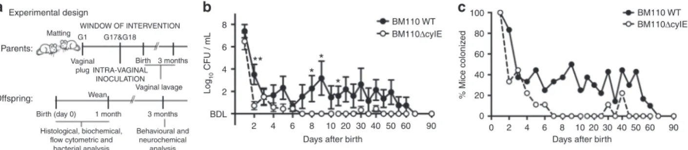
β-h/c expression favours colonisation of mouse vagina.
Knowing that human neonates acquire the bacterium by vertical
transmission, pregnant BALB/c mice were inoculated
intra-vaginally (i.vag.) with the serotype III hypervirulent strain CC17
BM110 or the isogenic attenuated strain BM110ΔcylE lacking
haemolytic/cytolytic activities. After several attempts, days 17 and
18 of gestation (G17 and G18) were defined as the specific
win-dow to administer the bacteria (Fig.
1
a). Vaginal colonisation was
monitored upon delivery by vaginal lavage and plating of
recovered bacteria (the delivery day was excluded due to excess
of blood and body
fluids). At day one after birth, and with both
GBS strains, the vaginal mucosa of all females presented high
bacterial load enabling the vertical transmission of the bacterium
(Fig.
1
b,c). Thereafter, the bacterial levels started to decrease, but
more abruptly with BM110ΔcylE mutant than with BM110 WT
(Fig.
1
b, c). After day 6 upon delivery, the levels of bacteria
remained almost exclusively below the detection threshold for the
attenuated strain, while a transient and intermittent colonisation
was observed with the hyper-virulent strain. At day 60, no
bac-terium was detected in the vaginal mucosa of all females in either
group (Fig.
1
b, c). Data from individual progenitors are depicted
in Supplementary Fig. 1a and b.
β-h/c expression induces a severe pathology to neonatal mice.
As GBS is a major cause of stillbirth, we monitored the foetal
death in pregnant mice colonised either with the hyper-virulent
or the attenuated GBS strain. Stillborn accounted for 14 or 5% of
WINDOW OF INTERVENTION 8 100 80 60 40 20 0 BM110 WT BM110ΔcyIE BM110 WT BM110ΔcyIE Log 10 CFU / mL % Mice coloniz ed 6 4
**
*
*
*
2 BDL 2 4 6 8 10Days after birth
20 30 40 50 60 90 0 2 4 6 8 10
Days after birth
20 30 40 50 60 90 Matting Vaginal plug Birth 3 months INTRA-VAGINAL INOCULATION Vaginal lavage G1 G17&G18 Parents: Birth (day 0) Wean 1 month 3 months Histological, biochemical,
flow cytometric and bacterial analysis Behavioural and neurochemical analysis Offspring: Experimental design
b
c
a
Fig. 1 Vaginal GBS colonisation of female mice. a A schematic illustration of the colonisation model is shown. Pregnant BALB/c mice were intra-vaginally
inoculated with 3 × 10⁴CFU of GBS BM110 WT or BM110ΔcylE at gestational days 17 and 18. b After birth, GBS vaginal colonisation levels were determined
at the indicated time-points in vaginal lavage by enumerating onto selective media GBS colony-forming units (CFU) [mean ± SEM,n = 10 (BM110 WT) and
n = 9 (BM110ΔcylE)]. Comparisons by two-way ANOVA, with Sidak’s multiple comparison. *P < 0.05, **P < 0.01. c The percentage of mice remaining
all pups born from BM110 or BM110ΔcylE-colonised females,
respectively, while none was observed in non-colonised mothers
(Fig.
2
a). Following delivery, approximately 40% of the pups born
from BM110-colonised females died after birth, 21% during the
first 24 h of life, while 12% of pups born from
BM110ΔcylE-colonised mothers died after birth, 6% in the
first 24 h (Fig.
2
b).
In the non-colonised control group, only 6% of the pups died
during the
first 24 h of life (Fig.
2
b). No death was recorded after
postnatal day (PND) 4 in all groups of mice (Fig.
2
b).
Measurement of weight gain, a sensitive marker of neonatal
well-being, was recorded during the
first 10 days of life. Body
weight gain was reduced in pups born from GBS-colonised
progenitors compared to those born from non-colonised mothers
(Fig.
2
c, d). This reduction was more pronounced with the pups
from BM110 WT- colonised mothers (8 days) compared to pups
from BM110ΔcylE-colonised mothers (4 days) (Fig.
2
c).
As pneumonia frequently heralds early stages of neonatal
GBS disease, we investigated bacterial load and signs of lung
a
e
i
m
n
j
k
l
f
g
h
b
c
d
P ercent stillbir th Log 10 CFU / organ Log 10 CFU / organ Log 10 CFU / organ Log 10 CFU / organ Log 10 CFU / mL % Mice coloniz ed % Mice coloniz ed P ercent sur viv al W eight gain / g 100 * ** **** *** ** **** **** **** ** * **** **** **** *** ** 80 60 40 20 0 8 8 7 6 5 4 4 100 80 60 40 20 0 3 2 1 Bdl 8 80 60.0 45.4 25.0 93.8 50.0 60 40 20 0 6 4 2 0 8 6 4 2 Bdl 3 Bdl Lungs Gut Brain BrainBlood Blood Liver
6 6 2 Bdl
3
Days after birth
1 5 7 15 30
3
Days after birth Days after birth
1 5 7 15 30
3
Days after birth
1 5 7 15 30
3
Days after birth
1 5 7 15 30 3
Days after birth
1 5 7 15 30
0 3 6 9 12 15 18 21 24 27 30
Days after birth 0 3 6 9 12 15 18 21 24 27 30 100 (46/49) (43/49) (39/94) 80 8 6 4 2 0 60 Uninfected BM110ΔcyIE BM110ΔcyIE BM110 WT BM110 WT Uninfected Uninfected 0 1 2 3456 7 01 2 3 4 5 6 7 01 2 3 4 5 6 7 BM110ΔcyIE BM110ΔcyIE BM110ΔcyIE BM110 ΔcyIE BM110 WT BM110 WT BM110 WT BM110 WT 40 20 ** * ******* ******** ******* *** 0 0 2 4 6 n.s. 10
Days after birth Days after birth
15 20 25 30 0 1 2 3 4 5 6 7 8 9 10 Uninf ected BM110 WT BM110 ΔcyIE
Fig. 2 GBS vertical transmission. Pregnant BALB/c mice were intra-vaginally inoculated with 3 × 10⁴CFU of GBS BM110 WT, BM110ΔcylE, or PBS
(uninfected) at gestational days 17 and 18.a Percentage of term stillbirths born from indicated dams. Each symbol indicates data from a single mouse
[mean,n = 15 (uninfected), n = 20 (BM110 WT) and n = 11 (BM110ΔcylE)]. Comparisons by one-way ANOVA, with Sidak’s multiple comparison.
*P < 0.05, **P < 0.01. b Kaplan–Meier survival curve of neonatal mice, monitored for 30 days. Numbers in parentheses represent surviving pups versus
total born [n = 49 (uninfected), n = 94 (BM110 WT) and n = 49 (BM110ΔcylE)]. Comparisons with log-rank (Mantel-Cox) test. **P < 0.01, ***P < 0.001
and n.s., not significant. c Body weight gain of mice following birth [mean ± SEM, n = 22 (uninfected), n = 34 (BM110 WT) and n = 12 (BM110ΔcylE)].
* represents the comparison between BM110 WT and uninfected pups,† BM110ΔcylE and uninfected pups, and ‡ BM110 WT and BM110ΔcylE pups.
Comparisons by two-way ANOVA, with Sidak’s multiple comparison. *,†P < 0.05, ***,†††,‡‡‡P < 0.001 and ****,‡‡‡‡P < 0.0001. d Overall appearance of an
offspring born from uninfected (left), BM110 WT- (middle), or BM110ΔcylE-colonised (right) progenitors at PND7. e, i, j, l, m GBS counts in lungs (e), gut
(i), blood (j), liver (l), and brain (m) in the offspring born from BM110 WT- or BM110ΔcylE-colonised progenitors at different time points after birth.
Each symbol indicates data from single pups [mean,n = 11 (PND1), n = 21 (PND2 and 7), n = 9 (PND3), n = 13 (PND15) and n = 13 (PND30) for BM110
WT;n = 8 (PND1), n = 9 (PND2 and 7), n = 15 (PND3), n = 6 (PND15 and 30) for BM110ΔcylE]. Comparisons by two-way ANOVA, with Sidak’s Multiple
comparison. *P < 0.05, **P < 0.01, ***P < 0.001 and ****P < 0.0001. f–h H&E staining of pulmonary tissue from uninfected (f), BM110 WT- (g), or
BM110ΔcylE- (h) colonised progenitors, at PND1. Representative micrographs are shown. Black arrow indicates interstitial inflammation and areas of
atelectasis. Arrowheads indicate oedema and severe haemorrhage. Red arrow indicates mild atelectasis areas. Scale bar, 100μm. k, n Percentage
inflammation, or loss of structure, in pups born from colonised
mothers. Higher and sustained GBS bacterial loads were detected
in the lungs of pups born from BM110 WT-colonised progenitors
compared to those born from BM110ΔcylE-colonised mothers
(Fig.
2
e). These bacterial loads were correlated with the observed
lung pathology (Fig.
2
f–h). Indeed, neonates born from BM110
WT-colonised mothers displayed significant lung pathology with
the presence of areas of atelectasis lung inflammation with
moderate neutrophil infiltration in the alveoli and interstitium,
narrowed airway lumen, oedema and severe haemorrhage. In
contrast, pups born from BM110ΔcylE-colonised mothers
showed reduced lung inflammation with mild atelectasis and
mild increased thickness of alveolar wall (Fig.
2
f–h). Gut GBS
colonisation was also quantified and only at PND1, the pups born
from BM110-colonised mothers presented higher levels of
bacteria than those born from BM110ΔcylE-colonised mothers
(Fig.
2
i). Thereafter, no significant differences were observed
between pups born from hyper-virulent or attenuated
GBS-colonised females with both groups presenting high levels of GBS
(Fig.
2
i).
As this bacterium can disseminate into the bloodstream, GBS
was also quantified in the blood of pups born from colonised
mothers. GBS were present in the blood of 60% of pups born
from BM110-colonised progenitors, reaching a maximum at
PND1, but were no longer detected at PND7 (Fig.
2
j, k). In
contrast, no bacterium was detected in the blood of pups born
from BM110ΔcylE-colonised mothers (Fig.
2
j, k). The bacterial
load was also quantified in the liver, an organ playing a key role
during sepsis. As expected, pups born from BM110 WT-colonised
mothers presented higher levels of GBS than those born from
BM110ΔcylE-colonised mothers (Fig.
2
l). GBS was also quantified
in the brain of pups and although the blood of neonatal mice
born from mothers colonised with the attenuated strain was
negative for GBS, bacteria were detected in their brains up to
PND15 (Fig.
2
m). The level of brain colonisation was always
slightly less than that observed in pups born from BM110
WT-colonised mothers, but the differences were not significant except
at PND5 (Fig.
2
m). The percentage of pups with brain
colonisation at PND5 was 94% (15 out of 16) for those infected
with BM110 and 50% (8 out of 16) for those infected with
BM110ΔcylE (Fig.
2
n). This indicates that almost all pups
surviving the infection caused by the hyper-virulent strain have
experienced meningitis.
To confirm that moribund animals infected with BM110 WT
were dying from sepsis-related multiple organ failure, serum levels
of several biochemical parameters were quantified at PND1 in
pups born from BM110-colonised mothers (BM110) and in pups
born from non-colonised mothers (Uninfected). The levels of
aspartate transaminase [AST (U L
−1), BM110: 685.0 ± 8.66 and
Uninfected: 32.50 ± 12.99], and creatine kinase [CK (U L
−1),
BM110: 7625 ± 4021 and Uninfected: 60.00 ± 23.09] were much
higher in the serum of pups infected with BM110 WT than in the
serum of uninfected pups. These results confirmed liver and heart
failure in the group of infected pups. No significant differences
were observed in the serum levels of creatinine [CREA (µmol L
−1),
BM110: 2.50 ± 0.87 and Uninfected: 0.625 ± 0.22] and urea
nitrogen [BUN (mmol L
−1), BM110: 16.00 ± 0.577 and
Unin-fected: 17.50 ± 1.44] between the infected and uninfected groups
indicating that the kidneys were functional.
The IAP administration of ampicillin or penicillin to
GBS-colonised pregnant women is routinely used to prevent neonatal
diseases. Therefore, BM110-colonised pregnant mice were
prophylactically treated with ampicillin added to their drinking
water from gestational day 20 (one day before delivery) until
PND1. A significantly increased survival was observed in the
group of pups born from IAP-treated mothers (87% survival), as
compared to those born from untreated progenitors (59%
survival) (Supplementary Fig. 2a). Moreover, at PND5, bacterial
counts in the organs tested were below detection level in almost
all pups born from IAP-treated mother, with only 1 out of 9 pups
displaying gut and brain colonisation (Supplementary Fig. 2b–f).
These results suggest that our mouse model can be used to test
the efficacy of new prophylactic treatments against neonatal GBS
infections.
GBS-induced meningitis in neonates born from colonised
mice. GBS-induced meningitis is often associated with long-term
NDI and epidemiological studies have revealed that it is
caused mainly by strains belonging to the hyper-virulent CC17
lineage
22–24. Therefore, we compared at PND5 the brain of pups
born from BM110-colonised mothers (infected pups) with those
born from non-colonised progenitors (uninfected pups). The
brain of infected pups exhibited the hallmarks of meningitis such
as meningeal congestion, vascular hyperaemia, and oedema
(Fig.
3
a). Moreover, haemorrhage and hyperaemia were also
found in the choroid plexus located in the dorsal third ventricle of
infected pups (Fig.
3
a). No histological alterations were observed
in the brain of infected pups (Fig.
3
a, c, e). Immunofluorescence
analysis revealed the presence of GBS within the brain
par-enchyma (Fig.
3
b). To investigate blood–brain barrier (BBB)
breakdown, its integrity was evaluated in the pups’ brains at
PND5 by the Evans blue (EB) dye assay. Higher values of vascular
permeability were detected in the brains of infected pups, as
compared to those observed in uninfected pups (Fig.
3
c, left). The
EB extravasation was further visualised by
fluorescence imaging
using the IVIS Lumina system (Fig.
3
c, right). Cortical thickness,
measured as indicated in Fig.
3
e, was significantly decreased in
brains from infected pups evidencing higher neuronal density and
poorer discrimination of the different cortical layers (Fig.
3
d).
This may result from the observed expansion of lateral ventricles
(Fig.
3
d), a likely consequence of GBS ventriculitis
29. However,
the thickness of the white matter (corpus callosum) of brains
from infected pups was not modified (Fig.
3
e) and regions of
abnormal necrotic death were not observed.
Microglia activation and neuronal apoptosis in infected brain.
To investigate whether brain inflammatory response was present,
cytokines known to be relevant for bacterial meningitis was
quantified in serum and brain of infected and uninfected pups at
PND5. The levels of tumour necrosis factor (TNF)-α, interleukin
(IL)−17 and interferon (IFN)-γ were significantly lower in the
serum of infected pups compared to uninfected pups (Fig.
4
a)
whereas the levels of keratinocyte chemoattractant (KC),
mac-rophage inflammatory proteins (MIP)−1α, IL-1β, IL-6 and IL-10
were similar (Fig.
4
a). No differences were observed in the levels
these 8 cytokines measured in brains of infected and uninfected
pups (Fig.
4
b). To gain insight into the mechanism responsible
for brain damage, we next analysed the leucocytes present at
PND5 in the brain of pups born from BM110-colonised mothers
together with the status of microglia activation. In this analysis, to
distinguish resident from infiltrating myeloid cells, we used a
common classification method based on CD45 expression, which
is higher on infiltrating cells
30. Cells were gated on CD45
+immune cells and among CD45
lowcells (resident cells) those
expressing CD11b
+were identified as resident myeloid cells
(commonly known as microglia), whereas the CD11b
+CD45
hicells were identified as infiltrating myeloid cells (Fig.
4
c). We
further gated CD45
hiCD11b
+to distinguish between
granulo-cytes (CD11b
+Ly6G
+) and monocytes/macrophages (CD11b
+Ly6G
−) (Fig.
4
c). A significant increase in the frequency of
resident myeloid cells was observed in brains from infected pups
compared with brains from uninfected pups while no significant
difference was detected in the infiltrating myeloid cells population
(Fig.
4
d, e). Higher expression of CD11b was observed in the
resident myeloid population of brains from infected pups
com-pared to those from uninfected pups (Fig.
4
f). Increased
expres-sion of CD11b levels is associated with resident myeloid cells
activation
31, indicating that the microglia of infected pups was
activated. No alterations were detected in the expression levels of
MHC class II and Ly6C in resident myeloid cells (Supplementary
Fig. 3a, b). Although no differences were observed in the levels of
the CD11b
+Ly6G
+population between brains from infected and
uninfected pups, the mean
fluorescence intensity of CD44 (an
important receptor for cell motility upregulated upon cell
acti-vation) was significantly increased in brains from infected pups
(Fig.
4
g), indicating neutrophil activation. A significant decrease
in Ly6C
hiinflammatory monocytes involved in brain
homo-eostasis
32was observed in brains from infected compared to those
from uninfected pups (Fig.
4
h). Concomitantly, the proportion of
Ly6C
int/lowmonocytes was increased in brain from infected pups.
Since pathological stimuli affect both microglia functional state
and anatomical structure
33, we used Iba1
+staining to visualise
changes in microglia morphology at PND5 in brains from
infected and uninfected pups. At this time-point, microglia is
usually uniform throughout the brain despite the presence of
microglia region-specific phenotypes
34. As expanded lateral
ventricles were observed in brains from infected pups, we focused
our attention on the striatum and hippocampus (CA3 region). In
both regions (Fig.
4
i), a shift towards an insult-responsive
phenotype was observed in infected pups, characterised by
reduced branching and increased soma size
33, which is coherent
with the observed activation state (Fig.
4
f).
Astrocytes
are
important
components
of
BBB
and
blood–cerebral spinal fluid (CSF) barrier and they respond to
GBS infection by releasing several inflammatory cytokines, which
may lead to reactive astrogliosis
35,36. Therefore, inflammatory
activation of astrocytes was evaluated at PND5 using GFAP
staining in the lateral ventricles and in the hippocampus of brains
from infected pups and brains from uninfected pups. Although
the GFAP intensity was not different between the groups (Fig.
4
j),
the area occupied by GFAP
+positive cells in the hippocampus
was larger in brains from infected than in brains from uninfected
pups (Fig.
4
j), indicating moderate reactive-astrogliosis. The
Uninfected BM110 WT LV WM Bregma 0.4 – 0.6 White matter V entr icular area Cor tical thic kness Epi-fluorescence 3.4 3.2 3.0 2.8 2.6 2.4 ×10⁸ Radiant efficiency ( )p/s/cm2/sr μW/cm2 Uninfected BM110 WT chp d3V Cor te x and meninges Uninfected BM110 WT Ev ans b lue leakage (μ g/mg dr y br ain) Cor tical thic kness/ μ m WM thic kness/ μ m V entr icular area / μ m 2 DAPI/BM110 WT
e
d
c
b
a
0.15 * ** *** 0.10 0.05 0.00 Uninf ected BM110 WT 1000 900 300,000 250,000 200,000 150,000 100,000 50,000 200 180 160 140 120 100 0 800 700 600 500 Uninf ected BM110 WTFig. 3 Prenatal-GBS infection leads to brain damage in the offspring. Pregnant BALB/c mice were intra-vaginally inoculated with 3 × 10⁴CFU of GBS BM110
WT or PBS (uninfected) at gestational days 17 and 18.a Sections of brain tissue were prepared at PND5 and subjected to H&E staining. Representative
micrographs of individual pups infected with GBS compared with uninfected control at indicated brain regions are shown. Arrows indicate oedema, vascular congestion and meningeal thickening in the cortex. Arrowheads depict vascular congestion and haemorrhage in the choroid plexus in the dorsal third
ventricle. Chp d3v, choroid plexus dorsal third ventricle. Scale bars are 100μm for top and bottom, and 20 μm for middle. b Immunofluorescence detection
of GBS in the brain of an offspring born from a GBS-colonised progenitor at PND5. Scale bar, 5μm. c BBB permeability measured by the quantitative
analysis of Evans blue leakage to the CNS at PND5. Vascular permeability of the Evans blue dye is expressed asµg of Evans blue per mg of dried
brain tissue using a standard curve. Representativefluorescence images of uninfected and infected pups’ brains analysed with the IVIS LUMINA LT are
shown. Each dot represents data from a single pup (mean,n = 4 for uninfected and BM110 WT). Comparison by unpaired Student’s t-test. *P < 0.05.
d Representative Nissl coronal sections showing cortical thickness, lateral ventricles enlargement and white matter (WM) thickness in pups infected with
GBS BM110 WT compared with uninfected controls.e These modifications were quantified in brains of uninfected and infected pups. Measures were
obtained as shown by red lines in representative sections (Image credit: Allen Institute). Scale bar, 100μm. Each dot represents data from a single pup
(mean,n = 5 for ventricular area and WM thickness, n = 6 for cortical thickness for both uninfected and BM110 WT). Comparisons by two-way ANOVA,
periventricular area occupied by GFAP
+cells could not be
compared between the groups due to ventricular expansion.
To evaluate whether apoptosis contributes to the
neuropathol-ogy, neuronal apoptosis was assessed at PND5 by TUNEL
nuclei in the hippocampus, striatum, motor and parietal cortical
regions, and posteriorly at the parietal associative cortex of brains
from infected and brains from uninfected pups. As illustrated
in Fig.
4
k, an increased apoptotic cell death was observed in all
assessed regions of the brains from infected pups.
Behavioural alterations in survivors of GBS meningitis. To
assess if, in our model, the offspring that survived infection with
Hoechst / TUNEL Hoechst / TUNEL
Uninfected BM110 WT Uninfected BM110 WT Str iatum Motor Cx Hippocampus P a rietal Cx DAPI / GFAP Uninfected Uninfected BM110 WT BM110 WT Later al v entr icle Hippocampus Str iatum Hippocampus DAPI / Iba1 Uninfected Uninfected BM110 WT BM110 WT L y6C % L y6C hi Ly6Chi Ly6Chi CD11b Uninfected BM110 WT Uninf ected BM110 WT Uninf ected BM110 WT Uninf ected BM110 WT Uninf ected BM110 WT Unstained cells CD44 Relativ e cell n umber Relativ e cell n umber GF AP intensity GF AP intensity TUNEL positiv e cells/field TUNEL positiv e cells/field TUNEL positiv e cells/field TUNEL positiv e cells/field GF AP + area (%) CD11b Granulocytes Ly6G Granulocytes Mono/ mφs CD11b Infil. Myeloid cells CD45 Res.Infil. CD45 CD45 CD11b SSC SSC
k
j
i
h
g
f
e
d
c
b
a
1000 *** ** *** * *** * ** pg / mL mg / organ % of total br ain imm une cells CD11b MFI (×10 3) MFI CD44 (×10 4) % of total CD45 high cells % of total CD45 high cells 800 600 Uninfected 300 250 200 150 100 50 0 BM110 WT Uninfected BM110 WT Uninfected Uninfected BM110 WT Uninfected BM110 WT BM110 WT 400 200 0 250 k 200 k 150 k 100 k 50 k 0 250 k 200 k 150 k 100 k 50 k 0 –103 104 105 103 0 –103 –103 104 104 104 105 105 105 103 103 0 –103 104 105 103 0 0 104 105 0 104 105 0 0 –103 104 105 103 0 –103 104 105 103 0 –103 104 105 103 0 KC MIP-1 α IL-1 β TNF-α IFN-γIL-6 IL-17 IL-10 KC
MIP-1 α IL-1 β TNF-α IFN-γ IL-6 IL-17 IL-10
Resident myeloid cells Infiltrating myeloid cells Mono/mφs (CD11b+ Ly6G– ) Granulocytes (CD11b+ Ly6G+ ) 100 70 10 8 6 4 2 0 60 50 40 30 80 60 8 6 10 2.0 25 20 15 10 5 0 1.5 1.0 0.5 0.0 8 6 4 2 Res.myeloid cells 0 4 2 0 50 40 30 20 10 0 50 40 30 20 10 0 50 20 15 10 40 30 20 10 0 5 0 20 15 10 15 10 5 0 5 0 * *** *** *** *** 40 30 20 10 0
the hyper-virulent strain experienced permanent NDI, as
observed in humans, we characterised their cognitive and motor
performance in adults. The learning and memory performances
were assessed using an 8-arm radial maze task
37. A significant
difference between GBS-Survivors (BM110-Survivors) and mice
born from non-colonised mother, control group (Uninfected),
was observed regarding the latency to enter to the
first arm in the
first two block sessions (Fig.
5
a), where the GBS-Survivors took
significantly more time to start exploring the maze. While the
Uninfected group presented a classical learning curve, with a
number of arm entries decreasing as time progressed, the
GBS-Survivors group did not alter the number of entries throughout
the test (Fig.
5
b). This is further evidenced in working and
reference memory performances, for which GBS-Survivors
pre-sent higher relative error rates at the
final sessions, indicating a
diminished ability to learn the task (Fig.
5
c, d). General motor
function and exploratory behaviour were assessed in the open
field test (OFT). GBS-Survivors display a significant decrease in
the total distance travelled in the OFT (Fig.
5
e). Importantly, both
the number of crosses between the peripheral and central area
and the time spent in the central area were significantly reduced
for the GBS-Survivors group (Fig.
5
f, g), which is a sign of
anxious-like behaviour. When analysing the exploratory activity
of GBS-Survivors, data revealed that these animals present a
significantly lower frequency and time spent in investigatory
behaviours such as rearing and exploration, when compared to
controls (Fig.
5
h, i). These animals also showed an increase of
immobility time and frequency (Fig.
5
h, i). Globally these results
show that GBS-Survivors presented compromised activity and
exploration abilities, as well as anxiety-related behaviours.
GBS meningitis leads to an altered neurotransmitter pattern.
To understand if the observed learning and behaviour changes in
GBS-Survivors mice are related to the levels of relevant
neuro-transmitters (dopamine and metabolites, norepinephrine,
gluta-mate, and GABA), these molecules were quantified in brain
regions involved in memory acquisition, learning and motor
activity by HPLC-ED.
In the hippocampus, a major glutamatergic region that receives
relevant dopaminergic input, we observed significantly reduced
levels of glutamate, dopamine and its metabolite DOPAC for the
GBS-Survivors (BM110-Survivors) (Fig.
5
j, k). In the striatum, a
predominantly GABAergic region modulated by nigral
dopami-nergic inputs and cortical glutamatergic projections, we observed
reduced levels of dopamine metabolites (DOPAC and HVA) for
the GBS-Survivors group (Fig.
5
l), which are indicative of
decreased dopaminergic turnover. Altered dopaminergic activity
in this region is associated with thalamic deregulation of
glutamate release
38. Accordingly, we observed in the thalamus a
significant reduction of glutamate levels for the GBS-Survivors
group (Fig.
5
m), which is also consistent with a decreased activity.
No other differences were observed in the levels of dopamine,
norepinephrine, glutamate or GABA in any of the evaluated brain
regions (hippocampus, striatum, prefrontal cortex, amygdala, and
cerebellum) (see Supplementary Tables 1 and 2).
Globally, mice born from BM110-colonised mothers present a
meaningful number of alterations in learning ability as well as in
the spontaneous behaviour, which is in agreement with the
observed decreases in glutamatergic and dopaminergic functions
in the hippocampus and the striatal-thalamic circuit.
Discussion
We here describe a novel mouse model that reproduces all steps
of GBS infection in humans. Transmission of this bacterium from
mother to the neonate results either from ascending spread of
bacteria or, alternatively, through ingestion or aspiration of
contaminated vaginal
fluids during delivery
2. Maternal vaginal
colonisation is thus the critical initial step of this invasive
neo-natal disease. To validate this model, we used the GBS strain
BM110 belonging to the hypervirulent lineage CC17 responsible
for approximately 80% of meningitis cases due to GBS in
neo-nates and the attenuated mutant derivative BM110ΔcylE not
expressing the
β-hemolysin/cytolysin. BM110 efficiently
colo-nised the vaginal mucosa of pregnant females and a transient and
intermittent vaginal colonisation was observed after delivery, as
observed in humans
39. Most importantly, the hypervirulent strain
colonises the vagina of pregnant mice more efficiently than the
attenuated strain. These results confirm previews studies showing
that
β-hemolysin/cytolysin expression is involved in adherence of
GBS to the murine vaginal tract of nonpregnant mice following
treatment with 17β-estradiol
40,41.
In human neonates, the lung is the primary organ infected by
GBS upon vaginal
fluids aspiration, with one-third to more than
half of infants showing lung symptoms within hours after birth
2.
In our murine model, 60% of pups infected with the
hyper-virulent GBS strain that survived (a percentage similar to that
reported in humans before IAP introduction) presented
respira-tory pathology. In addition, these pups had elevated GBS levels in
their lungs later in life, which is consistent with previous studies
reporting that throat and rectal cultures are the best sites for GBS
Fig. 4 Prenatal-GBS infection induces microglia activation and neuronal apoptosis in the offspring. Pregnant BALB/c mice were intra-vaginally
inoculated with 3 × 10⁴CFU of GBS BM110 WT or PBS (uninfected) at gestational days 17 and 18. Analyses were performed at PND5.a, b Levels of serum
(a) and brain (b) cytokines were quantified. Each dot represents data from one pup (mean, n = 6). Comparison by unpaired Student’s t-test. **P < 0.01,
***P < 0.001. c Representative flow cytometry plot scheme showing gating strategy. SSC: side scatter, Res: resident, Infil: infiltrating, Mono: monocytes,
mϕs: macrophages. d, e Quantification of CD45low/intCD11b+(resident myeloid cells), CD45highCD11b+(infiltrating myeloid cells), CD11b+Ly6G−(Mono/
mϕs) and CD11b+Ly6G+(Granulocytes) populations [mean,n = 4 (uninfected) and n = 5 (BM110 WT)]. Comparison by unpaired Student’s t-test.
*P < 0.05. f, g Quantification of CD11b on microglia (f) and CD44 on granulocytes (g), presented as median fluorescence intensity (MFI). Representative
histograms are shown. Black line, pups infected with BM110 WT; dotted line, uninfected pups; grey line, unstained cells. Mean,n = 4 (uninfected) and n = 5
(BM110 WT). Comparison by unpaired Student’s t-test. *P < 0.05, ***P < 0.001. h Quantification of Ly6Chion infiltrating Mono/mϕs with representative
dotplots [mean,n = 4 (uninfected) and n = 5 (BM110 WT)]. Comparison by unpaired Student’s t-test. **P < 0.01. i Representative images of striatal and
hippocampal microglia stained with Iba1. Arrows indicate relevant morphologies, as depicted in lateral panels of isolated cells. Nuclei were stained with
DAPI. Scale bar, 50µm. j GFAP-stained astrocytes in representative coronal sections of the lateral ventricles and hippocampus (CA3). GFAP intensity
and area were quantified to evaluate astrocytes reactivity. Nuclei were stained with DAPI. Scale bar, 50 µm. Each dot represents data from a single pup
(mean,n = 3 for uninfected and BM110 WT). Comparisons by two-way ANOVA with repeated measures. *P < 0.05. k Quantification and representative
coronal sections, processed for TUNEL assay, of motor and associative cortical regions, striatum and hippocampus (CA3). Nuclei were stained with
Hoechst. The arrow indicates TUNEL-positive nuclei showing DNA fragmentation and apoptosis. Scale bar, 50µm. Each dot represents data from a single
detection in childhood
42,43. As expected, pups infected with the
β-h/c toxin-deficient strain had low levels of systemic
colonisa-tion. However, both strains were detected in the gut during the
neonatal period and adulthood in animals, revealing that
GBS-commensalism is likely established during early life, without the
contribution of
β-h/c. These data support the hypothesis that the
gastrointestinal tract is the GBS reservoir and the most likely
source of vaginal colonisation in humans
44,45. Whether this form
of commensalism is a cause of transition to invasive niches
remains unknown. As reported in an adult murine model of
haematogenous GBS meningitis
25, bacteria were found in the
brains of neonatal mice infected with the WT or the
β-h/c-defi-cient mutant strain, albeit at lower levels with the mutant strain.
Consistently, screening of a large collection of human GBS
iso-lates led to the suggestion that
β-h/c production contributes to,
but is not essential for virulence, as non-haemolytic isolates were
found in strains originating from invasive diseases including
CC17 strains
46. Therefore, more exploration is needed to
understand the role of non-hypervirulent GBS infections in brain
damage and behavioural sequelae.
Previous mouse models of intra-vaginal infection during
pregnancy have also been described
26,47. However, to follow
a
d
g
j
k
l
m
h
i
e
f
b
c
Response latency (seconds per entr
y) Distance tr a v el (cm) Number of entr ies Ref erence memor y relativ e errors Time spent (s) Glutamate (ng / mg total protein) Glutamate (ng / mg total protein)
(ng / mg total protein) (ng / mg total protein)
Beha
viour (frequency) Beha
viour (s) Ref erence memor y relativ e errors Number of arms entr ies W o rking memor y relativ e errors W o rking memor y relativ e errors Uninf ected Centre Peripher al Centre Rear ing Rear ing Unmo ved Explor ation Peripher al BM110 WT -sur vivors Uninf ected Uninf ected BM110 WT -sur vivors Uninf ected BM110 WT -sur vivors Uninf ected BM110 WT -sur vivors BM110 WT -sur vivors 50 30 *** *** ** ** 5 4 3 2 1 0 4 3 Session 1 Session 7 Session 1 Session 7 2 1 0 4 4000 80 60 40 20 0 3000 2000 1000 0 3 2 1 0 25 20 15 10 5 0 ** ** * * * * * * * * * ** ** ** * ** * *** ** *** *** *** * * BM110 WT-survivors BM110 WT-survivors BM110 WT-survivors Uninfected Uninfected Uninfected BM110 WT-survivors Uninfected BM110 WT-survivors BM110 WT-survivors Uninfected Uninfected BM110 WT-survivors Uninfected BM110 WT-survivors Uninfected 40 30 20 10 5 4 3 2 1 250 120 100 80 60 40 20 0 100 80 60 40 20 2000 1.2 1.0 0.8 0.6 0.4 0.2 0.0 1600 1200 800 400 0 0 200 150 100 50 1.0 4.0 3.2 2.4 1.6 0.8 0.0 DA DA DOP AC DOP AC HVA
Hippocampus Hippocampus Striatum Thalamus
0.8 0.6 0.4 0.2 0.0 0 0 0 1 2 3 4 5 Block sessions 6 7 1 2 3 4 5 Block sessions 6 7 1 2 3 4 5 Block sessions 6 7 1 2 3 4 5 Block sessions 6 7 Unmo ved
Fig. 5 GBS-Survivors mice present behavioural sequelae and altered glutamatergic and dopaminergic function, in adulthood. Pregnant BALB/c mice were
intra-vaginally inoculated with 3 × 10⁴CFU of GBS BM110 WT or with PBS (uninfected) at gestational days 17 and 18. Offspring that survived to infection
(BM110 WT-survivors) and mice born from non GBS-colonised mothers (uninfected) were examined at PND90 for their capacity to execute tasks in the
radial arm maze (a–d) and tested in an open field (OF) test (e–i). a–d Latency (a), total number of arms entries (b), working memory errors (c) and
reference memory errors (d) in the radial arm maze test. e-i Total distance travelled in centimetres (e). Frequency (f) and time spent in the periphery and
centre of OF apparatus (g). Frequency (h) and time of exploratory behaviour (i). Data are presented as means ± SEM, or individually (n = 6 for uninfected
and BM110 WT-survivors). Comparisons by two-way ANOVA, with repeated measures for block sessions or unpaired Student’s t-test for the other
evaluations. *P < 0.05, **P < 0.01, ***P < 0.001. i-m Levels of Glutamate in the hippocampus (j) and thalamus (m), and DA and metabolites (DOPAC and
HVA) in the hippocampus (k) and striatum (l) of offsprings that survived to neonatal GBS infection (BM110 WT-survivors) or born from non GBS-colonised mothers (uninfected), were determined by HPLC-EC at PND90. HVA levels in the hippocampus were below the detection limit. Relative levels are shown
and normalised to total protein content. Each dot represents data from a single animal (mean,n = 6 for uninfected and BM110 WT-survivors). Comparison
ascending GBS infection to the foetus and preterm birth, a
pro-tocol with a high bacterial inoculum was used, leading to
infec-tion and severe intrauterine foetal demise with significant
maternal bacteraemia
26,47. Our model is different because
new-borns acquire the bacterium during delivery mainly by ingestion
or aspiration of vaginal secretions colonised by GBS, which allows
substantial neonatal survival and thus the study of meningitis
pathophysiology. Moreover, we confirmed that pregnant females
inoculated with a hypervirulent mutant strain CC17 lacking
β-h/c
showed a decrease in vaginal colonisation and vertical
transmis-sion to offspring, as compared to the haemolytic parental strain
26.
A minimum bacteraemia threshold is considered required for
meningeal invasion
48. However, we show that at PND1, 40% of
infected animals had bacteraemia below the detection threshold,
and this percentage decreases to 25% at PND5 although ~94% of
pups had bacteria in their brain. This observation is very
important as human blood cultures are negative in 15–38% of
cases
12. It should be noted that 50% of pups infected with the
mutant strain
β-h/c also had GBS in their brain with no bacteria
detected in the blood. Thus, our model summarises the
patho-genesis of human GBS infection and is able to discriminate the
effects of the hyper-virulent strain and an attenuated mutant
derivative. Hence, it is different from a model of GBS-induced
maternal infection leading to a deleterious neurodevelopmental
impact on uninfected offsprings, and which does not discriminate
between inactivated and live GBS
49. However, our model does not
allow the study of premature births and foetal deaths caused by
GBS and, because of the size of the animal, the collection of CSF
samples. Moreover, it does not clearly distinguish between the
early-onset and late-onset diseases. Lastly, it does not allow
investigating the possible transmission of GBS via infected breast
milk
50or hematogenous and peritoneal routes
51.
Although meningitis is classically defined as an inflammatory
disease of the meninges, it is not limited to these membranes, and
adverse consequences on brain dysfunction seem to be, at least
partially, associated with the host neuroinflammatory response
52.
Glia activation is one of the
first hallmarks of
neuroinflamma-tion
53, but the specific contribution of different cell types to brain
inflammation and cell death in GBS-induced meningitis remains
unknown. The initial inflammatory response is thought to
depend on activation of innate immune receptors
54. Direct RNA
sequencing analysis demonstrated that microglia express high
levels of several sensome genes, including the Toll-like receptor 2
(TLR2)
55and this receptor is up-regulated in the neonatal brain
under physiological conditions
56. Moreover, TLR2-signalling
might has detrimental effects in the immature brain
54, and
dur-ing neonatal sepsis
57. Peripheral administration of the TLR2
ligand Pam3CSK4 to neonatal mice induced a dramatic
accu-mulation of neutrophils and monocytes in the CSF, indicating
that systemic activation of TLR2 leads to an increased
inflam-matory response in the brain
58. Here, we did not observe the
recruitment of leucocytes into the brain at a time when systemic
infection persists. The absence of neutrophil infiltration was also
reported in a neonatal stroke model
59. However, we observed a
decrease in the proportion of Ly6G
-Ly6C
himonocytes and a
smaller increase in Ly6G
-Ly6C
int/lowmonocytes in the brains of
infected pups. Ly6C
himonocytes deficiency has been associated
with altered hippocampal neurogenesis
32and Ly6G
-Ly6C
in/lowmonocytes have recently been associated with impaired motor
learning and dendritic spinal plasticity related to learning
60.
Microglia undergoes rapid activation and proliferation upon
brain injury. Accordingly, we found increased microglia
activa-tion and a morphology shift towards a reactive phenotype
33that
was particularly evident in the dorsal striatum, near the lateral
ventricles in infected brains where apoptosis was also more
expressive.
Apoptosis and reactive astrogliosis, another hallmark of
neu-roinflammation, were observed in the hippocampus of infected
pups. This brain region is frequently associated with cognitive
neuronal sequelae induced by GBS meningitis
61. Expansion of
the lateral ventricles was also observed in the brain of infected
pups. This is a likely consequence of the on-going ventricular
neuroinflammation that may evolve to ventriculitis and
hydro-cephalus, two possible complications of GBS-induced
meningi-tis
29. It should be noted that the cortical thinning observed in the
brains of infected pups, likely resulting from an expansion of
lateral ventricles
29, may impair cognitive function at later stages.
We observed a lower concentration of some inflammatory
cyto-kines in the serum of infected pups that could be explained by the
unique features of the neonatal immune system highly regulated
by multiple factors
62.
No differences were observed for the levels of inflammatory
cytokines in the brain of infected or uninfected pups. Thus, brain
damage is unlikely due to local or systemic inflammatory
cyto-kines. Moreover, it is also unlikely that brain cells are directly
damaged by bacteria, as apoptosis was observed in several regions
with no detectable GBS. Therefore, neurotoxicity molecules such
as reactive oxygen species, nitric oxide, peroxynitrite,
metallo-proteinases, and excitatory amino acids, can be the responsible
for neuronal injury associated with GBS meningitis
63,64.
Alto-gether, these data support the hypothesis that local, rather than
systemic inflammation, contributes to NDI in GBS meningitis.
Though data are scarce concerning permanent NDI in
sur-viving neonates, a study showed that, despite reduced mortality
(5.5%), long-term outcomes of GBS meningitis were remarkably
similar to a report from 1985
9,11. These studies showed that
among the survivors of an initial episode of GBS meningitis,
one-half presented global or mild-to-moderate mental retardation,
including learning disabilities, and language deficits, with
asso-ciated neurologic abnormalities. More recently, a systematic
lit-erature review and meta-analysis for NDI outcomes due to GBS
meningitis showed that almost one-fifth of survivors presented
moderate to severe NDI
10. It is likely that this value is
under-estimated due to limited data from low and middle-income
populations, which accounts for more than 90% of the world’s
births and where health care systems are often poorly
devel-oped
10. In our experimental model, the GBS-Survivors mice
presented reduced exploratory activity in adulthood as well as
impairment in learning and memory as evaluated in the radial
maze. The reduced activity observed did not seem to affect the
learning process since the number of increased errors observed in
GBS-Survivors performance is not globally a reflex of reduced
arms entries. Altered neurotransmitter patterns in several brain
regions further sustained these differences. Glutamate, that plays
a crucial role in learning and memory formation
65, was decreased
in the hippocampus of GBS-Survivors mice and could reflect the
learning and memory disabilities found in children that survive
meningitis
66. The dopaminergic system is usually associated with
the control of movement and decision making
67,68.
Dopaminer-gic inputs into the hippocampus and the frontal cortex are also
relevant to learning and attention
67. In our model, the levels of
DA and its metabolite DOPAC were significantly decreased in the
hippocampus from GBS-Survivors, justifying the impairments
observed. The basal ganglia are involved in reward-based learning
and motor function
68. The major site of synaptic plasticity in the
basal ganglia is the striatum, which receives cortical glutamatergic
inputs and dense dopaminergic projections
69. Mice that survived
to neonatal GBS infection displayed reduced DA metabolism in
the striatum, which is in agreement with reduced global activity,
and a likely consequence of the postnatal inflammatory response
described above. Of note, this can lead to decreased influx of
glutamate from the thalamus to the motor cortex and result in
hypokinesia
70. In accordance, we detected significantly lower
glutamate levels in the thalamus from GBS-survivor animals,
which may also impact on other areas of the neocortex, since
glutamatergic afferents from the thalamic region serve as a
cortex relay
71.
Our experimental model closely mimics the human GBS
infection during birth and is the
first that enables a
mother-to-child transmission leading to GBS-induced diseases (Fig.
6
).
It constitutes a useful tool to study the pathophysiological
mechanisms of CNS inflammatory response during GBS
meningitis, as well as to study how bacteria circumvent the
blood–CNS barriers, aiming at reducing neurological and
neu-ropsychiatric morbidities in maturing newborns.
Methods
Bacterial strains and growth conditions. GBS strain BM110 (here referred to as BM110 WT), a capsular serotype III strain belonging to the hyper-virulent clonal complex 17 (CC17), is a well-characterised isolate from humans with invasive
infections72. The isogenic non-haemolytic (βH/C-deficient) ΔcylE mutant (here
referred to as BM110ΔcylE) was constructed by deleting in-frame the gene cylE
as previously described73. GBS BM110 WT and theΔcylE mutant were cultured at
37 °C in Todd-Hewitt broth or agar (Difco Laboratories) containing 5 µg/mL of
colistin sulphate and 0.5μg/mL of oxalinic acid (Streptococcus Selective
Supple-ment, Oxoid).
Animals and ethics statement. Six- to eight-week-old male and female BALB/c mice were purchased from Charles River. All animals were kept at the ICBAS animal facilities during the time of the experiments. All experiments were con-ducted in strict accordance with the recommendations of the European Convention for the Protection of Vertebrate Animals used for Experimental and Other Sci-entific Purposes (ETS 123) and Directive 2010/63/EU and Portuguese rules (DL 113/2013). All experimental protocols including animals were approved by the competent national authority Direcção Geral de Alimentação e Veterinária (DGAV), and by the ICBAS Animal Ethical Committee (No 113/2015). People
directly involved in animal experiments were also certified by DGAV. All efforts
were made to minimise animal suffering and to reduce the number of animals used. No animals were excluded from the analysis. Since our experiments were
designed to colonise progenitors during pregnancy, we did not randomise their litters. No blinding was carried out.
Gestation time and pregnancy monitoring. Since the presence of vaginal plug is
not a definitive indicator of true pregnancy, particularly in inbred mice74body
weight was used jointly to determine the time of gestation. Two to three females were housed with one male and examined daily for the presence of vaginal plug at the beginning of the light cycle. When the vaginal plug was observed, the female was separated and housed individually until delivery. Nest material was provided to each dam and no bedding changes were performed during the last days of preg-nancy. The day of the vaginal plug detection was considered as gestation (G) day one (G1) and the day of delivery designated as postnatal day (PND) 0. All animals were accustomed to the investigator, in order to reduce stress. Pregnant BALB/c mice were intra-vaginally (i.vag.) inoculated at G17 and G18.
Neonatal mouse model of GBS meningitis. Overnight GBS cultures were
sub-cultured 1:100, grown until mid-log phase (OD600~0.800), pellet and washed twice
with sterile phosphate-buffered saline (PBS). Pregnant BALB/c mice were i.vag.
inoculated with 40 µL containing 3 × 10⁴GBS cells using a micropipette. This
period of intervention to bacterial administration has been determined to be the optimal, as early administration did not allow pregnancy to reach term (data not shown). Pregnant females were allowed to deliver by spontaneous partum that occurred at G20-G21 and infected pups were kept with their mothers during the first 30 days after birth. When indicated, a group of females received ampicillin (Sigma) in their drinking water (1 mg/mL), from 1 day prior to delivery (G20) until
1 day after birth. Vaginal lavages were collected by vaginally instilling 50μL of
sterile PBS with a micropipette and repeatedly (10 times) removing and introdu-cing the PBS. This was repeated twice and 150 µL of vaginal lavage were recovered at the indicated time points. Vaginal lavages were serially diluted, plated on selective medium (CHROMagar StrepB agar) and incubated overnight at 37 °C.
The litter size and pup weight were assessed during thefirst 10 days after birth.
Survival curves were determined in a 30-day period monitored every day. To assess bacterial colonisation, the liver, lungs, gut and brain were aseptically removed at the indicated PND and homogenised in PBS. Serial dilutions were prepared in sterile saline, and plated for CFU counts. Blood was collected by decapitation to avoid extreme hypovolaemia associated with stress and pain. Samples were col-lected in heparinized containers and plated for CFU counts. At the indicated time points, gut was collected and recovered GBS were enumerated by light pink or mauve colonies on CHROMagar StrepB agar.
Blood–brain barrier permeability to Evan’s blue. The blood–brain barrier
integrity was investigated using Evan’s blue dye extravasation. At PND5 neonatal
mice were intraperitoneally injected with a 2% solution of Evans Blue in PBS (4 µl/g of body weight). Three hours (h) later, animals were deeply anaesthetised and perfused with saline. The whole brain was removed and wrapped in aluminium foil. The organs were dried for 48 h in an oven at 55 °C and Evans Blue was extracted by addition of formamide (8 µl / mg brain). After an additional 48 h period at 55 °C, Evans Blue stain was measured by spectrophotometer at 620 nm and quantified according to a standard curve. The results were presented as µg of
Evans Blue per mg of dried tissue. To visualise thefluorescence of Evans blue
brains were following perfusion andfixation with paraformaldehyde (PFA),
acquired with an IVIS Lumina LT (Perkin Elmer). The detection of thefluorescent
signal by the system resulted in the generation of signal maps automatically superimposed to the grey-scale photograph of the brain using the Living Image software (Perkin Elmer).
Blood biomarkers of organ injury. Serum samples from moribund BM110 WT-infected animals, or unWT-infected controls were collected at PND1. The following biochemical parameters were measured in the serum as markers of multiple organ failure: liver injury was assessed by measuring serum concentrations of aspartate transaminase (AST); kidney dysfunction was evaluated by measuring the increase in creatinine and blood urea nitrogen (BUN); and cardiac lesions were assessed by measuring creatine kinase (CK). The determinations were made using a com-mercial kit (Idexx laboratories).
Histopathology examination and immunohistochemistry. For histopathology
examination, brain and lungs were removed at the indicated time-points,fixed in
4% buffered formalin, routinely processed, and embedded in paraffin. Histologic 4–5 µm-thick sections were cut for staining or immune detection. Hematoxylin and eosin (H&E) staining were performed according to per standard protocols. H&E-stained sections were analysed by a pathologist (A.F.). Slides were assigned random numbers that had no relevance to the experiment for a blind analysis.
For immunohistochemistry analysis, animals were deeply anaesthetised, perfused with PBS, followed by 4% PFA in PBS. The brains were removed, post fixed by immersion in 4% PFA for 48 h, at 4 °C, washed with PBS and then cryoprotected using sucrose 30%. Samples were thereafter mounted in OCT
(Thermo Scientific) embedding medium, frozen and cryosectioned in the CM3050S
Cryostat (Leica Biosystems). Brain coronal sections (30μm) were collected in
Superfrost ultra plus slides (Thermo Scientific), and stored at −20 °C until Mouse model of GBS pathogenesis
Vertical transmission Pneumonia (lung pathology) Septicaemia (systemic spread) Meningitis (cognitive impairments) Maternal vaginal colonisation
Fig. 6 Neonatal mouse model of GBS pathogenesis. GBS colonises the vaginal tract of pregnant mice. The newborn acquires the bacteria during
birth, causing pneumonia, septicaemia, and meningitis. Scientific illustration