PHARMACOGENETIC ANALYSIS OF THE
PREGNANE X RECEPTOR IN A BREAST CANCER
CASE-CONTROL STUDY
Zhaneta Shala
Thesis submitted in fulfilment of the requirements for the degree of Master in Science
Supervised by:
Professor Doctor Vera Linda Ribeiro Marques (University of Algarve, Portugal)
ERASMUS MUNDUS MASTER IN QUALITY IN ANALYTICAL LABORATORIES UNIVERSITY OF CADIZ - SPAIN
UNIVERSITY OF BARCELONA - SPAIN UNIVERSITY OF ALGARVE – PORTUGAL
i
ABSTRACT
Breast cancer is the most common kind of cancer affecting women worldwide, with approximately 1.67 million new cancer cases diagnosed in 2012, which accounts for 25% of all cancers. On average, 1 in 8 women develop breast cancer during their lifetime.
In this work, results from a case-control study on breast cancer in Portuguese patients, recruited by the Research Unit in Human Molecular Genetics, Lisbon, Portugal, were analysed. DNA extraction and genotyping were performed in the Laboratory of Pharmacogenomics and Molecular Toxicology, Centre for Biomedical Research (CBMR), University of Algarve. Polymerase chain reaction-restriction fragment length polymorphism (PCR-RFLP) of a selected polymorphism in the Pregnane X receptor was employed in order to evaluate the potential role of this receptor in the susceptibility to the disease.
PXR allelic and genotype frequencies in individuals with and without breast cancer were analysed by means of Chi-square (χ2
) testing and calculation of Odds Ratios. According to the obtained results, there is a negative association of the PXR genotype A/A with breast cancer, and a positive association of the A/G genotype with the disese, with a 95% confidence level. On the other hand, the genotype G/G and the the alleles A and G considered individually have no significant association with the disease.
ii
CONTENTS
ABSTRACT ... I CONTENTS ... II ACKNOWLEDGEMENTS ... IV DEDICATIONS ... V ABBREVIATIONS ... VI 1. INTRODUCTION ... 1 1.1. Breast Cancer ... 11.1.1. Types of Breast Cancer ... 2
1.1.1.1. Carcinoma In Situ ... 3
1.1.1.2. Invasive Breast Carcinoma ... 5
1.1.2. Risk Factors ... 6
1.1.3. Treatments of breast cancer ... 11
1.1.4. Variability in response ... 13
1.2. Drug metabolism and clearance ... 14
1.2.1. Estrogen metabolism and breast cancer ... 15
1.2.2. Cytochrome P450s ... 17
1.2.3. Drug transporters ... 18
1.2.4. Regulation of expression ... 20
1.3. Nuclear receptors ... 21
1.3.1. The identification of Nuclear Receptor ... 21
1.3.2. Structure of Nuclear Receptors... 23
1.3.3. The role of Nuclear Receptors ... 23
1.3.4. Characteristics of PXR ... 24
1.3.5. Structure of PXR ... 25
1.3.6. Distribution of PXR ... 26
1.3.7. Function of PXR ... 26
1.3.8. PXR and drug metabolism ... 27
1.3.9. PXR and Chemotherapy ... 29
iii
1.4. Genotyping methods ... 32
1.4.1. Genotype and allele ... 34
1.5. Observational Studies ... 35
1.5.1. Case-Control Studies ... 35
1.5.2. Chi-Square Test of Contingency ... 37
1.5.3. Odds Ratio ... 38
1.6. Objectives of the study ... 39
2. EXPERIMENTAL ... 40
2.1. Patients and Method ... 40
2.2. Data treatment ... 40
3. RESULTS AND DISCUSSION ... 41
3.1. Genotype and Allele frequencies ... 41
3.2. Chi square analysis... 42
3.3. Odds ratio ... 43
3.4. Interpretation of results ... 44
4. CONCLUSIONS ... 45
5. COMMENTS AND FUTURE WORK ... 46
iv
ACKNOWLEDGEMENTS
Firstly, I would like to thank my supervisor, Professor Doctor Vera Linda Ribeiro Marques, from University of Algarve, for her guidance, without which the completion of this thesis would have not been possible.
I want to thank everyone that made Erasmus Mundus possible, in particular those responsible for the Cadiz and Barcelona editions of EMQAL I attended, the University of Cadiz edition director, Professor Doctor Miguel Palma; the former EMQAL coordinator, Professor Doctor Isabel Cavaco, the actual coordinator Professor Doctor Miguel Esteban; the director of EMQAL in University of Barcelona, Professor Doctor Angels Sahuquillo, and the fantastic team from the International Relations and Mobility Office of University of Algarve for their patience and for doing their best to provide an adequate learning environment while making us feel at home.
I also would like to express my gratitude to all my EMQAL lecturers, colleagues and friends. I believe that we all contributed to each other’s successful completion of the program, especially, Pedro Sousa, my best friend and husband, who gave me a great support during this academic journey.
Finally, I would like to thank my family, my mother Belkize, and my two brothers Erblin and Besart, for their constant love and support throughout my academic process and my life in general.
v
DEDICATIONS
I dedicate this manuscript to my father Agron, who does not live anymore; he was my motivation to achieve my goals in life, also, to my son André, who
was born while I was writing the thesis.
Zhaneta Shala Faro, 2015
vi
ABBREVIATIONS
ABC ATP Binding Cassette Transporter
ABCB1 ATP-Binding Cassette sub-family B member 1 ABCC2 ATP-binding cassette sub-family C member 2
AF Activation Function
AhR Aryl Hydrocarbon Receptor ALH Atypical Lobular Hyperplasia
AR Androgen Receptor
ATM Ataxia Telangiectasia Mutated BRCA1 Breast Cancer Gene 1, early onset BRCA2 Breast Cancer Gene 2, early onset BCRP Breast Cancer Resistance Protein CAR Constitutive Androstane Receptor CBMR Centre for Biomedical Research COMT Catechol-O-Methyltransferase
CYP Cytochrome P450
Da Dalton
DBD DNA Binding Domain
DCIS Ductal Carcinoma In Situ DME Drug Metabolizing Enzyme DNA Deoxyribonucleic acid
ER Estrogen Receptor
ERR Estrogen Related Receptor
FXR Farnesoid X Recepor
FSH Follicle-Stimulating Hormone GST Glutathione S-Transferase
Gy Grey Unit
hGR Glucorticoid hormone Receptor
IBC Invasive Breast Cancer
IDC Invasive Ductal Carcinoma
ILC Invasive Lobular Carcinoma
LBD Ligand Binding Domain
vii
LN Lobular Neoplasia
LXR Liver X Receptor
MATE Multidrug and Toxin Extrusion Transporter MDR Multi Drug Resistance
MR Mineralocorticoid Receptor
MRP Multidrug Resistance associated protein
NR Nuclear Receptor
OATP Organic Anion Transporting Polypeptide OCTs Organic Cation Transporter
PCR-RFLP Polymerase Chain Reaction Restriction Fragment Length Polymorphism
P53 Tumour Suppressor Gene
PD Pharmacodynamics
PEPT1 Peptide Transporter 1
Pgp P-glycoprotein
PK Pharmacokinetics
PPARs Peroxisome Proliferator Activated Receptor
PR Progesterone Receptor
PTEN Phosphatase and Tensin homolog
PXR Pregnane X Receptor
RAR Retinoic Acid Receptor
ST Sulphotransferase
SXR Steroid X Receptor
TR Thyroide Hormone Receptor
UGT Uridine Diphosphate Glucuronosyl Transferase
VDR Vitamin D Receptor
1
1. INTRODUCTION
1.1. Breast Cancer
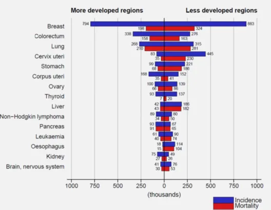
Breast cancer is the second most common cancer in the world and, by far the most frequent cancer among women with an estimated 1.67 million new cancer cases diagnosed in 2012, which accounts for 25% of all cancers. A slight majority of cases occur in women in less developed regions. Incidence rates vary nearly fourfold across the world regions, with rates ranging from 27 per 100,000 in Middle Africa and Eastern Asia to 96 in Western Europe. Breast cancer is the fifth cause of death from cancer overall (522,000 deaths) and while it is the most frequent cause of cancer death in women in less developed regions (324,000 deaths, 14.3% of total), it is now the second cause of cancer death in more developed regions (198,000 deaths, 15.4%) after lung cancer (Figure 1). [1]
Figure 1. Estimated numbers (thousands) of new cancer cases (incidence) and deaths (mortality) in
2
On average, one in eight women develops breast cancer during lifetime. Even if it is detected at an early stage, it still remains the second most common cause of cancer-related death in females after lung cancer. In spite of the advances over the last two decades in cancer diagnosis and treatments, breast cancer is still associated with high mortality and is a global health concern in both developed and developing countries. Previously to the year 2000, the incidence of breast cancer grew gradually since 1980. However, the rate reached a plateau between 2000 and 2003 for women of all ages, which coincides with the widespread use of mammograms as a standard screening method. Particularly, within this period, the incidence of breast cancer decreased dramatically among women between 50–69 years of age. This decrease has been associated with the reduced use of hormone replacement therapy in post-menopausal women during the early 2000s. [2]
1.1.1. Types of Breast Cancer
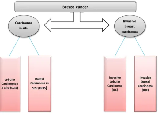
There are many risk factors that may be associated with breast cancer prevalence, which can affect the development and progression of the disease. Moreover, there are different types of breast cancer, which are described in this section. The majority of the cases are classified as either In Situ or Invasive, as represented in Figure 2.
3
Figure 2. Schematic division of the major types of breast cancer.
1.1.1.1. Carcinoma In Situ
Lobular Carcinoma In Situ (LCIS). Lobular carcinoma In Situ is located in the milk-producing glands (lobules) of the breast (Figure 3). Although it is not a cancer, being diagnosed with LCIS indicates an increased risk of developing breast cancer. It consists of an uncommon condition in which abnormal cells form in the breast lobules or milk glands, and is most commonly found in pre-menopausal women between the ages 40 and 50. [3]
4
Figure 3. Lobular Carcinoma In Situ (LCIS). Taken from [4].
Ductal Carcinoma In Situ (DCIS). Ductal carcinoma in situ is considered the earliest form of breast cancer. It consists on the presence of abnormal cells located in the milk ducts of the breast (Figure 4). DCIS is non-invasive, meaning that it does not spread out of the milk duct in other parts of the breast. DCIS is usually found through a mammogram, as part of breast cancer screening. Most cases of DCIS (~98%) do not become metastatic. However, around 50% evolve to invasive breast cancer (IBC). Most cases of DCIS are effectively treated with surgery and radiation. [3]
5 1.1.1.2. Invasive Breast Carcinoma
Invasive Lobular Carcinoma (ILC). Invasive Lobular Carcinoma (ILC) develops in the milk-producing glands (lobules) of the breast (Figure 5). ILC is able to advance to other parts of the body (mostly bone, brain, liver, and lungs), either through the bloodstream or the lymphatic system. ILC usually presents itself as an abnormal feeling in the breast (most often a thickening) and not as a hard mass that can be felt. Also, it is less likely to be detected in a mammogram. Women over the age of 40 have an increased risk of developing this kind of carcinoma, with most cases occurring at ages ranging 45-56. [3]
Figure 5. Invasive Lobular Carcinoma (ILC). Taken from [4].
Invasive Ductal Carcinoma (IDC). Invasive Ductal Carcinoma (IDC) is the most frequent type of invasive breast cancer, and is responsible for nearly 85% of the cases. IDC initiates in the milk ducts and grows into the surrounding tissue (Figure 6), being able to spread into other parts of the body, such as
6
bone, brain, liver and lungs, through the bloodstream or the lymphatic system. This kind of carcinoma is characterized by a hard lump that usually is detected a spiked mass on a mammogram. Women over the age of 40 have an increased risk of developing IDC, with around 50% of cases occurring in women over the age of 65. [3]
Figure 6. Invasive Ductal Carcinoma (IDC). Taken from [4].
1.1.2. Risk Factors
This big regional variation in breast cancer incidence rates is affected by a number of factors particularly related to lifestyle. A low number of children, giving birth at an older age, and lower acceptance of breastfeeding are some characteristics of women in more developed countries which are associated with the risk of breast cancer. [5]
Prospective studies have shown that a high risk of breast cancer is associated with early menarche, late menopause, obesity in postmenstrual
7 women, and high concentrations of endogenous estradiol. Reduction of risk is associated with childbearing, and especially with a greater protection from an early first birth or a larger number of births. Also, breastfeeding is a protection against breast cancer. Moreover, a small increase in breast cancer risk can arise from both oral contraceptives and hormonal therapy for menopause, which can be low once the using is stopped. Alcohol also increases the risk, whereas physical activity is assumed to be protective. Mutations in certain genes greatly increase breast cancer risk; however, these seem to account for a minority of cases. [6] A detailed list of the studied risk factors is presented below:
Age. While in reproductive years, breast cancer incidence seems to grow until women reach menopause; afterwards, over the age of 50, the rate is slower.
Menarche and the menstrual cycle. The risk of breast cancer decreases as the age of menarche increases (5% per year of delay). Other menstrual factors, such as cycle length and regularity, have not been consistently related to risk of breast cancer.
Childbearing. Childbearing has a dual effect on breast cancer risk, increasing in the period immediately right after a birth. However, this risk decreases gradually, and in long-term, bearing children has a protective effect against the disease. Women with at least one full-term pregnancy have, on average, around 25% reduction in breast-cancer risk. Moreover, an increased protection is observed as the number of pregnancies increases, reaching 50% risk with five or more children. Also, the risk of breast cancer can be lower as younger is the age of first birth.
8
Breastfeeding. The longer the time of breastfeeding, the higher will be the protection against breast cancer. Studies have revealed that women that have breastfed for total of 25 months have lowered the risk in about 33%. Also, the protective effect of breastfeeding is higher in younger than older women.
Menopause. Breast cancer risk can be higher for the women who have the menopause at older ages, with an increase of 3% for per year. Also, the risk is higher in premenopausal than in postmenopausal women with the same age.
Endogenous hormones. Studies have revealed that there is a positive association between the concentrations of serum oestradiol and risk of breast cancer in postmenopausal women. However, there is insufficient data for conclusions. Other sex hormones have been studied and it has been demonstrated that they also have positive associations with the risk of breast cancer.
Oral contraceptives. The risk of breast cancer is increased by around 25% in oral contraceptive users. However, this increased risk drops after cessation of use, with no significance increase of risk after ten or more years after the use stops. Also, the use of oral contraceptives at older ages increases the risk of the disease.
Hormonal therapy for the menopause. Women who are regular users of therapy with hormones have a higher risk of breast cancer than women who have never done these therapies.
9 Diet. In the countries with high-fat diet, the breast cancer risk has higher rates.
Alcohol and smoking. Provisional studies have repeatedly demonstrated that alcohol drinking is related with a moderate rising of the risk of breast cancer. A consumption of around 30 g alcohol (3 units) per day is related with an increase of about 30% in breast-cancer risk. Many studies on the relation between smoking and breast-cancer risk have been performed; however, overall there is no association.
Anthropometry. There seems to be a positive association between the height and the risk of breast cancer, with a 10% increase in risk for each 10 cm in height. However, the reasons for this association have not been established yet. Obesity increases the risk by 50% in postmenopausal women, on the other hand, in postmenopausal women, obesity seems to be protective. Some studies reported that the weight of birth is associated with the risk of breast cancer. This is related to factors such as hormone concentration during intrauterine life.
Exercise. Based in many studies, a lower risk of breast cancer is related to lightly physical activity. Few hours of activity during the week can reduce the risk of breast cancer around 30%.
Ionising radiation. Studies have revealed that the breast tissue is among the most sensitive tissues to the effects of ionising radiation. Also, studies on women younger than 40 reveal that a higher risk is observed with exposition. The negative effect of exposition can be the same for single or multiple exposures of the same dose.
10
Electromagnetic fields. Some studies suggest that being exposed to low dose of frequency electromagnetic fields and to artificial light at night may have been factors that can contributed in the increasing the incidence of breast cancer worldwide on the 20th century. However, the evidences are not persuasive.
Environmental estrogens. Some man-made chemicals, such as organochlorines or polychlorinated biphenyls, have structural similarities to endogenous estrogens and can bind to the estrogen receptors. Several prospective studies have been performed to examine the possible associations of these substances with breast-cancer risk; however, the results have not shown any association.
Family history and genetics. Apparently, environmental and lifestyle factors account for the most cases of breast cancer. Most women with the disease do not have a family history of it, and most women with affected relatives never develop it. On most studies on familial risk of breast cancer was found that, the risk is slightly higher for first-degree relatives (mothers, sisters, daughters), than with affected second-degree relatives (grandmothers, aunts, grand-daughters). However, the familial risk can be attributed both to share genes and to share physical environments and lifestyles.
High-risk mutations. Have been identified and localised at least five germline mutations that predispose to breast cancer. These include mutations in the genes BRCA1, BRCA2, P53, PTEN, and ATM. Mutations in BRCA1 and BRCA2 can cause high risks of breast cancer, and especially ovarian cancer. [6]
11 1.1.3. Treatments of breast cancer
The most common treatments for patients with breast cancer are surgery, radiotherapy and chemotherapy. The choice depends on several factors such as type of breast cancer, stage of evolving, location and especially the health status of the patient. Usually the goal of most of the treatment is to kill or to remove the cancer cells. Most of the treatments are applied in combination, either simultaneously or sequentially and the decision on which treatment is applied usually agreed between doctor and the patient. [7] The common types of cancer treatments are described shortly below:
Surgery: Usually is the treatment for many solid tumours. Surgery can be applied in the case of benign breast cancer when the disease is detected in the early stage. The goal is to remove all cancerous cells from the patient.
Radiation: The goal of radiation is to kill the cancer cells directly by damaging them with high energy beams. It can be applied in combination with surgery or drug treatment.
Chemotherapy: The goal of chemotherapy is to damage the dividing cells and to protect additional reproduction. Drug chemotherapy is a batch of drugs that is used to kill cancer cells.
Hormonal Treatments: These drugs are designed to prevent cancer cell growth by preventing the cells from receiving signals necessary for their continued growth and division.
12
Targeted Therapy: The goal of this treatment is to target the specific proteins and processes which are limited to cancer cells. The inhibition of these processes prevents the growth of cancer cell and division. This drug treatment is relatively new as a cancer treatment method.
Antibodies: The goal is to attack cancer cells where the mechanism of antibodies is to take away the cancer cells or causing the direct death of cells. Because antibodies are naturally appearing proteins in our body, they can be used as treatment against cancer and are manufactured for use as drugs. Because of their specificity, antibodies may be regarded as a type of specific inhibitor.
Biological Response Modifiers: These treatments involve the use of naturally occurring normal proteins that stimulate the body’s own defences against cancer.
Vaccines: Vaccines include proteins found or produced by cancer cells. The aim of those proteins is to increase the body response against the cancer cells. The goal of cancer vaccines is to activate the protection of the body against cancer.
Complementary and Alternative Medicines: These treatment methods are not practiced by conventional western medicine. They can include herbal, animal derived, and mind-body approaches of treating cancer. [8,9]
13 1.1.4. Variability in response
In oncology, the treatment with anticancer drugs is one of the major strategies to control the disease. Anticancer drugs should be active on cancer cells without causing toxicity in normal tissues. However, there is a thin line between suboptimal treatment of the anticancer drugs and their toxicity, and actually anticancer drugs tend to be an aggressive treatment. The clinical use of these drugs contains both weighing benefits and toxicity, trying to find a favourable therapeutic index. Because there is a reasonable variability in human response to anticancer drugs, this can happen by inter-individual variability in drug absorption, distribution, metabolism and excretion (pharmacokinetics) or by differences in the sensitivity of target tissues (pharmacodynamics). Both the genetic status of the tumour and the genetic profile of the patient can influence the individual effect and safety of the drugs used. [7]
The differences between individuals in drug metabolism may be associated to demographic factors such as gender, age, pregnancy. Drug metabolism and clearance of drugs can be greatly affected by drug-drug interactions or pathological alteration such as hepatic, renal and cardiac diseases. [7]
14
1.2. Drug metabolism and clearance
The most important removal pathway of xenobiotics from the organism is through biotransformation, which usually happens in the liver. Because of this process, the water solubility of these substances increases and their elimination from the organism becomes easier through urine or bile. [7]
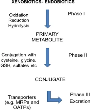
These reactions can be divided into three phases. The schematic illustration of phases I, II, and III of xenobiotic substance materialization is presented in Figure 7.
Figure 7. Schematic illustration of phases I, II, III of xenobiotic metabolizing systems. (adapted from [10])
Phase I reactions (also known as functionalization reactions) involve oxidations (e.g., hydroxylation and deamination), reductions (e.g., addition of hydrogen atoms), and hydrolysis (e.g., splitting of ester and amide bonds). Phase II reactions (also known as conjugation reactions) involve the conjugation of the drug with hydrophilic endogenous substrates. [7]
15 The purpose of these two main phases (I and II) during drug metabolism is to change the structure and inactivate the drug, facilitating its elimination from the body through urine or faeces in phase III. [11]
Nearly all drugs and their metabolites are eventually eliminated from the body in urine or in bile. Generally, drugs with less molecular weight, water soluble, and that have been slowly bio–transformed slowly, are eliminated in urine, however drugs with high molecular weight (>500 Da) are eliminated in bile. [7]
The transformation of drugs in Phase II involves conjugation of a drug or metabolite, initially transformed in Phase I, with endogenous substrates. These reactions include acetylation, amino acid conjugation, glucuronidation, sulfate conjugation and glutathione conjugation. The kidneys can successfully excrete these Phase II hydrophilic conjugates. [11]
The enzymes catalysing these reactions are known as drug metabolizing enzymes (DME). [7]
1.2.1. Estrogen metabolism and breast cancer
Lately, much attention has been given to the effect of estrogens on breast carcinogenesis, since a number of risk factors are associated with endogenous estrogen levels, suggesting that estrogens have an important role in breast cancer. [12]
Many different biological effects such as female sexual differentiation and development, arterial vasodilation, the maintenance of bone density, and neuroprotective actions are regulated by estrogens. Most of these effects result from direct interaction between estrogen and the estrogen receptor (ER), which
16
activates the expression of target genes encoding proteins with important biological functions. However, estrogens also contribute to the development and evolution of breast cancer. Exposure to estrogens continuously is considered a crucial etiological factor for the induction of estrogen-associated cancers. [13]
Estradiol and estrone are the main estrogens circulating in the human body, as well as 16-hydroxyestradiol (estriol). Estriol is usually the most concentrated estrogen present in pregnant women, and also in the urine of all women, however, estradiol is the most biologically active estrogen, primarily secreted by ovarian granulosa cells regulated by follicle-stimulating hormone (FSH). [12]
The elimination of estrogens from the body occurs by metabolic transformation to estrogenically inactive metabolites that are excreted in the urine and/or faeces. The metabolism of estrogens involve oxidation (mainly hydroxylation) by cytochrome P450s (CYPs), glucuronidation by UDP-glucuronosyltransferase, sulfation by sulfotransferase, and O-methylation by catechol O-methyltransferase (COMT). The first step in the metabolism of estrogens, hydroxylation, is mediated by CYP enzymes. CYP1, CYP2, and CYP3 are three families that primary catalyse the oxidative metabolism of exogenous and endogenous compounds. Some isoforms in these CYP families are also responsible for the metabolism of estrogens. Because most CYP isoforms are mainly expressed in liver, the metabolism of estrogens occurs mainly in this organ. [13]
The expression level of the estrogen-metabolizing CYP enzymes is controlled by many factors in the liver and target tissues. The metabolism of estrogen not only alters the intensity of its action but may also change the profile of its physiological effects in target tissues. [13]
17 1.2.2. Cytochrome P450s
Cytochromes P450 (CYP) enclose a complex family of isoenzymes, which are involved in drug metabolism (phase I enzymes), and are present in different tissues, mainly in the liver and intestine. [11]
In order to inactivate (or activate) drugs and toxic combinations they are able to catalyse the oxidative biotransformation for most of the drugs and other xenobiotics, therefore they also play a pivotal role in clinical pharmacology also. [13] CYP1, CYP2, CYP3 and CYP19 are the four most distinguished P450 families, which are involved in metabolism of anticancer drugs, and other xenobiotics. [11]
The classification of CYPs is based on their amino acid sequence into gene families and subfamilies. The enzymes in families 1-3 participate in xenobiotics metabolism, while the others mostly metabolize endogenous compounds (e.g. steroids, bile acids, cholesterol). [7]
CYP3A4, CYP2D6 and CYP2C9 dominate drug metabolism. CYP3A4 (CYP belonging to family 3, subfamily A) has the widest substrate specificity and is the most abundant CYP in the body. It has been estimated that CYP3A4, CYP2D6 and CYP2C9 are respectively involved in the metabolism of about 50, 25 and 15% of clinically used drugs. [7]
18
1.2.3. Drug transporters
Drug-metabolizing enzymes (DMEs) and transporters play very important roles in the detoxification of many unknown and endogenous chemicals. [14] Accumulation of carcinogens, environmental pollutants, therapeutic drugs or any other kind of xenobiotics in the body can affect human health. The detoxification of these chemicals, involve four main stages:
absorption/permeability, distribution,
metabolism and excretion.
These stages are mediated by a large number of DMEs and transporters. [15] Drug absorption, delivery and their secretion across biological membranes profoundly affect the pharmacokinetics, which in its turn are affected by the drug transporters. [11]
Functionally, transporters are proteins that eliminate endogenous compounds (such as bile acids, lipids, sugars, amino acids, steroids, hormones, and electrolytes) and xenobiotics (such as drugs and toxins) through the biological membranes in order to control the cellular and physiologic concentrations and to keep the fluid balance. Therefore, they support as a better mechanism for detoxification of any dangerous foreign substances in cells. Also, transporter proteins can influence drug absorption in the small intestine and drug elimination in the liver and/or kidney, conducting these substances in and out of the intestinal enterocytes, hepatocytes, or renal tubular cells. Another important role of transporters is to limit the penetration of drugs into brain, placenta, tumour, T-cells, and others. The reduction in the transporter’s functions can increase the toxicity. [16]
19 The ABC transporters (ATP-Binding Cassette transporters) contain a family of transmembrane proteins which use ATP hydrolysis as a power source for transport activity. Seven major subfamilies of the ABC transporters genes exist, however, only three of them (ABCB, ABCC, ABCG) are engaged in drug transport including anticancer drugs. [11]
The ABCB1 (MDR1/P-glycoprotein), ABCC (MRP), and ABCG2 (MXR/BCRP) are active transporters and have a main interest to pharmacologists since they are responsible for both the uptake and the efflux of drugs and are essential elements of the pharmacokinetic characteristics of a drug. However, it has now become clear that transporters are crucial for the uptake, accumulation, distribution and efflux of drugs. For example, drug efflux transporters including the P-glycoprotein pump (Pgp), the multidrug-resistant protein-1 (MRP1) and the BCRP actively pump drugs such as chemotherapeutics out of the cells, therefore are used for reducing their intracellular accumulation and making the cell insensitive to different drugs.
Among the major known ABC transporters, ABCB1 gene, also known as MDR1, encoding Pgp is by far the best characterized and understood efflux transporter. Pgp plays an important role in limiting intestinal drug absorption and brain penetration as well as in facilitating renal or biliary excretion of drugs. The MRPs are involved in the drug efflux from the liver or kidney into the peripheral blood (e.g. MRP1, MRP3, and MRP6), or from the liver, kidney and small intestines into the bile urine and intestinal lumen respectively (MRP2). [17]
20
1.2.4. Regulation of expression
The inter-individual variability in drug metabolism and transport is due to genetic factors or to variable expression of the relevant genes in different physiological or pathological circumstances. Several xenobiotics and endobiotics are known to change expression of target genes, mainly by regulating the rate of transcription. An essential concept about gene expression in the disease state, and the effects of xenobiotics or any other foreign pollutants, is that often the events observed are cell-, species-, sex-, and development-specific. This is due to the fact that combinational gene expression is typical of eukaryotes. Generally, a combination of multiple gene-regulatory proteins, rather than a single protein, decide where and when a gene is transcribed. A single gene may be essential but not enough to change the phenotype of a cell. [18]
A key component of many complex phenomena including cellular development, differentiation, maintenance, and injury or death is the differential gene expression. The subgroup of genes which are expressed determines the phenotype of that cell. Also, a failure of control of differential gene expression determines many disease states, including cancer. The understanding of the function of these genes can be helpful in the identification of genes that are being expressed in one cell type versus another (i.e., control versus treated; tumour versus normal). Therefore, the identification of differentially expressed genes has been followed for different influences such as responses to biological programs, physical agents (i.e., UV irradiation, X-rays), and chemical agents (hormones and xenobiotics). [18]
21
1.3. Nuclear receptors
Drug-metabolizing enzymes (DMEs) and transporters play a central role in the metabolism and clearance of xenobiotics. To face chemical challenges, the expression of many DMEs and transporters is controlled by a group of ligand-activated transcription factors designated as nuclear receptors (NRs). [14]
1.3.1. The identification of Nuclear Receptor
The first NR was identified biochemically in the 1960s by Ron Evans and co-workers Pierre Chambon and Geoffrey Greene. Since then, NRs have become identified as a superfamily of transcription factors, and the NR research field has grown very fast and covers areas ranging from structural and functional analyses to the molecular mechanisms of transcription regulation. [19] . To date, 48 NRs have been recognized in humans (Table 1). [20]
22
23 1.3.2. Structure of Nuclear Receptors
All the nuclear receptors have common structural characteristics (Figure 8), which contain a central DNA binding domain (DBD) responsible for targeting the receptor to highly specific DNA sequences. The ligand binding domain (LBD) is involved in the C-terminal half of the receptor and recognizes specific hormonal and nonhormonal ligands directing specificity to the biologic response. These receptors contain variable N-terminal and C-terminal domains, as well as a variable length hinge region between the DBD and LBD. [21]
Figure 8. Structure and functional domains of the nuclear receptors (adapted from [22])
1.3.3. The role of Nuclear Receptors
Nuclear receptors regulate specific target genes involved in different functions of the body, such as homeostasis, metabolism, development, reproduction and other physiological processes. [20,23]
NRs are members of a superfamily of ligand-regulated (and orphan) transcription factors that transduce different endocrine hormones such as steroid, retinoid, thyroid, and lipophilic into specific physiological responses. [24] NRs were identified as receptors for their cognate ligands and they primarily function as ligand-activated DNA-binding transcription factors. As transcription factors, NRs regulate directly the expression of hormone response genes.
24
There are different functions associated with different kinds of hormone receptors, e.g., the progesterone (PR), androgen (AR), and estrogen (ERα and ERβ) receptors operate in reproduction and target tissue growth; the thyroid hormone receptors (TR) control oxidative metabolism; the glucocorticoid receptor (GR) regulates glucose metabolism, inflammation, and stress; and peroxisome proliferator-activated receptors (PPARs) have central roles in regulating energy and lipid metabolism. [24]
Nuclear receptors regulate specific target genes involved in metabolism, development reproduction and other physiological processes. [19] The significance of NRs in xenobiotic metabolism and clearance is best demonstrated by xenobiotic receptors: pregnane X receptor (PXR, NR1I2). [14]
1.3.4. Characteristics of PXR
Xenobiotics including drugs and environmental chemicals have a very negative impact on human health. [14] Therefore the nuclear pregnane X receptor (PXR; NR1I2) is an important component of the body’s protection mechanism against toxic substances including foreign chemicals. PXR is activated by a large number of endogenous and exogenous chemicals including steroids, antibiotics, antimycotics, bile acids, and the herbal antidepressant. [25]
25 1.3.5. Structure of PXR
Pregnane X receptor (PXR), also known as the steroid and xenobiotic sensing nuclear receptor (SXR) or nuclear receptor subfamily 1, group I, member 2 (NR1I2) is a protein that in humans is encoded by the NR1I2 (nuclear receptor subfamily 1, group I, member 2) gene. [26] The pregnane X receptor is an approximately 434- amino acid, 50-kDa protein, primarily expressed in the liver and intestine. [14]
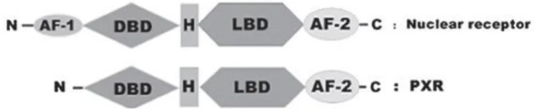
The PXR protein contains a DNA-binding domain (DBD), a hinge region (H), a ligand-binding domain (LBD) a ligand-dependent transactivation function 2 (AF-2) (Figure 9). PXR does not have a ligand-independent activation function 1 domain (AF-1), in comparison with other nuclear receptors. PXR is an orphan nuclear receptor that is activated by binding to various chemically and structurally distinct endobiotics and xenobiotics including clinically-used chemotherapeutic drugs (tamoxifen, doxorubicin, taxol, and vincristine) and environmental chemicals. [27]
Figure 9. Schematic comparison of the domain structures of a typical nuclear receptor and PXR. AF‑1, activation function-1; DBD, DNA-binding domain; H, hinge region; LBD, ligand-binding domain; AF‑2, transactivation function-2; PXR, pregnane X receptor (taken from [28])
26
1.3.6. Distribution of PXR
PXR is mainly expressed in the liver, small intestine and colon in humans. PXR is expressed not only in normal tissues, but also in numerous types of human cancer, including breast, osteosarcoma, colon, endometrial, ovarian, prostate and esophageal cancers. Most significantly, the expression levels of PXR in these cancer tissues are usually higher than in non‑neoplastic tissues. [28]
PXR co-ordinately regulates a large proportion of genes and proteins in the liver, intestine and other organs that are involved in all aspects of the detoxification and elimination of xenobiotics and drugs. [28]
1.3.7. Function of PXR
The pregnane X receptor (PXR) is a nuclear receptor which main function is to sense the presence of foreign toxic substances (e.g. xenobiotics), and in response regulates the expression of proteins involved in the detoxification and clearance of these substances from the body. [25]
PXR is the main transcriptional regulator of many enzymes that metabolize xenobiotics, such as P450s, and drug transporters (Figure 10). PXR is a member of the nuclear receptor superfamily of ligand activated transcription factors that controls the expression of their target genes by binding to the gene’s promoter. [27]
27
Figure 10. hPXR mediated induction of drug metabolizing enzymes of phases I,II and III (taken from [29])
1.3.8. PXR and drug metabolism
The human Pregnane X Receptor (hPXR) is the master transcription factor for cytochrome P450 3A4 (CYP3A4) and multidrug resistance protein 1 (MDR1). The hPXR is able to be activated by structurally different ligands. [30]
Many steps in xenobiotic metabolism can be regulated by PXR in collaboration with other nuclear receptors such as:
constitu-tive androstane receptor (CAR), aryl hydrocarbon receptor (AhR),
peroxisome proliferation activated receptor α and γ (PPAR), liver X receptor (LXR),
farnesoid X receptor (FXR), and vitamin D receptor (VDR).
hPXR is able to regulate the expression of the main phase I DMEs, such as CYP2B6, CYP2C8, CYP2C9, CYP2C19, CYP3A4/5, and CYP3A7; therefore, involving about >60% metabolism of endobiotics and xenobiotics. It
28
also organizes phase II DMEs such as UGT (Uridine-diphospho-glucuronosyltransferase), sulfotransferase, acetyltransferase, methyltransferase, and glutathione-S-transferase, as presented in Figure 11. hPXR also can cause the stimulation of phase III efflux transporters of ABC (ATP binding cassette) superfamily, such as Pgp (p-glycoprotein), BCRP (breast cancer resistance protein), and MRP (multiple resistance drug protein). [30]
Figure 11. hPXR mediated induction of drug metabolizing enzymes, transporter and their role in drug
interaction. Abbreviations: PXR, pregnane X receptor; RXR, retinoid X receptor; DNA, deoxyribonucleic acid; mRNA, messenger ribonucleic acid; MRP, multiple resistance drug protein; MDR, multidrug resistance protein; BCRP, breast cancer resistance protein; CYP3A4, cytochrome P-450 monooxygenase 3A4; CYP2B6, cytochrome P-450 monooxygenase 2B6; UGTs, uridine 5’- diphospho- glucuronosyltranferases; SULTs, sulfotransferases; GSTs, glutathione-S-transferases; ALDHs, aldehyde dehydrogenases (taken from [30])
The cytochrome P450 CYP3A4 and the ATP binding cassette (ABC) transporter P-glycoprotein (Pgp) are two main factors that regulate the exposure to a large range of xenobiotics. The transcription of the corresponding genes CYP3A4 and multidrug resistance (MDR1) is activated by xenobiotics through activation of at least one common nuclear receptor, the PXR. [31]
29 1.3.9. PXR and Chemotherapy
One of the most common treatments for cancer is chemotherapy. However, due to multidrug resistant cancer cells, the efficiency of this treatment is narrow or limited. However drug metabolizing enzymes (DMEs) and efflux transporters can help in promotion of the metabolism, excretion, and detoxification of chemotherapeutic agents. Due to this, high levels of DMEs and efflux transporters, decrease the efficacy of chemotherapeutics and usually may cause failing of the therapeutic treatment. During chemotherapy, nuclear receptors, in particular pregnane X receptor (PXR, NR1I2) and constitutive androstane activated receptor (CAR, NR1I3), are known for their pivotal role in metabolism and clearance of xenobiotic and also for their importance in the development of multidrug resistance (MDR). [15]
Multidrug resistance (MDR) is an important barrier to the pharmacokinetics and pharmacodynamics of anticancer drugs, mostly as a result of the induction potential of anticancer drugs for drug metabolizing enzymes (DMEs) and efflux transporters through nuclear receptors. [30]
30
1.3.10. Pharmacogenetics of PXR polymorphisms
Single nucleotide polymorphism (SNP) is an inherited change in the DNA sequence, usually substituting one nucleotide for another (Figure 12). [7]
Figure 12. Genotyping looks at single letter substitutions in the genetic code. These genetic variation are
called Single Nucleotide Polymorphism (SNPs). [32]
A single nucleotide polymorphism (SNP) is a nucleotide variation at a specific location in the genome. Typically, SNPs are bi-allelic and by definition found in more than 1% of the population. In practice, tri- or tetra-allelic SNPs, insertions, deletions and variations found in less than 1% of the population are also considered as SNPs. SNPs are the most abundant variations in the human genome. [33]
Like mutations, SNPs can cause changes within a gene (coding SNP), predicted to change an amino acid (a non-synonymous SNP). Rather, the SNP can change a nucleotide within a gene, which is not predicted to change an amino acid (a synonymous SNP). [7]
Single nucleotide polymorphisms (SNPs) are used as markers in linkage associated to studies to detect which regions in the human genome may be
31 involved in diseases. SNPs in coding and regulatory regions may be involved in disease themselves. In the main interest are non-synonymous SNPs, which cause an amino acid change in the protein product, and these amino acid substitutions are considered to be responsible for approximately half of the known gene lesions responsible for human inherited disease. [34]
Related to this argument, as we mentioned above in the drug metabolism and clearance section, the effectiveness of the steps in drug metabolism, as well as the host response to the drug treatment is mainly dependent on the genetic polymorphisms in the drug metabolizing enzymes, patients' age and the health status of drug detoxifying organs. Therapy toxicity or drug resistance can be a result of any failure in this machinery caused by these factors, with a special distinction of SNP genotypes present in the drug responsive genes. Also, genetic polymorphisms can influence transporters causing inter patient differences in drug response. [11] The NR1I2 gene, which encodes PXR, contains 10 exons and is located on chromosome 3q13-21. Different SNPs have been reported in NR1I2 and some are associated with changes in PXR function. [35]
To date, 227 SNPs of hPXR have been deposited in dbSNP. [36] The SNP that was investigated in this study, SNP NR1I2 7635A>G, is present in intron 5 and the 7635G allele has been associated with increased expression of CYP3A4 in the presence of rifampicin. [35]
32
1.4. Genotyping methods
Different SNP genotyping techniques have been reported in the last years. One of the most common methods employed to discover novel SNPs is sequencing DNA fragments amplified by polymerase chain reaction (PCR). PCR primers are designed to amplify both strands of DNA from genes or other single copy genomic sequences (Figure 13). PCR products are sequenced, allowing for novel SNP identifications. [33]
Figure 13. General steps in a PCR procedure (taken from [37])
Since the invention of PCR by Karry Mullis in the 1980s, the application of the PCR method has provided great contributions in molecular genetic research. This simple and sensitive enzymatic technique for the amplification of DNA fragments has been used for many different purposes, but especially for the detection of nucleic acid polymorphisms in order to find the biological significance in genetic variation and molecular divergence in living organisms. [38]
Denaturation
Annealing
33 The majority of polymorphisms in the human genome are SNPs, which are considered for more than 90% of sequence variations, and are applied as an important tool for the study of the structure and history of our genome. There are more than 27 million SNPs in the human genome that have been recorded in a database. This immense amount of data contribute with important information for SNP-based studies, identification of candidate genes involved with complex genetic diseases, pharmacogenetic analysis, drug development, population genetics, evolutionary studies and forensic investigations. In the last twenty years, many different methods have been developed for SNP genotyping by PCR, including hybridization, allele-specific PCR, primer extension, oligonucleotide ligation, direct DNA sequencing and endonuclease cleavage, after amplification of the subjected genomic region by PCR. Recent methods for SNP genotyping, such as Taqman method, Invader, MALDI-TOF and GeneChips have high performance; however, they are relatively expensive. The PCR technique has evolved for better understanding of the causes of genetic variants and mutations. Polymerase chain reaction-restriction fragment length polymorphism (PCR-RFLP) is a technique applied on the detection of single nucleotide polymorphisms (SNPs). PCR-RFLP allows a fast detection of point mutations after the genomic sequences is amplified by PCR. The mutation is discriminated by digestion with specific restriction endonucleases and is identified by gel electrophoresis after staining with ethidium bromide (EtBr) or other fluorescent indicator, such as Green Safe. [38]
PCR-RFLP is a simple, relatively inexpensive and precise method for SNP genotyping, and especially helpful in small basic research studies of complex genetic diseases. It is based on endonuclease digestion of PCR-amplified DNA. The specific restriction endonuclease recognizes and cleaves the DNA in the
34
region of the point mutation of the PCR products. The SNP type is easily identified by using gel electrophoresis to confirm and separate the sizes of the smaller DNA fragments generated by the endonuclease digestion. This method is available for genotyping not only a SNP but also insertion/deletion polymorphisms, and multiple mutations [38]
1.4.1. Genotype and allele
In the living cell, genes are organized in linear order along microscopic threadlike bodies called chromosomes. A typical chromosome may consist of several thousand genes. The position of a gene along a chromosome is called the locus of the gene. In higher organisms, each cell has two copies of each type of chromosome. Such organism, in which the chromosome are present in pairs, are said to be diploid. In each pair of chromosomes, one member is inherited from the mother through the egg and the other is inherited from the father through sperm. At each locus, accordingly, diploid organism contains two alleles, one each at corresponding positions in the maternal and paternal chromosomes. If the two alleles at a locus are chemically identical (in the sense of having the same nucleotide sequence along the DNA), the organism is said to be homozygous at the locus under consideration; if the two alleles at a locus are chemically different, the organism is said to be heterozygous at the locus. The term gene is a general term usually used in the sense of locus. Geneticists make an essential differentiation between the genetic structures of an organism and the physical or biochemical attributes of the organism. The genetic structure of an organism is called genotype; genotype thus refers to the
35 particular alleles present in an organism at all loci that affect the trait in question. [39]
1.5. Observational Studies
Observational studies are a fundamental part of epidemiology and account for many of the research papers published in specialty research journals. We can apply epidemiology to study any specific population. In epidemiology is assumed that health, disease, and illness are not random, and there are characteristics that protect us and that predispose us to harm, different diseases, and other occurrences. These characteristics may be genetic, environmental exposures, or other factors. Typically, the distributions of characteristics or events are evaluated, or possible associations between certain characteristics and health outcomes are investigated. [40]
1.5.1. Case-Control Studies
A case–control study is an observational study in which study subjects are chosen who meet a “case” definition (e.g. individuals with breast cancer) and also subjects that decidedly are not “cases” (e.g. individuals without any neoplastic disease). Case–control studies are typically retrospective studies because the approach is to identify persons with the disease of interest and then look backward in time to identify factors that may have caused it. Possible associations between the disease of interest and one or more hypothesized risk
36
factors are analysed in case-control-studies. The general strategy is to compare the frequency or level of potential risk factors between a representative group of diseased subjects, or cases, and a representative group of disease-free persons, or controls, derived from the same population. Three important criteria for case–control studies help to minimize the potential for bias: [40]
The selection of cases must be representative of all patients who develop the disease.
The controls must be representative of the general healthy population who do not develop the disease.
The information must be collected from cases and controls in the same way.
Case–control studies that require invasive procedures for diagnosis typically cannot expose controls to the risk of these procedures unless indicated clinically (e.g. coronary angiography or tissue biopsy). As in all other observational study designs, it is important to have as high a participation rate as possible to minimize biases resulting from nonresponse. Cost and accessibility should be considered when selecting controls because it is generally more difficult to motivate disease-free persons to join a study than those with disease who have a strong interest in determining its cause. The validity and reproducibility of measurement techniques in both cases and controls should be established or assessed during the study. Observations from case–control studies are frequently presented in a “2 × 2” table with the exposure status in rows and the case–control status in columns (Table 2). Cell “A” represents the number of exposed cases, cell “B”, the number of exposed
37 controls, and the row total, “A + B”, all exposed subjects. Column totals “A + C” and “B + D” are the numbers of cases and controls, respectively. [40,41]
Table 2. Presentation of Findings in a “2 × 2” Contingency Table.
1.5.2. Chi-Square Test of Contingency
Testing a contingency table assesses the independence between categories in rows and columns. Independence implies that the variables in the rows are not related to the variables in the columns. Dependence implies the existence of association. However, this does not provide information about the strength of this association. The strength of association can be assessed by the means of odds ratios, which is described in the next section. The chi-square test of contingency is based on the differences between the observed values and those that would be expected if the variables were independent. If these differences are small, there is little dependence between the variables, whereas large differences indicate dependence. The expected values are calculated by multiplying the corresponding row sum by the column sum, divided by the total sum (N). In general, for row i and column j, the expected value of the ijth entry is given by equation 1. The expected values calculated from the observed values in Table 2 are represented in Table 3. The chi-square statistic is the sum of
Total Present A B A+B Absent C D C+D Total A+C B+D N Number with disease Number without disease Presence of Disease Exposure
38
squares of these differences in ratio to the expected value (equation 2). A small chi-square statistic arises if the observed values are close to the values we would expect if the two variables were unrelated. A large chi-square statistic arises if the observed values are rather different from those we would expect from unrelated variables. [41]
Eij= ni×nj N (1) χ2= ∑ (Oi-Ei)2 Ei i (2)
Table 3. Expected Values from a “2 × 2” Contingency Table.
1.5.3. Odds Ratio
Odds ratio (OR) is a measure of the strength of association between an exposure factor (risk factor) and an event (disease). In observational studies, it represents the ratio between the odds for the occurrence of an event in a group exposed to a factor and the odds for the occurrence of the same event in a group exposed to a different factor (or not exposed). The calculation of the odds ratio is obtained with equation 3, where the values A, B, C, D are the observed
Present (A+B)(A+C)/N (A+B)(B+D)/N
Absent (C+D)(A+C)/N (C+D)(B+D)/N Exposure Presence of Disease Number with disease Number without disease
39 cases from Table 2. The confidence interval with 95% confidence level for the odds ratio is given by equation 4. [41,42]
OR=A⁄B C
D
⁄ (3)
exp [ln(OR)±1.96√A1+B1+C1+D1] (4)
1.6. Objectives of the study
Given that estrogens are a well-established risk factor for breast cancer, and that these molecules are metabolized mainly by CYP3A4, it is conceivable that variations in CYP3A4 activity may be associated with individual susceptibility for the disease. Since PXR is the major transcriptional regulator of CYP3A4 expression, we hypothesized that variability in PXR may contribute to inter-individual variability in risk. As such, in this work, we aimed at evaluating the existence of an association between the PXR 7635 SNP and risk of disease, in a case-control study for breast cancer in the Portuguese population.
40
2. EXPERIMENTAL
2.1. Patients and Method
The A7635G polymorphism of PXR was investigated in a case-control study in a sample of the Portuguese population. Blood samples were obtained from 100 female patients with breast cancer, and from 114 female individuals without any neoplastic disease, recruited by the Research Unit in Human Molecular Genetics, Lisbon, Portugal.
Written consents were obtained from all individuals before the blood analysis. This study was approved by the ethical boards of the involved clinical institution and followed the recommendations of the Declaration of Helsinki, promulgated in 1964.
DNA extraction and genotyping were performed in the Laboratory of Pharmacogenomics and Molecular Toxicology, Centre for Biomedical Research (CBMR), University of Algarve. The genotyping was performed by means of PCR–RFLP.
2.2. Data treatment
PXR allelic and genotypes frequencies in individuals with and without breast cancer with 95% confidence intervals (CI) were assessed using the Chi-square (χ2
41
3. RESULTS AND DISCUSSION
3.1. Genotype and Allele frequencies
The presence of the polymorphism A7635G was investigated in breast cancer patients and its frequency compared with that found in healthy individuals, in order to evaluate its potential association with individual risk. The A7635G polymorphism in PXR was obtained from 100 female patients with breast cancer, and from 114 female individuals without any neoplastic disease in a sample of the Portuguese population. The PXR genotype and allele frequencies are represented in Figure 14.
Figure 14. Frequencies of PXR7635 genotypes and alleles in 100 individuals with breast cancer
42
3.2. Chi square analysis
The chi-square and p-values access the significance in the association between the disease and the genotypes and alleles studied. The expected values, calculated from the observed cases using a contingency table (Table 3) and equations 1 and 2, and are presented in Table 4 in. These results are also displayed in a Chi-square distribution function (Figure 15).
Table 4. Contingency tables (2x2) with the observed cases the calculated expected values, Chi
square and p-values (calculated in Excel).
Figure 15. χ2 and p-values of the genotypes and alleles association with the Breast Cancer disease, represented in a χ2
distribution with one degree of freedom.
Breast Cancer Controls Breast Cancer Controls
27 47 34.6 39.4 73 67 65.4 74.6 60 52 52.3 59.7 40 62 47.7 54.3 13 15 13.1 14.9 87 99 86.9 99.1 114 146 121.5 138.5 86 82 78.5 89.5 86 82 78.5 89.5 114 146 121.5 138.5 2.21 0.137 4.42 0.036 0.973 0.00 2.21 0.137
With Genotype A/A With Genotype A/G
Without Allele A With Allele G Without Allele G
OBSERVED VALUES
Without Genotype A/A Without Genotype A/G Without Genotype G/G With Genotype G/G With Allele A 4.77 0.029 χ2 (1 d.f.) p-value EXPECTED VALUES
43
3.3. Odds ratio
The odds ratios and the confidence intervals with 95% confidence level were calculated using equations 3 and 4 respectively. These results are presented in Table 5 and displayed in Figure 16.
Table 5. Genotypic and allelic frequencies of PXR7635 in individuals with Breast Cancer and
individuals without any neoplastic diseases, Odds Ratios, and p-values of the Chi-Square distribution with one degree of freedom.
Figure 16. Odds ratios and confidence intervals (95% confidence level) of the associations
between individuals with each genotype and allele and the Breast Cancer disease.
A/A 27.0 41.2 27 47 0.029 0.53 0.296 - 0.940 A/G 60.0 45.6 60 52 0.036 1.79 1.038 - 3.081 G/G 13.0 13.2 13 15 0.973 0.99 0.445 - 2.187 Total 100 114 A 57.0 64.0 114 146 0.137 0.74 0.504 - 1.099 G 43.0 36.0 86 82 0.137 1.34 0.910 - 1.982 Total 200 228 OR Confidence Interval (95%) p-value Alleles
Genotypes Breast Cancer (%) Controls (%) Breast Cancer (N) Controls (N)
44
3.4. Interpretation of results
The associations of the genotypes A/A and A/G with the disease are significant (p < 0.05); on the other hand, and according to the results, the genotype G/G and the alleles A and G have no association with the disease, with a 95% level of confidence. Small χ2 values and large p-values indicate that the observed and expected cases are not significantly different, and there is a large probability of a case occurring by chance alone (p > 0.05), whereas, large χ2 values and small p-values indicate that the observed and expected cases are significantly different, and there is a small probability of a case occurring by chance alone (p < 0.05).
The odds ratios provide information on the kind of association existing between the presence of the genotypes or alleles with the disease. As observed in Figure 16, the odds ratio for the genotype A/A is lower than the critical value of 1, and also its confidence interval, which represents a negative association between this genotype and the disease.
The odds ratio regarding genotype A/G is higher than the critical value, including the 95% confidence interval, which represents and positive association with the disease. The genotype G/G has an odds ratio very close to one, which means no association, either positive or negative, and considering the alleles A and G individually, they appear to have negative and positive association respectively; however, the 95% confidence intervals make these assumptions inconclusive.
45
4. CONCLUSIONS
According to the results obtained from the investigation of patients with breast cancer compared with control cases study, we can conclude:
The associations of the genotypes A/A and A/G with the disease are statistically significant with 95% confidence level (p < 0.05). The genotype G/G and the alleles A and G considered individually have no significant association with the disease, with a 95% level of confidence. Individuals with genotypes A/A have low risk of having breast cancer
disease (OR = 0.53, CI=[0.296 - 0.940], 95% CL), whereas the ones with genotype A/G have high risk to contract the disease (OR = 1.79, CI=[1.038 - 0.940], 95% CL).
Individuals with genotype G/G have no association with the disease (OR = 0.99, CI = [0.445-2.187], 95 CL), since it’s very close to OR = 1. However, with the confidence interval, both positive and negative associations can be possible with 95% confidence level.
Considering the alleles, positive and negative associations are verified with the odds ratios. However, this conclusion is affected by the confidence intervals. Therefore it is inconclusive, like for genotype G/G.
46
5. COMMENTS AND FUTURE WORK
Lately, in the last 16 years, after many studies about hPXR, the pivotal role in the activation of cytochromes P450s and efflux transporters leading to MDR in cancer is documented in the scientific literature. In view of the development of inhibitors and to restrain the spreading of the cancer, especially targeting the MDR in chemotherapeutics, the hPXR is a hopeful goal. A new strategy in anticancer drug development is necessary, in order to avoid the limitations in the present cancer chemotherapy and MDR. The hPXR is a novel target and its inhibitors can be used as adjuvants in the chemotherapy. [24]
In the future work I would like to work in molecular biology and genetic molecular respectively in cancer research groups, in order to employ the techniques that were used in this study, and also new methods used nowadays, for helping the society specially women gender for the detection of breast cancer disease in the early stages, and to evaluate the potential role of PXR receptor in the susceptibility to this disease, as a hopeful goal in chemotherapeutics.
During this study, because of my pregnancy circumstances, I was not able to work in the laboratory because of the presence of ethidium bromide, which is a dangerous chemical substance especially for my case. However I enjoyed searching, studying and writing the topic of my thesis, which I think is very interesting and it will be helpful in the future studies too.
![Figure 3. Lobular Carcinoma In Situ (LCIS). Taken from [4].](https://thumb-eu.123doks.com/thumbv2/123dok_br/18838064.928552/14.892.213.571.106.387/figure-lobular-carcinoma-in-situ-lcis-taken-from.webp)
![Figure 5. Invasive Lobular Carcinoma (ILC). Taken from [4].](https://thumb-eu.123doks.com/thumbv2/123dok_br/18838064.928552/15.892.325.650.533.914/figure-invasive-lobular-carcinoma-ilc-taken-from.webp)
![Figure 6. Invasive Ductal Carcinoma (IDC). Taken from [4].](https://thumb-eu.123doks.com/thumbv2/123dok_br/18838064.928552/16.892.236.573.308.698/figure-invasive-ductal-carcinoma-idc-taken-from.webp)
![Table 1. List of mammalian Nuclear Receptor (taken from [20])](https://thumb-eu.123doks.com/thumbv2/123dok_br/18838064.928552/32.892.163.645.153.1154/table-list-mammalian-nuclear-receptor-taken.webp)
![Figure 8. Structure and functional domains of the nuclear receptors (adapted from [22])](https://thumb-eu.123doks.com/thumbv2/123dok_br/18838064.928552/33.892.220.774.539.617/figure-structure-functional-domains-nuclear-receptors-adapted.webp)
![Figure 10. hPXR mediated induction of drug metabolizing enzymes of phases I,II and III (taken from [29])](https://thumb-eu.123doks.com/thumbv2/123dok_br/18838064.928552/37.892.222.670.116.358/figure-hpxr-mediated-induction-metabolizing-enzymes-phases-taken.webp)
