ABC efflux transporters and its relevance on the
bioavailability and detoxification of polycyclic aromatic
hydrocarbons (PAHs)
Joana Faria da Costa
Tese de doutoramento em Ciências Biomédicas
Joana Faria da Costa
ABC efflux transporters and its relevance on the
bioavailability and detoxification of polycyclic aromatic
hydrocarbons (PAHs)
Tese de Candidatura ao grau de Doutor em
Ciências Biomédicas submetida ao Instituto de
Ciências Biomédicas Abel Salazar da Universidade
do Porto.
Orientadora:
Professora Doutora Maria Armanda Reis Henriques
Professora Catedrática, Instituto de Ciências
Biomédicas de Abel Salazar
Directora do Laboratório de Toxicologia Ambiental,
Centro Interdisciplinar de Investigação Marinha e
Ambiental (CIIMAR)
Co-orientadora:
Doutora Marta Sofia Sá Ferreira
Investigadora auxiliar
Laboratório de Toxicologia Ambiental, Centro
Interdisciplinar
de
Investigação
Marinha
e
Ambiental (CIIMAR)
Esta tese foi financiada por uma bolsa da Fundação para a Ciência e a
Foram muitas as pessoas que, de alguma forma, contribuíram para a realização deste trabalho. Esta tese foi também o resultado de um trabalho de equipa. Uma equipa de excelentes profissionais e grandes amigos que, de uma forma ou outra, foram condicionando que este fosse o resultado final de uma jornada de quatro anos de trabalho, que me permitiram crescer muito em termos pessoais e profissionais.
Os meus primeiros agradecimentos vão para a minha orientadora, Professora Maria Armanda Reis-Henriques, por me ter dado a oportunidade de trabalhar no seu laboratório, e por me ter guiado e apoiado ao longo destes quatro anos de trabalho. De seguida, é impreterível agradecer à Marta que, com minha co-orientadora, desempenhou um papel essencial para a concretização deste trabalho. Obrigada Marta pela infinita paciência que sempre tiveste para comigo! Sem a tua ajuda nada disto seria possível.
Quero agradecer também ao Miguel Santos, Filipe Castro e Jonathan Wilson, por sempre estarem disponíveis para ajudar e aconselhar. Esta ajuda foi muito importante para a realização de todo o trabalho prático desenvolvido. Um agradecimento especial ao Jonathan, e aos membros do Laboratório de Ecofisiologia, por disponibilizarem o seu espaço e equipamentos sempre que foi necessário.
Agradeço a todos os membros “mais jovens” do LETox, Angeliki, Raquel, Teresa, Daniela, Joana, Virginia, Sandrine, Ana André, Miguel e Ricardo, pela amizade e pelo fantástico espírito de grupo que transmitiram em todos os momentos. Foi um grande prazer poder trabalhar com todos.
Quero também agradecer a todos os que foram passando pelo LETox, Ledi, Manuel, Sami, Filipa Ferreira, Inês Coelho e Marisa, por toda a ajuda e amizade.
Agradeço à equipa do Boga, Hugo, Olga, Ricardo e Carlos Rosa, pela ajuda prestada, que permitiu com que todas as experiências decorressem da melhor forma possível. Obrigada também aos meninos da informática, Pedro e Hélder, por responderem a todos os meus pedidos de socorro.
Obrigada à Nádia, ao Pedro e ao André por me terem ajudado em parte do trabalho prático desenvolvido, e por com eles ter aprendido um pouco do que é ensinar.
Agradeço às companheiras do almoço Daniela, Ledi, Inês, Odete, Filipa Gonçalves e Catarina pelos bons momentos partilhados na cozinha do CIIMAR.
ii
Um agradecimento especial à Ledi, por toda a amizade e pelos bons momentos nas duras tarefas de amostragens e manutenção dos aquários, que os tornou inesquecíveis e muito divertidos.
Um obrigada especial à Daniela e à Filipa por estarem sempre disponíveis para ajudar, apoiar e aconselhar, e com quem partilhei muito do que foi toda esta experiência.
Agradeço ao Abel, por tudo! Pelo amor, carinho, paciência e dedicação. Por nunca me deixar desmotivar e sempre saber como me fazer ver a luz ao fundo do túnel!
Um muito muito obrigada à minha família, pelo que sou hoje. Por sempre acreditarem em mim, por sempre me apoiarem nas minhas escolhas, e pelo constante incentivo em nunca desistir de lutar pelo que se deseja.
Nunca serei capaz de exprimir por palavras o quanto a vossa ajuda foi importante para mim!
iii
De acordo com o disposto no nº 2, alínea a, do artigo 31º do decreto lei nº 230/2009, utilizaram-se neste trabalho resultados já publicados ou em vias de publicação, que a seguir se enumeram:
1. Costa, J., Ferreira, M., Rey-Salgueiro, L., Reis-Henriques, M.A., 2011. Comparision of the waterborne and dietary routes of exposure on the effects of Benzo(a)pyrene on biotransformation pathways in Nile tilapia (Oreochromis
niloticus). Chemosphere 84, 1452-1460.
2. Rey-Salgueiro, L., Costa, J., Ferreira, M., Reis-Henriques, M.A., 2011. Evaluation of 3-hydroxy-benzo[a]pyrene levels in Nile tilapia (Oreochromis
niloticus) after waterborne exposure to Benzo[a]pyrene. Toxicological &
Environmental Chemistry 93, 2040-2054.
3. Costa, J., Reis-Henriques, M.A., Castro, L.F.C., Ferreira, M., 2012. Gene expression analysis of ABC efflux transporters, CYP1A and GSTα in Nile tilapia after exposure to benzo(a)pyrene. Comparative Biochemistry and Physiology Part C: Toxicology & Pharmacology 155, 469-482.
4. Costa, J., Reis-Henriques, M.A., Wilson, J.M., Ferreira, M. 2012. Tissue distribution and response patterns of Pgp and CYP1A proteins in Nile tilapia (Oreochromis niloticus) after waterborne exposure to Benzo(a)pyrene (BaP). Submitted for publication in Toxicology and applied pharmacology.
5. Costa, J., Reis-Henriques, M.A., Castro, L.F.C., Ferreira, M. 2012. ABC transporters, CYP1A and GSTα gene transcription patterns in developing stages of the Nile tilapia (Oreochromis niloticus). Gene. DOI: 10.1016/j.gene.2012.06.092
v
Abstract
The detoxification mechanisms of living organisms are crucial for their normal growth and survival. Aquatic organisms are in face of a particularly difficult task since their habitats have long been the last receptacle for several different types of pollutant compounds, mainly resultant from anthropogenic sources. One of the best studied mechanisms of detoxification is the biotransformation of the xenobiotic compounds into more easily excreted forms, in a two phase (I and II) process of enzymatic reactions. In recent years, the cellular efflux of pollutants and/or their metabolites by some proteins of the ATP-Binding Cassette Superfamily (ABC) has also been considered as an important defence mechanism. ABC proteins were first recognized for their role in multidrug resistance (MDR) in chemotherapeutic treatments, which is a major impediment for the successful treatment of many forms of malignant tumors in humans. These proteins, found to be highly conserved throughout vertebrate species, were later related to cellular detoxification and accounted as responsible for protecting aquatic organisms from xenobiotic insults in the so-called multixenobiotic resistance mechanism (MXR). Moreover, recent studies in different mammalian models have provided some evidence that, ABC efflux transporters and biotransformation enzymes, act in coordination, resulting in an effective global mechanism of cellular detoxification. Hence, the full knowledge of the functionality of these detoxification processes is needed, both for environmental as for human health.
Nevertheless, despite the importance of these mechanisms, and although ABC efflux transporters have been identified in several fish species, information regarding their mode of action is still scarce, namely on the integration of these transporters in the detoxification pathway, and on the cooperation with the biotransformation enzymes. This study was delineated to fill these gaps, by assessing the responses of the three phases of cellular detoxification after in vivo exposures of a fresh water species (Nile tilapia, Oreochromis niloticus) to a highly toxic polycyclic aromatic hydrocarbon (PAH), Benzo(a)pyrene (BaP).
The biotransformation of BaP by phase I and phase II enzymes was first investigated in Nile tilapia in order to determine the effectiveness of their biotransformation system. Based on the obtained results, we concluded that the pathway of detoxification was dependent on the route of exposure to the contaminant. Different barrier tissues were primarily involved in BaP metabolism whether the exposure occurred through the water
vi
(liver, gills and intestine) or through the ingestion of contaminated food (intestine). These findings are of sovereign value for environmental risk assessment studies since they imply that, besides liver, the study of extra-hepatic tissues, as gills and intestine, may provide valuable information on the major sources of the contamination in the field. The phase I enzyme cytochrome P4501A (CYP1A) was demonstrated to display a paramount role on the metabolism of BaP in the different barrier tissues (liver, gills and intestine), at gene, protein and activity levels. Moreover, phase II enzymes (Glutathione-S-Transferases, GSTs and UDP-glucuronyl transferases, UGTs) were shown to be capable of neutralizing the toxic phase I metabolites, including 3-OH-BaP, since since the majority of BaP metabolites found in bile and plasma of Nile tilapia were phase II conjugates. Therefore, our findings have shown that Nile tilapia has a well-developed biotransformation system, capable of providing an effective detoxification pathway to BaP, despite the main route of exposure.
In order to assess the cooperation between ABC transporter genes and drug metabolizing enzymes, ecotoxicologically relevant ABC efflux transporter genes (ABCB1b, ABCB11, ABCC1, ABCC2 and ABCG2) were isolated, and their modulation, along with phase I and II biotransformation enzymes, to BaP was evaluated after dietary and waterborne exposure routes in Nile tilapia. Although ABCB1b was found not to be altered by BaP exposure (indicating that BaP should not be a substrate for Pgp), both at gene at protein level, ABCC2 and ABCG2a could be related to the efflux of phase II metabolites, and they probably display major roles in gills and proximal intestine of fish. Thus, our results clearly reflected the cooperation between efflux transporters and biotransformation enzymes in BaP detoxification in Nile tilapia. Evidence of the crucial role of these transporters and biotransformation enzymes in the protection of organisms were further provided, as they were found to be expressed since the onset of Nile tilapia embryos development. ABCB1b, ABCC1, CYP1A and
GSTα genes were maternally transmitted to Nile tilapia, which suggests that they are
essential for the embryos survival. ABCB11, ABCC2 and ABCG2 gene transcription was initiated later in the development, but preceding the highly sensitive period of hatching, indicating that the higher defence levels that may be necessary at this stage can be assured by these particular proteins. These findings are, therefore, important to support the highly valuable role of ABC transporters and biotransformation enzymes in the protection against xenobiotics in fish and reflect that these groups of proteins cooperate to provide aquatic organisms with an effective mechanism of cellular detoxification.
vii
Resumo
Os mecanismos de destoxificação de todos os seres vivos são cruciais para o seu normal desenvolvimento e sobrevivência. Os organismos aquáticos estão perante uma tarefa particularmente difícil uma vez que, ao longo dos anos, o seu habitat tem sido o recipiente final para um vasto conjunto de poluentes, principalmente resultantes de fontes antropogénicas. Um dos mecanismos de destoxificação mais estudado é a biotransformação de compostos xenobióticos, em metabolitos mais facilmente excretáveis, através de reacções enzimáticas em duas fases (fase I e fase II). Recentemente, o efluxo cellular de poluentes e/ou dos seus metabolitos, por acção de proteínas pertencentes à superfamília ATP-Binding Cassette (ABC), tem sido também considerado um importante mecanismo de defesa. As proteínas ABC foram inicialmente reconhecidas pelo seu papel na resistência a multi-drogas (MDR) durante tratatamentos quimioterapêuticos, factor que tem sido responsável pela diminuição da taxa de sucesso no que respeita ao tratamento de várias formas de tumores malignos em humanos. Estas proteínas, que são muito conservadas ao longo das várias espécies de vertebrados, foram, mais tarde, relacionadas com a destoxificação celular, e indicadas como responsáveis pela proteção de organismos aquáticos contra danos causados por xenobióticos, num mecanismo de resistência multi-xenobiótica (MXR). Adicionalmente, estudos recentes em diferentes modelos animais de mamíferos têm fornecido evidências de que os transportadores ABC e as enzimas de biotransformação actuam em cooperação, formando um mecanismo efectivo de destoxificação celular. Desta forma, o conhecimento da funcionalidade destes mecanismos de destoxificação torna-se necessário, tanto para a saúde ambiental como para a saúde humana.
No entanto, apesar da importância destes mecanismos, e embora os transportadores de efluxo ABC tenham vindo a ser identificados em diferentes espécies de peixes, há ainda pouca informação sobre o seu mecanismo de acção, nomeadamente na integração destes transportadores nas vias de destoxificação, e na cooperação com as enzimas de biotransformaçao. Este estudo foi delineado para preencher essas lacunas, através da avaliação das respostas das três fases de destoxificação celular após exposições in vivo, de uma espécie de água doce (Tilápia do Nilo, Oreochromis
niloticus), a um hidrocarboneto aromático policíclico (PAH) com propriedades
altamente tóxicas, o Benzo(a)pireno (BaP)
A biotransformação do BaP pelas enzimas de fase I e fase II foi investigada na tilápia do Nilo por forma a determinar a eficácia mecanismo de biotransformação desta
viii
espécie. Com base nos resultados obtidos, concluímos que a via de destoxificação está dependente da via de exposição ao contaminante. Diferentes tecidos barreira estiveram primeiramente envolvidos no metabolismo do BaP consoante a exposição ocorreu através da água (fígado, brânquia e intestino), ou através da ingestão de alimento contaminado (intestino). Estes resultados são importantes em estudos de avaliação de risco ambiental uma vez que implicam que, para além do fígado, o estudo de tecidos extra-hepáticos, tais como a brânquia e o intestino, pode fornecer informação de valor sobre as fontes maioritárias de contaminação ambiental. A enzima de fase I, Citocromo P4501A (CYP1A), demonstrou desempenhar um importante papel no metabolismo do BaP nos diferentes tecidos barreira (fígado, brânquia e intestino), ao nível do gene, da proteína e da actividade catalítica. Adicionalmente, as enzimas de fase II (Glutationa-STransferases, GSTs e UDP-Glucoronil transferases, UGTs) mostraram ser capazes de neutralizar os metabolitos tóxicos de fase I, incluindo o 3-OH-BaP, visto que a maioria dos metabolitos de BaP encontrados na bílis e no plasma eram conjugados da fase II. Assim, foi possível demonstrar que a tilápia do Nilo possui um sistema de biotransformação bem desenvolvido, capaz de fornecer uma via de destoxificação efectiva para o BaP, independentemente da via de exposição.
De forma a avaliar a cooperação entre os transportadores ABC e as enzimas de biotransformação, os genes para transportadores ABC com relevância ecotoxicológica (ABCB1b, ABCB11, ABCC1, ABCC2 and ABCG2a) foram isolados e a sua modulação, juntamente com a das enzimas de biotransformação de fases I e II, foi avaliada após exposição ao BaP pela água e pela dieta na tilápia do Nilo. Apesar de a expressão génica do ABCB1b não se ter alterado após exposição ao BaP, tanto a nível do gene como da proteína (indicando que, provavelmente, o BaP não é um substrato para a Pgp), o aumento da expressão génica de ABCC2 e ABCG2a pode ser relacionado com o efluxo de metabolitos de fase II, indicando que, provavelmente, estes transportadores desempenham importantes papéis na brânquia e intestino dos peixes. Estes resultados reflectem claramente a cooperação entre os transportadores de efluxo e as enzimas de biotransformação na destoxificação do BaP na tilápia do Nilo.
Indicações adicionais sobre o papel crucial destes transportadores e das enzimas de biotransformação na protecção dos organismos foram fornecidas, já que os genes que codificam para estas proteínas são expressos desde o início do desenvolvimento embrionário da tilápia do Nilo. Os genes de ABCB1b, ABCC1 e CYP1A e GSTα são
transmitidos maternalmente, sugerindo que serão essenciais para a sobrevivência dos embriões. A transcrição génica de ABCB11, ABCC2 e ABCG2 teve início mais tardiamente, mas precedendo um período muito sensível do desenvolvimento, a
ix
eclosão, o que indica que, nesta fase, serão necessários níveis mais elevados de protecção, que serão assegurados por estas proteínas em particular. Assim, estes resultados são importantes para suportar o importante papel dos transportadores ABC e das enzimas de biotransformação na protecção dos peixes contra xenobióticos e reflectem que estes grupos de proteínas actuam em cooperação para prover os organismos com um mecanismo efectivo de destoxificação celular.
xi
Index
1.
General Introduction ... 3
1.1. The superfamily of ABC efflux transporters ... 4
1.2. Multidrug resistance (MDR) associated ABC transporters ... 7
1.2.1. Subfamily ABCB ... 7
1.2.2. Subfamily ABCC (MRPs – Multiresistance associated proteins) ... 10
1.2.3. Subfamily ABCG – ABCG2, BCRP, MXR or ABCP... 12
1.3. From MDR to MXR - ABC efflux transporters in aquatic organisms ... 13
1.4. Biotransformation enzymes ... 19
1.4.1. Phase I enzymes ... 19
1.4.2. Phase II enzymes ... 22
1.5. From phase 0 to phase III – a complete pathway for cellular detoxification ... 24
1.6. PAHs (Polyciclic aromatic hydrocarbons) ... 27
1.7. Model species – Oreochromis niloticus (Nile tilapia) ... 29
1.8. Objectives ... 31
1.9. References ... 32
2.
Comparision of the waterborne and dietary routes of
exposure
on
the
effects
of
Benzo(a)pyrene
on
biotransformation pathways in Nile tilapia (Oreochromis
niloticus) ... 53
2.1. Abstract ... 53
2.2. Introduction ... 54
2.3. Materials and methods ... 55
2.3.1. Chemicals ... 55
2.3.2. Animals ... 56
2.3.3. Stock solutions of BaP and preparation of contaminated food ... 56
xii
2.3.5. Sampling ... 57 2.3.7. Biochemical analysis ... 58 2.3.8. BaP metabolites in fish bile ... 59 2.3.9. Statistical analysis ... 60 2.3.10. Ethics statement ... 60 2.4. Results ... 60 2.4.1. Water exposure ... 62 2.4.2. Diet exposure ... 65 2.5. Discussion ... 70 2.6. Acknowledgements ... 73 2.7. References ... 73
3.
Evaluation of 3-hydroxy-benzo[a]pyrene levels in Nile
tilapia (Oreochromis niloticus) after waterborne exposure
to Benzo[a]pyrene ... 85
3.1. Abstract ... 85 3.2. Introduction ... 86 3.3. Material and methods ... 87 3.3.1. Chemicals and materials ... 87 3.3.2. Animals and exposure experiment ... 87 3.3.3. Sampling ... 88 3.3.4. Biochemical analysis ... 88 3.3.5. Determination of BaP ... 89 3.3.6. Determination of free and conjugates of 3-OH-BaP ... 89 3.3.7. Methods validation ... 91 3.3.8. Chromatographic conditions ... 93 3.3.9. Statistical analysis ... 93 3.3.10. Ethics statement ... 93 3.4. Results ... 94 3.4.1. BaP concentration in water ... 94
xiii
3.4.2. Bile and plasma analysis ... 95 3.4.3. UGT activities and correlations between biotransformation enzymes and 3-OH-BaP levels ... 97 3.5. Discussion ... 99 3.6. Conclusion ... 101 3.7. Acknowledgements ... 101 3.8. References ... 101
4.
Gene expression analysis of ABC efflux transporters,
CYP1A and GSTα in Nile tilapia after exposure to
Benzo(a)pyrene... 109
4.1. Abstract ... 109 4.2. Introduction ... 110 4.3. Materials and methods ... 112 4.3.1. Animals ... 112 4.3.2. Stock solutions of BaP and preparation of contaminated food ... 112 4.3.3. Xenobiotic exposures ... 113 4.3.4. BaP determination in water and food samples ... 113 4.3.5. Sampling ... 114 4.3.6. RNA isolation, RT-PCR, cloning and sequence analysis ... 114 4.3.7. Quantitative real-time PCR (qRT-PCR) ... 117 4.3.8. Statistical analysis ... 118 4.3.9. Ethics statement ... 119 4.4. Results ... 119 4.4.1. Identification of ABC transporters related genes ... 119 4.4.2. Quantification of ABCB1b, ABCB11, ABCC1, ABCC2, ABCG2,
CYP1A and GSTα mRNA expression in liver, gill and proximal intestine of
Nile tilapia, after BaP exposures ... 126 4.5. Discussion ... 132
4.5.1. Identification and tissue distribution of ABC transporters, CYP1A and GSTα ... 132
xiv
4.5.2. ABC transporters CYP1A and GSTα transcriptional responses upon BaP exposure ... 134 4.6. Conclusion ... 137 4.7. Acknowledgements ... 137 4.8. References ... 138
5.
Tissue distribution and response patterns of Pgp and
CYP1A proteins in Nile tilapia (Oreochromis niloticus) after
waterborne exposure to Benzo(a)pyrene (BaP) ... 149
5.1. Abstract ... 149 5.2. Introduction ... 150 5.3. Materials and methods ... 152 5.3.1. Animals ... 152 5.3.2. Water exposure to BaP ... 152 5.3.3. Sampling ... 153 5.3.4. Membrane vesicle and microsomes preparation ... 153 5.3.5. Antibodies ... 153 5.3.6. SDS-Page and Western blotting ... 154 5.3.7. Immunofluorescence microscopy ... 155 5.3.8. Statistical analysis ... 156 5.3.9. Ethics statement ... 156 5.4. Results ... 156 5.4.1. Western blot detection of P-glycoprotein ... 156 5.4.2. Cellular localization of P-glycoprotein ... 157 5.4.3. Western blot detection of CYP1A ... 162 5.4.4. Cellular localization of CYP1A ... 165 5.5. Discussion ... 171 5.6. Conclusions ... 175 5.7. Acknowledgements ... 175 5.8. References ... 175
xv
6.
ABC
transporters,
CYP1A
and
GSTα
gene
transcription patterns in developing stages of the Nile
tilapia (Oreochromis niloticus) ... 185
6.1. Abstract ... 185 6.2. Introduction ... 186 6.3. Materials and methods ... 188 6.3.1. Biological material ... 188 6.3.2. Embryos collection and rearing conditions ... 188 6.3.3. RNA isolation and cDNA synthesis ... 189 6.3.4. Quantitative real-time PCR (qRT-PCR) ... 189 6.3.5. Statistical analysis ... 190 6.3.6. Ethics statement ... 190 6.4. Results ... 192 6.5. Discussion ... 198 6.6. Acknowledgments... 201 6.7. References ... 202
7.
Final remarks ... 211
7.1. General discussion ... 211 7.2. Conclusions ... 219 7.3. Future perspectives ... 220 7.4. References ... 222Appendix ... 227
xvii
Acronyms list
3MC 3-methylcholantrene 4MU 4-methylumbelliferone 7-ER 7-ethoxyresorufin aa aminoacidABC Adenosine tris-phosphate Binding Cassette ABCP Placenta specific ABC protein
afu arbitrary fluorescence units AhR Aryl-hydrocarbon Receptor ANOVA Analysis of variance
Arnt Aryl-hydrocarbon Receptor translocator ATP Adenosine Tris-Phosphate
ATSDR Agency for Toxic Substances and Disease Registry BaP Benzo(a)pyrene
BCRP Breast Cancer Resistance Associated Protein BSA Bovine Serum Albumine
BSEP Bile Salt Export Pump
BWSF Biodegradated water-soluble fraction CAR Constitutive Androstane Receptor cDNA Complementar Deoxyribonucleic Acid CDNB 1-chloro-2,4-dinitrobenzene
CF Condition Factor
CIIMAR Centro Interdiciplinar de Investigação Marinha e Ambiental CYP Cytochrome P-450
CYP1A Cytochrome P450 1A
DABCO 1,4-diazabicyclo[2.2.2]octane DAPI 4´6-diamino-2-phenylindole DGV Direccção Geral de Veterinária
xviii DIC Differential Interference Contrast DNA Deoxyribonucleic acid
DNTPs Deoxyribonucleotide dpf days post-fertilization
EDTA Ethylenediamine tetraacetic acid EF1 Elongation factor 1
EPA United States Environmental Protection Agency ER Endoplasmatic reticulum
EROD Ethoxyresorufin-O-deethylase EST Expressed Sequenced Tags FACs Fluorescent Aromatic Compounds FAO Food and Agriculture Organization FCT Fundação para a Ciência e Tecnologia FD Fluorescence detector
FF Fixed wavelength fluorescence FXR Farnesoid X Receptor
GA Glucuronic acid
GADPH Glyceraldehyde 3-phosphate dehydrogenase GSH Glutathione
GSTs Glutathione- S-Transferases GSTα Glutathione-S-Transferase α
hpf hours post-fertilization
HPLC High-performance liquid chromatography HRP Horseradish Peroxidase
HSI Hepatic Somatic Index HSP90 Heat Shock Protein 90 IHC Immunohistochemistry
IPTG Isopropyl β-D-1-thiogalactopyranoside LOD Limit of detection
xix LOQ Limiti of quantification
LXR Liver X Receptor mAb Monoclonal Antibody MDR Multidrug Resistance MFO Mixed-Function Oxidase miRNAs Micro ribonucleic acids
MO Monooxygenase enzymes
mRNA Messenger Ribonucleic Acid
MRPs Multiresistance Associated Proteins MSDs Membrane Spawning Domains MW Molecular Weight
MXR Multixenobiotic Resistance MZT Maternal to zygote transition
NADPH Nicotinamide Adenine Dinucleotide Phosphate-Oxidase NBDs Nucleotide Binding Domains
NCBI National Centre for Biotechnology Information PAHs Polycyclic Aromatic Hidrocarbons
PBS Phosphate Buffer Saline PC Pyruvate Carboxylase PCBs Polychlorinated Biphenyls PCR Polymerase Chain Reaction PFA Paraformaldehyde
PFIC Progressive Familiar Intrahepatic Cholestasis Pgp Permeability glycoprotein
PMSF Phenylmethanesulfonylfluoride POPs Persistent Organic Pollutants PPAR Proliferator activated Receptor
PPCPs Pharmaceuticals and Personal Care Products PTFE Polytetrafluoroethylene
xx PXR Pregnane X Receptor
qRT-PCR Quantitative reverse transcription polymerase chain reaction RNA Ribonucleic acid
rRNA Ribossomal ribonucleic acid RXR Retinoid X Receptor
SDS Sodium Dodecyl Sulphate
SDS-Page Sodium Dodecyl Sulphate – Polyacrilamide Gel Spgp Sister of permeability glycoprotein
TAE Tris-acetate-ethylenediamine tetraacetic acid TBS Tris Buffer Saline
TM Transmembrane
TMH Transmembrane Helice
UASEs Ultrasound AssistedSolvent Extractions UGTs Uridine diphosphate glucuronyl transferases UTAD Universidade de Trás-os-Montes e Alto Douro
UV Ultraviolet
WB Western Blot
XRE Xenobiotic Responsive Elements
xxi
IIustrations índex
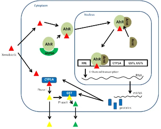
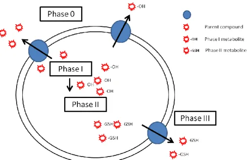
Figure 1.1 – Schematic representation of a typical ABC protein and characteristic amino acid sequences found in Nucleotide Binding Domains (NBDs) ... 6 Figure 1.2 – Predicted structures of MDR associated members of ABC transporters ... 9 Figure 1.3 - Schematic representation showing probable localization of ABC transporters in liver and intestine of fish, as determined in mammals. ... 17 Figure 1.4 - Schematic representation of the mechanisms involved in biotransformation of xenobiotic compounds in cells. ... 21 Figure 1.5 - Schematic representation of drugs/chemicals/xenobiotics induced stress response, leading to the activation of specific receptor-mediated gene expression of phase I enzymes, phase II enzymes, other stress enzymes and phase 0 and phase III transporters, which result in the enhancement of xenobiotic detoxification. ... 25 Figure 1.6 - Schematic representation of the possible cooperation of ABC efflux transporters (phase 0 and phase III) and biotransformation enzymes (phase I and phase II) in cellular detoxification (adapted from Bard et al., 2000)... Figure 1.7 - Biotransformation pathways of BaP. Source: IARC, 1983 ... 29 Figure 1.8 - Nile tilapia, Oreochromis niloticus, the model specie used in the present study. ... 30 Figure 2.1 - EROD activities in liver (a), gill (b) and intestine (c) of Nile Tilapia after waterborne exposure to BaP (Solvent, 10, 25, 50 and 100 µg/L). ... 61 Figure 2.2 - GST activities in liver (a), gill (b) and intestine (c) of Nile Tilapia after waterborne exposure to BaP (Solvent, 10, 25, 50 and 100 µg/L). ... 63 Figure 2.3 – BaP type metabolites in bile after waterborne exposure to BaP (a), first dietary exposure to BaP (b) and second dietary exposure to BaP (c). ... 64 Figure 2.4 – EROD activities in liver (a), gill (b) and intestine (c) of Nile tilapia after the first dietary exposure to BaP (Solvent, 1 and 10 µg of BaP/g of food). ... 66 Figure 2.5 – GST activities in liver (a), gill (b) and intestine (c) of Nile tilapia after the first dietary exposure to BaP (Solvent, 1 and 10 µg of BaP/g of food). ... 67 Figure 2.6 – EROD activities in liver (a), gill (b) and intestine (c) of Nile tilapia after the second dietary exposure to BaP (Solvent, 100 and 200 µg of BaP/g of food)... 68 Figure 2.7 – GST activities in liver (a), gill (b) and intestine (c) of Nile tilapia after the second dietary exposure to BaP (Solvent, 100 and 200 µg of BaP/g of food)... 69
xxii
Figure 3.1 - BaP (in percentage) in water at 0, 8 and 24 h after the addition of 10 and 100 µg of BaP L-1 of water... 94 Figure 3.2 - Free (a), total (b) and glucuronide (c) 3-OH-BaP levels in bile after waterborne exposure to BaP (10 and 100 µg L-1) for 7 and 14 d. ... 96 Figure 3.3 - Total 3-OH-BaP levels in plasma after waterborne exposure to BaP (10 and 100 µg L-1) for 7 and 14 d. ... 97 Figure 4.1 - Multiple alignments of the deduced amino acid sequences for
ABCB1b (a), ABCB11 (b), ABCC1 (c), ABCC2 (d) and ABCG2 (e) in N. tilapia with
other fish species and human. ... 122 Figure 4.2 – Predicted topologies of Nile tilapia partial ABCB1b (a), ABCB11 (b), ABCC1 (c), ABCC2 (d) and ABCG2 (e) proteins with membrane spanning domains (MSDs) and nucleotide binding domains (NBDs) according to the Polyphobius algorithm. ... 123 Figure 4.3 - Phylogenetic trees based on the multiple alignments (Clustal W) of closely related proteins from ABCB, ABCC and ABCG subfamilies found in other species. ... 125 Figure 4.4 – Relative mRNA expression of ABC transporters, CYP1A and GST α in Nile tilapia liver (a), gill (b) and proximal intestine (c). ... 127 Figure 4.5 – Relative mRNA expression of ABCB1b (a), ABCC1 (b), ABCC2 (c),
ABCG2 (d), CYP1A (e) and GSTα (f) in liver, gill and proximal intestine of animals
exposed to waterborne BaP. ... 129 Figure 4.6 - Relative mRNA expression of ABCB1b (a), ABCC1 (b), ABCC2 (c),
ABCG2 (d), CYP1A (e) and GSTα (f) in liver, gill and proximal intestine of animals
exposed to dietary BaP. ... 131 Figure 5.1 - Western blot of membrane vesicles from liver, gill and proximal intestine of Nile tilapia. ... 157 Figure 5.2 - Representative immunofluorescence results of liver, gill and proximal intestine in Nile tilapia probed with mammalian Pgp antibodies, C219 and C494. ... 161 Figure 5.3 - Representative immunoblots of Nile tilapia CYP1A protein in liver, gills and proximal intestine after waterborne exposures to BaP (10µg/L, 25µg/L and 50µg/L) for 7 and 14 days. ... 161 Figure 5.4 - Western blot results of CYP1A protein fold induction over control after Nile tilapia waterborne exposure to BaP in liver (a), gill (b) and proximal intestine (c). ... 163 Figure 5.5 - Representative CYP1A immunostaining in liver samples of Nile tilapia exposed to BaP. ... 167
xxiii
Figure 5.6 - Representative CYP1A immunostaining in gill samples of Nile tilapia exposed to BaP. ... 169 Figure 5.7 - Representative CYP1A immunostaining in proximal intestine of Nile tilapia exposed to BaP. ... 171 Figure 6.1 – Relative mRNA expression of ABCB1b (a), ABCC1 (b), CYP1A (c) and GSTα (d) during the first five stages (1-5) of embryonic development in O.
niloticus comprising the zygote and cleavage periods. ... 193
Figure 6.2 - Relative mRNA expression of ABCB1b (a), ABCC1 (b), CYP1A (c) and GSTα (d) during the blastula and gastrula periods of embryonic development in O. niloticus. ... 195 Figure 6.3 - Relative mRNA expression of ABCB1b (a), ABCC1 (b), CYP1A (c) and GSTα (d) during the last embryonic (segmentation to hatching) and larval periods (early and late larvae) in O. niloticus development. ... 196 Figure 6.4 – Relative mRNA expression of ABCB11 (a), ABCC2 (b) and ABCG2a (c) during the embryonic and larval periods (zygote to late larvae) in O. niloticus development. ... 197 Figure 6.5 – Schematic representation of the main results achieved in this study, showing the temporal pattern of mRNA expression for the genes in study (ABCB1b, ABCB11, ABCC1, ABCC2, ABCG2a, CYP1A and GSTα) in Nile tilapia developmental stages. Adapted from Kimmel et al. (1995) and Fujimura and Okada (2007). ... 201
xxiv
Tables index
Table 1.1– List of known ABC genes, functions and number of members found in human and zebrafish genomes ... 5 Table 1.2 – ABC transporters involved the efflux of toxic compounds, fish species where they were identified and tissue distribution pattern in mammals and fish. ... 15 Table 3.1 - Recoveries ± RSD, instrument linear dynamic ranges, determination coefficients (r2) and limits of detection (LOD) and quantification (LOQ) in μg L-1 (n=5) ... 92 Table 3.2 - EROD, GST and UGT activities in liver, intestine and gill of Nile tilapia after waterborne exposure to BaP (10 and 100 µg L-1) ... 98 Table 4.1 – Primer sequences, sequence lengths, annealing temperatures, GenBank accession numbers and homology percentages with other animal species of ABC transporters identified in Nile tilapia. ... 116 Table 4.2 – Primer sequences, amplicon lengths and efficiency of reaction, used in ABC transporters, CYP1A, GSTα and 18S rRNA gene expression quantification by qRT-PCR in Nile tilapia ... 118 Table 5.1 - Cellular localization of Pgp in liver, gill and proximal intestine of Nile tilápia exposed to different concentrations of waterborne BaP. ... 159 Table 5.2– Cellular localization of CYP1A in liver, gill and proximal intestine of Nile tilápia exposed to different concentrations of waterborne BaP. ... 164 Table 6.1 – Primer sequences, amplicon lengths and efficiency of reaction, used in ABC transporters, CYP1A, GSTα and 18S rRNA gene expression quantification by qRT-PCR in Nile tilapia ... 191
Chapter 1
3
1.
General Introduction
The aquatic ecosystems have unpaired importance for the maintenance and support of the overall environmental health, due to the enormous economic and aesthetic value of aquatic biodiversity. Humans have long depended on aquatic resources for food, medicines, materials, as well for recreational and commercial purposes, such as fishing and tourism. As a result declining levels of aquatic biodiversity, in both freshwater and marine environments, are recognized. Several factors, as species overexploitation, pollution from urban, industrial and agricultural areas, as well as habitat loss and alteration have been contributing to the situation of aquatic environment “health” decline. Although aquatic pollution has a long history, significant international laws to counter it were only enacted in the twentieth century. The aquatic environments were for many years, and still are, the ultimate destination for different type of pollutants from anthropogenic origins. Toxic heavy metals and nanomaterials resultant from industrial processes, persistent organic pollutants (POPs) from agricultural and industrial sources and polycyclic aromatic hydrocarbons (PAHs) mainly resultant from oil spills are among the sources of major concern regarding water pollution, due to their known toxic effects on the aquatic biota. More recently, a highly diverse group of bioactive chemicals, denominated pharmaceuticals and personal care products (PPCPs), have been receiving attention, since these compounds are continually introduced into the aquatic environment as complex mixtures via a number of routes, but primarily by both untreated and treated sewage (Daughton and Ternes, 1999). In order to survive in polluted environments, living organisms have well developed strategies of protection to the adverse effects of pollutants. Whenever possible, organisms avoid or escape from the polluted areas; additionally, chronically exposed organisms use detoxification mechanisms in order to reduce the toxic effects of pollutants. These mechanisms include the activity of specific proteins to preclude the permanence of toxic compounds or their metabolites in the cells - some members of the ATP-Binding Cassette (ABC) superfamily - and enzymatic systems to transform the chemicals into a more easily excreted form - phase I and phase II biotransformation enzymes. Thus, the knowledge of the functionality of these detoxification pathways is of critical importance, to maintain the balance of nature and support the availability of the aquatic resources for future generations.
The main focuses of this PhD thesis are the detoxification mechanisms adopted by fish species, namely the ecotoxicologically relevant ABC efflux transporters and
4
biotransformation enzymes of phase I and phase II. Recent studies have raised the possibility of a coordinated action between these three groups of proteins, resulting in a powerful and effective mechanism of cellular detoxification, and the cooperation of these systems will be the main focus of this dissertation. To achieve this goal, we have developed in vivo assays of exposure of the freshwater fish Nile tilapia (Oreochromis
niloticus) to Benzo(a)pyrene (BaP), a toxic polycyclic aromatic hydrocarbon (PAH) that
is a ubiquitous contaminant of the aquatic environments, and whose carcinogenic and mutagenic effects to the aquatic biota are well recognized. The presence and functionality of ABC transporters and biotransformation enzymes was subsequently evaluated in Nile tilapia barrier tissues (liver, gills and intestine) at gene, protein and activity levels, in order to assess the cooperation between these groups of proteins in the detoxification of this pollutant.
1.1.
The superfamily of ABC efflux transporters
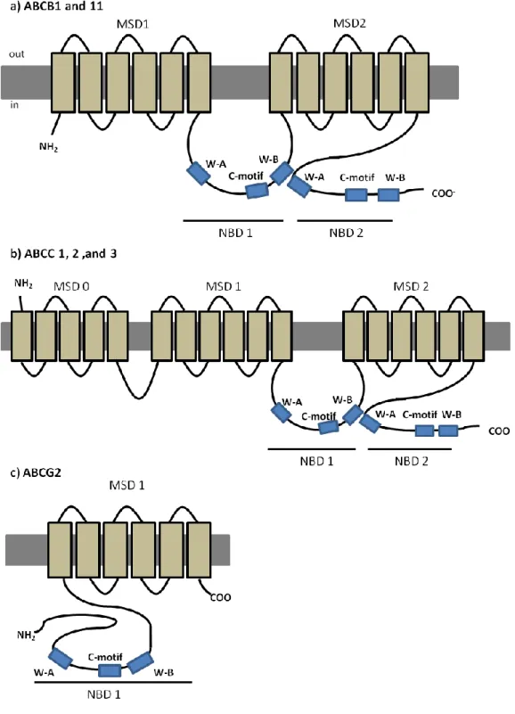
The ABC genes represent the largest family of transmembrane proteins encoded in the human genome. These proteins bind to ATP and use that energy to drive the transport of a wide variety of molecules across cellular membranes (table 1.1) (Higgins, 1992; Dean et al., 2001). So far, 56 members of the ABC family have been described, including 48 human ABC genes and 9 additional genes found in other animal species. From the 56 genes, 68% are present in all vertebrate genomes, suggesting that theirs proteins´ structures and functions have been largely conserved throughout the evolution of vertebrate species (Dean and Annilo, 2005). Based on the sequence and the organization of the ATP-binding domains, also known as nucleotide binding domains (NBDs), ABC proteins were grouped into eight subfamilies in eukaryotes (A– H), with seven of these (A–G) present in the human genome (Dean and Annilo, 2005), as shown in table 1.1. The NBDs are located in the cytoplasm and transfer the necessary energy to transport the substrate across the cellular membrane. These domains contain characteristic motifs with specific amino acid sequences (Walker A, Walker B and C-motif) described in all ABC proteins (Walker et al., 1982; Hyde et al., 1990) (figure 1.1b).
5
Table 1.1– List of known ABC genes, functions and number of members found in human and zebrafish genomes
Subfamily Members Functions Human Zebrafish*
ABCA ABCA1 to ABCA13 Cholesterol efflux, phosphatidil choline efflux, N-retinylidiene-PE efflux 12 members 7 members ABCB ABCB1 to ABCB11 Peptide transport; iron transport; Fe/S cluster transport; bile salt transport; xenobiotics transport 11 members 9 members ABCC ABCC1 to ABCC13 Organic anion efflux, nucleoside transport, chloride ion channel, sulfonylurea receptor, potassium channel regulation,
xenobiotics transport
13 members 11 members
ABCD ABCD1 to ABCD4 Very long chain fatty acids transport regulation 4 members 4 members
ABCE ABCE1 Elongation factor complex 1 member 1 member
ABCF ABCF1 to ABCF3 Unknown function 3 members 3 members
ABCG ABCG1 to ABCG5 Cholesterol transport, sterol transport, toxin transport 5 members 5 members
ABCH ABCH1 Unknown function no members 1 member
6
Typically, a functional protein contains two NBDs and two membrane spawning domains (MSDs), the latter being composed by 6-10 membrane spawning α-helices that confer the substrate specificity (figure 1.1a) (Locher 2009). Eukaryotic ABC proteins are organized either as full transporters (containing two NBDs and two MSDs), or as half transporters (containing one NBD and one MSD), that have to form homo- or heterodimers in order to constitute a functional protein (Dean et al., 2001). Some variation exists in protein structure in the different subfamilies, as it will be described further ahead, but a high degree of structural and sequence homology is shared among all ABC transporter proteins (Higgins, 2007). The linker region is a central sequence that connects the two homologous halves of the protein, and plays an important role due to its flexible secondary structure that coordinates the functioning of both halves, which is required for the proper interaction of the two ATP binding sites. Evidence exist that, for the efficient ATP hydrolysis, the two NBDs have to interact and form a sandwich dimer, so that C-motif of one NBD comes in contact with Walker-A of the other NBD, forming a nucleotide-binding pocket (Lee et al., 2008).
Figure 1.1 – Schematic representation of a typical ABC protein and characteristic amino acid sequences found in Nucleotide Binding Domains (NBDs)
a) lipidic bilayer is shown in grey, membrane spawning domains (MSDs) in light brown and nucleotide binding domains (NBDs) in blue ( W-A - Walker A and W-B - Walker B), dashed line represents the linker region; b) aminoacid (aa) sequences characteristic from the conserved domains Walker A (x represents any aa), C-motif and Walker-B (ф represents hydrophobic residues). Adapted from Dean et al. 2001.
7
1.2.
Multidrug resistance (MDR) associated ABC transporters
Resistance to multiple anticancer agents is a major impediment for the successful treatment of many forms of malignant tumours. Juliano and Ling (1976) described a multidrug resistance (MDR) phenomenon, found in tumor cell lines of mammals, as the result of low intracellular accumulation of anti-cancer drugs, and related it with the overexpression of a transmembranar protein, responsible for an ATP-dependent efflux of those drugs into the extracellular medium. This protein denominated as Permeability glycoprotein (Pgp) and encoded by the ABCB1 gene was, for that reason, the first ABC transporter to gain importance and later related to cellular detoxification. From this point, several other members of the ABC transporter efflux family have been identified as capable of transporting endo- or xenobiotic compounds, including members of the ABCC and ABCG subfamilies (Cole et al., 1992; Doyle et al., 1998). Due to their ecotoxicological role, these three subfamilies of transporters will be addressed in higher detail in the following sections.
1.2.1. Subfamily ABCB
1.2.1.1. ABCB1 – P-glycoprotein (Pgp)
The best characterized ABC transporter is Pgp (subfamily B, member 1: ABCB1; MDR1). In humans, Pgp is encoded by two different isoforms of the gene, MDR1 and
MDR3, and the class 1 has been implicated in drug resistance whereas the function of class
III isoform is still unknown (Georges et al., 1990). ABCB1 (MDR1) was the first eukaryotic ABC member identified in result of its implication in MDR of cancer cells to chemotherapy (Gottesman and Ling, 2006). Further evidence of its MDR abilities included decreased drug accumulation in cells transfected with ABCB1 gene (Ueda et al., 1987), and increased drug accumulation in gene knockouts organisms, compared to the wild-type organism (Schinkel et al., 1994). In humans, ABCB1 is a 170 - 180 kDa protein containing ~1280 amino acids, with a predicted four-domain structure, typical of most eukaryotic ABC transporters, with two NBDs each preceded by a MSD composed of six transmembrane (TM) helices (Loo and Clarke, 1999) (figure 1.2a). MDR provided by Pgp is a consequence of its remarkable non-specificity with respect to the substrates. Several researchers have focused their attention on the understanding of the promiscuity of this transporter regarding its substrates, and
8
have indicated common characteristics among them, such as moderate hydrophobicity, small size and positively charged or neutral domains, and include natural products, chemotherapeutic drugs or steroids (reviewed in Schinkel and Jonker, 2003). Pgp can also interact with modulators that are able to reverse MDR by blocking or saturating Pgp binding locations, called chemosensitizers (described in section 1.3).
Besides being expressed in cancerous tissues ABCB1 is also expressed in normal tissues, such as kidney, intestine, lung, brain, placenta, adrenal cortex, testis, uterus, lymphocytes and hematopoietic cells of mammals, in most cases adopting an apical localization in the cells (reviewed in Szakács et al., 2008). Due to this cellular localization determined in mammals, it is believed that ABCB1 functions in three main areas: 1) limiting the drug entry into the body after oral drug or toxin administration, due to its expression in the apical membrane of enterocytes; 2) once the xenobiotic has reached the blood circulation, ABCB1 promotes drug elimination into bile and urine, as a result of its expression in the canalicular membrane of hepatocytes and luminal membrane of proximal tubule cells in the kidneys, respectively; and, 3) once a xenobiotic has reached the systemic blood-circulation, Pgp limits drug penetration into sensitive tissues, like the blood-brain-barrier (BBB) (Fromm, 2004) where its localized in both luminal and abluminal membranes of capillary endothelial cells, pericytes and astrocytes (Bendayan et al., 2006). These different localizations of ABCB1 in barrier tissues strongly support the role of this transporter as a key player in the cellular defence mechanisms, emphasizing its ecotoxicological relevance.
Besides mammals, highly conserved MDR genes have been described in animals from diverse taxa including nematode worms (Broeks et al., 1995), fruit fly (Vaché et al., 2006), plants (Wang et al., 1996) and bacteria (van Veen et al., 1998).
9
Figure 1.2 – Predicted structures of MDR associated members of ABC transporters
a) predicted structure of ABCB1 and ABCB11; b) predicted structure of “long-chain” ABCC proteins, ABCC1, 2 and 3; c) predicted structure of ABCG2.
10 1.2.1.2. ABCB11 – Spgp or BSEP
Sister of P-glycoprotein (Spgp, ABCB11, BSEP) was first cloned from porcine liver (Childs et al., 1995), and named for its close sequence homology with Pgp. It follows the same predicted structure as ABCB1 (figure 1.2a). Recently, this transporter has become more widely known as the bile salt export pump (BSEP), since studies in rats showed predominant expression in liver, canalicular localization, and high affinity to transport primary and secondary bile salts (Gerloff et al., 1998). ABCB11 is thus positioned to mediate active, ATP dependent, bile acid secretion across the canalicular membrane of hepatocytes, limiting intracellular bile acid concentrations and maintaining the enterohepatic circulation of bile acids. Conclusive evidence that BSEP is indeed the bile salt export pump in liver came from a study in which mutations in the human ABCB11 gene resulted in type 2 progressive familial intrahepatic cholestasis (PFIC2), a disease characterized by an interruption in hepatic bile salt secretion (Strautnieks et al., 1998). Despite its predominance in liver, ABCB11 was also found in other porcine tissues, such as large and small intestinal mucosa and in brain (Török et al., 1999). This suggests that ABCB11 may perform additional, extrahepatic functions, possibly limiting other tissues exposure to bile salts. As well as its endogenous role as a hepatic bile salt export pump, studies in mammals cell lines transfected with murine ABCB11 showed that ABCB11 was capable of transporting some Pgp substrates (vinblastine and calcein-acetoxymethyl ester), although it was unable to efflux others (rhodamine 123, vincristine, daunorubicin, paclitaxel, and digoxin) (Lecureur et al., 2000). Thus the role of ABCB11 in drug disposition, if any, is considerably more limited than that of its close relative, Pgp.
1.2.2. Subfamily ABCC (MRPs – Multiresistance associated proteins)
After the discovery of ABCB1, the study on cancer cells displaying MDR phenotype not associated with ABCB1 expression, led to the discovery of ABCC1, the founding member of the ABCC subfamily (Cole et al., 1992). So far, this subfamily includes a total of 13 members, most of which are active ATP-dependent membrane transporters for organic anions of therapeutic compounds (Honorat et al., 2009). Among its members, at least five (ABCC1, ABCC2, ABCC3, ABCC4 and ABC5) are potentially involved in mediating drug resistance (Cole et al., 1992; Kool et al., 1997; Evers et al., 1998). At present, there are no high-resolution structural data available on any eukaryotic ABCC transporter, but considering predicted membrane topologies, phylogenetic position and domain
11
arrangements, members of the ABCC subfamily can fall in one of two different subclasses, “short” and “long” (Kast and Gros, 1997; Bakos and Homolya, 2007; Honorat et al., 2009). The so called “long” ABCC transporters (ABCC1, 2, 3 and 6) present an additional N- terminal MSD (MSD0), of approximately 250 amino acids, a unique feature of these specific transporters (figure 1.2b) in comparison to the “short” transporters. ABCCs are ~190kDa proteins, and share only 14-25% amino acid identity with ABCB proteins (Cole et al., 1992; Keppler and Konig, 1997). From the ABCC subfamily, ABCC1 and ABCC2 are the best characterized transporters with existing evidence from animal models to have a role in tissue defence, while other members like ABCC3, 4 and 5, are far less studied and characterized. The toxicological relevance of ABCC1 and ABCC2 will be evaluated in this dissertation. ABCC2 gene was identified based on its similarity to ABCC1 and absence of its expression of homozygously ABCC2-deficient rats and humans (Mayer et al., 1995; Keppler and KÖNig, 2000). The absence of the ABCC2 gene in hepatocytes canalicular membrane causes the Dubin–Johnson syndrome (Konig et al., 1999), and affected individuals suffer from a conjugated hyperbilirubinemia, since ABCC2 normally mediates the hepatobiliary excretion of bilirubin (König et al., 1999). As for ABCB1, ABCC1 and ABCC2 are also expressed in non-malignant tissues. In mammals, ABCC1 shows a ubiquitous expression throughout the different tissues, with relatively high levels found in kidney, lung, testis, skeletal muscle, peripheral blood mononuclear cells, while relatively low levels were found in liver (Cole et al., 1992; Flens et al., 1996; Stride et al., 1996). Similarly, ABCC2 has also been found in tissues important for the pharmacokinetics of substrate drugs, namely liver, kidney, placenta, lungs, intestine and BBB (for a review see Schinkel and Jonker, 2003). Although without a complete overlap, many similarities exist between the spectrum of compounds transported by ABCC1 and ABCC2, which are mainly composed by amphipathic anionic drugs and endogenous compounds, encompassing GSH-, glucoronide- and sulphate- conjugates (reviewed in Schinkel and Jonker, 2003). In most tissues, ABCC1 efflux transporter is localized in the basolateral surface of the cells, which, in certain tissues, results in the efflux of its substrates into the blood (Evers et al., 1996). In contrast to ABCC1, and similarly to ABCB1 and ABCB11, ABCC2 is localized in the apical membrane of the cells in which it is expressed (Evers et al., 1998).
12
1.2.3. Subfamily ABCG – ABCG2, BCRP, MXR or ABCP
The second member of the ABCG subfamily, ABCG2, is a ~72kDa efflux transporter, whose overexpression in permanent cell-lines has been associated with high levels of resistance to a variety of anticancer drugs, such as mitoxantrone, doxorubicin, and daunorubicin, without evidence of expression of the well-characterized genes for ABCB1 or
ABCCs therefore contributing to a MDR phenotype. Moreover reduced accumulation of
such drugs could be reversed by incubation of the cells in ATP-depleting conditions, indicating the presence of an ATP-dependent transporter (Ross et al., 1999; Bates et al., 2001; Allen and Schinkel, 2002). This protein is also known as breast resistance associated protein (BCRP) (Doyle et al., 1998), mitoxantrone-resistance protein (MXR) (Miyake et al., 1999) or placenta-specific ABC protein (ABCP) (Allikmets et al., 1998) since it was cloned independently by 3 different groups. Members of the ABCG subfamily have a unique domain organization; unlike the remaining subfamilies, these are half-transporters, composed by one single NBD followed by one MSD (figure 1.2c). In addition, they also present a unique protein configuration, in which the NBD precedes the MSD, whereas ABCBs and ABCCs have an opposite domain arrangement, that is, the MSD is followed by the NBD (figure 1.2c). There are increasing evidence to suggest that these proteins may operate either as homodimers or heterodimers (Graf et al., 2003; Xu et al., 2004). ABCG2 is believed to function probably as a homodimer (Doyle et al., 1998; Miyake et al., 1999; Kage et al., 2002) or homotetramer (Xu et al., 2004).
Functional characterization studies have demonstrated that ABCG2 can transport a wide range of substrates, from chemotherapeutic agents to organic anion conjugates (reviewed in Mao, 2005). Moreover, it seems that ABCG2 has higher affinity to transport sulphated conjugates of steroids and xenobiotics over GSH and glucoronide metabolites (Chen et al., 2003). High levels of ABCG2 have been found in placenta, brain, prostate, intestine, liver and ovary, mostly in an apical localization (Rocchi et al., 2000; Scheffer et al., 2000; Maliepaard et al., 2001).This tissue localization is consistent with the ability of ABCG2 to function as a protective efflux pump, limiting absorption of drugs and increasing elimination of its substrates.
13
1.3.
From MDR to MXR - ABC efflux transporters in aquatic organisms
Aquatic organisms are able to survive and thrive in heavily polluted environments, showing surprisingly low accumulation of pollutants in body tissues (Kurelec and Pivčević, 1989). Kurelec and coworkers were the first to demonstrate that aquatic organisms adopt strategies of xenobiotic transport, in order to improve adaptation to pollutants in their habitats. To this phenomenon of resistance, Kurelec coined the term of multixenobiotic resistance (MXR) (Kurelec, 1992), and identified its biochemical basis as similar to the one adjacent to the MDR phenotype. The presence of a drug transporter resembling Pgp (ABCB1) sensitive to verapamil and trypsin (known inhibitors of human Pgp) was described in two bivalves species (Anodonta cygnea and Mytilus galloprovinciallis) (Kurelec and Pivčević, 1989, 1991). This study was the starting point for the identification and characterization of the MXR phenotype in several other species of aquatic organisms, such as sponges (Kurelec et al., 1992), molluscs (McFadzen et al., 2000; Minier et al., 2002; Smital et al., 2003; Luckenbach and Epel, 2008; Faria et al., 2011), crabs (Kohler et al., 1998) and sea urchins (Toomey and Epel, 1993; Hamdoun et al., 2002). In fish, the presence of ABC efflux transporters have been described for an increasing number of species, such as winter flounder (Chan et al., 1992), rock cod (Zucchi et al., 2010), rainbow trout (Zaja et al., 2008; Fischer et al., 2010; Loncar et al., 2010), zebrafish (Long et al., 2011a; Long et al., 2011b), mullet (Diaz de Cerio et al., 2012) and killifish (Paetzold et al., 2009) (table 1.2).
Identification of ABC transporters and characterization of the MXR mechanism has been achieved by the use of several detection methods, including quantitative or semi-quantitative reverse transcription polymerase chain reaction (qRT-PCR or RT-PCR) and in
situ hybridization to evaluate mRNA expression of genes, immunochemical techniques for
protein detection applying mammals monoclonal antibodies to ABCB1 (western blotting or immunohistochemistry), northern-blotting and activity assays (through the measurement of efflux or accumulation of fluorescent model substrates).
Most studies aiming to determine tissue distribution pattern of ABC transporters in aquatic organisms have been directed to Pgp by the use of the mammalian anti-Pgp monoclonal antibody (mAb) C219. This mAb recognizes an epitope common to all known Pgps (in human MDR1 and MDR3) and also to Spgp (ABCB11) (Georges et al., 1990; Childs et al., 1995; van den Elsen et al., 1999). After probing with the mammalian mAb C219, an immunologically related to the mammalian MDR protein was identified in embryos and gills of a few aquatic invertebrates (Toomey and Epel, 1993; Cornwall et al., 1995). In
14
fish, a positive reaction was seen in the hepatic bile canaliculi (Kleinow et al., 2000; Doi et al., 2001; Bard et al., 2002a; Bard et al., 2002b; Klobučar et al., 2010), apical membrane of enterocytes (Kleinow et al., 2000; Doi et al., 2001; Bard et al., 2002a), kidney (Kleinow et al., 2000) and in endothelial capillaries of brain (Miller et al., 2002). A negative reaction of the mAb C219 was patent in gills, kidney, gonad, brain, spleen, heart of killifish and blenny (Bard et al., 2002a; Bard et al., 2002b). In summary, the suggested Pgp localization in fish reveals a pattern similar to that described in mammals, with Pgp present in epithelial tissues involved in secretion, absorption or serving a barrier function. However, due to the known cross-reactivity of the mAb C219 with ABCB11 (Childs et al., 1995), results from these studies should be addressed with care, since until the development of fish-specific Pgp probes, ABCB1 definitive tissue localization cannot be demonstrated in fish (Sturm and Segner 2005).
For other ABC efflux transporters no fish-functional antibodies are available, thus information on their distribution pattern is quite limited and restricted to the mRNA expression among the various fish tissues. As in mammals, ABCB11 was found to be almost exclusively expressed in trout liver, with low expression in proximal intestine, and very low expression in other tissues as gills, brain, gonads and kidney (Loncar et al., 2010), supporting its role in the efflux of bile salts from hepatocytes into the bile. In trout tissues, low ABCC1 expression was found, and kidney was the only tissue showing notable ABCC1 expression while significant expression levels of ABCC2 in major detoxification tissues were reported (Loncar et al., 2010). High ABCC2 expression was also previously reported in kidney, intestine and liver of little skate (Cai et al. 2003) and in apical membrane of proximal kidney tubule of killifish (Miller et al., 2007). Regarding ABCG2, Loncar and coworkers (2010) have examined the tissue distribution pattern of this protein in trout tissues, and found high expression levels in gonads and moderate expression in distal part of intestine, kidney and brain. Relatively high expression of ABCG2 was reported in trout liver and primary hepatocytes (Zaja et al., 2008) (table 1.2).
Based on these results and on mammal studies, investigators believe that, in aquatic organisms, ABC eflux transporters should follow a similar distribution pattern and cellular localization, with expression in tissues involved in secretion, absorption or serving a barrier function, like liver and intestine (figure 1.3). Thus, ABCB1, ABCB11, ABCC2 and ABCG2 should be localized in the apical membrane of polarized cells, pumping substrates into the intestinal lumen or bile canaliculi, while ABCC1 localization in basolateral membrane results in the export of its substrates into the blood. Additionally, ABCB1 should efflux mostly unmodified compounds, while ABCCs and ABCG2 should efflux organic anions conjugated by phase II enzymes (figure 1.3) (reviewed in Epel et al., 2008).
15
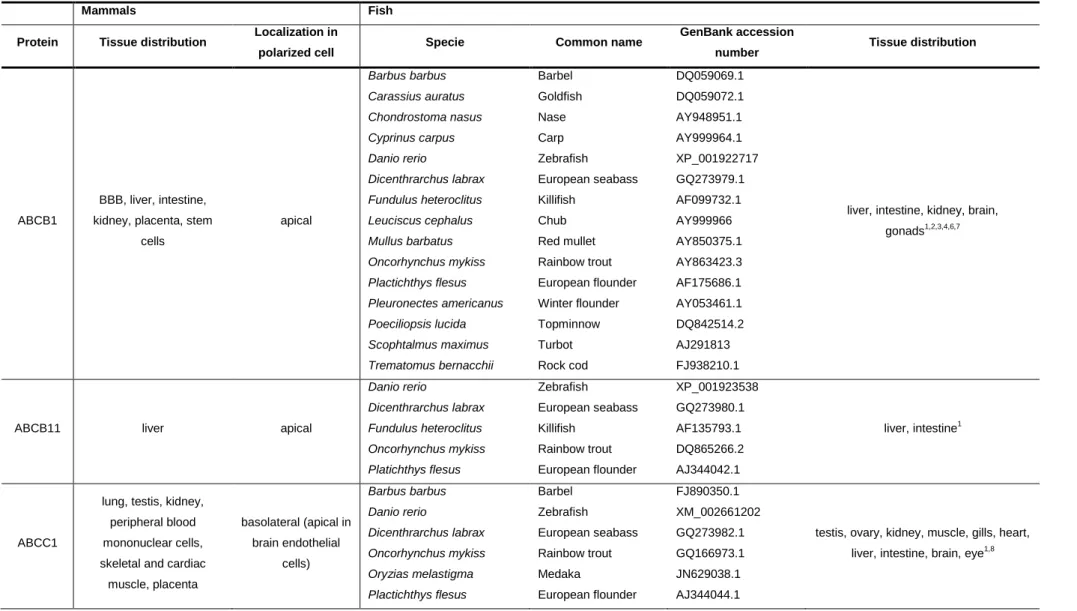
Table 1.2 – ABC transporters involved the efflux of toxic compounds, fish species where they were identified and tissue distribution pattern in mammals and fish.
Mammals Fish
Protein Tissue distribution Localization in
polarized cell Specie Common name
GenBank accession
number Tissue distribution
ABCB1
BBB, liver, intestine, kidney, placenta, stem
cells apical Barbus barbus Carassius auratus Chondrostoma nasus Cyprinus carpus Danio rerio Dicenthrarchus labrax Fundulus heteroclitus Leuciscus cephalus Mullus barbatus Oncorhynchus mykiss Plactichthys flesus Pleuronectes americanus Poeciliopsis lucida Scophtalmus maximus Trematomus bernacchii Barbel Goldfish Nase Carp Zebrafish European seabass Killifish Chub Red mullet Rainbow trout European flounder Winter flounder Topminnow Turbot Rock cod DQ059069.1 DQ059072.1 AY948951.1 AY999964.1 XP_001922717 GQ273979.1 AF099732.1 AY999966 AY850375.1 AY863423.3 AF175686.1 AY053461.1 DQ842514.2 AJ291813 FJ938210.1
liver, intestine, kidney, brain, gonads1,2,3,4,6,7
ABCB11 liver apical
Danio rerio Dicenthrarchus labrax Fundulus heteroclitus Oncorhynchus mykiss Platichthys flesus Zebrafish European seabass Killifish Rainbow trout European flounder XP_001923538 GQ273980.1 AF135793.1 DQ865266.2 AJ344042.1 liver, intestine1 ABCC1
lung, testis, kidney, peripheral blood mononuclear cells, skeletal and cardiac
muscle, placenta basolateral (apical in brain endothelial cells) Barbus barbus Danio rerio Dicenthrarchus labrax Oncorhynchus mykiss Oryzias melastigma Plactichthys flesus Barbel Zebrafish European seabass Rainbow trout Medaka European flounder FJ890350.1 XM_002661202 GQ273982.1 GQ166973.1 JN629038.1 AJ344044.1
testis, ovary, kidney, muscle, gills, heart, liver, intestine, brain, eye1,8
16 Poeciliopsis lucida Trematomus bernacchii Topminnow Rock cod HM102361.1 FJ938212.1
ABCC2 BBB, liver, intestine,
kidney, placenta, lung apical
Carassius auratus Chondrostoma nasus Cyprinus carpio Danio rerio Dicenthrarchus labrax Leuciscus cephalus Mullus barbus Oncorhynchus mykiss Plactichthys flesus Poeciliopsis lucida Trematomus bernacchii Raja erinácea Goldfish Nase Carp Zebrafish European seabass Chub Barbel Rainbow trout European flounder Topminnow Rock cod Little skate FJ890349.1 AY948950 AY679169 NM_200589.1 GQ273983.1 FJ890348.1 AY275434.1 NM_001124655.1 AJ344045.1 HM102360.1 FJ938211.1 AF486830
liver, kidney, intestine, brain, muscle1,9,10,11
ABCG2
BBB, placenta, liver, intestine, breast, stem
cells apical Chelon labrosus Dicenthrarchus labrax Oncorhynchus mykiss Poeciliopsis lucida Salmo salar Mullet European seabass Rainbow trout Topminnow Atlantic salmon HM467811.1 GQ273981.1 EU163724.1 HM102358.1 NM_011736655.1
liver, kidney, gonads, intestine, gills1
1(Loncar et al., 2010), 2 (Kleinow et al., 2000), 3(Doi et al., 2001), 4(Bard et al., 2002a), 5(Bard et al., 2002b), 6(Klobučar et al., 2010), 7(Miller et al., 2002), 8(Long et al., 2011a), 9(Long et al., 2011b), 10(Miller et al., 2007), 11 (Cai et al., 2003)
17
Figure 1.3 - Schematic representation showing probable localization of ABC transporters in liver and intestine of fish, as determined in mammals.
(a) liver hepatocytes, b) enterocytes. Parent xenobiotic compounds, phase I metabolites and phase II metabolites are shown as red, yellow and green circles, respectively. Adapted from Schinkel and Jonker 2003, Brand et al., 2006.
The bulk of the studies conducted in aquatic organisms, on the interaction of environmental contaminants with ABC efflux transporters have been mostly performed on Pgp. Some compounds, as Pgp model reversal agents (verapamil and/or cyclosporine A) and xenobiotics (like the insecticide malathion) were shown to inhibit Pgp activity in fishes, resulting in an increase of the bioaccumulation of toxic compounds like 2-aminoanthracene, hydrocarbon-rich Diesel-2 oil, and/or model Pgp substrates like rhodamine B (Kurelec et al., 1992; Kurelec, 1995; Smital and Sauerborn, 2002). Other studies have demonstrated the ability of some xenobiotics to induce Pgp expression in fish, such as cadmium (Zucchi et al., 2010), the organophosphate insecticide chloropyrifos and the carcinogen N-nitrosodiethylamine (Albertus and Laine, 2001). Similarly, resistance-killifish populations from highly polluted sites also demonstrated enhanced Pgp expression (Cooper et al., 1999; Bard et al., 2002a) suggesting a protective role of Pgp. Nevertheless, in another study performed in killifish from a heavily polluted site with polycyclic aromatic hydrocarbons (PAHs), polychlorinated biphenyls (PCBs) and heavy metals, Paetzold and coworkers (2009) found no up-regulation of hepatic ABCB1 transcripts. Moreover, after in vivo exposures of fishes to different xenobiotics, such as prochloraz, nonylphenol diethoxylate, ß-naphthoflavone, BaP and 3,4,3´,4´-tetrachlorobiphenyl no changes were seen in the levels of hepatic and/or intestinal Pgp (Doi et al., 2001; Sturm et al., 2001).