FACULDADE DE MEDICINA VETERINÁRIA
CLINICAL AND MOLECULAR CHARACTERIZATION OF FELINE MAMMARY CARCINOMAS OVEREXPRESSING HER2 PROTO-ONCOGENE (FMC-HER2+) – NEW
STRATEGIES FOR EFFECTIVE DIAGNOSTIC AND CANCER THERAPY
Maria João da Costa Soares da Silva
Orientador(es): Professor Doutor Fernando António da Costa Ferreira Professor Doutor Jorge Manuel de Jesus Correia
Tese especialmente elaborada para obtenção do grau de Doutor em Ciências Veterinárias na Especialidade de Ciências Biológicas e Biomédicas
Júri:
Presidente: Doutor Rui Manuel de Vasconcelos e Horta Caldeira Vogais:
- Doutora Maria de Fátima Rodrigues Moutinho Gartner - Doutora Raquel Maria Garcia dos Santos Chaves - Doutor António José de Freitas Duarte
- Doutor Rui Pedro Brás Martins Faísca - Doutor Jorge Manuel de Jesus Correia - Doutor Fernando António da Costa Ferreira
Para os meus pais, Maria Custódia e César Soares por serem os meus maiores mentores e o meu maior e mais bonito exemplo de Vida
Para o Tiago Silva, porque és o meu anjo aqui na Terra
Para o meu irmão, Pedro Soares, porque te preocupas
―Cancer, unlike politics and religion, is not a topic of controversy. No one is for it. Cancer is not another word for death. Neither is it a single disease for which there is one
cure. Instead, it takes many forms, and each form responds differently to treatment.‖
Samantha Mooney In ―A snowflake in my hand‖
A Vida tem me abençoado com várias e maravilhosas oportunidades. A possibilidade de fazer um doutoramento, num tema que me diz tanto, é um privilégio que foi apenas possível com o trabalho e dedicação de várias pessoas. Entre elas, o Professor Doutor Fernando Ferreira teve um papel fundamental. Por ter idealizado este projecto, ter acreditado nele e por me ter dado a oportunidade de realizar o meu doutoramento sob a sua orientação, o meu Muito Obrigada. Obrigada por tudo o que me ensinou, por todas as oportunidades que me proporcionou, por me ter contagiado com toda a sua dedicação, preseverança e capacidade de trabalho. Sou uma pessoa indubitavelmente mais rica, pessoal e profissionalmente, devido ao seu exemplo e ensinamentos. Obrigada.
Ao Professor Doutor Jorge Correia, o meu Muito Obrigada por ter aceite ser meu co-orientador. Obrigada pelo apoio que me deu ao longo destes anos, pela paciência e carinho com que sempre acolheu as minhas dúvidas e os meus pedidos. Por todo o conhecimento que me transmitiu, profissional e pessoalmente, o meu mais profundo Obrigada.
Agradeço à Professora Doutora Conceição Peleteiro a possibilidade de trabalhar no Departamento de Anatomia Patológica da Faculdade de Medicina Veterinária da Universidade de Lisboa (FMV-ULisboa). A sua experiência, capacidade de trabalho e alegria são uma verdadeira inspiração para todos os que a rodeiam. Obrigada por ter tido o privilégio de contar com a sua ajuda sempre que precisei.
O meu agradecimento estende-se ainda a todos os funcionários do Departamento de Anatomia Patológica da FMV-ULisboa, que me acolheram sempre com muito carinho. Entre eles, devo um agradecimento especial à Mestre Sandra Carvalho, que esteve sempre disponível para ajudar o grupo e a mim particularmente. Por todas as horas perdidas ao micrótomo, por todos os conselhos e conhecimentos partilhados, o meu Muito Obrigada.
Entre as várias pessoas que contribuiram para a realização desta tese, não posso deixar de destacar a Doutora Margarida Simões. Caminhámos juntas durante esta etapa e a sua generosidade, sinceridade, amizade, carinho e, sobretudo, o seu exemplo enriqueceram-me e fizeram de mim uma pessoa melhor. Obrigada por toda a ajuda, por toda a dedicação por todo o conhecimento, enfim, Obrigada por tudo.
À Professora Doutora Luísa Mendes Jorge o meu obrigada por toda a ajuda que sempre nos proporcionou.
Ao Professor Doutor João Ferreira, do Instituto de Medicina Molecular (IMM) da Universidade de Lisboa e à Professora Doutora Luísa Mateus da FMV-ULisboa, que gentilmente cederam os seus equipamentos para que pudesse realizar ensaios em cultura de células. À Doutora Vukosava Mills Torres, do Instituto Ricardo Jorge, pela cedência da linha celular SKBR3 e ao Professor Doutor Nobuo Sasaki e Professor Doutor Takayuki Nakagawa, da Graduate School of Frontier Sciences, Universidade de Tóquio, Japão pela cedência das linhas celulares felinas.
À Doutora Elizabete Silva, da FMV-ULisboa, ao Dr. Bruno D’Almeida, da IZASA Portugal, ao Dr. Pedro Gonçalo Rodrigues, do Hospital de Santa Maria, ao Dr. José Cabeçadas e ao Dr. João Matos, do Instituto Português de Oncologia de Lisboa, agradeço toda a ajuda e disponibilidade durante a otimização dos protocolos para detecção do HER2 felino. Especialmente ao Dr. Pedro Gonçalo Rodrigues, obrigada por todo o conhecimento transmitido, por toda a paciência e preseverança.
deram na aquisição de imagens de microscopia de fluorescência.
Ao Departamento de Genética e Biotecnologia da Universidade de Trás dos Montes e Alto Douro (UTAD) o meu profundo agradecimento pela maneira como me acolheram, pela vossa disponibilidade e amabilidade. À Professora Doutora Raquel Chaves e à Professora Doutora Filomena Adega obrigada pela oportunidade de aprender num grupo tão rico como o vosso. À Doutora Susana Meles e à Dra. Daniela Ferreira fica o meu agradecimento por todo o carinho e ajuda que me deram.
Sendo um trabalho prospectivo, este doutoramento exigiu a generosa participação de várias clínicas e hospitais, que colaboraram connosco e que foram fundamentais para a realização de todos os trabalhos. Assim, o meu sincero obrigado para o Professor Doutor António Ferreira, que autorizou a recolha e acompanhamento dos casos clínicos no Hospital Escolar da FMV-ULisboa. A todos os médicos-veterinários, enfermeiros e auxiliares do Hospital Escolar, obrigada porque sempre me abriram as portas e sempre estiveram disponíveis para mim. Em particular, quero agradecer ao Dr. Rodrigo Bom e à Dra. Ana Murta do Departamento de cirurgia e ao Dr. Óscar Gamboa, Dr. António Almeida e Professora Doutora Sandra Jesus, do Departamento de Imagiologia do Hospital, obrigada por todo o esforço e paciência que tiveram para comigo.
Obrigada a todos os auxiliares e médicos-veterinários da Clínica Veterinária Zoomédica, à Dra. Ana Mota, ao Dr. Tiago Rafael e ao Dr. Manuel Mestre em particular, por todo o conhecimento que partilhou comigo e por ter mantido sempre as portas abertas para mim, o meu sincero obrigado. À Dra. Telma Almeida e à Dra. Verónica Azevedo do Hospital Sul do Tejo, à Dra. Rafaela Lalanda e ao Dr. Miguel Caninhas da Clínica Veterinária Mvet, à Dra. Ana Filipa Dinis e à Dra. Carolina Guardado do Hospital da Estefânia o meu obrigado.
Finalmente, quero também agradecer à Dra. Ana Mafalda Lage, da Clínica Veterinária Villa Animal, por toda a ajuda que sempre me deu, primeiro na Zoomédica e depois na Villa Animal. Por tudo, obrigada.
A todos os meus colegas do CIISA, com quem aprendi e que me ajudaram a crescer enquanto pessoa e enquanto investigadora, obrigada: à Andreia Valença, que nunca te queixaste das músicas e constantes correrias à volta do gabinete, ao David Ramilo, Marcos Santos e Cátia Marques, que me acolheram sempre com um sorriso e uma gargalhada, à Sara Madeira e Rita Ribeiro, por todo o conhecimento que me transmitiram nas nossas reuniões de estatística e análise de dados, a todos os alunos que estagiaram comigo Nuno Coelho, Tiago Silva, André Beselga, Angelina Pedrosa, Marta Tavares e Marta Pereira, obrigada por me terem enriquecido com a vossa presença, as vossas dúvidas e o vosso conhecimento, ao Núcleo dos BolseirosCIISA, em especial ao Daniel Murta e ao Samuel Francisco, obrigada.
Agradeço ainda às minhas amigas Joana Figueiredo, Ana Rodrigues e Margarida Simões, pela revisão do inglês e à Teresa Ribeiro, pela revisão do português.
Porque os amigos são a família que escolhemos, quero também deixar um agradecimento a cada um deles. O vosso apoio incondicional e constante é uma benção para mim, Obrigada!
Finalmente, não posso deixar de agradecer aos meus Pais, por quem tenho uma admiração sem fim. São o maior exemplo de Amor que conheço e a minha certeza absoluta na Vida que sou muito afortunada. Ao meu irmão, Pedro, por todos estes anos de convivência, amizade e amor. E ao Tiago,
FUNDING
I thank the Portuguese Foundation for Science and Technology (FCT) for the financial support of my PhD fellowship SFRH/BD/70720/2010
Considering the scarce data available in feline mammary carcinoma (FMC) and despite its importance in veterinary clinical practice, this thesis emerges in order to increase the knowledge of this tumor type, especially the FMC-HER2 positive.
In the first two studies, the protocols for detection and quantification of the fHER2 and Ki-67 biomarkers were optimized and validated. These studies demonstrated that, in cats, the incidence of fHER2 overexpression were similar to women (about 30%), although no gene amplification was detected. It was also demonstrated that high levels of Ki-67 index were associated with a worse prognosis.
Using a panel of protein biomarkers, the FMC were divided into six different groups that demonstrated prognostic value, similarly to what is described in women. In fact, cats with triple negative basal-like or HER2-positive subtypes were associated with shorter overall survival, contrasting with cats presenting luminal A tumors. Moreover, these studies also indicated that luminal B and triple negative basal-like subtypes are the most common in cats. When the metastatic lesions were evaluated, a marked loss of receptor expression was found, which was associated with an increase of the triple negative basal-like subtype, highlighting the importance of immunophenotyping all lesions (primary and metastatic) in cats. Considering these results, the development of diagnostic methodologies that allows the continuous follow-up of the patients would be very useful. Therefore, the last study presented in this thesis evaluates the fHER2 serum levels in cats with FMC using two different immunoassays (ELISA and dot blot). The serum levels of fHER2 were significantly associated with the fHER2 in tissue samples of FMC (assessed by IHC). This is consistent to what is described for humans and suggests that serum quantification could be an important tool for monitoring cats after the surgery.
In sum, the results presented herein provide new diagnostic and prognostic tools for veterinary oncology. Considering the high prevalence and similarities with the human counterpart, cat can also represent a potential animal model for the study of luminal B and triple negative subtypes. Considering fHER2-positive FMC more studies are required in order to determine the aetiology of the protein overexpression.
Keywords: feline mammary carcinoma; molecular classification; HER2; metastatic disease;
Os tumores mamários felinos (TMF) são umas das neoplasias mais comuns em Oncologia Felina, com uma incidência que pode atingir os 40%, pelo que assumem um papel relevante na prática clínica veterinária. Estes tumores apresentam habitualmente uma etiologia maligna (carcinomas) e um comportamento agressivo, estando associados a um prognóstico reservado. Atualmente, existem poucas opções terapêuticas que permitam aumentar a qualidade e a esperança média de vida dos animais afectados por esta neoplasia. Assim, estudos que permitam uma melhor caracterização dos tumores, identificando potenciais biomarcadores que possam ser utilizados como factores de prognóstico ou preditivos, são fundamentais para o desenvolvimento da Medicina Felina. Por outro lado, os carcinomas mamários que ocorrem espontaneamente nos animais de companhia têm sido sugeridos como potenciais modelos biológicos para o estudo do Cancro da Mama, com vantagens, comparativamente aos animais de laboratório que são atualmente utilizados. De facto, nesses animais, os tumores são quimicamente induzidos ou xenotransplantados, pelo que são considerados modelos mais artificiais. Deste modo, esta tese de doutoramento surge com o objetivo de aumentar o conhecimento sobre os tumores mamários felinos, com especial interesse no recetor transmembranar para o fator de crescimento epidérmico de tipo 2 (HER2), quer numa perspetiva clínica, de forma a melhorar a qualidade de vida destes animais de companhia, abrindo portas a novos meios de diagnóstico e potenciais novos alvos terapêuticos, quer no sentido de investigar a viabilidade de a Gata ser um bom modelo animal para o estudo do Cancro da Mama na Mulher.
Relativamente à Mulher, a investigação oncológica desenvolveu grandes esforços para encontrar biomarcadores que permitam otimizar o diagnóstico e o tratamento do cancro da mama. Entre estes, encontra-se a proteína HER2, uma oncoproteína que pode estar sobreexpressa em vários tumores da espécie humana (mama, pâncreas, cólon, próstata, bexiga, entre outros) e que lhes confere elevada agressividade e prognóstico reservado. No Cancro da Mama, estima-se que entre 10 a 40% dos tumores apresentem amplificação do gene HER2, o que se traduz na sobreexpressão da proteína. Estas alterações são rotineiramente detetadas através de duas técnicas moleculares, a hibridação in situ (ISH) e a imunohistoquímica (IHC), respetivamente. A avaliação do status da proteína HER2 é importante, não só pelo seu valor de prognóstico, mas também como fator preditivo, já que pacientes com sobreexpressão desta proteína são elegíveis para tratamento com terapêuticas específicas dirigidas contra o HER2, tais como os anticorpos anti-HER2 (sendo o trastuzumab o mais conhecido), o que veio aumentar consideravelmente a sobrevida destas doentes.
Contrastando com a medicina humana, a literatura disponível em medicina veterinária apresenta ainda escassos estudos sobre a importância da proteína HER2 nos TMF, ou
Assim, os primeiros estudos apresentados nesta tese permitiram a otimização do protocolo de deteção da proteína fHER2 através de IHC, de onde se concluiu que existe uma percentagem relevante de TMF que sobreexpressam fHER2 (cerca de 30%), semelhante à incidência descrita na Mulher. Para além deste dado, estes estudos indicaram que a sobreexpressão do fHER2 não tem origem na amplificação do gene felino, demonstrando que a sobreexpressão proteíca terá uma etiologia diferente da que está descrita para a Mulher.
Durante o segundo estudo, em que foi identificado o melhor valor de referência (cut-off) para a avaliação do índice de Ki-67 nos TMF, foram estabelecidas associações com a proteína fHER2. O índice de Ki-67 é um importante biomarcador de proliferação celular em medicina humana, com valor de prognóstico. Adicionalmente, este índice também é importante nos sistemas de classificação molecular dos tumores mamários, uma vez que permite diferenciar os tumores de subtipo luminal A e luminal B. Foi ainda estabelecido como cut-off para o índice de Ki-67 o valor de 14%, que é o cut-off mais utilizado em medicina humana. Foi ainda observado que o índice de Ki-67 apresenta valor de prognóstico nos TMF, uma vez que gatas com tumores mamários com elevados índices de Ki-67 (≥14%) foram associadas a uma diminuição do tempo de sobrevida e do tempo livre de doença. Em relação ao fHER2, foi demonstrada uma associação com índices mais baixos de Ki-67 (<14%) e, portanto, com apresentações clínicas menos agressivas o que, aparentemente, contradiz o descrito em medicina humana.
O terceiro estudo caracteriza uma população de TMF (n=229) diagnosticados num grupo com cerca de 100 gatas, de acordo com a classificação molecular recomendada por um painel internacional de especialistas (St Gallen International Expert Consensus Panel), dividindo-a em 6 subtipos moleculares: luminal A, luminal B/HER2-negativo, luminal B/HER2-positivo, HER2-positivo, triplo negativo do tipo basal e triplo negativo do tipo normal. Neste estudo, o subtipo mais comum foi o luminal B (em primeiro o luminal B/HER2-negativo e em segundo o luminal B/HER2-positivo), sendo seguido pelo triplo B/HER2-negativo do tipo basal, o que difere com o descrito em medicina humana, onde o subtipo luminal A é o mais comum. O estudo demonstrou ainda que, à semelhança da Mulher, o subtipo luminal A encontrou-se associado a características mais benignas, em oposição ao subtipo triplo negativo do tipo basal, que foi associado às características tumorais mais agressivas, tais como tumores de maiores dimensões, com maior grau de malignidade, presença de necrose tumoral e maior índice de Ki-67. Relativamente ao fHER2, este estudo demonstrou que a grande maioria dos tumores que sobreexpressam o fHER2 pertencem ao subtipo luminal B/positivo (cerca de 84% dos tumores fHER2 positivos) e não ao subtipo HER2-positivo, o que poderá justificar as associações encontradas no estudo anterior (capítulo II) e
sobrevivência univariada e multivariada demonstrou que, à semelhança do que está descrito na Mulher, os subtipos triplo negativo do tipo basal e HER2-positivo apresentam menores tempos de sobrevida, em comparação com os outros subtipos moleculares, de onde o luminal A é o que apresenta melhor prognóstico.
Neste trabalho de doutoramento, foram também avaliadas e classificadas as lesões primárias e metastáticas de 23 animais que morreram no decurso do estudo, em consequência da progressão da doença oncológica. Em relação à distribuição das metástases distantes, o pulmão foi o orgão mais afetado, sendo seguido pelos linfonodos não regionais e pelo fígado. Foi ainda observada uma elevada heterogeneidade entre o perfil molecular dos tumores primários e das lesões metastáticas, com uma clara tendência para a perda da expressão dos biomarcadores estudados (fHER2, recetor do estrogénio e recetor da progesterona) e um consequente aumento do subtipo triplo negativo do tipo basal nas metástases dos animais avaliados. Tal como havia sido observado no segundo estudo, o índice de Ki-67 demonstrou uma tendência para aumentar os seus níveis de expressão nas lesões metastáticas comparativamente aos tumores primários. Desta forma, a alteração do perfil molecular no decurso da progressão da doença metastática exige que todas as lesões (primárias e secundárias) sejam avaliadas, de forma a delinear o melhor protocolo terapêutico, sobretudo no caso do desenvolvimento de terapêuticas dirigidas para estes animais. Isto constitui uma importante limitação, uma vez que a caracterização molecular das lesões secundárias pode ser inviável (dependendo da localização da metástase e do estado clínico do animal) e corresponde a um acréscimo nos custos do maneio da doença. Assim, a utilização de outros métodos de diagnóstico que permitam o controlo da doença de forma menos invasiva, mais acessível e mais rápida, tais como a determinação dos níveis séricos de biomarcadores, poderá ser uma alternativa interessante para o seguimento de gatas com tumores mamários.
No último estudo, os níveis de fHER2 sérico foram determinados, utilizando duas técnicas, o ELISA e o dot blot. Foi estabelecido como valor de referência a concentração de 10 ng/ml e os níveis séricos de fHER2 foram positivamente associados a animais com TMF fHER2-positivos, o que é idêntico ao descrito para a mulher. Tendo em conta a elevada discordância entre a expressão de fHER2 em tumores primários e metástases, o desenvolvimento destas técnicas poderá ser importante no maneio do paciente felino após a cirurgia, sendo necessários mais estudos, de forma a perceber se os níveis séricos de fHER2 podem ser correlacionados com a progressão da doença metastática ou com a resposta ao tratamento, sobretudo se forem utilizadas terapêuticas dirigidas contra o fHER2. Em conclusão, os estudos aqui apresentados fornecem metodologias otimizadas para avaliação de biomarcadores em Oncologia Felina e o estudo prospetivo permitiu a
diagnóstico, quer no sentido do desenvolvimento de novas abordagens terapêuticas. Paralelamente, estes resultados podem ainda ser úteis em oncologia comparada, uma vez que os TMF apresentam várias semelhanças com os tumores de mama na mulher. Desta forma, as gatas com TMF poderão constituir um bom modelo animal para o estudo de tumores de mama, sobretudo do subtipo luminal B e triplo negativo, que são os subtipos mais resistentes e com menos terapêuticas efetivas em medicina humana.
Palavras chave: tumores mamários felinos; classificação molecular; recetor transmembranar para o fator de crescimento epidérmico de tipo 2 (HER2); doença metastática; valor de prognóstico.
INTRODUCTION ... 1
THESIS OUTLINE AND OVERVIEW ... 3
LITERATURE REVIEW ... 5
1. Feline mammary tumors ... 5
1.1. Anatomy, histology and physiology of the normal mammary gland ... 5
1.2. Epidemiology ... 6
1.3. Dysplastic lesions and benign mammary tumors... 7
1.4. Malignant mammary tumors ... 8
1.4.1. Malignancy grade ... 9 1.5. Clinical presentation ... 10 1.6. Treatment options ... 13 1.7. Prognostic factors ... 15 1.7.1. Clinical features ... 15 1.7.2. Histologic features ... 16 1.7.3. Immunohistochemical features ... 17 1.7.3.1. Hormone receptors (HRs) ... 17
1.7.3.2. Human epidermal growth factor receptor-2 (HER2) ... 17
1.7.3.3. Proliferative markers ... 17
1.7.3.4. Other biomarkers ... 18
1.8. Progression of the disease ... 18
2. HER2 or HER2/neu or c-erbB-2 ... 19
2.1. Function ... 20
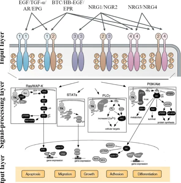
2.2. HER network ... 21
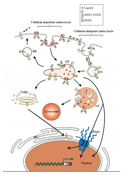
2.3. Tumorigenesis of HER2 ... 27
2.4. HER2 and breast cancer ... 30
2.4.1. Chromossome 17 Polysomy ... 31
2.4.2. Methods of HER2 testing ... 31
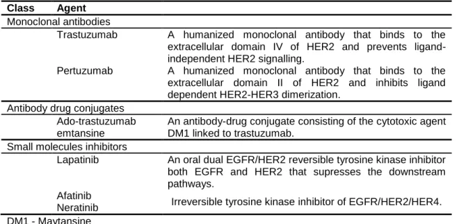
2.4.3. HER2 target therapies ... 35
2.4.3.1. Monoclonal antibodies ... 35
2.4.3.2. Small molecules inhibitors ... 38
2.4.3.3. Other HER2 target therapies ... 39
2.4.4. HER2 metastatic disease ... 40
2.5. Feline homologue of HER2 (fHER2) ... 40
3.1.1. Luminal A ... 43
3.1.2. Luminal B ... 44
3.2. Non-luminal subgroup ... 45
3.2.1. HER2-positive – non luminal ... 45
3.2.2. Triple negative ... 46 3.2.2.1. Basal-like ... 46 3.2.2.1.1. BRCA ... 47 3.2.2.2. Normal-like ... 48 3.2.3. Claudin-low ... 48 3.3. Other subtypes ... 49
3.4. Feline mammary tumors ... 49
4. Animal models ... 50
5. Aims ... 52
EXPERIMENTAL WORK ... 55
Chapter I – Feline HER2 Protein Expression Levels and Gene Status in Feline Mammary Carcinoma: Optimization of Immunohistochemistry (IHC) and In Situ Hybridization (ISH) Techniques ... 57
1. Abstract ... 57
2. Introduction ... 58
3. Material and Methods ... 59
3.1. Tumor Sample Collection and Histology ... 59
3.2. Immunohistochemical Studies... 59
3.3. ISH Studies ... 61
4. Results ... 62
4.1. fHER2 Overexpression Evaluation in FMC... 62
4.2. ISH Study ... 63
5. Discussion... 64
6. Conclusions ... 66
Chapter II – Ki-67 as prognostic factor in feline mammary carcinoma – what is the optimal cut-off value? ... 67
1. Abstract ... 67
2. Introduction ... 68
3. Material and Methods ... 69
3.1. Animal population ... 69
4.1. Animal population data ... 72
4.2. Ki-67 index of primary tumor is strongly and positively correlated with Ki-67 index of regional and distant metastasis ... 72
4.3. The optimal cut-off value of Ki-67 index is 14% in feline mammary carcinoma ... 73
4.4. Animals with high Ki-67 index (>14%) have higher probability of dying within the 2 years after surgery ... 75
4.5. Ki-67 index ≥14% is significantly associated with unfavorable features ... 75
5. Discussion... 75
6. Supplemental material ... 77
Chapter III – Molecular based subtyping of feline mammary carcinomas and clinicopathological characterisation ... 83
1. Abstract ... 83
2. Introduction ... 84
3. Material and Methods ... 85
3.1. Study population ... 85
3.2. Tumor histological classification ... 86
3.3. Immunohistochemistry ... 86
3.4. Statistical analysis ... 87
4. Results ... 88
4.1. Clinicopathological features ... 88
4.2. Associations between FMC molecular subtypes and clinicopathological features ... 94
4.3. Survival analysis ... 95
4.4. Concordance of the molecular subtypes of primary tumors from the same animal ... 95
5. Discussion... 96
6. Conclusions ... 98
Chapter IV – St Gallen molecular subtypes in feline mammary carcinoma and paired metastases-disease progression and clinical imlications from a 3-year follow-up study ... 99
1. Abstract ... 99
2. Introduction ... 100
3. Material and Methods ... 101
3.4. Statistical analysis ... 104
4. Results ... 105
4.1. Animal population ... 105
4.2. Distribution pattern of subtypes and metastases ... 108
4.3. Survival analysis ... 109
4.4. Multivariable analysis ... 111
4.5. Associations and concordance of the expression pattern of biomarkers ... 113
4.5.1. Estrogen receptor (ER) ... 113
4.5.2. Progesterone receptor (PR) ... 113 4.5.3. fHER2 ... 113 4.5.4. Cytokeratin 5/6 (CK5/6) ... 114 4.5.5. Ki-67 index... 114 4.5.6. Molecular subtypes ... 115 5. Discussion... 116 6. Conclusions ... 117 7. Supplemental material ... 118
Chapter V – Serum HER2 levels are increased in cats with mammary carcinomas and predict tissue HER2 status ... 137
1. Abstract ... 137
2. Introduction ... 138
3. Material and Methods ... 139
3.1. Cat study population ... 139
3.2. Tissue HER2, ER and PR status and Ki-67 assessment ... 139
3.3. Quantitative immunoassays to measure sHER2 levels ... 141
3.3.1. Enzyme-linked immunosorbent assay (ELISA) ... 141
3.3.2. Dot blot assay ... 142
3.4. Western blotting analysis ... 142
3.5. Statistical analysis ... 143
4. Results ... 144
4.1. Animal study population ... 144
4.2. Cats with mammary carcinoma show elevated sHER2 levels ... 144
4.3. Serum HER2 levels predict the tumor HER2 status ... 146
4.4. Serum HER2 molecules contain a portion of the intracellular receptor domain ... 146
4.6. Elevated sHER2 levels are associated with a less aggressive tumor phenotype ... 149 5. Discussion... 150 GENERAL DISCUSSION ... 155 CONCLUSIONS ... 163 FUTURE PERSPECTIVES ... 164 REFERENCES ... 165 ANNEX I ... 202 ANNEX II ... 209 ANNEX III ... 216 ANNEX IV ... 224 ANNEX V ... 236
Figure 1. Schematic representation of mammary glands in cat ... 6 Figure 2. Dysplastic lesions and benign mammary tumors ... 8 Figure 3. Feline malignant mammary tumors ... 9 Figure 4. Clinical presentations for mammary tumors ... 11 Figure 5. Metastatic lesions from mammary carcinomas in cats ... 12 Figure 6. Imagiologic findings ... 13 Figure 7. Fine-needle aspirate of a solid mammary carcinoma ... 14 Figure 8. Conformation of HER proteins and homo and heterodimerization ... 20 Figure 9. The HER signalling network ... 22 Figure 10. Trafficking of EGFR family ... 25 Figure 11. Extracellular and intracellular HER2 target therapies ... 36 Figure 12. Evaluation of fHER2 by IHC ... 63 Figure 13. fHER2 and fTOP2A genes were not amplified in FMC-HER2+ ... 64 Figure 14. Cribriform carcinoma, mammary gland, cat. Nuclear Ki-67 staining... 73 Figure 15. Kaplan-Meier overall survival (OS) curves using different cut-off values of Ki-67 74 Figure 16. Immunohistochemical expression of the different proteins studied in FMC ... 93 Figure 17. Immunohistochemical expression of the different proteins studied in FMC ... 93 Figure 18. Immunohistochemical expression of the different proteins studied in FMC ... 94 Figure 19. Immunohistochemical classification of FMC into six subtypes on the basis of their
ER, PR, fHER2, CK5/6, and Ki-67 expression ... 109
Figure 20. Kaplan-Meier curves illustrating the OS (a) and DFS (b) of cats subjected to
mastectomy (without chemotherapy) stratified according to the mammary tumor subtype . 110
Figure 21. Standard curve for sHER2 measurements using a commercial ELISA kit ... 146 Figure 22. Box plot diagrams representing the sHER2 levels in control cats (healthy group)
and in cats with mammary carcinoma (cancer group) determined by ELISA (A) and dot blot (B) assay ... 147
Figure 23. Box plot diagrams showing that tumor HER2 status correlates with sHER2 levels
as assessed by both ELISA (A) and Dot-blot assay (B) ... 147
Figure 24. Soluble truncated HER2 forms carry a portion of the ICD and are quantifiable by
Dot blot assay. (A) ... 148
Figure 25. Receiver-operating characteristic (ROC) curve for sHER2 levels for ELISA (A)
and Dot blot assay (B) ... 150
Figure 26. Evaluation of fHER2 (green signal) and fTOP2A (red signal) gene expression by
FISH ... 156
Figure 27. HER2 gene copy number in mammary carcinomas and normal mammary feline
tissue samples ... 157
Table 1. Elston and Ellis grading systems for evaluation of invasive mammary carcinoma in
female cats. ... 10
Table 2. Mills et al. (2015) grading systems for evaluation of invasive mammary carcinoma in
female cats. ... 10
Table 3. Staging of Feline Mammary Tumors. ... 12 Table 4. HER2 targeted therapy approved in HER2 positive breast cancer ... 35 Table 5. Summary of the IHC protocols used for fHER2 detection. ... 60 Table 6. HER2 IHC scoring criteria (HercepTest interpretation manual, DAKO). ... 60 Table 7. HER2 and TOP2A gene amplification assessment (ASCO guidelines, Tanner et al.,
2006; Wolff et al., 2007). ... 62
Table 8. Results of IHC using A0485, CB11 and 4B5 as primary antibodies. ... 65 Table 9. Optimised protocol steps for HER2 detection in feline mammary carcinomas by IHC
and FISH techniques. ... 66
Table 10. Univariate and Multivariate analysis of Ki-67 proliferation index ... 73 Table 11. Allred Score guidelines for ER and PR staining ... 86 Table 12. fHER2 immunohistochemistry scoring criteria ... 87 Table 13. Immunohistochemical definitions of the molecular subtypes in FMC ... 87 Table 14. FMC clinical and pathological characteristics and associations with the molecular
subtype ... 89
Table 15. FMC characteristics and associations with the molecular subtype ... 91 Table 16. Primary antibodies and immunohistochemical protocols used for ER, PR, fHER2,
CK5/6 and Ki-67 detection. ... 102
Table 17. Clinical features of the cats diagnosed with mammary carcinoma (n=61). ... 106 Table 18. Tumor features found in the studied cats. ... 107 Table 19. Location and number of metastases found at necropsy of 23 cats with mammary
carcinoma. ... 108
Table 20. Multivariate cox regression analysis for OS in cats that underwent only to surgery
(n=42). ... 1122
Table 21. Multivariate cox regression analysis for DFS in cats that underwent only to surgery
(n=36). ... 112
Table 22. HER2 immunohistochemistry scoring criteria ... 140 Table 23. IHC semi-quantitative scoring system for ER/PR assessment (Harvey et al., 1999;
Mohsin et al., 2004) ... 141
Table 24. Clinicopathological features of 60 female cats with mammary carcinoma ... 145 Table 25. Serum HER2 levels in cats with mammary carcinoma ... 149
aa – amino acids
ADAM – A Disintegrin and Metalloproteases ADCC – Antibody-dependent cellular cytotoxicity ADCs - Antibody-drug conjugates
AgNOR – Argyrophilic nucleolar organiser regions AI – Aromatase inhibitors
ADR – Androgen receptor AgR – Antigen retrieval Akt – see PKB
ANOVA – Analysis of variance AR – Amphiregulin
ASCO – American Society of Clinical Oncology ATP – Adenosine triphosphate
AUC – Area under the curve
AVMA – American Veterinary Medicine Association bp – base pair
Bcl-2 – B cell lymphoma 2
BIRC5 – Survivin or Baculoviral inhibitor of apoptosis repeat-containing 5 BRCA1 – Breast cancer susceptibility 1
BRCA2 – Breast cancer susceptibility 2 BSA – Bovine serum albumin
BTC – Betacellulin
cDNA – complementary Deoxyribonucleic acid CEP – centromere evaluation probe
CI – Confidence interval
CISH – Chromogenic in situ hybridization CK – Cytokeratin
CNS - Central Nervous System COX-2 – Cyclooxygenase-2 CT – Computed tomography CTCs – Circulating tumor cells CTF – Carboxyl-terminal fragment DAB – diaminobenzidin-tetrahydrochlorid
DAPI – 4’, 6-Diamidino-2-phenylindole, dihydrochloride DEP-1 – Density-enhanced phosphatase-1
DFS – Disease free survival DM – Distant metastases DM1 – Maytansine
DNA – Deoxyribonucleic acid DSH – domestic shorthaired e.g. – exempli gratia
ECD – Extracellular domain
EDTA – Ethylenediamine tetraacetic acid EE – Elston & Ellis
EGF – Epidermal growth factor
EGFR – Epidermal growth factor receptor EGR – Epidermal growth receptor
ELISA – Enzyme-Linked Immunosorbent Assay EMT – Epithelial to mesenchymal transition EPG – Epigen
EPR – Epiregulin ER – Estrogen receptor
fHER2 – feline epidermal growth factor receptor type 2 FISH – Fluorescence in situ hybridization
FITC – fluorescein isothiocyanate FMC – Feline mammary carcinomas FMT – Feline mammary tumors FMV – Faculty of Veterinary Medicine FNA – Fine-needle aspiration
FOXA1 – Forkhead box protein A1 FOXM1 – Forkhead box protein M1
fTOP2A – feline topoisomerase II alpha gene
GATA3 – GATA-binding protein 3 or Trans acting T-cell specific transcription factor GGI – Genomic Grade Index
GRB7 – Growth factor receptor-bound protein 7 H&E – Hematoxylin & eosin
HB-EGF – Heparin-binding EGF-like growth factor HDACi – Histone deacetylase inhibitor
HE – Hematoxylin & Eosin HER - Epidermal growth receptor
HER1 – Epidermal growth factor receptor
HER2 – Human epidermal growth factor receptor-2 HER2+ - HER2-positives
HER2- - HER2 negative
HER2-ECD – extracellular domain fragment of HER2 HER3 – Human epidermal growth factor receptor-3 HER4 – Human epidermal growth factor receptor-4 HP – Histopathological
HPF – High power fields HSP90 – Heatshock protein-90 HR – Hazard ratio
HRP – Horseradish peroxidase HRs – Hormone receptors
I.C.V.G.A.N. – International Committee on Veterinary Gross Anatomical Nomenclature ICD – Intracellular domain
i.e. – id est
IGF – Insulin-like growth factor IgG – Immunoglobulin G IHC – Immunohistochemistry
IMM – Instituto de Medicina Molecular ISH – in situ hybridization
JAK – Janus kinase kDa – kilo Dalton L – Ligand
Lc. a – Axillary lymph center
Lc. I – Inguinalfemoral lymph center LI – Lymphocitic infiltration
LNS – Lymph node status LOH – Loss of heterozygosity LR – Local relapse
M – Metastasis
mAbs – monoclonal antibodies
MAPK – Mitogen-activated protein kinase MBC – Metastatic breast cancer
MC – Mammary carcinoma
MCh – Mastectomy plus chemotherapy
MMP – Matrix Metallo Proteases mRis – microRNAs
mRNA – messenger ribonucleic acid MT – mammary tumors
mTOR – mammalian Target of rapamycin MW – Microwave
N – Lymph node
NFC – Norwegian Forest Cat NPV – Negative predictive value NRG – Neuregulins
O/N – overnight OR – Odds ratio OS – Overall survival
p-AKT – Phosphorylated protein kinase B PARP-1 – poly-ADP ribose-polymerase-1 PBS – Phosphate buffered saline
PC – pressure chamber
PCNA – Proliferating cell nuclear antigen PCR – Polymerase chain reaction PI3K – Phosphatidylinositol 3 kinase
PI3K-PKB/Akt – Phosphatidylinositol 3 kinase-protein kinase B
PIK3CA – Phosphatidylinositol 4,5-bisphosphate 3-kinase catalytic subunit alpha isoform PLC-PKC – Phospholipase C-protein kinase C
PKB – Protein kinase B
PPV – Positive predictive value PR – Progesterone receptor PT – Primary tumors
PTEN – Phosphatase and tensin homolog PTP1B – Protein tyrosine phosphatase-1B qPCR – Quantitative polymerase chain reaction
qRT-PCR – Quantitative real time reverse transcript polymerase chain reaction Ras-MAPK – Ras-mitogen-activated protein kinase
RE – Endoplasmatic resticulum
rHER2-ECD – recombinant human HER2-ECD RIPA – Radioimmunoprecipitation assay RM – Regional metastases
RNA – Ribonucleic acid
ROC – Receiver-operating characteristics RR – Relative risk
RT – room temperature
RT-PCR – Reverse transcription polymerase chain reaction RTU – ready-to-use
SC – Squamous cell SD – Standard deviation SDS – Sodium deoxycolate
SDS-PAGE – Sodium dodecyl sulphate-polyacrylamide gel electrophoresis SERMs – Selective estrogen receptor modulators
sHER – serum HER2
SISH – Silver-enhance in situ hybridization SPSS – Social Package for Social Sciences SSC – Saline sodium citrate
STAT – Signal transducer and activator of transcription T - Tumor
T4 – Thyroxine
TGF-α – Transforming growth factor-α TK – Tyrosine kinases
TKIs – Tyrosine kinase inhibitors TM – Transmembrane domain
TMB – 3,3′,5,5′-tetramethyl-benzidine TMF – Tumores mamários felinos TN – Triple Negative
TOP2A – topoisomerase II alpha gene TP – Tubulopapillary
UK – United Kingdom
ULisboa – University of Lisbon
uPA – Urokinase-type plasminogen activator USA – United States of America
UTAD – Universidade de Trás dos Montes e Alto Douro v-erbB – Avian erythroblastosis virus
VEGF – Vascular endothelial growth factor WB – water bath
WHO – World Health Organization v. – vessels
INTRODUCTION
T
HESIS OUTLINE AND OVERVIEW―In 2010, about six hundred thousand Americans, and more than 7 million humans around the world, will die from cancer. In the United States, one in three women and one in two men will develop cancer during their lifetime. A quarter of all American deaths, and about 15 per cent of all deaths worldwide, will be attributed to cancer. In some nations, cancer will surpass heart disease to become the most common cause of death.‖
Siddhartha Mukherjee In ―The Emperor of all Maladies: A Biography of Cancer‖
Cancer is one of the most common causes of death worldwide, with a high incidence in developed countries, despite the substantial progress on prevention, treatment and basic knowledge for cancer (Jemal et al., 2011; Global Burden of Disease Cancer Collaboration, 2015; World Health Organization [WHO], 2015). This alarming advance results from the aging and growth of the world population as well as the adoption of cancer-associated risk factors, as smoking, physical inactivity/obesity and dietary patterns (Jemal et al., 2011; Global Burden of Disease Cancer Collaboration, 2015). The global burden of cancer has extensive economic implications for society, added to the obvious social and individual effects (American Cancer Society, 2015). In 2012, there were estimated 14.1 million cancer cases around the world (7.4 million cases in men and 6.7 million in women) and this number is expected to increase to 24 million by 2035 (WHO, 2015).
Additionally, breast cancer represents the highest incidence in women, is the second leading cause of cancer death and foremost, this type of cancer has been continuously increasing (Global Burden of Disease Cancer Collaboration, 2015), leading to efforts for a better understanding of breast cancer biology and the development of more effective cancer treatments and prevention.
Therefore, breast cancer has been the target of several studies by scientific community. Research allowed the passage from classification systems exclusively based on morphological features to systems that include molecular markers, the development of target therapies and the improvement of the predictive and prognostic factors of the disease (Vuong et al., 2014).
Human epidermal growth factor receptor-2 (HER2) is one molecular marker that has demonstrated particularly importance in breast cancer, due to its prognostic and therapeutic
implications (Benusiglio, 2007). Once breast cancer presents a well known heterogeneity, readily molecular classification systems were proposed, taking several molecular markers into account while different prognostic and predictive subgroups were determined (Gama et al., 2008; Voung et al., 2014).
The research work presented in this thesis aimed to evaluate the role of HER2 protein in feline mammary carcinomas (FMC), assuming this protein as a single molecular marker and also using a molecular classification system with several molecular markers usually used in breast cancer studies.
In this study, the cat was selected mainly for two main reasons. First the possibility to contribute for the advance in the understanding of breast cancer biology and to improve treatment options, prevention and quality of life of our companion animals. Secondly, to assess whether FMC could be used as animal model in breast cancer research.
The research outcome of this work resulted in five manuscripts that were published in international peer-reviewed journals and represent five chapters of the experimental work include in this thesis, as follows:
Chapter I: Feline HER2 Protein Expression Levels and Gene Status in Feline Mammary Carcinoma: Optimization of Immunohistochemistry (IHC) and In Situ Hybridization (ISH) Techniques
Soares, M., Correia, J., Rodrigues, P., Simões, M., de Matos, A., Ferreira, F. (2013). Microscopy and Microanalysis, 19(4): 876-82.
Chapter II: Ki-67 as prognostic factor in feline mammary carcinoma – what is the optimal cut-off value?
Soares, M., Ribeiro, R., Carvalho, S., Peleteiro, M., Correia, J., Ferreira, F. (2016). Veterinary Pathology, 53(1): 37-43.
Chapter III: Molecular based subtyping of feline mammary carcinomas and clinicopathological characterization
Soares, M., Madeira, S., Peleteiro, M., Correia, J., Cardoso, F., Ferreira, F. (2016). The Breast Journal, 27: 44-51.
Chapter IV: St Gallen molecular subtypes in feline mammary carcinoma and paired metastases-disease progression and clinical imlications from a 3-year follow-up study
Soares, M., Correia, J., Peleteiro, M.C., Ferreira, F. (2015). Tumor Biology [Epub ahead of print]
Chapter V: Serum HER2 levels are increased in cats with mammary carcinomas and predict tissue HER2 status
Soares, M., Ribeiro, R., Najmudin, S., Gameiro, A., Rodrigues, R., Cardoso, F., Ferreira, F. (2016). Oncotarget [Epub ahead of print]
L
ITERATURER
EVIEW1. Feline mammary tumors
1.1.
Anatomy, histology and physiology of the normal mammary gland
As mammals, cat species (Felis catus, Linnaeus, 1758) have mammary glands to produce and secrete milk for the nourishment of their new born offspring. Moreover, they are also important to provide passive immunity through the colostrum (Cunningham, 2002).
Histologically, the mammary gland is classified as a modified sudoriparous apocrine gland, which is formed by several compound tubuloalveolar glands (Burkitt et al., 1994). For the micro and macro development of this gland, hormones like estrogen, progesterone and prolactin are essential (Cunningham, 2002).
Typically in cats, we observe eight mammary glands arranged in two mammary chains (Figure 1), one on each side, extending from the pectoral region to the inguinal region (Raharison & Sautet, 2006). Usually they are identified as two thoracic pairs (T1, T2) and two abdominal pairs (A1, A2) of mammary glands (Raharison & Sautet, 2006) or as thoracic (one pair), abdominal (two pairs, cranial and caudal) and iguinal (one pair) mammary glands (Dyce et al., 2010). Occasionally, accessory mammary glands can be present (Raharison & Sautet, 2006).
According to the vascularization, the mammary glands are supply through the follow arteries (Figure 1A): T1 and T2 mammary glands are irrigated by lateral thoracic and intercostal vessels (laterally), and internal thoracic vessels (medially), A1 mammary gland is irrigated by epigastric cranialis superficialis vessels, and A2 mammary glands receive blood from epigastric caudalis superficialis arteries (König & Liebich, 2004; Giménez et al., 2010). The veins of cat mammary glands closely follow the arteries, except some small veins that cross the mideline and are thought to be responsible for the spread of malignant cell tumors between the contralateral mammary glands (Giménez et al., 2010). Similarly, the venous vascularization of the thoracic glands through the chest wall (via the internal thoracic or the intercostal veins), could allow the tumor spreading to thoracic cavity (Silver, 1966).
Finally, the lymphatic drenage of mammary glands are the major responsible of the spread of neoplastic cells with origin in this organ. The two thoracic mammary glands lymphatic drainage comprises the axillary lymph center (composed by the axillary of first rib lymph node, proper axillary lymph node and accessory axillary lymph node) and the sternal cranial
lymph node. The caudal epigastric lymph nodes and the superficial inguinal lymph node, that constitute the inguinalfemoral lymph center are responsible for the lymphatic drainage of the two abdominal mammary glands (Raharison & Sautet, 2006; Raharison & Sautet, 2007; Dyce & Wensing, 2010; International Committee on Veterinary Gross Anatomical Nomenclature [I.C.V.G.A.N.], 2012). Conversely to the venous circulation, the lymphatic vessels do not cross the midline (Raharison & Sautet, 2007) but the T1 and A2 mammary glands (Figure 1) drain for the axillary and inguinalfemoral lymph centers, which is important for surgery management (Raharison & Sautet, 2006).
Figure 1. Schematic representation of mammary glands in cat
A, The four pairs of mammary glands in the cat and the corresponding irrigation. B, General schematic
drawing of the lymphatic drainage of the glands and their respective lymph nodes. The large black arrow represents the drainage of T1 and T2 for the axillary lymph center and the large grey arrow represents the drainage of A1 and A2 for the inguinalfemoral lymph center, whereas the small black arrow represents the drainage of T2 to the inguinalfemoral lymph center and the small grey arrow represents the drainage of A1 for the axillary lymph center. v., vessels; Lc. a, axillary lymph center; Lc. I, inguinalfemoral lymph center. Adapted from Raharison & Sautet (2006); Raharison & Sautet (2007); Giménez et al. (2010).
1.2.
Epidemiology
In the epidemiology field there are few studies regarding the incidence of mammary neoplasia in cats. In addition, researchers believe that the available data underestimate the true incidence of the disease, because most of the studies are archaic and outdate (Dorn et al., 1968, MacVean et al., 1978). According to Dorn et al. (1968), mammary tumors are the third most common tumor type in female cats (after skin tumors and lymphoma),
representing 12% of tumors in cats indepedent of sex. More recently, Vascellari and collegues (2009) reported that mammary tumors were the second most common tumor in cats and represent 16.3% of the tumors in cats independently of the sex (and 25.3% of the tumors in female cats).
Feline mammary tumors are almost exclusive of the female sex, such as human breast cancer (Sorenmo et al., 2013). In humans, there are some risk factors well-established, like age, obesity, sedentary behaviour, alcohol consumption and smoking, shift work, hormonal or genetic status that may be implicated in mammary tumorigenesis (American Cancer Society, 2015).
In cats, only age, breed, and hormonal influence were already identified as risk factors for FMC. According to the literature, and as is described for women and bitch, feline mammary carcinoma incidence increases with age, and disease is predominantly seen in middle-aged to older cats. The mean age of diagnosis is between 10 to 12 years of age, with increasing risk up to 14 years of age, and carcinomas are more likely to occur in older cats, when compared with benign mammary tumors (Weijer & Hart, 1983; Sorenmo et al., 2013; Zappulli et al., 2015).
Based on the breed, two studies registered that Siamese cats appear to be overrepresented when compared to other breeds (Hayes et al., 1981; Ito et al., 1996). Finally, the exposure to ovarian hormones is also strongly implicated in mammary tumorigenesis in cat. Overley et al. (2005) concluded that sexually intact cats have a higher risk than spayed cats to develop mammary carcinomas (p=0.001, odd ratio [OR] 2.7) and also that the risk of the disease increase with age. Moreover, cats spayed prior to 6 months of age had a 91% reduction of the risk of develop mammary tumors (MT) and when the cat is spayed prior to one year the reduction of the risk lowed for 86%. Besides the ovarian hormones, there is also an increased risk of developing mammary cancer in queens subjected to regular administration of progestogens (Misdorp et al., 1991). Finally, Overley and collegues (2005) concluded that parity did not affect mammary tumor development.
1.3.
Dysplastic lesions and benign mammary tumors
Less than 15% of the feline mammary lesions are dysplastic lesions and benign neoplasias (Figure 2). The dysplastic lesions include ductal hyperplasia, lobular hyperplasia (epithelial hyperplasia, adenosis and fibroadenomatous change), cysts, duct ectasia and fibrosclerosis. Fibroadenomatous change (fibroepithelial hyperplasia, fibroepithelial hyperthrophy or feline mammary hyperthrophy) is a hormonal induced proliferation of interlobular ducts and periductal stromal cells (Misdorp et al., 1999).
Benign tumors (Figure 2) in cat are uncommon and include adenoma (simple and complex), fibroadenoma, benign mixed tumor and duct papilloma (Misdorp et al., 1999).
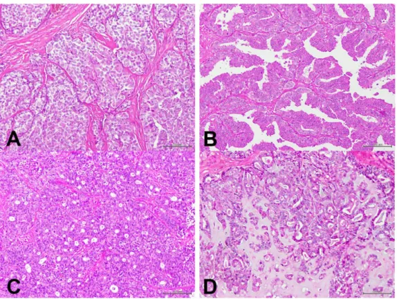
Figure 2. Dysplastic lesions and benign mammary tumors (Hematoxylin & Eosin [HE]).
A (20x) and B (100x) images represent a simple tubulopapillary adenoma in a female cat. The lesion
is composed of a single layer of cells arranged in tubules that contain an amorphous amphophilic secretion. In C (40x) a feline mammary gland with duct ectasias is illustrated. Ectasias affect the tubules and also the acini. In the figure, dilated tubules that contain fluid are observed. D (40x) presents a fibroepithelial hyperplasia in a female cat. The lesions are clearly circumscribed, without being capsulated. There is a extremely marked proliferation of the intralobular duct system as well as of the intralobular connective tissue.
1.4.
Malignant mammary tumors
Most of mammary tumors in cats are malignant (85% to 95%), and present an aggressive biological behaviour, with early lymphatic invasion and lymph node metastases. The malignant tumors types in cats are predominantly adenocarcinomas, specially the simple carcinomas like tubulopapillary and solid carcinomas (Figure 3A and 3B). Others types of mammary tumors include noninfiltrating (in situ) carcinoma, cribriform carcinoma (Figure 3C), invasive micropapillary carcinoma, squamous cell carcinoma, mucinous carcinoma (Figure 3D), lipid rich carcinoma and inflammatory mammary carcinoma (Misdorp et al., 1999; Pérez-Alenza et al., 2004; Kamstock et al., 2005; Seixas et al., 2007; Millanta et al., 2012). Carcinosarcomas, sarcomas and other non-epithelial neoplasias are very rare mammary tumors in cats (Misdorp et al., 1999; Sorenmo et al., 2013).
Figure 3. Feline malignant mammary tumors (100x, HE).
A, Solid mammary carcinoma of a female cat with 7,5 years old. It is evident that lobules are changed
into solid tumor cell masses with reduced glandular structure. B, Tubulopapillary mammary carcinoma in a female cat. The tubules are predominantly arranged in a pedunculated papillary fashion. C, Cribriform mammary carcinoma. In the figure is evident the sievelike arrangement of the neoplastic epithelial cells, characteristic of this hystope. D, Mucinous mammary carcinoma in a female cat with 11 years old. It is an uncommon type of mammary carcinoma, characterized by the abundant mucin production.
1.4.1. Malignancy grade
In Human Medicine, Elston and Ellis method is the most common histological grading method for invasive carcinomas, and is correlated with the prognosis of the disease (Elston & Ellis, 1998).
This system, that was also adapted for feline mammary tumors, is based on the assessment of three morphological features: (1) the degree of glandular differentiation assessed by tubular formation; (2) nuclear pleomorphism and (3) mitotic activity (Table 1). Recently, another classification was suggested by Mills et al., 2015, that consider lymphovascular invasion, nuclear form and mitotic counting (Table 2).
Notwithstanding, the two classification systems have already proven its prognostic value in cats with mammary carcinoma (Castagnaro et al., 1998a; Seixas et al., 2011; Mills et al., 2015).
Table 1. Elston and Ellis grading system for evaluation of invasive mammary carcinoma in female cat.
Histologic feature Score
Tubule formation
tumor had more than 75% tubules 1
10-75% of tumor had tubule formation 2
<10% tubules 3
Nuclear pleomorphism
small regular uniform cells 1
moderate nuclear size and variation 2
marked nuclear variation 3
Number of mitoses per 10 high power fields (HPF)
0-5 mitoses per 10 HPF 1
6-10 mitoses per 10 HPF 2
>11 mitoses per 10 HPF 3
Point total Grade
3-5 Grade I, well differentiated or low grade
6-7 Grade II, moderately differentiated or intermediate grade 8-9 Grade III, poorly differentiated or high grade
HPF, High power fields
Table 2. Mills et al. (2015) grading system for evaluation of invasive mammary carcinoma in female cat.
Histologic feature Score
Lymphovascular invasion Absent 0 Present 1 Nuclear form ≤5% abnormal 0 >5% abnormal 1
Mitotic count per 10 high power fields (HPF)
≤62 0
>62 1
Point total Grade
0 Grade I, low grade
1 Grade II, intermediate grade 2-3 Grade III, high grade
1.5.
Clinical presentation
As indicated above (chapter 1.2), cats with mammary carcinomas are often old and may be sexually intact or spayed. Tumors are usually easy to detect on physical examination as they appear as firm and discrete masses in the mammary gland that can be detached or attached to the underlying tissue (Figure 4).
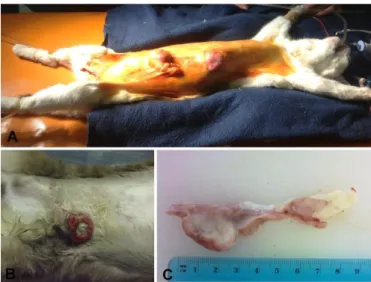
Multiple tumors are common (Figure 4A) and 60% of cats have more than one tumor at the time of diagnosis, according to Hayes & Mooney (1985). All glands are susceptible to tumor development although anterior glands are less commonly affected (Weijer & Hart, 1983). For this reason, a careful examination of the remaining mammary glands is always recommended in the clinical examination of the oncologic animal and for each mass the following features should be evaluated: size of the mass, consistency, mobility and the presence of skin ulceration (Weijer & Hart, 1983).
Figure 4. Clinical presentations for mammary tumors.
A, Multiple mammary tumors of a domestic shorthaired (DSH) female cat with 11 years. The masses
were malignant (tubulopapillary carcinomas) and the animal also presented regional lymph node invasion by metastatic cancer cells (stage 3, T3, N1, M0). B, Siamese cat with 13 years old presenting a large ulcerated mammary tumor (stage 3, T3, N1, M0). C, Macroscopic examination of the mammary mass in picture B (sagittal cut, in the left), and a completely altered lymph node by metastatic lesions (sagittal cut, in the right).
Tumor size is a very important feature as it presents prognostic value and depends on how early the tumor is detected and how aggressive the tumor behaves. Usually, larger tumors may become ulcerated, inflamed and infected (Weijer & Hart, 1983; MacEwen et al., 1984; Ito et al., 1996).
FMC are typically very aggressive and present high capability to metastasize to regional lymph nodes and to distant organs such as lungs and pleura (Figure 5), which are the organs most commonly affected (Weijer & Hart, 1983). Moreover, authors report that 50% to 93% of cats with mammary carcinoma show metastasis at necropsy, making the inspection of the local lymph nodes in the clinical examination crucial (Hayden & Nielsen, 1971; Hahn et al., 1994).
Considering the high incidence and the morbidity of FMC, a clinical and complete work-up is recommended and includes: a complete physical examination, a complete blood count, serum biochemical profile, serum T4 concentration, urinalysis, three-view thoracic radiographs (ventrodorsal, right and left lateral views), abdominal ultrasound or a computed tomography (CT) scan (Figure 6). All mammary masses and any palpable regional lymph node should be subjected to a fine-needle aspiration (FNA) or to a biopsy (Figure 7). Ulcerated lesions should be scraped and fluids from the affected glands should be examined, in order to achieve a diagnosis before surgery. All the mammary lesions and corresponding regional lymph nodes should be analyzed by histopathology.
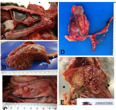
Figure 5. Metastatic lesions from mammary carcinomas in cats.
A, Necropsy of a 15 year old DSH female cat with pleural effusion and metastases in the pleura and B, lung. C, Necropsy of a 9 year old female cat with metatases in the pancreas, D, iliac lymph node
and kidney, and other organs not represented in the picture (lung, liver and muscle). E, Necropsy of a Norwegian Forest Cat (NFC) 11 years old that presented liver metastases of a primary mucinous mammary carcinoma.
Similarly to humans and dogs, the mammary disease in cats is also staged, using a modification of the original system published by Owens (McNeill et al., 2009), as is shown in Table 3. This staging system should not be used for mammary gland sarcomas.
Finally, inflammatory mammary carcinoma is a rare but a clinical important type of mammary tumor, which is also described in women and bitch. In these cases, the entire mammary chain or the mammary gland affected may appear edematous, swollen, warm and painful. The clinical identification of this type of carcinoma is very important once these animals are poor surgical candidates, due to the secondary postsurgical complications like nonhealing incisions, oedema and suture rejections (Pérez-Alenza et al., 2004).
Table 3. Staging of Feline Mammary Tumors.
Stage Tumor size Lymphnode status Metastasis
Stage 1 T1: <2cm N0 M0 Stage 2 T2: 2-3cm N0 M0 Stage 3 T1 or T2 T3: >3cm N1 (positive) N0 or N1 M0 M0 Stage 4 Any N0 or N1 M1
Legend: T, tumor; N, lymph node; M, metastasis; 0, absence of metastasis; 1, presence of
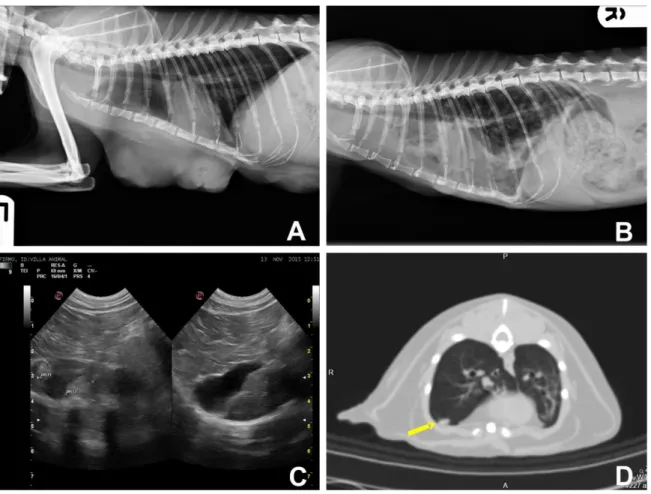
Figure 6. Imagiologic findings.
Left lateral (A) and right lateral (B) thoracic radiographs from a 14 year old queen with a solid mammary carcinoma before the surgery (A) and three-months after the surgery (B). A, Radiograph shows masses in the thoracic mammary region. B, In these radiograph, it is visible an extensive pleural effusion and numerous ill-defined round soft tissue opacities that are scattered through the lung fields consistent with pulmonary metastasis. C, Abdominal ultrasound in a cat with a tubulopapillary mammary carcinoma. There are two ill-defined rouded hyperechoic lesions in the hepatic parenquima, consistent with hepatic metastasis. D, CT transverse image of a cat with tubular mammary carcinoma. In the CT is visible a soft tissue nodule in the pulmonary parenchyma (yellow arrow), consistent with pulmonary metastasis, which was subsequently confirmed at necropsy.
1.6.
Treatment options
Surgery is traditionally the required treatment for FMC and the surgical extension has already been associated to the disease free survival (DFS) period (MacEwen et al., 1984). Cats with mammary lesions are usually subjected to a simple lumpectomy, mastectomy, regional mastectomy, or a chain mastectomy that can be unilateral or bilateral. The goal of surgical intervention is to remove the current tumor or tumors with clean margins, in order to prevent the emergence of new tumors in the remaining mammary glands.
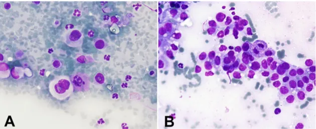
Figure 7. Fine-needle aspirate of a solid mammary carcinoma (Giemsa, 40x).
A, The cytological smear displayed multinucleated tumor cells and pleomorphic tumor cells, with
marked anisocytosis, anisokaryosis and multiple preominent nucleoli suggesting a high level of tumor aggressiveness. Mitoses are also present. B, Presence of a homogeneous population of epithelial cells with high nuclear:citoplasmatic ratios, pleomorphic cells and the presence of mitoses.
In cats, radical mastectomy (unilateral for cats possessing a single tumor or a 2-staged bilateral chain mastectomy for animals with bilateral tumors) results in a significant larger DFS when compared with cats receiving a conservative chain mastectomy (MacEwen et al., 1984). Thus, unilateral or staged bilateral mastectomy is the recommended treatment for FMC. In some cases a bilateral mastectomy could be performed in a single surgery, if the postsurgical tension is minimal (when the animal presents excessive mammary or adipose tissue, for example). For tumors that are fixed, muscular fascia or portions of the body wall should also be resected (Sorenmo et al., 2013).
Aggressive assessment of the regional lymph node is justified by the high metastatic potential of FMC and by the poor prognosis that is associated to the presence of lymph node metastasis. This could be assessed with an ultrasound-guide FNA or through the histopathological evaluation of the resected regional lymph node during surgery (Sorenmo et al., 2013).
Early detection and aggressive surgery (including prophylactic chain mastectomy) can result in long-term survival in cats with early stage mammary tumors. However, cats with late diagnosis or later stages of the disease are not treated effectively with surgery alone. In those cases the use of adjuvant doxorubicin-based chemotherapy should be considered (Novosad et al., 2006; McNeill et al., 2009). In one multi institutional retrospective study, a comparison between cats with mammary carcinomas receiving surgery versus cats receiving surgery with adjuvant chemotherapy demonstrated that the subset of cats having unilateral chain mastectomy followed by chemotherapy presented an longer overall survival (OS) than the other group of animals (McNeill et al., 2009).
Alternative chemotherapy protocols include a combination of doxorubicin and cyclophosphamide or doxorubicin-based chemotherapy and a nonsteroidal anti-inflammatory drug (meloxicam). However, survival studies based on these protocols have not been performed, so their efficiency cannot be determined in cats with mammary tumors (Mauldin et al., 1988; Sorenmo et al., 2013; Borrego et al., 2009).
Concerning others treatments, as hormonal therapy, they are unlike to be effective once studies indicate that FMC presents a low expression of hormonal receptors (de las Mulas et al., 2002; Millanta et al., 2005b; Millanta et al., 2006a; Burrai et al., 2010) and studies that evaluate this therapy in cats with FMC were not found (Sorenmo et al., 2013).
Recently, promising results were obtained using oncolytic virotherapy using FMC cell lines (Adelfinger et al., 2014). This therapy is based on the capacity of oncolytic viruses infecting and promoting cancer cells lysis without extensively damaging the surrounding normal tissue.
Finally, and despite the high incidence of distant metastasis in these animals, there are almost no recent studies to search for effective adjuvant systemic treatments for FMC.
1.7.
Prognostic factors
Prognosis is generally assessed using the one-year postsurgical rate of survival/remission in cats, which is comparable to the ten-year postsurgical survival/remission rate generally used in humans. DFS and postsurgical remission rate at a fixed interval are considered better prognostic indicators, at least in cats, since the overall survival time might be influenced by other factors such as concomitant diseases or euthanasia (MacEwen et al., 1984; Matos et al., 2012).
There are some studies related to survival in cats with feline mammary carcinomas. For example, a review by Zappulli et al. (2015) indicated that survival time after primary tumor detection rounds the 12 months in cats depending on the clinical staging and on the tumor excision. Additionally, several parameters are considered important prognostic indicators for FMC such as age, size of the primary tumor, malignancy grade, lymph node involvement, number of mitoses, extent of necrosis, completeness of surgical resection and type of invasion (MacEwen et al., 1984; Ito et al., 1996; Sarli et al., 2003).
1.7.1. Clinical features
Usually, age is an associated indicator since old animals show lower survival rates (Weijer & Hart, 1983; Ito et al., 1996). More recently, another study correlate cats with old age with a shorter DFS and OS but the prognostic significance was lost when multivariate analysis was performed (Seixas et al., 2011).
Clinical staging of FMC, based on the TNM classification of malignant tumors system, proved to be an important prognostic factor. According to Hughes & Dobson (2012) the cats with
![Figure 2. Dysplastic lesions and benign mammary tumors (Hematoxylin & Eosin [HE])](https://thumb-eu.123doks.com/thumbv2/123dok_br/18895666.934573/37.892.183.756.82.509/figure-dysplastic-lesions-benign-mammary-tumors-hematoxylin-eosin.webp)