https://doi.org/10.1007/s10620-019-06029-z
ORIGINAL ARTICLE
Severity of Ascites Is Associated with Increased Mortality in Patients
with Cirrhosis Secondary to Biliary Atresia
Renata R. Guedes1 · Carlos O. Kieling2 · Jorge L. dos Santos3 · Carolina da Rocha4 · Fernando Schwengber5 ·
Marina R. Adami2 · Marcio F. Chedid6 · Sandra M. G. Vieira1
Received: 27 August 2019 / Accepted: 22 December 2019
© Springer Science+Business Media, LLC, part of Springer Nature 2020
Abstract
Background Very few prior studies have investigated the presence of ascites as a prognostic factor in children with cirrhosis. To the best of our knowledge, there are no prior studies evaluating the relationship between severity of ascites and patient survival in children with biliary atresia and cirrhosis.
Aims To evaluate the association between severity of ascites and survival of children with cirrhosis and biliary atresia. Methods All children with cirrhosis secondary to biliary atresia evaluated at our institution from 2000 to 2014 were included in this study. Patients were classified into four groups: NA = no ascites; A1 = grade 1 ascites; A2 = grade 2 ascites; and A3 = grade 3 ascites. The primary endpoint of the study was mortality within the first year after patient inclusion. Ninety-day mortality was also evaluated. Prognostic factors related to both endpoints also were studied.
Results One-year patient survival for NA was 97.1%, versus 80.8% for A1, versus 52% for A2, versus 13.6 for A3 (p < 0.001). The presence of ascites increased mortality by 17 times. In the multivariate analysis, clinically detectable ascites (HR 3.14, 95% CI 1.14–8.60, p = 0.026), lower sodium (HR 1.15, 95% CI 1.04–1.27, p = 0.006), higher bilirubin (HR 1.06, 95% CI 1.00–1.12, p = 0.023), and higher PELD score (HR 1.05, 95% CI 1.02–1.08, p = 0.001) were all associated with decreased sur-vival. Lower serum sodium (HR 1.20, 95% CI 1.09–1.32, p < 0.001) and higher PELD score (HR 1.03, 95% CI 1.001–1.063,
p = 0.043) were associated with increased 90-day mortality.
Conclusions Clinically detectable ascites is associated with decreased 1-year survival of children with biliary atresia. These patients should be treated with caution and prioritized for liver transplantation.
Keywords Ascites · Pediatric patients · Portal hypertension · Survival · Chronic liver disease · Liver transplant
Introduction
Biliary atresia (BA) is the leading cause of chronic liver dis-ease (CLD) and the most common indication for liver trans-plantation (LT) in children. It is a rare disorder characterized by an inflammatory cholangiopathy, the etiology of which has not yet been clarified [1–3]. Hepatoportoenterostomy (HPE) is the primary therapeutic option for BA. If BA is left untreated, cirrhosis leads to death within 2 years [3, 4]. Following HPE, approximately 50% of children still develop long-term complications associated with cholangitis, biliary cirrhosis, and portal hypertension [5].
Although recent reports have demonstrated high mortality among infants with BA on liver waiting lists [6], few studies have reported the impact of decompensated cirrhosis in chil-dren and establishing prognosis for these patients remains a challenge [7, 8]. In adult cirrhotic patients, ascites is often the first sign of CLD decompensation and is usually consid-ered a landmark sign of cirrhosis aggravation [9–12]. Devel-oped in the 1960s, the Child–Pugh score included presence of ascites as one of the variables related to worse clinical status [13]. In the 1980s, Malatack et al. [14] developed a system for assessing prognosis in cirrhotic children. In this scoring system, presence of ascites was also one of the vari-ables associated with poor prognosis before LT.
Pugliese et al. [8] showed that ascites and serum sodium were important variables associated with waiting list mortal-ity in a sample of more than 500 Brazilian patients. Sponta-neous bacterial peritonitis (SBP) is a common complication
* Renata R. Guedes rguedes@hcpa.edu.br
of ascites and one that our research team has studied. In prior work, we demonstrated identification of bacterial DNA in children with nonculture, non-neutrocytic ascites [15], and more recently, we reported that very poor prognosis was related to a first episode of SBP [16].
To the best of our knowledge, there are no prior studies evaluating the relationship between severity of ascites and survival of children with cirrhosis and BA. We hypothesize that the severity of ascites may be strongly associated with worse prognosis in pediatric patients with cirrhosis second-ary to BA. The aim of this study was to analyze presence and severity of ascites as prognostic factors in survival of children with BA and cirrhosis.
Methods
This study was approved by the Hospital de Clinicas de Porto Alegre Institutional Review Board. Since the analysis was retrospective, informed consent waiver was obtained for all individual participants included in the study.
All children with cirrhosis secondary to BA, aged 12 years or younger, seen at the Pediatric Gastroenterology and Hepatology Unit at the Hospital de Clinicas de Porto Alegre from March 2000 to July 2014 were included in this historical cohort. Patients had previous or no Kasai surgery and were studied retrospectively. For patients who never developed ascites, inclusion was performed at the time of diagnosis of cirrhosis. For patients who developed ascites, the inclusion was accomplished on its first episode. Accord-ingly, for each patient, the severity of ascites was recorded as it was on its first episode.
We excluded patients with other previous abdominal sur-gery, gastrointestinal bleeding due to portal hypertension, SBP, or sepsis or other systemic infection within a four-week span before ascites diagnosis.
After enrollment, patients were classified into four groups: (1) NA = no ascites; (2) A1 = grade 1 ascites; (3) A2 = grade 2 ascites; and (4) A3 = grade 3 ascites. Ascites was diagnosed by abdominal ultrasound and classified according to the international criteria [17]. Most of those patients (53/72 with and 13/34 without ascites) were on the waiting transplant list.
The primary outcome was mortality occurring at any time within the first year after patient inclusion. The secondary outcome was mortality occurring within the first 90 days. Prognostic factors related to both outcomes were analyzed.
Since mortality was the event of interest, patients who underwent LT were removed from the analysis (censored statistically). No investigations or interventions were per-formed for the purpose of the study, and no patients were included more than once.
All patients underwent routine diagnostic assessment according to a routine diagnostic protocol which included complete blood count, coagulation tests (platelets, prothrom-bin time estimated by the international normalized ratio [INR]), liver and renal biochemical tests (total and conju-gated serum bilirubin, aspartate aminotransferase, alanine aminotransferase, alkaline phosphatase, gamma glutamyl-transferase, serum albumin, creatinine, urea), and blood cul-tures. The SBP treatment protocol was in accordance with international guidelines [17].
Cumulative probability of survival by groups was calcu-lated using the Kaplan–Meier method, and curves were com-pared using the log-rank test. A p value of less than 0.05 was considered significant. Univariate analysis was performed to identify variables related to decreased survival for both time endpoints using the Cox regression method. Variables analyzed included: age at inclusion, Pediatric End-Stage Liver Disease (PELD) score [18], nutritional status as deter-mined by height/age z-score, prothrombin time as assessed by INR, total bilirubin, serum albumin, and serum sodium. For statistical analysis, age was categorized as 0–1 years versus ≥ 1 year. Malnutrition was defined as a height/age
z-score < − 2. The distribution of continuous numeric
vari-ables was assessed using the Shapiro–Wilk test. Continuous variables with normal distribution were expressed as mean and standard deviation and compared using the T-test and/ or ANOVA as appropriate. Continuous variables without normal distribution were expressed as median and interquar-tile interval (perceninterquar-tiles 25 and 75) and compared using the Kruskal–Wallis and Mann–Whitney tests.
Categorical variables were presented as percentages and were compared using the Chi-square test. A multivariate analysis was performed to identify independent predictors of survival. Variables with a p value equal to or lower than 0.05 in univariate analysis were subsequently included in this multivariate proportional hazards Cox analysis. Data analysis was conducted using the Statistical Package for Social Sciences, version 18.0 (SPSS Inc. Released 2009. PASW Statistics for Windows, Version 18.0. Chicago-USA).
Results
One hundred and twenty consecutive children with BA were reviewed. Fourteen of these were excluded due to missing data. Thus, a total of 106 patients were included in this study. Eighty-nine of those 106 patients had undergone a prior HPE. Thirty-nine out of the 89 patients had biliary drainage following HPE. Sixteen of those developed decompensations with ascites. The remaining 50 patients failed the Kasai pro-cedure. Forty-one of those were enlisted to liver transplan-tation. From these, 29 were under 1 year old and required early liver transplantation. (We defined early transplantation
as the procedure performed during the first year of patient’s life and HPE failure as total bilirubin > 2 mg/dL post-HPE.)
Demographic data and clinical characteristics of the study population are summarized in Table 1. Most children were under 1 year old when included in the study. Only 31 of 106 were over 1 year old. The oldest patient was 7 years old.
The prevalence of ascites was 68% (72 patients). Thirty-four of the whole sample of 106 patients (32.1%) had no ascites. Of the 72 patients with ascites, 26.4% had mild ascites, 28.3% had moderate ascites, and 13.2% had severe ascites (Table 1). Thirteen of the 34 patients without ascites and 53 of the 72 patients with ascites were on the waiting transplant list.
Table 2 shows patient data by presence of ascites. Patients with ascites were more commonly younger than 1 year, had
worse nutritional status, more severe hepatic synthetic dys-function, lower serum sodium, and higher PELD scores.
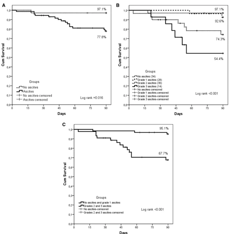
During the first year of observation, 31 of the 106 patients (26.4%) underwent LT. Figure 1a illustrates transplant-free 1-year patient survival of the entire cohort (patients who underwent LT during the first year of observation were cen-sored). Twenty-seven of the 75 patients who did not have LT died during the first year of follow-up (mortality rate = 36%).
Figure 1b shows a survival comparison according to presence of ascites. One-year patient survival for patients without ascites was 97.1% versus 56.9% for patients with ascites (p = 0.001).
Figure 1c shows transplant-free survival for the four patient groups (NA, A1, A2 and A3). For the NA group, 1-year survival was 97.1%, versus 80.8% for the A1 group, versus 52% for the A2 group, versus 13.6 for the A3 group (p < 0.001).
There was no difference between survival in NA and A1 groups (p = 0.079) or between A2 and A3 groups. Thus, patients without ascites (NA) were combined into one group with patients with mild ascites (A1) and A2 patients were also combined with A3 patients into a single group. One-year survival in the NA + A1 group was compared to that in the A2 + A3 group (Fig. 1d). One-year patient survival in the NA + A1 group was 89.9% versus 39.3% in the A2 + A3 group (p < 0.001).
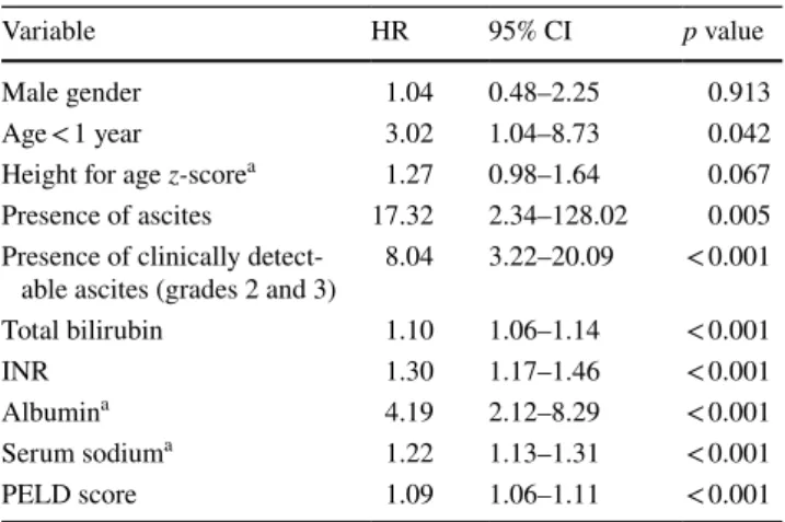
Table 3 shows variables related to decreased survival according to Cox proportional hazards regression. Mortal-ity of patients was increased by 17 times by the presence of ascites (HR 17.32, 95% CI 2.34–128.02, p = 0.005). Clini-cally, detectable ascites (A2 and A3 groups) increased mor-tality by 8 times (HR 8.04, 95% CI 3.22–20.09, p < 0.001). Lower albumin was associated with a fourfold increase in 1-year mortality (HR 4.19, 95% CI 2.2–8.29, p < 0.001). Age younger than 1 year was associated with a threefold decrease in 1-year survival (HR 3.02, 95% CI 1.04–8.73, p = 0.042).
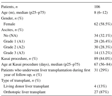
Table 1 Demographic data for 106 children with biliary atresia (n = 106) Patients, n 106 Age (m), median (p25–p75) 8 (6–12) Gender, n (%) Female 62 (58.5%) Ascites, n (%) No (NA) 34 (32.1%) Grade 1 (A1) 28 (26.4%) Grade 2 (A2) 30 (28.3%) Grade 3 (A3) 14 (13.2%) Kasai procedure, n (%) 89 (84.0%)
Age at Kasai procedure (days), median (p25–p75) 67 (56–84) Patients who underwent liver transplantation during first
year of follow-up, n (%) 31 (29%)
Type of transplant, n (%)
Living donor liver transplant 4 (13%)
Orthotopic liver transplant 27 (87%)
Table 2 Patient data by presence of ascites
INR international normalized ratio, PELD score Pediatric End-Stage Liver Disease score, IQR interquartile range Ascites
72 (67.9%) No ascites34 (32.1%) Total106 (100%) p value
Age < 1 year, n (%) 57 (79.2%) 18 (52.9%) 75 (70.8%) 0.011
Female gender, n (%) 43 (59.7%) 19 (55.9%) 62 (58.5%) 0.833
Kasai procedure, n (%) 59 (81.9%) 30 (88.2%) 89 (84%) 0.573
Total bilirubin ≤ 2 mg/dL post-Kasai, n (%) 16 (27.1%) 23 (76.7%) 39 (43.8%) < 0.001
Height for age z-score, median [IQR 25–75] − 2.08 [− 2.90 to − 1.32] − 0.93 [− 1.94 to − 0.33] − 1.87 [− 2.68 to − 0.88] < 0.001 Albumin (g/dL), median [IQR 25–75] 2.8 [2.3 to 3.18] 3.6 [3.3 to 4.2] 3.5 [2.5 to 3.1] < 0.001 Total bilirubin (mg/dL), median [IQR 25–75] 13.4 [6.5 to 17.5] 1.9 [0.7 to 9.5] 10.4 [2.1 to 16.4] < 0.001 INR, median [IQR 25–75] 1.36 [1.19 to 1.65] 1.10 [1.02 to 1.27] 1.30 [1.10 to 1.55] < 0.001 Serum sodium (mEq/L), median [IQR 25–75] 135 [132 –137.8] 138 [134.8 to 138.3] 136 [133 to 138] 0.018 PELD score, median [IQR 25–75] 18 [13 to 25] 3.5 [− 7.3 to 11.3] 15 [5 to 22] < 0.001
The other variables related to the decreased 1-year survival were elevated serum bilirubin, elevated INR, and higher PELD. Lower serum sodium was also related to increased mortality (Table 3).
Albumin, bilirubin, INR, age, and nutrition status are all part of the PELD score. Therefore, combining any of these factors with the PELD score in the same multivariate model
would create a potential for collinearity and so two differ-ent multivariate models were constructed. In the first (not using PELD score, but using age, nutrition status, bilirubin, INR, and albumin), the three variables independently related to increased mortality were moderate and severe ascites (groups A2 and A3) (HR 3.14, 95% CI 1.14–8.60, p = 0.026), low sodium (HR 1.15, 95% CI 1.04–1.27, p = 0.006), and
Fig. 1 a One-year patient survival of the 106 patients (patients who underwent LT were censored). b One-year patient survival, by presence of ascites (patients who underwent LT were censored) (p < 0.001). c One-year patient survival for the four patient groups
(NA, A1, A2, and A3) (patients who underwent LT were censored) (p < 0.001). d Comparison of 1-year transplant-free patient survival in two groups: patients without ascites (NA) or with grade 1 ascites (A1) versus patients with grade 2 or 3 ascites (A2 and A3) (p < 0.001)
elevated bilirubin (HR 1.06, 95% CI 1.00–1.12, p = 0.023) (Table 4).
In the multivariate model that included the PELD score, presence of moderate and severe ascites (groups A2 and A3) was also the strongest predictive variable (HR 3.24, 95% CI 1.17–8.96, p = 0.023) (Table 5). Lower serum sodium was associated with a higher mortality risk (HR 1.13, 95% CI 1.03–1.25, p = 0.009). Interestingly, the PELD score was only associated with a marginal increase in mortality risk (HR 1.05, 95% CI 1.02–1.08, p = 0.001).
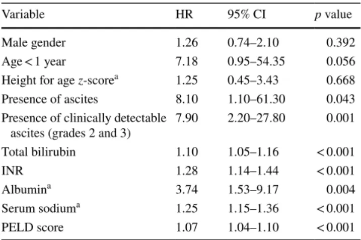
The same statistical analyses were employed to evalu-ate 90-day survival. Patients with and without ascites had statistically different survival rates. Patients with ascites (A1 + A2 + A3) had lower 90-day survival (Fig. 2a). The 90-day survival for patients with clinically detectable ascites was significantly lower than survival among patients without ascites (Fig. 2b, c).
Univariate analyses revealed that presence of ascites, presence of clinically detectable ascites (A2 and A3 groups),
lower albumin, increased serum bilirubin, increased INR, higher PELD, and also decreased serum sodium also were all related to increased mortality (Table 6). The multivariate model that did not include the PELD score revealed that only lower serum sodium was associated with increased mortal-ity (HR 1.21, 95% CI 1.10–1.33, p < 0.001) (Table 7). The multivariate model that did include PELD score revealed that both lower serum sodium (HR 1.20, 95% CI 1.09–1.32,
p < 0.001) and PELD score were associated with increased
mortality (HR 1.03, 95% CI 1.001–1.063, p = 0.043) (Table 8).
Discussion
Most children with BA will eventually develop CLD [4,
5]. In the first years of life, progression to cirrhosis occurs in patients with late diagnosis of BA or failure after HPE. These patients develop severe hepatic synthetic dysfunction during a period of immature hepatic function and vulner-ability to nutritional compromise [19–21]. Children with ascites, and especially infants, are prone to organ dysfunc-tion [8]. It has been proposed that systemic inflammation plays an important role in the course of ascites and is associ-ated with decompensation in adult patients [9]. The response to inflammation is individual, and certain aspects are age related.
The impact of ascites on morbidity and mortality is well known in adult cirrhotic patients [11, 12, 17], in whom 1-year and 2-year mortality are nearly 40% and 50%, respec-tively [17]. Only a very few prior studies have investigated presence of ascites as a prognostic factor in children with cirrhosis [8, 22, 23]. Pugliese et al. [8] studied 522 pedi-atric patients with end-stage liver disease listed for LT due to several etiologies. They detected that presence of ascites was independently associated with a lower survival rate at 90 days (58.8% vs. 83.8%). Specifically with children with BA, Jiang et al. analyzed a total of 133 patients who had undergone Kasai procedures. They detected that presence of ascites was one of the variables related to poor prognosis [22]. They also proposed a scoring system to predict which
Table 3 Univariate analysis of variables related to 1-year patient sur-vival, by Cox proportional hazards regression
INR international normalized ratio, PELD score Pediatric End-Stage Liver Disease score, HR hazards ratio
a For these variables, lower values were associated with worse out-comes
Variable HR 95% CI p value
Male gender 1.04 0.48–2.25 0.913
Age < 1 year 3.02 1.04–8.73 0.042
Height for age z-scorea 1.27 0.98–1.64 0.067 Presence of ascites 17.32 2.34–128.02 0.005 Presence of clinically
detect-able ascites (grades 2 and 3) 8.04 3.22–20.09 < 0.001
Total bilirubin 1.10 1.06–1.14 < 0.001
INR 1.30 1.17–1.46 < 0.001
Albumina 4.19 2.12–8.29 < 0.001
Serum sodiuma 1.22 1.13–1.31 < 0.001
PELD score 1.09 1.06–1.11 < 0.001
Table 4 Variables related to 1-year patient survival, by multivariate Cox proportional hazards regression (model not including PELD)
INR international normalized ratio, PELD score Pediatric End-Stage Liver Disease score, HR hazards ratio
Variable HR 95% CI p value
Ascites grade (NA + A1 groups
versus A2 + A3 groups) 3.14 1.14–8.60 0.026 Albumin 1.98 0.86–4.52 0.105 Total bilirubin 1.06 1.00–1.12 0.023 INR 1.03 0.87–1.21 0.742 Serum sodium 1.15 1.04–1.27 0.006 Age < 1 year 1.40 0.46–4.24 0.552
Table 5 Variables related to 1-year patient survival, by multivariate Cox proportional hazards regression (Model including PELD)
PELD score Pediatric End-Stage Liver Disease score, HR hazards ratio
Variable HR 95% CI p value
Ascites grade (NA + A1 groups
versus A2 + A3 groups) 3.24 1.17–8.96 0.023
Serum sodium 1.13 1.03–1.25 0.009
patients should be considered for LT. However, none of these studies stratified the degree of ascites.
To the best of our knowledge, this is the first study report-ing that moderate and severe ascites is related to increased mortality in children with cirrhosis. Ascites was the main independent adverse prognostic factor in a cohort of 106 patients with end-stage liver disease secondary to BA. Pres-ence of grades 2 and 3 ascites was related to a threefold
increase in 1-year mortality. Although no prior studies have evaluated the relationship between degree of ascites and mortality in children, Zipprich et al. did study a large cohort (n = 443) of adult cirrhotic patients stratified by degree of ascites. They observed that even patients with subclinical ascites (grade 1) had lower survival than patients without ascites [12]. In contrast to these findings, our study did not detect mild ascites as an adverse prognostic factor. This is
Fig. 2 a 90-day patient survival, by presence of ascites (p = 0.016). b 90-day transplant-free patient survival for all four patient groups (NA, A1, A2 and A3) (p < 0.001). c Comparison of 90-day transplant-free
patient survival in two groups: patients without ascites (NA) or with grade 1 ascites (A1) versus patients with grade 2 or 3 ascites (A2 and A3) (p < 0.001)
likely to be due to the relatively small sample size of our study.
Lower sodium and increased bilirubin were indepen-dently related to decreased 1-year and 90-day mortality. Lower sodium was associated with a 15% increase in mortality. PELD score was also associated with a mild increase in 1-year mortality. Pugliese et al. also identi-fied ascites, total bilirubin level, and low serum sodium
level as independently related to 90-day mortality. They also found that low serum sodium levels were related to a minimal increase in mortality [8].
Although age younger than 1 year was identified as an adverse prognostic factor in the univariate analysis, asso-ciated with a threefold increase in 1-year mortality, the multivariate analysis failed to confirm this finding. The younger the patient, the higher the inflammatory response, the more impaired the immunological tolerance, or both [21].
Our study revealed that three-month mortality was only increased by lower sodium and higher PELD scores. Inter-estingly, the results of the present study revealed that the PELD score was only associated with a very small increase in 1-year mortality (5%) and an even lower increase in 90-day mortality. In contrast, presence of clinically detect-able ascites was associated with an increase in 1-year mor-tality exceeding 300%. This association was also detected in the univariate analysis, as an eightfold increase in mortality. However, this association was not confirmed in the multi-variate analysis of 90-day survival. There may be a chance that our study was underpowered to detect a difference.
A recent study evaluating adult patients with cirrhosis detected that ascites had better prognostic accuracy than the MELD score alone in patients with low MELD scores [24]. However, presence of ascites is not part of the MELD score and so the current system for allocation of livers from deceased donors to pediatric patients based on the PELD score does not consider the presence of ascites.
This study has some limitations. The first is its retrospec-tive, single-center nature. Nevertheless, the study was based on a database that is populated prospectively. While the size of the sample is not large, it is composed exclusively of BA patients. These aspects may strengthen the homogeneity of the population and the selection criteria, increasing the validity of the study.
In conclusion, our study revealed that clinically detect-able ascites was independently associated with a threefold increase in 1-year mortality of cirrhotic patients with BA. Moreover, moderate and severe ascites were the strong-est adverse prognostic factors with much higher odds for mortality than the PELD score and the few other adverse variables. Conversely, this association was not confirmed for 90-day mortality, likely due to our study’s small sample size. The PELD score was associated with a minimal increase in both 1-year and 90-day mortality of children with BA. Larger studies are warranted to investigate whether ascites is related to increased 90-day mortality of children with BA. Therefore, children with BA and clinically detectable ascites should be listed for LT. Living donor LT should be strongly considered as a suitable alternative for this patient population.
Table 6 Univariate analysis of variables related to 90-day patient sur-vival, by Cox proportional hazards regression
INR international normalized ratio, PELD score Pediatric End-Stage Liver Disease score, HR hazards ratio
a For these variables, lower values were associated with worse out-comes
Variable HR 95% CI p value
Male gender 1.26 0.74–2.10 0.392
Age < 1 year 7.18 0.95–54.35 0.056
Height for age z-scorea 1.25 0.45–3.43 0.668
Presence of ascites 8.10 1.10–61.30 0.043
Presence of clinically detectable
ascites (grades 2 and 3) 7.90 2.20–27.80 0.001
Total bilirubin 1.10 1.05–1.16 < 0.001
INR 1.28 1.14–1.44 < 0.001
Albumina 3.74 1.53–9.17 0.004
Serum sodiuma 1.25 1.15–1.36 < 0.001
PELD score 1.07 1.04–1.10 < 0.001
Table 7 Variables related to 90-day patient survival, by multivariate Cox proportional hazards regression (Model not including PELD)
INR international normalized ratio, PELD score Pediatric End-Stage Liver Disease score, HR hazards ratio
Variable HR 95% CI p value
Ascites grade (NA + A1 groups
versus A2 + A3 groups) 2.37 0.57–9.93 0.238 Albumin 1.69 0.55–5.24 0.363 Total bilirubin 1.06 0.98–1.14 0.128 INR 0.99 0.83–1.20 0.967 Serum sodium 1.21 1.10–1.33 < 0.001 Age < 1 year 2.78 0.35–22.30 0.336
Table 8 Variables related to 90-day patient survival, by multivariate Cox proportional hazards regression (model including PELD)
PELD score Pediatric End-Stage Liver Disease score, HR hazards ratio
Variable HR 95% CI p value
Ascites grade (NA + A1 groups
versus A2 + A3 groups) 3.18 0.79–12.92 0.105
Serum sodium 1.20 1.09–1.32 < 0.001
Funding Fundo de Incentivo à Pesquisa e Eventos do Hospital de Clínicas de Porto Alegre (FIPE), Porto Alegre, Brazil.
Compliance with Ethical Standards
Conflict of interest The authors declare that they have no conflict of interest.
References
1. Lakshminarayanan B, Davenport M. Biliary atresia: a comprehen-sive review. J Autoimmun. 2016;73:1–9.
2. Muraji T, Ohtani H, Ieiri S. Unique manifestations of biliary atre-sia provide new immunological insight into its etiopathogenesis. Pediatr Surg Int. 2017;33:1249–1253.
3. Petersen C. Biliary atresia: unity in diversity. Pediatr Surg Int. 2017;33:1255–1261.
4. Nightingale S, Stormon MO, O’Loughlin EV, et al. Early posth-epatoportoenterostomy predictors of native liver survival in biliary atresia. J Pediatr Gastroenterol Nutr. 2017;64:203–209. 5. Lee WS, Ong SY, Foo HW, et al. Chronic liver disease is universal
in children with biliary atresia living with native liver. World J Gastroenterol. 2017;23:7776–7784.
6. van der Doef HPJ, van Rheenen PF, van Rosmalen M, Rogiers X, Verkade HJ, for pediatric liver transplantation centers of Euro-transplan. Wait-list mortality of young patients with biliary atre-sia: competing risk analysis of a Eurotransplant registry-based cohort. Liver Transpl. 2018;24:810–819.
7. de Magnée C, Veyckemans F, Pirotte T, et al. Liver and systemic hemodynamics in children with cirrhosis: impact on the surgical management in pediatric living donor liver transplantation. Liver Transpl. 2017;23:1440–1450.
8. Pugliese R, Fonseca EA, Porta G, et al. Ascites and serum sodium are markers of increased waiting list mortality in children with chronic liver failure. Hepatology. 2014;59:1964–1971.
9. Bernardi M, Moreau R, Angeli P, Schnabl B, Arroyo V. Mecha-nisms of decompensation and organ failure in cirrhosis: from peripheral arterial vasodilation to systemic inflammation hypoth-esis. J Hepatol. 2015;63:1272–1284.
10. D’Amico G, Morabito A, D’Amico M, et al. New concepts on the clinical course and stratification of compensated and decompen-sated cirrhosis. Hepatol Int. 2018;12:34–43.
11. Fortune B, Cardenas A. Ascites, refractory ascites and hypona-tremia in Cirrhosis. Gastroenterol Rep. 2017;5:104–112.
12. Zipprich A, Seufferlein T, Dollinger MM. Subclinical ascites defines an intermediate stage between compensated and decom-pensated cirrhosis. Z Gastroenterol. 2012;50:996–1001. 13. Child CG, Turcotte JG. Surgery and portal hypertension. Major
Probl Clin Surg. 1964;1:1–85.
14. Malatack JJ, Schaid DJ, Urbach AH, et al. Choosing a pedi-atric recipient for orthotopic liver transplantation. J Pediatr. 1987;111:479–489.
15. Vieira SM, da Silveira TR, Matte U, et al. Amplification of bacte-rial DNA does not distinguish patients with ascitic fluid infection from those colonized by bacteria. J Pediatr Gastroenterol Nutr. 2007;44:603–607.
16. Vieira SMG, Schwengber FP, Melere M, Ceza MR, Souza M, Kieling CO. The first episode of spontaneous bacterial peritoni-tis is a threat event in children with end-stage liver disease. Eur J Gastroenterol Hepatol. 2018;30:323–327.
17. European Association for the Study of the Liver. EASL Clinical Practice Guidelines for the management of patients with decom-pensated cirrhosis. J Hepatol. 2018;69:406–460.
18. Barshes RN, Lee TC, Udell IW, et al. The pediatric end-stage liver disease (PELD) model as a predictor of survival benefit and post-transplant survival in pediatric liver post-transplant recipients. Liver Transpl. 2006;12:475–480.
19. Moller S, Bernstein F. Cirrhotic multiorgan syndrome. Dig Dis Sci. 2015;60:3209–3225.
20. Clària J, Stauber RE, Coenraad MJ, et al. Systemic inflammation in decompensated cirrhosis: characterization and role in acute-on-chronic liver failure. Hepatology. 2016;64:1249–1264.
21. Wood JH, Patrick DA, Johnston RB Jr. The inflammatory response to injury in children. Curr Opin Pediatr. 2010;22:315–320. 22. Jiang CB, Lee HC, Yeung CY, et al. A scoring system to predict
the need for liver transplantation for biliary atresia after Kasai portoenterostomy. Eur J Pediatr. 2003;162:603–606.
23. Gunadi, Gunawan TA, Widiyanto G, Yuanita A, Mulyani NS, Makhmudi A. Liver transplant score for prediction of biliary atre-sia patients’ survival following Kasai procedure. BMC Res Notes. 2018;11:381.
24. Prohic D, Mesihovic R, Vanis N, Puhalovic A. Prognostic sig-nificance of ascites and serum sodium in patients with low meld scores. Med Arch. 2016;70:48–52.
Publisher’s Note Springer Nature remains neutral with regard to jurisdictional claims in published maps and institutional affiliations.
Affiliations
Renata R. Guedes1 · Carlos O. Kieling2 · Jorge L. dos Santos3 · Carolina da Rocha4 · Fernando Schwengber5 ·
Marina R. Adami2 · Marcio F. Chedid6 · Sandra M. G. Vieira1
Carlos O. Kieling ckieling@hcpa.edu.br Jorge L. dos Santos kapars56@gmail.com Carolina da Rocha carolinaroosrocha@gmail.com Fernando Schwengber fernandopschwengber@gmail.com Marina R. Adami mradami@hcpa.edu.br Marcio F. Chedid mchedid@hcpa.edu.br Sandra M. G. Vieira smvieira@hcpa.edu.br
1 Pediatric Liver Transplantation Unit, Pediatric Service, Hospital de Clínicas de Porto Alegre, Postgraduation Program in Gastroenterology and Hepatology Sciences,
Universidade Federal do Rio Grande do Sul, Rua Ramiro Barcelos, 2350, Sala 1143, Porto Alegre, RS 90035-903, Brazil
2 Pediatric Liver Transplantation Unit, Pediatric Service, Hospital de Clínicas de Porto Alegre, Rua Ramiro Barcelos, 2350, Sala 1143, Porto Alegre, RS 90035-903, Brazil 3 Health Science Research Centre, University of Beira Interior
(CICS, UBI), Universidade da Beira Interior, R. Marquês de Ávila e Bolama, 6201-001 Covilhã, Portugal
4 Pediatric Service, Hospital de Clínicas de Porto Alegre, Rua Ramiro Barcelos, 2350, Sala 1045, Porto Alegre, RS 90035-903, Brazil
5 Internal Medicine Service, Hospital de Clínicas de Porto Alegre, Rua Corte Real 82, Porto Alegre, RS 90630-080, Brazil
6 Postgraduation Program in Surgical Sciences, Universidade Federal do Rio Grande do Sul, Rua Ramiro Barcelos, 2350, Sala 743, Porto Alegre, RS 90035-903, Brazil