1. Division of Clinical Immunology and Rheumatology, University Hospital Center Zagreb
2. Clinical department of Pathology and Cytology, University Hospital Center Zagreb
IntroductIon
Antiphospholipid syndrome (APS) is characterized by arterial, venous and small vessel thrombosis and/or pregnancy morbidity occurring in patients with per-sistently positive antiphospholipid antibodies (aPL).
Catastrophic antiphospholipid syndrome (CAPS) is the most severe, life-threatening variant of APS1. It is
characterized by small vessel thrombosis developing over a short period of time in multiple organs, resul ting in multiorgan dysfunction and often failure, with very high mortality rates. In 2002 the proposed preliminary classification criteria for CAPS were accepted (Table I)2.
Etiology and the pathogenesis of CAPS are unknown at present. It has been hypothesized that a specific trigger or multiple triggers, like infection, anticoagula-tion withdrawal followed by surgery or biopsy, tissue necrosis or thrombosis in patients with positive aPL can lead to cytokine overproduction with development of systemic inflammatory response syndrome and diffuse small vessel thrombosis resulting in CAPS3.
Early and correct diagnosis and improvement in therapy has led to decrease in mortality rates from 53% in patients diagnosed before 2001 to 33% in those di-agnosed between 2001 and 20054. Improvement in
therapy is the consequence of using combined therapy comprised of anticoagulation, glucocorticoids, plasma exchange and/or intravenous immunoglobulins with identification and treatment of any precipitating factor as a first line of therapy5. The use of cyclophosphamide
is associated with increased survival rates in patients with systemic lupus erythematosus associated CAPS (SLE-CAPS) and has a worsening effect on the survival of patients with primary CAPS (P-CAPS). Generally pa-tients with SLE-CAPS have a higher mortality risk than patients with P-CAPS6. Despite the use of first line
ther-apy there is still a significant proportion of patients who will suffer from recurrent episodes of CAPS or who will
Catastrophic antiphospholipid syndrome associated
with systemic lupus erythematosus treated with
rituximab: case report and a review of the literature
Sukara G1, Baresic M1, Sentic M1, Brcic L2, Anic B1
AbstrAct
Catastrophic antiphospholipid syndrome (CAPS) is a rare, acute, life-threatening form of antiphospholipid syndrome. In the last several decades there has been a significant improvement in the treatment of patients with CAPS, but the overall mortality is still very signi -ficant. The use of rituximab has been reported in the treatment of refractory cases of CAPS but the data are still scarce and inconclusive. We report a case of 47--year-old male patient with long standing SLE and secon dary APS who presented with acute thromboem-bolic incident (partial thrombosis of superior mesen-teric artery). During the first week of his hospitalization he met the criteria for probable CAPS. He was treated with anticoagulants, glucocorticoids, intravenous im-munoglobulins and systemic antibiotics. Finally he was treated with rituximab. There was no response to the implemented treatment and he eventually died. Au-topsy showed evidence of small vessel thrombosis in the lung microvasculature. With this the criteria for definitive CAPS were fulfilled. To our knowledge, at present time, this is the first ever reported case of defini-tive CAPS associated with SLE treated with rituximab. There is a great need for further investigation to evalu-ate the effectiveness of rituximab in treatment of CAPS. Keywords: Systemic Lupus Erythematosus; Rituximab; Antiphospholipid Syndrome
die from it being defined as having refractory CAPS7.
New biologic drugs such as rituximab, defibrotide and eculizumab have been used in treatment of patients with CAPS and refractory CAPS but there are no stu -dies or guidelines for the usage of these agents.
Rituximab is a chimeric murine/human monoclo nal antibody directed against the B-lymphocyte surface antigen CD20. There are no available studies about the use of rituximab in the treatment of CAPS. There are only a few case reports and one systematic review so it is still not possible to determine the influence of ritu -ximab on the mortality rate of patients with CAPS. Nevertheless there is a general agreement that it could have a role in its treatment8. We present a case of CAPS
treated with rituximab in patient with SLE. To our knowledge, our patient is the first ever to be reported with definite CAPS associated with SLE and treated with rituximab.
cAsE rEPort
We are presenting a 47-year-old male patient with a 13 year history of systemic lupus erythematosus and secondary antiphospholipid syndrome. He initially presented with deep venous thrombosis of right leg,
nonerosive arthritis, discoid rash, lymphopenia, posi -tive titers of nuclear antibodies (ANA) and anti--dsDNA, anticardiolipin IgG and IgM antibodies and positive lupus anticoagulant (LAC). Despite combined immunosupresive drugs (chloroquine, moderate to high dose of prednisone and cyclophosphamide dur-ing one period for suspected central nervus system lupus) and adequate anticoagulant therapy he expe -rienced seve ral arterial and venous thrombotic events over the time. Given the refractory course of APS com-bination of oral anticoagulant (warfarin) and an-tiplatelet (acetylsalicylic acid) therapy was imple-mented.
Three months before his last hospitalization, he de-veloped acute pain and lividness of his second left toe. Angiography was performed and occlusion of distal part of left anterior tibial artery was found. Amputation of ischemic toe was eventually performed. Taking into account the extremely high titers of antiphospholipid antibodies (positive LAC; anticardiolipin IgG anti-bodies - highly positive; anticardiolipin antianti-bodies IgM - weakly positive; beta2-glycoprotein-I IgG anti-bodies - highly positive) and refractory course of the disease with limited further therapeutic options, two plasmapheresis were performed with the goal of low-ering the titers of aPLs. Patient recovered well and was tAbLE I. PrELImInAry cLAssIfIcAtIon crItErIA for cAtAstroPhIc AntIPhosPhoLIPId syndromE*
Preliminary classification criteria for catastrophic antiphospholipid syndrome
Criteria:
1. Evidence of involvement of three or more organs, systems and/or tissues. 2. Development of manifestations simultaneously or in less than a week.
3. Confirmation by histopathology of small vessel occlusion in at least one organ or tissue.
4. Laboratory confirmation of the presence of antiphospholipid antibodies (lupus anticoagulant and/or anticardiolipin antibodies and/or anti-Beta2-glcyoprotein I antibodies)
Definite catastrophic APS
All four criteria
Probable catastrophic APS
All four criteria, except for only two organs, systems and/or tissues involvement
All four criteria, except for the absence of laboratory conformation at least six weeks apart due to the early death of a patient never tested for aPL before the catastrophic APS
• 1, 2 and 4
• 1, 3 and 4 and the development of a third event in more than a week but less than a month, despite anticoagulation
APS, antiphospholipid syndrome; aPL, antiphospholipid antibodies *adapted from Asherson2
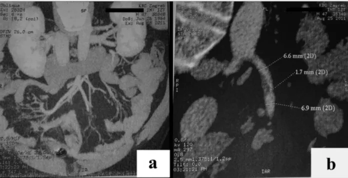
fIGurE 1.Multi-slice computer tomography (MSCT) angiography of the abdomen and pelvis (with i.v. contrast agent):
A) Normal diameter and flow through superior mesenteric, splenic and portal veins. Normal size and structure of the kidneys,
spleen, pancreas and biliary tract. B) Partial thrombosis, with narrowing of the lumen (from approx. 6.6 to approx. 1.7 cm), of the superior mesenteric artery approx. 2 cm from the starting point.
At that time the decision was made that the urgent surgical or percutaneous endovascular intervention would not be carried out. The option of thrombolysis was also discussed but was rejected. He was treated with wide spectrum antibiotics, pulses of methylpre -dnisolone and periodic transfusions of packed red blood cells. Warfarin was replaced with low molecular weight heparin. During the next few days pain in the abdomen persisted but there was no signs of acute peri-tonitis or intestinal necrosis. There was a gradual de-cline in acute phase reactants accompanied with an signi ficant elevation in liver enzymes and creatinine with mild proteinuria. There was a severe decline in the platelets count (min. 6x10⁹/L requiring transfusion)
and hemoglobin (min. 75 g/L when transfusion was conducted) with prolongation of aPTT. LAC and IgG anticardiolipine and beta2-glycoprotein I anti-bodies were again positive in high titer. Analysis of the bone marrow was performed and showed a regular tri-lineage hematopoesis. Autoimmune hemolytic anemia and heparine induced thrombocytopenia were ex-cluded. Initial blood smear showed no schistiocytes but regarding the above, mentioned and elevated le -vels of lactate dehydrogenase, microangiopathic ane-mia was suspected. There were no clinical or laborato-dismissed with the recommendation for oral
methyl-prednisolone (0.75 mg per kg/day with slow ta pering of the dose to 0.5 mg per kg/day), cyclo -phosphamide (2 mg per kg/day), acetylsalicylic acid (100 mg per day) and warfarin (targeted INR 2-3).
One month later he was admitted due to very strong acute abdominal pain. On first examination in the emergency department he was cardiorespiratory sta-ble, afebrile and his abdomen was distended, painful but there were no clinical signs of acute peritonitis. Laboratory results showed anemia (Hb 70 g/L, normal range 138-175 g/L), thrombocytopenia (T 78x109/L,
normal range 158-424x109/L), prolonged activated
partial thromboplastin time (aPTT 55.1 s, normal range 24-33 s), prothrombin time in therapeutic range (INR 2.2) with normal creatinine and urea with mildly ele-vated aminotransferases (AST 49 U/L, normal range 11-38 U/L and ALT 82 U/L, normal range 12-48 U/L) and g-GT (56 U/L, normal range <177 U/L) and nor-mal alkaline phosphatase and total bilirubin with ele-vated C-reactive protein (67,7 mg/L, normal range 0-5 mg/L). Additional tests were performed and mesente -ric CT angio graphy showed a partial thrombosis of su-perior mesenteric artery with 75% lumen occlusion (Fi gure 1).
ry sings of SLE activity. The diagnosis of CAPS was ma -de (Table II) and intravenous immunoglobulines in do se of 0.4 g/kg/day for five days were added to the therapy.
With combined therapy for CAPS the patient was stable but the permanent deterioration in renal function was recorded with permanent refractory thrombocy-topenia. Three weeks after IVIGs were introduced to the therapy two doses of rituximab (500 mg i.v.) were administered within a 7-day interval. There was no therapeutic response on administered therapy and fur-ther deterioration of renal function with persisting thrombocytopenia and anemia were recorded. Six day after the administration of the second dose of ritu ximab
patient become acutely dyspneic developing partial res-piratory insufficiency with the rise in serum acute phase reactants so he had to be moved to the intensive care unit. Computed tomography of the lungs was made and bilateral ground-glass infiltrates with zones of dense consolidation were verified. Bronchoscopy was also made and Candida albicans and Streptococcus species were isolated from lavage fluid. Cytome -galovirus DNA (1.78x10³copies/ml) was detected in
plasma using quantitative polymerase chain reaction (PCR) test. Therapy with piperacillin plus tazobactam, teicoplanin and fluconazole was initiated. Because of the possibility of CMV pneumonia therapy with gan-tAbLE II. crItErIA for dIAGnosIs of dEfInItIvE cAPs In our PAtIEnt
Involvement of three or more organs, systems Laboratory Confirmation by
and/or tissues (criteria 1) simultaneously or in less than confirmation of the histopathology of
a week (criteria 2) presence of aPLs small vessel
(LAC and/or aCLs) occlusion in at
– criteria 3 least one organ or
tissue – criteria 4
Kidney Reference Admission Relevant Positive LAC, ab2- GPI small vessel change in aCL-IgG (65 occlusion in lung first week GPL-U/ml) and ab2- sample from autopsy creatinine (79-125 mmol/L) 96 160 GPI (112 RU/ml)
urea (2.8-8.3 mmol/L7,6) 13,2 proteinuria (negative++) (0,75 g/L)
Hepatic AST (11-38 U/L) 49 184
ALT (12-48 U/L) 82 243
g-GT (< 177 U/L) 56 84
T. bilirubine (3-20 mmol/L) 9 26 Partial
thrombosis
Confirmed with MSCT angiography at admission of superior (Figure 1) mesenteric artery Other findings
prolonged activated partial thromboplastin time suporting
microangiopathic anemia and thrombocytopenia the diagnosis
of CAPS
FULFILLMENT OF 1st AND 2nd CRITERIA FULFILLMENT OF FULFILLMENT OF 3rd CRITERIA 4th CRITERIA DIAGNOSIS OF PROBABLE CATASTROPHIC ANTIPHOSPHOLIPID SYNDROME
DIAGNOSIS OF DEFINITIVE CATASTROPHIC ANTIPHOSPHOLIPID SYNDROME
CAPS, catastrophic antiphospholipid syndrome; aPL, antiphospholipid antibodies; aCL, anticardiolipin antibodies; LAC, lupus anticoagulant; ab2-GPI, antib2 glycoprotein 1; AST, aspartate aminotransferase; ALT, alanin aminotransferase; g-GT, gamma-glutamyl transferase; MSCT, multislice computed tomography
cyclovir was also applied. Partial clinical improvement with gradual decline in acute phase reactants was achieved but with persisting prolongation of aPTT, se-vere thrombocytopenia and anemia demanding peri-odic transfusions of blood products. Few days later sig-nificant decline in renal function with hyperkalemia, demanding the start of hemodyalisis, with severe pan-cytopenia (L 0.5, Hb 76, T 10) and raise in acute phase reactants were recorded. Transfusions of blood prod-ucts were given together with granulocyte colony-stim-ulating factor (G-CSF) filgrastim. Quantitative CMV PCR plasma test was repeated and came back negative so gancyclovir was excluded from thera py. Despite all measures taken, the patient was permanently pancy-topenic, in poor general condition. Soon he became hemodynamically unstable and despite supportive measures eventually died.
The autopsy was performed and evidence of small vessel thrombosis was found in the lung microvascu-lature (Figure 2). The criteria for the definite CAPS were fulfilled.
dIscussIon
At the present time, there are no formal recommendations and guidelines and no studies on the use of ri tuximab in the treatment of CAPS. In the current lite
-rature 11 reports of 13 patients with CAPS treated with rituximab can be found8-19. Berman et al. in their
sys-temic review included 9 more patients whose clinical cases were never published8. Only 2 of the published
patients had CAPS associated with known or suspect-ed systemic lupus erythematosus12,15. None of them met
the criteria for definite CAPS. Our patient is the first ever to be reported with definite CAPS associated with SLE and treated with rituximab. Reported patient did not respond to the first line therapy for CAPS (gluco-corticoides, anticoagulation and IVIGs). Treatment with rituximab was also ineffective (Table III) and the patient eventually died from refractory CAPS and com-plications.
It is important to emphasize a number of factors which probably contributed to the ineffectiveness of the used therapeutic protocol. As already mentioned, patients with CAPS associated with SLE have higher mortality rates than patients with primary CAPS. Treat-ment with cyclophosphamide has a favourable effect in the treatment of this subgroup of patients. In the des cribed patient, cyclophosphamide and plasma-pheresis were not used in the first line of therapy. Con-sidering triple positive aPLs in high titers, not using the plasmapheresis could be an important contributing fac-tor to the ineffectiveness of therapy. In addition it is im-portant to say that the initially verified partial throm-bosis of superior mesenteric artery was not resolved during the course of disease. This could be a very im-portant factor in the sustaining of pathogenetical mecha nisms responsible for development of CAPS con-sequently leading to ineffectiveness of used therapy. Furthermore, time between the first line of the thera-py and the introduction of rituximab was relatively long. Prolonged course of the disease has lead to the de-velopment of infectious complications that probably also contributed to sustaining pathogenetical mecha-nisms responsible for development of CAPS.
Whether more “aggressive” approach in dealing with sustaining factors and simultaneous use of all current-ly available therapeutic options as first line therapy would have led to a greater effect or would have re-sulted in earlier death is not possible to conclude at this time.
concLusIon
We presented the first ever reported case of definitive CAPS associated with SLE treated with rituximab. In re-fIGurE 2.Hematoxylin and eosin stain, objective 10x, inlet
objective 40x. Presentation of infarcted (necrotic) area of the lung where parenchymal architecture can hardly be recognized, some alveolar spaces in the upper parts are filled with
abundant fibrin, interalveolar septa completely necrotic, somewhere even completely missing. Small blood vessel with the remnant of fibrinous thrombus in the middle of inlet figure, favoring thrombosis as the most probable cause of pulmonary hemorrhagic infarction in our patient.
ported patient combined therapeutic protocol (sys-temic antibiotics, glucocorticoids, anticoagulation, IVIGs) with rituximab was ineffective. We listed a num-ber of factors that could have influenced the final result. The right timing for the introduction of rituximab is still uncertain. Whether the course of the disease would have been changed if the rituximab was started earlier is also unknown. There is a great need for further in-vestigation to evaluate the effectiveness of rituximab in treatment of CAPS and to define the optimal regime and timing of implementation. It is also necessary to de-termine possible differences in effectiveness on patients with primary CAPS and CAPS associated with SLE and to determine possible other prognostic factors that could influence therapeutic decisions and results.
corrEsPondEncE to
Sukara G
Omladinska 24, 10310, Ivanic Grad E-mail: gsukara@gmail.com
rEfErEncEs
1. Asherson RA. The catastrophic antiphospholipid (Asherson’s) syndrome. Autoimmun Rev 2006; 6:64–67.
2. Asherson RA, Cervera R, de Groot PG, et al. Catastrophic anti-phospholipid syndrome: international consensus statement on classification criteria and treatment guidelines. Lupus 2003; 12(7):530–534.
3. Espinosa G, Cervera R, Asherson RA. Catastrophic antiphos-pholipid syndrome and sepsis. A common link? J Rheumatol 2007; 34(5):923–926.
4. Bucciarelli S, Espinosa G, Cervera R, et al. Mortality in the ca-tastrophic antiphospholipid syndrome: causes of death and prognostic factors in a series of 250 patients. Arthritis Rheum 2006; 54:2568-2576.
5. Espinosa G, Bucciarelli S, Asherson RA, et al. Morbidity and mortality in the catastrophic antiphospholipid syndrome: pa-tophysiology, causes of death, and prognostic factors. Semin Thromb Haemost 2008; 34:288-292.
6. Bayraktar UD, Erkan D, Bucciarelli S, et al. Catastrophic Anti-phospholipid Syndrome Project Group. The clinical spectrum of catastrophic antiphospholipid syndrome in the absence and presence of lupus. J Rheumatol 2007; 34(2):346-352. 7. Espinosa G, Berman H, Cervera R. Management of refractory
ca-ses of catastrophic antiphospholipid syndrome. Autoimmun Rev 2011; 10:664–668.
8. Berman H, Rodríguez-Pintó I, Cervera R, et al. Rituximab use in the catastrophic antiphospholipid syndrome: Descriptive analysis of the CAPS registry patients receiving rituximab. Au-toimmun Rev 2013; 12(11): 1085-1090.
9. Rubenstein E, Arkfeld DG, Metyas S, et al. Rituximab treatment for resistant antiphospholipid syndrome. J Rheumatol 2006;33(2):355-357.
10. Manner H, Jung B, Tonassi L, et al. Successful treatment of ca-tastrophic antiphospholipid syndrome (CAPS) associated with splenic marginal-zone lymphoma with low-molecular weith he-parin, rituximab and bendamustine. Am J Med Sci 2008;335(5): 394-397.
11. Van Wissen S, Bastiaansen BA, Stroobants AK, et al. Catastro -phic antiphospholipid syndrome mimicking a malignant pan-creatic tumour - a case report. Lupus 2008;17(6):586-590. 12. Haque W, Kadikoy H, Pacha O, et al. Osteonecrosis secondary
to antiphospholipid syndrome: a case report, review of the lite-rature, and treatment strategy. Rheumatol Int 2010;30(6):719--723.
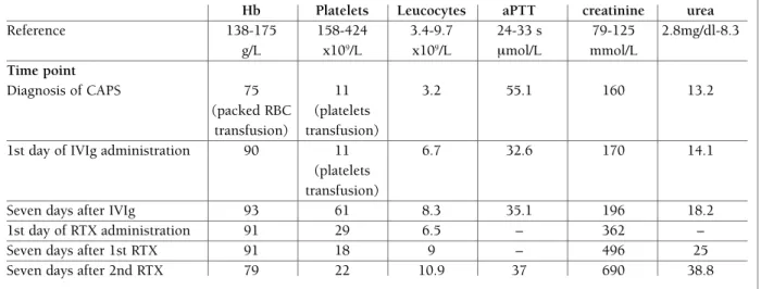
tAbLE III. rELEvAnt LAborAtory rEsuLts bEforE And AftEr thE APPLIcAtIon of IntrAvEnous ImmunoGLobuLIns And rItuxImAb suGGEstInG thE fAILurE of thErAPy
Hb Platelets Leucocytes aPTT creatinine urea
Reference 138-175 158-424 3.4-9.7 24-33 s 79-125 2.8mg/dl-8.3 g/L x109/L x109/L mmol/L mmol/L Time point Diagnosis of CAPS 75 11 3.2 55.1 160 13.2 (packed RBC (platelets transfusion) transfusion)
1st day of IVIg administration 90 11 6.7 32.6 170 14.1
(platelets transfusion)
Seven days after IVIg 93 61 8.3 35.1 196 18.2
1st day of RTX administration 91 29 6.5 – 362 –
Seven days after 1st RTX 91 18 9 – 496 25
Seven days after 2nd RTX 79 22 10.9 37 690 38.8
Hb, hemoglobin; aPTT, activated partial thromboplastin time; CAPS, catastrophic antiphospholipid syndrome; IVIg, intravenous immunoglobulin
13. Asherson RA, Espinosa G, Menahem S, et al. Relapsing catas-trophic antiphospholipid syndrome: report of three cases. Se-min Arthritis Rheum 2008;37(6):366-72.
14. Nageswara Rao AA, Arteaga GM, Reed AM, et al. Rituximab for successful management of probable pediatric catastrophic an-tiphospholipid syndrome. Pediatr Blood Cancer 2009;52(4): 536-538.
15. Ketari Jamoussi S, Zaghdoudi I, Ben Dhaou B, et al. Catastro -phic antiphospholipid syndrome and rituximab: a new report. Tunis Med 2009; 87(10):699-702.
16. Iglesias-Jimenez E, Camacho-Lovillo M, Falcon-Neyra D, et al. Infant with probable catastrophic antiphospholipid syndrome successfully managed with rituximab. Pediatrics 2010;125(6): e1523-1528.
17. Haskin O, Amir J, Schwarz M, et al. Severe abdominal pain as a presenting symptom of probable catastrophic antiphospholi-pid syndrome. Pediatrics 2012;130(1):e230-235.
18. Camacho-Lovillo S, Bernabeu-Wittel J, Iglesias-Jimenez E, et al. Recurrence of cutaneous necrosis in an infant with probable ca-tastrophic antiphospholipid syndrome. Pediatr Dermatol 2013;30(4):e63-64.
19. Shiber S, Molad Y. Catastrophic antiphospholipid syndrome: a case series. IMAJ 2013;15:549-552.