B C E L L S
:
F R O M T H E B E N C H T O T H E C L I N I C A L P R A C T I C ERita Moura,
*Ana Água-Doce,
**Pamela Weinmann,
*Luís Graça,
**João Eurico Fonseca
*,***apenas um tipo de anticorpo quando estimulada por um antigénio, tendo a capacidade de se multi-plicar e originar várias células B todas com a mes-ma especificidade antigénica (teoria da selecção clonal). Contudo, o seu importante papel nas res-postas imunitárias adaptativas deve-se à capaci-dade surpreendente de reconhecer um conjunto ilimitado de antigénios, devido a mecanismos de diversidade de anticorpos, como a recombinação V(D)J, mudança de classe e hipermutação somáti-ca. As células B são também células apresentado-ras de antigénio que activam as células T, aumen-tando a eficácia da resposta imune. A manutenção da tolerância imunitária através de delecção clonal, anergia e edição do receptor é necessária para evi-tar condições patológicas, como doenças nes. As células B podem contribuir para a autoimu-nidade através da produção de autoanticorpos, sín-tese de citocinas, apresentação de antigénios, ac-tivação de células T e desenvolvimento de folículos. Palavras-Chave: Células B; Medula óssea; Anticor-po; Tolerância; Autoimunidade.
Introduction
The human body contains approximately 2×10¹² lymphocytes, of which 5-15% are B cells. B lym-phocytes are small white blood cells (6-10 µm) with a dense nucleus and little cytoplasm that origina-te and mature in the bone marrow expressing a membrane-bound immunoglobulin. Discovered in the early 1960’s, these cells are important mediators of our immune system, more precisely the humo-ral response, by the production and secretion of proteins collectively called immunoglobulins (Ig) or antibodies. The main function of an antibody is to recognize a foreign antigen exhibited by an in-vading pathogen or at the surface of an altered cell, like a tumour cell, and facilitate the clearance and elimination of that antigen. Antibodies can elimi-nate pathogens by several mechanisms including
*Unidade de Investigação em Reumatologia, Instituto de Medicina Molecular, Faculdade de Medicina da Universidade de Lisboa **Unidade de Imunologia Celular, Instituto de Medicina Molecular, Faculdade de Medicina da Universidade de Lisboa
***Serviço de Reumatologia e de Doenças Ósseas Metabólicas do Hospital de Santa Maria
Abstract
B cells develop in bone marrow and undergo anti-gen-induced activation and terminal differentiati-on in germinal centres of secdifferentiati-ondary lymphoid or-gans. Each B cell is a clone, which means that an in-dividual B cell has a unique genetic code and pro-duces only one type of antibody when stimulated by antigen, being able to multiply itself and origi-nate several B cells with the same antigen specifi-city (clonal selection theory). However, their im-portant role in adaptive immune responses is sup-ported by the remarkable capacity of recognizing an unlimited array of antigens, due to mechanisms of antibody diversity, such as V(D)J recombination, class switching and somatic hypermutation. B cells can also function as antigen presenting cells that can activate T cells, improving the effectiveness of the immune response. Immune B cell tolerance surveillance through clonal deletion, anergy and receptor editing is also necessary to avoid patholo-gical conditions, like autoimmune diseases. B cells can contribute to autoimmunity by autoantibody production, cytokine synthesis, antigen presenta-tion, T cell activation and ectopic lymphogenesis. Key-Words: B Cells; Bone Marrow; Antibody; Tole-rance; Autoimmunity.
Resumo
As células B desenvolvem-se na medula óssea e di-ferenciam-se terminalmente nos centros germi-nais dos órgãos linfóides secundários. Cada célula B é um clone, o que significa que uma célula B in-dividual tem um codigo genético único e produz
B C E L L S: F R O M T H E B E N C H T O T H E C L I N I C A L P R A C T I C E
opsonization, complement activation and direct lysis of bacteria and neutralization of viruses and toxins to prevent their entry into host cells. B cells can also function as antigen presenting cells (APCs) and interleukin (IL) producing cells that can further activate T cells and contribute to the development of an effective immune response.
Origin and B cell development
Bone marrow
B cells develop in the bone marrow in mammals, but in birds, where they first were studied, these cells de-velop and mature in a special organ called «Bursa of
Fabricius» (hence, the designation «B»), situated in
the cloaca of the animals.1,2In humans, B cells are primarily produced in the fetal liver before birth and in the bone marrow afterwards. B cell development can be detected in human bone marrow from 20 weeks of gestation and continues throughout life.3 B cells arise from a lymphoid stem cell in the bone marrow and proceed through several maturation stages, during which they express different cell sur-face markers (Figure 1).4-7The earliest distinctive B--lineage cell is the progenitor B cell (pro-B cell).8-11 Pro-B cells proliferate in the bone marrow, filling the extravascular spaces between large sinusoids in the shaft of a bone, and further differentiate into precursor B cells (pre-B cells).12,13Human pre-B cells can be generally subdivided into large proliferating cells (designated pre-BI) and small postmitotic cells (designated pre-BII) on the basis of cell-cycle analy-sis.14-16The bone marrow phase of B cell develop-ment culminates in the production of an immatu-re B cell17 that is not fully functional. All B cell diffe-rentiation stages in the bone marrow do not requi-re antigen, this correqui-responding to the antigen--independent phase of B cell development.
Periphery
Organization and structure of secondary lymphoid organs
LYMPHNODES
Lymph nodes (LNs) are specialized organs for trap-ping antigen from local tissues supplied by lympha-tic vessels. LNs can be divided in three regions: the cortex, paracortex and medulla.18,19During lymph node development, hematopoietic lymphoid pro-genitor cells interact with high endothelial venules (HEV) and mesenchymal cells. This interaction is mediated by adhesion molecules and leads to
lo-cal release of chemoattractants, like CXCL13, that attract B cells into the nascent organ through HEV.20-22These naïve B cells are then organized into follicles, which are spheroid aggregates located in the cortex of lymph nodes, surrounded by an ou-ter T cell zone.23In the presence of T cell help, B cells can proliferate and differentiate into GC B cells within follicles or short-lived plasma cells, ge-nerated outside follicles.
SPLEEN
The spleen is a large organ specialized in the des-truction of old erythrocytes and trapping antigens in the circulating blood.24The spleen is divided into two zones: the red pulp, mainly populated by ma-crophages and red blood cells, where old and de-fective erythrocytes are removed from the circula-tion; and the white pulp, that comprises B cell fol-licles, T cell zones (also designated periarteriolar lymphoid sheaths, or PALS) and a marginal zone, in the interface of the white and red pulps, that contains macrophages and B cells.19,25In humans, the spleen has an additional region, the perifolli-cular zone, situated outside the marginal zone, that contains capillaries and blood-filled spaces that belong to the open splenic circulation.26 Lympho-cytes are believed to enter the white pulp through Figure 1. B cell developmental stages in bone marrow (BM) and periphery. During B cell development, B lymphocytes express different cell surface markers, specific for each stage.The BM phase culminates with the expression of IgM and IgD. B cells then leave BM and circulate through the blood and lymph towards secondary lymphoid organs (lymph nodes, spleen and Peyer’s Patches), where they reside until they are activated by an antigen. B cells can then differentiate into antibody secreting plasma cells, or memory B cells.
R I TA M O U R A E C O L.
the perifollicular zone. Circulating naïve B cells mi-grate to the spleen, where they reside until they are activated by antigens and start a GC reaction. PEYER’SPATCHES
Peyer’s Patches (PPs) are organized lymphoid fol-licles similar to lymph nodes located in the muco-sa and submucomuco-sa of the small intestine, especially the ileum. Since the lumen of the gastrointestinal tract constitutes a way of entry of potentially pa-thogenic microorganisms, PPs are fundamental for the establishment of the immune surveillance in the gut. Human PPs can be even centimetres big and have a large number of follicles.27PPs contain specialized cells called M («microfold») cells, lo-cated in the intestinal epithelium over lymphoid follicles.28The M cell basolateral membrane is de-eply invaginated to form a large intraepithelial «pocket» containing B and T lymphocytes29and occasional dendritic cells. M cells can internalize antigens from the lumen and transfer them effi-ciently to the underlying APCs that can further ac-tivate T cells. Acac-tivated T cells produce cytokines, such as TGF-β1, that induce B cells to differentia-te into immunoglobulin A (IgA)-secreting plasma cells.30,31IgA is the major antibody present in the in-testinal secretions32,33and it constitutes the first line of defence against pathogenic microorganisms in mucosal surfaces by interfering with adhesion and invasion properties of microbes. Signals through chemokine receptors expressed by B cells, namely CXCR4, CXCR5 and CXCR7, contribute to B cell homing towards PPs.20Moreover, CXCL13, expressed by HEVs in PPs, is required for B cell en-try into these structures.21,22
The Germinal Centre (GC) reaction
When full maturation is achieved, signalled by the co-expression of IgM and IgD on the membrane, B cells leave the bone marrow. These naïve B cells, which have never encountered an antigen, circu-late in the blood and lymphatic systems where they are carried to secondary lymphoid organs, where they reside. In the follicles of the secondary lym-phoid organ, after encountering an antigen, matu-re B cells transform into large B-blasts that may follow two different pathways. Some proliferate and differentiate into short-living, IgM producing plasma cells, responsible for the early production of antibodies and, thus, first-line defence against the antigen. A minority of the B-blasts differentia-te to form the follicle centre or germinal centre
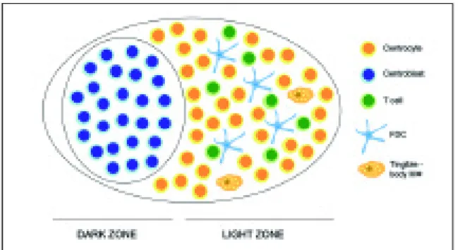
(GC).34,35The non-antigen-triggered naïve B cells form the follicle mantle or mantle zone. The folli-cle, containing a GC and a mantle zone, is known as a secondary follicle. The inner part of the man-tle zone of the B cell follicle is designated the lym-phocytic corona and is mainly composed of ma-ture, naïve B cells.36The outer part of the mantle zone, called the marginal zone, is populated by dif-ferent types of cells: macrophages, some T cells, granulocytes, plasma cells and marginal zone B cells.37GC are the specialized sites for memory B cell generation, plasma cells differentiation and where affinity maturation of serum antibodies takes place.38,39GC first appear at day 4-5 post-im-munization, achieve their maximum cell number by day 10-11 and decline after about three weeks.34 B cells that form the GC are divided into centrob-lasts (large noncleaved cells) and centrocytes (small or large cleaved cells). Centroblasts are lar-ge, proliferating cells that lack surface immuno-globulin expression and accumulate at one pole of the GC, forming the dark zone (Figure 2). These cells can differentiate themselves into non-proli-ferating centrocytes that re-express surface immu-noglobulin and accumulate at the opposite pole of the GC, known as the light zone.40Activation of GC B cells is made by follicular dendritic cells (FDC), which have a pivotal role in promoting B cell pro-liferation and differentiation in GC.41,42FDC not only are very efficient in trapping and retaining an-tigen-antibody complexes for long periods of time and present them to B cells, but also are involved
Figure 2. V(D)J recombination process in light and heavy chains. During VDJ recombination, the gene segment rearrangements that occur in both light and heavy chains are possible due to specific enzymes, such as RAG proteins, that initiate the process. Further diversity is increased by nucleotide additions by Terminal deoxynucleotidyl transferase (TdT).
in B cell recruitment to secondary lymphoid or-gans with consequent follicle formation and orga-nization through the secretion of CXCL13, a B-lym-phocyte chemoattractant.35,43,44Positive selection of B cells in GC occurs through high-affinity bin-ding to antigens trapped by FDC. These high-affi-nity B cells receive survival signals from both FDC42 and T cells45located in the light zone, whereas low-affinity B cells non-triggered by antigens die by apoptosis. More than 90% of the GC B cells die as a result of apoptosis46and are then phagocytosed and digested by the so-called tingible body ma-crophages.47-49Only the GC B cells that are stimu-lated and able to synthesize high-affinity antibo-dies survive and may differentiate into long-lived plasma cells or memory B cells. Long-lived plasma cells are thought to reside mainly in the bone mar-row and organs that are directly exposed to foreign antigens (gastrointestinal tract, lungs…),50 wheas memory cells reside in the follicle mantle or re-circulate freely to survey for secondary antigen ex-posure. Memory B cells are long-lived and are res-ponsible for the ability of the immune system to le-arn. During a second encounter with an antigen, memory B cells are able to recognize the antigen to which the organism was previously exposed and induce a more rapid and effective immune respon-se.51Since B cell activation and differentiation in the periphery require antigen, this stage compri-ses the antigen-dependent phase of B cell develop-ment. In the absence of antigen-induced activa-tion, naïve B cells in the periphery have a short life span, dying within a few weeks by apoptosis. B-1 and B-2 cell lineages
B cells comprise two main subsets, B-1 and B-2, a concept based on the discovery that a small pro-portion of B cells in mice expressed the T-cell mar-ker CD5.52-55B-1 cells are the CD5+subpopulation, whereas B-2 cells represent the conventional B cell subpopulation. B-1 cells arise before B-2 cells, are long-lived, emerge early in development, du-ring fetal life, are self-renewing and predominate in the follicular mantle zone, being minor popula-tions in secondary tissues such as lymph nodes and spleen and are virtually absent in human adult bone marrow.56B-1 cells can be subdivided in B-1a cells, which express CD5, and B-1b cells, that are CD5 negative. B-1a cells are largely responsible for the production of circulating immunoglobulin M (IgM) antibodies known as «natural antibodies».57,58 These low affinity antibodies are the first line of
defence against bacterial and viral infections.59The antibodies produced by B-1b cells are induced af-ter antigenic exposure and are required for long-lasting protective immunity to pathogens.60 B-2 cells constitute the major group of B cells in hu-mans and mice. They are CD5, surface and mRNA, negative cells. B-2 cells derive from resident bone marrow progenitors throughout life and undergo maturation primarily in the spleen. Although the developmental origin of B-1 cells has remained an unresolved issue, it was demonstrated that bone marrow CD45Rlo-negCD19+cells include a B-1 cell--specified progenitor,61which supports the idea of the existence of distinct developmental pathways for B-1 cells. CD5 expression was identified in ma-lignant human B cells62and later shown to mark a minority of B lymphocytes in the normal blood.52 In fact, in humans, 5-30% of the circulating B cells are B-1a in adults; 4-6% are B-1b and 65-89% are B-2 cells.63 Nevertheless, a separate lineage for CD5+B cells still awaits a demonstration in hu-mans. Some lines of evidence suggest that in the human case CD5 might be an activation marker ra-ther than a cell lineage feature, since ra-there are a va-riety of stimuli capable of inducing the expression of CD5 on conventional B cells.64,65It remains to be determined, nonetheless, the criteria that will de-fine whether human B cell subsets are homolo-gous to the mouse B-1a, B-1b and B-2 lineages.
Antibody production
Structure of an antibody
Antibodies, also called immunoglobulins, are the antigen-binding proteins present on the B cell membrane and secreted by plasma cells. An an-tibody molecule has a common structure of four peptide chains that consist of two identical light (L) chains and two identical heavy (H) chains, which are linked by disulfide bonds. The first 100-110 amino acids of the amino-terminal region of a light or heavy chain vary greatly among antibodies. The-se The-segments of highly variable The-sequence are called
variable (V) regions: VL in light chains and VHin heavy chains. The carboxyl-terminal region of both light and heavy chains consists of relatively cons-tant sequences, designated as constant (C) regions:
CLon the light chain and CHon the heavy chain. Most of the differences among antibodies fall wi-thin areas of the V regions called Complementary--Determining Regions (CDRs) and these CDRs, on
both light and heavy chains, constitute the antigen binding site of the antibody molecule. There are two light chain types, kappa (κ) and lambda (λ). In humans, 60% of the light chains are kappa and 40% are lambda.66A single antibody molecule contains only one light chain type, either κ or λ, never both. The heavy chain of a given antibody molecule de-termines the class of that antibody, or isotype. The-re aThe-re five diffeThe-rent heavy-chain constant The-region sequences: µ, δ, γ, ε and α; hence, in humans, the five major classes of antibodies: IgM (µ), IgG (γ), IgA (α), IgD (δ) and IgE (ε), each with different structu-ral and functional properties (Table I).
Mechanisms of antibody diversity
One of the most remarkable features of our immu-ne system is its ability to respond to an apparently limitless array of foreign antigens. The major me-chanisms that are responsible for the enormous diversity of antibodies (>1010possibilities) produ-ced by our immune system are random rearrange-ments in genes that codify the variable region (an-tigen binding domain) of immunoglobulins. Anti-body diversity is achieved by V(D)J recombination, somatic hypermutation and class switching. V(D)J recombination
Immunoglobulin light and heavy chains are
enco-ded by three separate multigene families, situated on different chromosomes. In germ-line DNA, each of these multigene families contains several coding sequences, called gene segments, separa-ted by noncoding regions. During B cell matura-tion, these gene segments are rearranged and brought together to form functional immunoglo-bulin genes.67,68The κ and λ light-chain families contain V (for variable), J (for joining) and C (for constant) gene segments and the rearranged VJ
segments encode the variable region of the light chains. The heavy-chain family contains V, D (for
diversity), J and C gene segments and the
rearran-ged VDJ gene segments encode the variable region of the heavy chain. In each gene family, C gene seg-ments encode the constant region. V(D)J recombi-nation is an ordered site-specific DNA rearrange-ment process69that occurs in developing lympho-cytes in the bone marrow initiated by two lym-phocyte-restricted specific proteins RAG-1 and RAG-2,70-73which together form an endonuclease responsible for DNA double-stranded breaks at re-combination signal sequences (RSS).74,75RAG pro-teins are expressed at high levels during ontogeny, but their expression diminishes in immature B cells and is usually absent in recirculating mature naïve B cells.76,77In certain pathologies, however, like autoimmune diseases, RAG expression may be
Table 1. Classes of immunoglobulins and their biological functions.
Class of Serum
Antibody levels Structure Biological functions
Membrane-bound immunoglobulin on the surface of immature and mature B cells
IgM 5% Monomer First antibody produced in a primary response to an antigen Pentamer First antibody produced by the fetus
Efficient in binding antigens with many repeating epitopes, such as viruses Classical complement activation
IgD 0.3% Monomer Membrane-bound immunoglobulin on the surface of mature B cells No biological effector function known
Predominant antibody class in secretions (saliva, tears, breast
IgA 7-15% Monomer milk) and mucosa
Dimer First line of defence against infection by microorganisms Most abundant class with four isotypes - IgG1, IgG2, IgG3, IgG4
IgG 85% Monomer Crosses the placenta
Opsonization
Defence against parasite infections
IgE 0.02% Monomer Associated with hypersensitivity reactions (allergies) Found mainly in tissues
up-regulated.78,79The importance of RAG for V(D)J recombination is supported with the finding that mutations in RAG genes abolish recombination activity and block differentiation of both B and T cells, leading to different forms of immune defi-ciency.80-82RSS, recognized by RAG in the initial event of the recombination process, are comprised of a highly conserved heptamer and nonamer se-quences, separated by a relatively nonconserved spacer region of either 12 or 23 base pairs (bp).75,83 A gene segment with 12-bp spacer can only join with a segment flanked by 23-bp spacer (12-23 bp rule).74,84During variable-region gene rearrange-ments the heavy-chain genes rearrange first, fol-lowed by light-chain rearrangements. The genera-tion of a funcgenera-tional immunoglobulin heavy-chain gene requires two separate rearrangement events within the variable region. The first step of recom-bination is a D to J association. The resulting D-J segment then moves next to and joins a V segment to generate a VDJ unit that encodes the entire va-riable region. Light-chain DNA rearrangements oc-cur by the joining of a functional V segment to a functional J-C segment (Figure 3). The combinato-rial diversity generated by V(D)J recombination is further augmented by another enzyme, terminal deoxynucleotidyl transferase (TdT),85-87that ran-domly adds up to 20 nucleotides, called N addi-tions, to D-J and V-DJ junctions.
Somatic Hypermutation
Somatic Hypermutation (SHM) is a major mecha-nism for producing antibody diversity and
increa-sing antibody affinity, which occurs in GC of secon-dary lymphoid organs.88-90SHM is a process that in-troduces point mutations, mainly nucleotide subs-titutions, but also occasional deletions and du- pli-cations, at a very high rate into the DNA of heavy and light-chain variable-region genes, which may alter the specificity of the encoded antibodies.91-93 SHM introduces mutations at a rate of ~10-3 mu-tations per base pair per cell division, which is 106 -fold higher than the spontaneous mutation rate in somatic cells. As a result, following exposure to an-tigen, these B cells with higher-affinity receptors will be preferentially selected for survival. This dif-ferential selection is due to an increase in antibody affinity for an antigen, a process called affinity ma-turation. Thus, only the B cells with the best fitting receptors are selected for rapid proliferation and further maturation. SHM appears early in the phylogeny of vertebrate immune system and ope-rates in all studied vertebope-rates.94Although the me-chanism of SHM remains unknown, excitement in this field was triggered by the discovery of a requi-rement for activation-induced cytidine deaminase (AID), which has homology with the RNA-editing cytidine deaminase APOBEC-1.95,96
Class Switching
Class or isotype switching is a deletional DNA re-combination process that occurs in mature B cells and consists in the replacement of an expressed heavy-chain constant-region gene (usually Cµfor IgM) with another one with a different biological function, allowing expression of IgG, IgA or IgE.97 The deleted sequence is excised in a form of circu-lar DNA.98In this way, the antigen binding specifi-city of the antibody remains unaltered, but its ef-fector functions are changed. This process is me-diated by a recombination event that deletes the DNA between repeated sequences («switch re-gions») located upstream of the constant-region genes involved. AID catalyzes the hydrolytic dea-mination of cytidine to uridine, either as a free nu-cleotide or in the context of RNA and participates in CSR.99,100In fact, AID-deficient mice, generated by gene-targeted mutation, lack completely the ability to undergo CSR and only form antibodies belonging to IgM isotype.95Moreover, in a group of immunodeficient patients with hyper-IgM syndro-me, who had impaired CSR, the human gene en-coding AID was mutated,96so their B cells only pro-duced IgM molecules. AID is expressed specifically in GC-centroblasts,99but its exact role is unknown. Figure 3. Structure of a germinal centre (GC). GCs are
divided in a dark zone, where centroblasts predominate, and a light zone, mainly populated by centrocytes, follicular dendritic cells (FDC),T cells and tingible – body
macrophages (FDC and tingible – body macrophages symbols were adapted from Rheugulation Database, a knowledge base on rheumatoid arthritis,
Importantly, it was demonstrated that AID defi-ciency also affects SHM in mice and humans, which supports the idea that AID is involved in both mechanisms, CSR and SHM.95,101
B cell activation
B cell receptor (BCR) and B cell co-receptor
Signals propagated through the BCR are vital for the development and survival of B lymphocytes in both the bone marrow and the periphery. The BCR of mature B cells is a multiprotein structure con-taining an antigen-binding membrane immuno-globulin, noncovalently associated with signal transducing elements Ig-α (CD79a) and Ig-β (CD79b), that form an heterodimer, localized mostly or wholly within the cell.102,103The cytoplas-mic tails of both Ig-α and Ig-β contain an 18-resi-due motif termed the immunoreceptor tyrosine-based activation motif (ITAM).104BCR signalling is initiated upon binding of antigen to membrane immunoglobulin, which induces a subsequent phosphorylation of ITAMs of Ig-α and Ig-β that, in turn, transduce the stimulus into an effective in-tracellular signal.104-106The activating signals trans-mitted through the BCR during B cell stimulation can also be amplified by a B cell co-receptor. The B cell co-receptor is a complex of three proteins: CD19, CR2 (CD21) and CD81.107-109CD19 is a mem-ber of the immunoglobulin superfamily; CR2 is a receptor for C3d, a breakdown product of the com-plement system, which is an important effector mechanism for destroying invaders; and CD81 is another transmembrane protein, also designated as a target for anti-proliferative antigen-1 (TAPA--1). Following antigen cross linking of the BCR and activation of B cell co-receptors, a cascade of phosphorylations is initiated by protein tyrosine kinases (PTKs)103,110-113that culminate in the activa-tion of transcripactiva-tion factors114and the release of intracellular Ca2+ 113,115that leads to B cell activation. In addition to the stimulatory co-receptor, there are also inhibitory receptors expressed on B cells. Each inhibitory receptor contains one or more im-munoreceptor tyrosine-based inhibitory motifs (ITIMs) within its cytoplasmic domain, essential for generation and transduction of inhibitory sig-nals. The surface molecules CD22 and FcγRIIb are examples of inhibitory B cell surface receptors. CD22 is a member of the immunoglobulin gene superfamily that is first expressed in the cytoplasm
of pro-B and pre-B cells and, later, is expressed on the surface of mature B cells. CD22 functions as a B cell inhibitor,116,117although there are studies that indicate that it has a dual role in B cell activation,118 FcγRIIb is also a member of the immunoglobulin superfamily and it is a low-affinity receptor for the Fc portion of IgG.119Fc
γRIIb is exclusively expres-sed on B cells and has the potential to terminate B cell signal transduction. Coligation of FcγRIIb to the BCR leads to tyrosine phosphorylation of the ITIM by the tyrosine kinase Lyn, recruitment of phosphatase SHIP and inhibition of Ca2+influx and proliferation.120
B cell – T cell interactions
For an efficient activation of B cells by soluble pro-tein antigens, an involvement of T cells is required. Binding of an antigen to the BCR does not, by it-self, induce an effective competent signal without additional interactions between numerous recep-tor-ligand pairs on the two cell types. Antigen cap-ture by membrane immunoglobulin on B cells ini-tiates signalling through the BCR (signal 1) that le-ads to an up-regulation of the expression of class II Major Histocompatibility Complex (MHC) mo-lecules121,122and costimulatory B7 molecules.123-125 Antigen is internalized, processed and degraded to peptides that are then presented to the cell sur-face as MHC-peptide complexes.126,127T cells can re-cognize these MHC-peptide complexes on B cell membrane. Both T and B cells interact to form a T-B conjugate that ultimately leads to T cell-depen-dent B cell activation.23,125,128This interaction sti-mulates the expression of CD40-L (CD154) on the T cells, which is the ligand for CD40, expressed on B cells.129,130CD40, expressed on B-lineage cells (pro-B through plasma cells), belongs to the tu-mour necrosis factor (TNF) family of cell-surface proteins and soluble cytokines that regulate cell proliferation. CD40L, expressed on T cells, belongs to the TNF receptor (TNFR) family. Interaction of CD40-L with CD40 on the B cell delivers a signal (signal 2)131-133to the B cell that, in concert with the signal generated by the BCR crosslinkage, induces the expression of costimulatory molecules of the B7 family.134,135The B7 costimulatory family has two ligands, B7-1 (CD80) and B7-2 (CD86) that can ac-tivate or inhibit a T-cell response, depending on the membrane molecule on T cell with which they interact, CD28 or CTLA-4.136,137CD28 is expressed by both resting and activated T cells and is the predominant receptor for B7 on resting T cells.
CTLA-4 is expressed only on activated T cells. Upon binding of a B7-costimulatory molecule with CD28, a positive costimulatory signal is generated and the T cell is activated, while signalling through CTLA-4 inhibits the response of the T cell.138So, for an efficient T-cell-dependent B cell activation a B7-CD28 interaction has to occur. The Inducible Costimulator Molecule (ICOS), a member of the CD28 family of costimulatory molecules expres-sed by T cells,139is also a key regulator of humoral immunity and B cell homeostasis and functions. ICOS binds to a B7-like molecule, B7RP-1,140,141 ex-pressed constitutively on B cells.142A major role for the ICOS/B7RP-1 pathway in the T cell-dependent B cell responses is supported by studies with both ICOS-/-and B7RP-1-/-mice that have decreased le-vels of serum antibodies and defective GC forma-tion.143-146Once activated, B cells begin to express membrane receptors for various cytokines produ-ced by the interacting T cell. The signals produprodu-ced by these cytokine-receptor interactions induce a number of intracellular signalling pathways that ultimately result in changes in gene expression that support B cell proliferation and can induce diffe-rentiation into plasma and memory cells, class switching and affinity maturation. Also, it has been demonstrated that activated B cells by sequential BCR and CD40 stimulation proliferate and secrete TNF-α, lymphotoxin and IL-6, cytokines that can act not only as autocrine growth and differentiati-on factors, but also serve to amplify the differentiati-ongoing immune response.147
B cells and Toll-like receptors
Toll-like receptors (TLRs) are pattern recognition receptors that sense molecular patterns specific of invading pathogens. Eleven TLRs have been iden-tified so far, although in humans TLR11 is thought to be non-functional.148Recent evidence suggest that the generation of T-dependent B cell respon-ses requires the activation of TLRs in B cells.149,150 The regulated expression of selected TLRs in B cells may play an important role in linking innate to adaptive immune responses. Human B lympho-cytes express a distinct TLR expression profile in which TLR9 and TLR10 predominate.151In naïve B cells most TLRs (TLR1-2 and TLR6-10) are expres-sed at low to undetectable levels, but the expressi-on of TLR9 and TLR10 is rapidly induced following BCR triggering. In contrast, memory B cells express several TLRs at constitutively high levels.152 Un-methylated CG-dinucleotides with certain
sequen-ce contexts (CpG DNA), which function as TLR9 li-gand, are recognized by our immune system as fo-reign DNA (bacterial or viral) and it has been de-monstrated that CpG DNA motifs can trigger pro-liferation and activation of primary human B cells.153In fact, CpG DNA increases B cell expres-sion of costimulatory molecules (CD80, CD86) and enhances the expression of both class I and class II MHC proteins.154Furthermore, memory B cells proliferate and differentiate to immunoglobulin secreting cells in response to CpG DNA, while naï-ve B cells only do so if simultaneously triggered through the BCR.152,155So, in human B cells TLRs play distinct functions in primary responses and immunologic memory: on the one hand, the BCR induced expression of TLRs in naïve B cells pre-vents polyclonal activation in a primary response, because it restricts stimulation to antigen-specific B cells and, on the other hand, the constitutive ex-pression of TLRs in memory B cells allows polyclo-nal activation of the entire memory pool, thus sus-taining serologic memory.152Importantly, B cell ac-tivation by TLRs occurs when the signal is delivered to B cells by a variety of microbial products (TLR agonists) acting directly on the TLRs expressed on B cells or indirectly through activation of dendri-tic cells that release cytokines, namely 6 and IL--12, that sustain B cell activation.155Besides the ef-fect on B cell proliferation and plasma cell differen-tiation, it has been suggested that TLR stimulation is also required for induction of CSR, a fact suppor-ted by the up-regulation in the mRNA levels of AID on B cells stimulated by BCR, T cell help and CpG simultaneously.155
B cells and antigens
T cell independent (TI) and T cell dependent (TD) antigens
After generation and migration of B cells from the bone marrow, activation, proliferation and diffe-rentiation occur in the periphery in a process that requires antigen. An antigen can be classified as T cell independent (TI) or T cell dependent (TD). TI antigens do not require T cell help to induce an immune response and do not produce immuno-logic memory. These antigens are generally polysaccharides and, as a result, cannot be pre-sented to T cells via MHC molecules. They can be divided into two classes, TI-1 and TI-2.156,157TI-1 antigens, such as bacterial lipopolysaccharide
(LPS),158are potent B cell mitogens that probably act through the activation of the Toll-like recep-tors159which leads to a non-specific polyclonal ac-tivation of B cells, that is, they are able to activate B cells regardless of their antigenic specificity.160In contrast, TI-2 antigens consist of large molecular weight, repetitive structures, such as capsular polysaccharides from bacteria and, unlike TI-1 an-tigens, they do not function as B cell mitogens. These antigens are resistant to degradation in vivo
and are poorly internalized by B cells. TD antigens consist of proteins or peptides that require immu-ne stimulation from helper T cells to elicit an im-mune response. Such antigens are presented to T cells in the context of MHC molecules expressed on B cells and other antigen presenting cells, following pathogen exposure.23The subsequent activation of T cells induces cytokine production that can fur-ther stimulate B cell responses. Importantly, TD antigens are more effective at inducing a lasting immune response, since they lead to the produc-tion of high affinity antibodies of multiple isotypes and memory B cells.
B cells as antigen presenting cells (APCs)
Binding of antigen to BCR leads to antigen inter-nalization and presentation to T cells, a critical pro-cess in the initiation of the humoral immune res-ponse.161,162B cells can internalize and present an-tigen that has been encountered in soluble form, as particles or when tethered to a non-internaliza-ble surface.163The dependence of the efficiency of presentation differs for these three forms of anti-gen and it relies on antianti-gen-BCR affinity.164After encountering antigen, an APC can internalize it by three means: phagocytosis, fluid-phase pinocyto-sis and receptor-mediated endocytopinocyto-sis. B cells have the ability to present antigen efficiently, sin-ce they can find T sin-cells in secondary lymphoid or-gans shortly after antigen entrance and BCR-me-diated endocytosis allows them to concentrate small amounts of specific antigen.165-167In fact, BCR affinity is directly proportional to the capacity of B cells to present antigen to CD4+T cells. It has been demonstrated that cells with a BCR of low affinity required ten times more antigen to induce CD4+T cell proliferation than cells with a BCR of very high affinity, while presentation after uptake by fluid phase pinocytosis needed concentrations about 5000 times higher.163So, the high affinity of the BCR for its specific antigen allows its efficient internali-zation even in the presence of small amounts of
an-tigen.166Also, BCR signalling up-regulates MHC-II expression and, consequently, the generation of peptide-MHC II complexes,127,168which enhances the efficiency of antigen presentation. Given the complexity of immune responses and the diver-sity of antigens relevant to human disease, B cells should participate as APC in many situations.
Immune B cell tolerance
Upon encountering an antigen, the immune sys-tem can either develop an immune response or enter a state of unresponsiveness called tolerance. The development of immunity or tolerance must be carefully regulated since an inappropriate res-ponse, whether it is immunity to self-antigens (au-toimmunity) or tolerance to a potential pathogen, can have serious and possibly life-threatening con-sequences. Every time an antigen is introduced in the human body, important regulatory decisions determine the branch of the immune system (hu-moral or cell-mediated immunity) to be activated, the intensity of the response and its duration. The mechanisms that play a role in the induction of B cell tolerance are anergy, clonal deletion and recep-tor-editing.
Clonal Deletion and Anergy
During B cell development in the bone marrow, 50-75% of B cells produced are autoreactive and must be silenced.169Tolerance is achieved by the clonal deletion (apoptosis) of self-reactive lym-phocytes expressing receptors with high avidity for self.170,171Another mechanism that is responsible for silencing a significant proportion of autoreac-tive B cells that arise in the bone marrow or that cir-culate in the periphery is anergy.172-174A cell is said to be anergic if it gives no activation response when exposed to an antigen that binds to its receptor. The decision of which silencing mechanisms are used depends upon receptor affinity and autoan-tigen avidity. Higher avidity favors clonal deletion and, hence, cell death. Low avidity interactions in-voke anergy.
Receptor Editing
In the bone marrow, newly formed B cells expres-sing autoreactive receptors can either be deleted by apoptosis, or escape cell death by modifying their antigen receptors by receptor editing.175Receptor editing is a process that alters antigen receptors by
allowing secondary V(D)J rearrangements (usually on light chains), that change the receptor specifi-city.176,177So, signalling through an autoreactive an-tigen receptor promotes further receptor-gene re-arrangements, which can destroy the receptor gene that confers autoreactivity, thereby eliminating the autoreactivity of the cell without eliminating the cell itself.178,179This process is the dominant toleran-ce mechanism for developing B toleran-cells and it occurs at the immature B cell stage, in the bone marrow.177 If a B cell with a forbidden receptor fails to edit to a less self-reactive receptor, cell death occurs wi-thin 1-2 days, either in the bone marrow or short-ly after arriving in the spleen.180Importantly, about 25% of the light chains found on the surface of de-veloping B cells in vivo are produced by receptor editing,181, a fact that can emphasize the important contribution of this process to the normal anti-body repertoire.
B cells and Autoimmunity
Autoimmune diseases result from disorders in the immune system that erroneously recognizes self antigens. Autoreactive B cells can arise in the bone marrow or in the periphery and, if not properly in-hibited or eliminated, autoimmune diseases may develop. In fact, the pathogenic roles of B cells in autoimmunity can be due to several mechanisms that include autoantibody production and immu-ne complex formation, cytokiimmu-ne synthesis, che-mokine-mediated functions, antigen presentati-on, T cell activation and ectopic lymphogenesis. These effects are part of the process of antibody formation and their exact roles in human autoim-munity are in fact unclear. Nevertheless, there is a strong correlation between autoimmune diseases and autoantibody production: in Rheumatoid Arthritis (RA), rheumatoid factors (RF) against the Fc portion of Ig are produced and form immune complexes that are deposited in the joints;182,183 Systemic Lupus Erythematosus (SLE) is a systemic autoimmune disease with autoantibodies direc-ted against a vast array of tissue antigens;184,185 Myasthenia gravis is an autoimmune disease that ultimately leads to a progressive weakening of the skeletal muscles due to autoantibodies that bind the acetylcholine receptors;186 or Idiopathic Thrombocytopenia Purpura (ITP), that results from the production of autoantibodies against pla-telets.187,188Evidence indicating an important role
for B cells in directing T cell function in autoimmu-nity is provided by experiments involving CD40/ /CD40L blockade. In fact, it has been demonstra-ted that the use of anti-CD40L monoclonal antibo-dies is beneficial in several mouse models of SLE, inflammatory arthritis and colitis.189,190Importantly, it is necessary to realize that a careful extrapolation to the human case has always to be considered, since the aetiology of experimental autoreactivity in animal models is not likely to be the same which applies in spontaneous autoimmunity in humans. B cells also secrete cytokines and chemokines, in-cluding IL-16, MIP1α and MIP1β, that can modu-late DC migration and function.191,192 Lymphoid-like follicles have also been described in several autoimmune diseases, like the synovium in RA pa-tients,193,194inflamed salivary glands in Sjögren’s Syndrome,43,195the ventricular-meningeal com-partments in Multiple Sclerosis (MS),196,197thyroid lobes in autoimmune thyroiditis198,199and kidneys in lupus nephritis patients.200Within these structu-res, B cells are fundamental for the generation of inflammatory signals and for activating T cells and DC, contributing to inflammatory disease. For ins-tance, lymphotoxin α/β, which is expressed by B cells, is essential for the differentiation of FDC in secondary lymphoid organs and the development of effective lymphoid architecture.201Defects in B cell signalling can also be responsible for autoim-mune conditions. For example, deficiency in CD22 is sufficient to predispose to development of high-affinity autoantibodies.202Moreover, over-expressi-on of CD19 correlates with autoimmunity by the induction of hyperresponsive B cells.203,204 Further-more, alterations in the expression of genes that re-gulate B cell survival can lead to the development of autoimmunity. MRL mice homozygous for mu-tations in the Fas gene, a death-inducing receptor required for normal regulation of B cell lifespan, develop a spectrum of autoreactivity resembling that found in human SLE and other autoimmune diseases.205,206BAFF, also known as BlyS, TALL-1, THANK or zTNF4, is a member of the TNF family of cytokines produced by DC, monocytes and ma-crophages that has a fundamental role in B cell growth, differentiation and survival. Mice transge-nic for BAFF have SLE-like disease, with anti-DNA antibodies, elevated serum IgM, vasculitis and glo-merulonephritis.207,208Importantly, several of the autoimmune diseases might result from the defi-cient B cell tolerance associated, in part, with the over-production of BAFF that might lead to
inap-propriate survival of autoreactive B cells. In fact, elevated BAFF levels can be found in the sera of pa-tients with autoimmune diseases, particularly tho-se with SLE, RA, Sjögren’s syndrome, myasthenia gravis and ITP.195,209-211Having in mind the role of B cells in autoimmunity, the development of B cell depletion therapies is of great importance in order to stop or attenuate the disease. The introduction of B cell depletion therapy in RA patients using Ri-tuximab (RTX), a monoclonal antibody directed to CD20, has confirmed and reinforced the impor-tance of these cells in established RA. In fact, fol-lowing B cell depletion in patients with RA, there is clinical and serological improvement which pa-rallels a decrease in RF levels.212Moreover, RTX the-rapy was also beneficial in the treatment of ITP213 and SLE.214,215Furthermore, the efficacy of B cell depletion therapy was also observed in animal mo-dels: B cell deprived MRL/lpr mice not only lack the immune deposit manifestations of nephritis, but also have no cellular infiltrates in the kidney or vas-culitis, which implies an essential role for B cells in this animal model of lupus.216However, in spite of the beneficts that B cell depletion therapies may have in patients suffering with an autoimmune condition, clinical relapses are observed.183This might indicate that new and more effective me-thods for treating autoimmune diseases are nee-ded and further research in this area is fundamen-tal and necessary.
Conclusions
The life of a B cell is a complex process that has to be tightly regulated. During B cell development in the bone marrow and later in the periphery in se-condary lymphoid organs, immune B cell toleran-ce mechanisms are fundamental in order to avoid pathologies like autoimmune diseases that, if not properly diagnosed and treated, can have life-thre-atening consequences. In fact, B cell signalling and activation, B-T cell interactions and processes to generate antibody diversity can be responsible for the development of abnormal immune conditions when not efficiently regulated and monitored. So, research areas that concern B cell targeted thera-pies are of great importance to stop or, at least, to attenuate the effects of diseases with a B cell ori-gin, such as autoimmune diseases, or B cell lymphomas. Nevertheless, B cells do play an essen-tial role in our immune system, most notably for
the astonishing capacity to produce antibodies to an apparently limitless array of antigens and by in-teracting with T cells, thus enabling a more effec-tive immune response.
Ackowledgements
This work was supported by a grant from Socieda-de Portuguesa Socieda-de Reumatologia/Schering-Plough 2005 and by the project research fellowship SFRH/ /BD/30247/2006 by Fundação para a Ciência e a Tecnologia (FCT).
Correspondence to:
João Eurico Fonseca
Unidade de Investigação em Reumatologia Instituto de Medicina Molecular
Faculdade de Medicina da Universidade de Lisboa Edifício Egas Moniz
Av. Professor Egas Moniz 1649-028 Lisboa, Portugal E-mail: jefonseca@netcabo.pt
References:
1. Glick, B., The bursa of Fabricius and immunoglobu-lin synthesis. Int Rev Cytol, 1977. 48: p. 345-402. 2. Cooper, M.D., et al., The functions of the thymus
system and the bursa system in the chicken. J Exp Med, 1966. 123(1): p. 75-102.
3. Nunez, C., et al., B cells are generated throughout life in humans. J Immunol, 1996. 156(2): p. 866-872. 4. Loken, M.R., et al., Flow cytometric analysis of
hu-man bone marrow. II. Normal B lymphocyte deve-lopment. Blood, 1987. 70(5): p. 1316-1324.
5. LeBien, T.W., et al., Multiparameter flow cytometric analysis of human fetal bone marrow B cells. Leuke-mia, 1990. 4(5): p. 354-358.
6. Rossi, M.I., et al., B lymphopoiesis is active throug-hout human life, but there are developmental age-related changes. Blood, 2003. 101(2): p. 576-584. 7. Hystad, M.E., et al., Characterization of early stages
of human B cell development by gene expression profiling. J Immunol, 2007. 179(6): p. 3662-3671. 8. Li, Y.S., et al., Identification of the earliest B lineage
stage in mouse bone marrow. Immunity, 1996. 5(6): p. 527-535.
9. Tudor, K.S., et al., Functional assessment of precur-sors from murine bone marrow suggests a sequence of early B lineage differentiation events. Immunity, 2000. 12(3): p. 335-345.
10. Johnson, S.E., et al., Murine and human IL-7 activate STAT5 and induce proliferation of normal human pro-B cells. J Immunol, 2005. 175(11): p. 7325-7331. 11. Taguchi, T., et al., Interleukin-7 contributes to
hu-man pro-B-cell development in a mouse stromal cell-dependent culture system. Exp Hematol, 2007. 35(9): p. 1398-1407.
12. Kline, G.H., et al., Pre-B cell receptor-mediated se-lection of pre-B cells synthesizing functional mu
he-avy chains. J Immunol, 1998. 161(4): p. 1608-1618. 13. Hardy, R.R., et al., Resolution and characterization of
pro-B and pre-pro-B cell stages in normal mouse bone marrow. J Exp Med, 1991. 173(5): p. 1213-1225. 14. Rolink, A., et al., IL-2 receptor alpha chain (CD25,
TAC) expression defines a crucial stage in pre-B cell development. Int Immunol, 1994. 6(8): p. 1257-1264. 15. Ghia, P., et al., Ordering of human bone marrow B lymphocyte precursors by single-cell polymerase chain reaction analyses of the rearrangement status of the immunoglobulin H and L chain gene loci. J Exp Med, 1996. 184(6): p. 2217-2229.
16. Karasuyama, H., et al., The expression of Vpre-B/lambda 5 surrogate light chain in early bone mar-row precursor B cells of normal and B cell-deficient mutant mice. Cell, 1994. 77(1): p. 133-143.
17. Ghia, P., et al., Immature B cells from human and mouse bone marrow can change their surface light chain expression. Eur J Immunol, 1995. 25(11): p. 3108-3114.
18. Moe, R.E., Electron Microscopic Appearance of the Parenchyma of Lymph Nodes. Am J Anat, 1964. 114: p. 341-369.
19. Crivellato, E., A. Vacca, and D. Ribatti, Setting the stage: an anatomist’s view of the immune system. Trends Immunol, 2004. 25(4): p. 210-217.
20. Okada, T., et al., Chemokine requirements for B cell entry to lymph nodes and Peyer’s patches. J Exp Med, 2002. 196(1): p. 65-75.
21. Ebisuno, Y., et al., Cutting edge: the B cell chemokine CXC chemokine ligand 13/B lymphocyte chemoat-tractant is expressed in the high endothelial venules of lymph nodes and Peyer’s patches and affects B cell trafficking across high endothelial venules. J Im-munol, 2003. 171(4): p. 1642-1646.
22. Kanemitsu, N., et al., CXCL13 is an arrest chemokine for B cells in high endothelial venules. Blood, 2005. 106(8): p. 2613-2618.
23. Garside, P., et al., Visualization of specific B and T lymphocyte interactions in the lymph node. Scien-ce, 1998. 281(5373): p. 96-99.
24. Weiss, L., The structure of fine splenic arterial ves-sels in relation to hemoconcentration and red cell destruction. Am J Anat, 1962. 111: p. 131-179. 25. Weiss, L., A scanning electron microscopic study of
the spleen. Blood, 1974. 43(5): p. 665-691.
26. Steiniger, B., P. Barth, and A. Hellinger, The perifolli-cular and marginal zones of the human splenic whi-te pulp : do fibroblasts guide lymphocywhi-te immigra-tion? Am J Pathol, 2001. 159(2): p. 501-512.
27. Cornes, J.S., Peyer’s patches in the human gut. Proc R Soc Med, 1965. 58(9): p. 716.
28. Owen, R.L. and A.L. Jones, Epithelial cell specializa-tion within human Peyer’s patches: an ultrastructu-ral study of intestinal lymphoid follicles. Gastroente-rology, 1974. 66(2): p. 189-203.
29. Farstad, I.N., et al., Heterogeneity of M-cell-associa-ted B and T cells in human Peyer’s patches. Immu-nology, 1994. 83(3): p. 457-464.
30. Islam, K.B., et al., TGF-beta 1 induces germ-line transcripts of both IgA subclasses in human B lym-phocytes. Int Immunol, 1991. 3(11): p. 1099-1106. 31. Nilsson, L., et al., Structure of TGF-beta 1-induced
human immunoglobulin C alpha 1 and C alpha 2 germ-line transcripts. Int Immunol, 1991. 3(11): p. 1107-1115.
32. Crabbe, P.A. and J.F. Heremans, The distribution of immunoglobulin-containing cells along the human gastrointestinal tract. Gastroenterology, 1968. 54(4): p. 822, 825.
33. Crago, S.S., et al., Distribution of IgA1-, IgA2-, and J chain-containing cells in human tissues. J Immunol, 1984. 132(1): p. 16-18.
34. MacLennan, I.C., Germinal centers. Annu Rev Im-munol, 1994. 12: p. 117-139.
35. Allen, C.D., et al., Germinal center dark and light zone organization is mediated by CXCR4 and CX-CR5. Nat Immunol, 2004. 5(9): p. 943-952.
36. van den Oord, J.J., C. de Wolf-Peeters, and V.J. Des-met, The composite nodule. A structural and functi-onal unit of the reactive human lymph node. Am J Pathol, 1986. 122(1): p. 83-91.
37. Tierens, A., et al., Marginal-zone B cells in the hu-man lymph node and spleen show somatic hyper-mutations and display clonal expansion. Blood, 1999. 93(1): p. 226-234.
38. Berek, C., A. Berger, and M. Apel, Maturation of the immune response in germinal centers. Cell, 1991. 67(6): p. 1121-1129.
39. Han, S., et al., Distinctive characteristics of germinal center B cells. Semin Immunol, 1997. 9(4): p. 255-260. 40. Camacho, S.A., M.H. Kosco-Vilbois, and C. Berek,
The dynamic structure of the germinal center. Im-munol Today, 1998. 19(11): p. 511-514.
41. Husson, H., et al., Functional effects of TNF and lymphotoxin alpha1beta2 on FDC-like cells. Cell Im-munol, 2000. 203(2): p. 134-143.
42. Lindhout, E., et al., Direct evidence that human folli-cular dendritic cells (FDC) rescue germinal centre B cells from death by apoptosis. Clin Exp Immunol, 1993. 91(2): p. 330-336.
43. Barone, F., et al., Association of CXCL13 and CCL21 expression with the progressive organization of lym-phoid-like structures in Sjogren’s syndrome. Arthri-tis Rheum, 2005. 52(6): p. 1773-1784.
44. Gunn, M.D., et al., A B-cell-homing chemokine made in lymphoid follicles activates Burkitt’s lymphoma receptor-1. Nature, 1998. 391(6669): p. 799-803.
45. Breitfeld, D., et al., Follicular B helper T cells express CXC chemokine receptor 5, localize to B cell folli-cles, and support immunoglobulin production. J Exp Med, 2000. 192(11): p. 1545-1552.
46. Han, S., et al., In situ studies of the primary immune response to (4-hydroxy-3-nitrophenyl)acetyl. IV. Af-finity-dependent, antigen-driven B cell apoptosis in germinal centers as a mechanism for maintaining self-tolerance. J Exp Med, 1995. 182(6): p. 1635-1644.
47. Surh, C.D. and J. Sprent, T-cell apoptosis detected in situ during positive and negative selection in the thymus. Nature, 1994. 372(6501): p. 100-103. 48. Baumann, I., et al., Impaired uptake of apoptotic
cells into tingible body macrophages in germinal centers of patients with systemic lupus erythemato-sus. Arthritis Rheum, 2002. 46(1): p. 191-201. 49. Gaipl, U.S., et al., Impaired clearance of dying cells
in systemic lupus erythematosus. Autoimmun Rev, 2005. 4(4): p. 189-194.
50. Arce, S., et al., CD38 low IgG-secreting cells are pre-cursors of various CD38 high-expressing plasma cell populations. J Leukoc Biol, 2004. 75(6): p. 1022-1028. 51. Vora, K.A., K. Tumas-Brundage, and T. Manser, Con-trasting the in situ behavior of a memory B cell clo-ne during primary and secondary immuclo-ne respon-ses. J Immunol, 1999. 163(8): p. 4315-4327.
52. Caligaris-Cappio, F., et al., Infrequent normal B lym-phocytes express features of B-chronic lymphocytic leukemia. J Exp Med, 1982. 155(2): p. 623-628. 53. Hayakawa, K., et al., Ly-1 B cells: functionally
dis-tinct lymphocytes that secrete IgM autoantibodies. Proc Natl Acad Sci U S A, 1984. 81(8): p. 2494-2498. 54. Hayakawa, K., et al., Progenitors for Ly-1 B cells are
distinct from progenitors for other B cells. J Exp Med, 1985. 161(6): p. 1554-1568.
55. Hardy, R.R. and K. Hayakawa, Development and physiology of Ly-1 B and its human homolog, Leu-1 B. Immunol Rev, 1986. 93: p. 53-79.
56. Rothstein, T.L., Cutting edge commentary: two B-1 or not to be one. J Immunol, 2002. 168(9): p.4257-4261. 57. Bos, N.A., et al., The influence of exogenous
antige-nic stimulation on the specificity repertoire of back-ground immunoglobulin-secreting cells of different isotypes. Cell Immunol, 1988. 112(2): p. 371-380. 58. Mackenzie, L.E., et al., Auto- and polyreactivity of
IgM from CD5+ and CD5- cord blood B cells. Scand J Immunol, 1991. 33(3): p. 329-335.
59. Haas, K.M., et al., B-1a and B-1b cells exhibit distinct developmental requirements and have unique functional roles in innate and adaptive immunity to S. pneumoniae. Immunity, 2005. 23(1): p. 7-18. 60. Alugupalli, K.R., et al., B1b lymphocytes confer T
cell-independent long-lasting immunity. Immunity, 2004. 21(3): p. 379-390.
61. Montecino-Rodriguez, E., H. Leathers, and K. Dor-shkind, Identification of a B-1 B cell-specified proge-nitor. Nat Immunol, 2006. 7(3): p. 293-301.
62. Boumsell, L., et al., Some chronic lymphocytic leukemia cells bearing surface immunoglobulins share determinants with T cells. Eur J Immunol, 1978. 8(12): p. 900-904.
63. Kasaian, M.T., H. Ikematsu, and P. Casali, Identifica-tion and analysis of a novel human surface CD5- B lymphocyte subset producing natural antibodies. J Immunol, 1992. 148(9): p. 2690-2702.
64. Morikawa, K., F. Oseko, and S. Morikawa, Induction of CD5 antigen on human CD5- B cells by stimulati-on with Staphylococcus aureus Cowan strain I. Int
Immunol, 1993. 5(8): p. 809-816.
65. Wortis, H.H., et al., B-cell activation by crosslinking of surface IgM or ligation of CD40 involves alternati-ve signal pathways and results in different B-cell phenotypes. Proc Natl Acad Sci U S A, 1995. 92(8): p. 3348-3352.
66. Hieter, P.A., et al., Human immunoglobulin kappa light-chain genes are deleted or rearranged in lamb-da-producing B cells. Nature, 1981. 290(5805): p. 368-372.
67. Hozumi, N. and S. Tonegawa, Evidence for somatic rearrangement of immunoglobulin genes coding for variable and constant regions. Proc Natl Acad Sci U S A, 1976. 73(10): p. 3628-3632.
68. Tonegawa, S., Somatic generation of antibody diver-sity. Nature, 1983. 302(5909): p. 575-581.
69. Papavasiliou, F., et al., V(D)J recombination in matu-re B cells: a mechanism for altering antibody matu- res-ponses. Science, 1997. 278(5336): p. 298-301. 70. Oettinger, M.A., et al., RAG-1 and RAG-2, adjacent
genes that synergistically activate V(D)J recombina-tion. Science, 1990. 248(4962): p. 1517-1523. 71. McBlane, J.F., et al., Cleavage at a V(D)J
recombinati-on signal requires recombinati-only RAG1 and RAG2 proteins and occurs in two steps. Cell, 1995. 83(3): p. 387-395. 72. Grawunder, U., et al., Down-regulation of RAG1 and
RAG2 gene expression in preB cells after functional immunoglobulin heavy chain rearrangement. Im-munity, 1995. 3(5): p. 601-608.
73. Difilippantonio, M.J., et al., RAG1 mediates signal sequence recognition and recruitment of RAG2 in V(D)J recombination. Cell, 1996. 87(2): p. 253-262. 74. Sawchuk, D.J., et al., V(D)J recombination:
modulati-on of RAG1 and RAG2 cleavage activity modulati-on 12/23 substrates by whole cell extract and DNA-bending proteins. J Exp Med, 1997. 185(11): p. 2025-2032. 75. Hiom, K. and M. Gellert, A stable RAG1-RAG2-DNA
complex that is active in V(D)J cleavage. Cell, 1997. 88(1): p. 65-72.
76. Constantinescu, A. and M.S. Schlissel, Changes in locus-specific V(D)J recombinase activity induced by immunoglobulin gene products during B cell de-velopment. J Exp Med, 1997. 185(4): p. 609-620. 77. Girschick, H.J., et al., RAG1 and RAG2 expression by
B cell subsets from human tonsil and peripheral blood. J Immunol, 2001. 166(1): p. 377-386.
78. Zhang, Z., et al., Expression of recombination-acti-vating genes and terminal deoxynucleotidyl transfe-rase and secondary rearrangement of immunoglo-bulin kappa light chains in rheumatoid arthritis synovial tissue. Arthritis Rheum, 2001. 44(10): p. 2275-2284.
79. Girschick, H.J., et al., Expression of recombination activating genes 1 and 2 in peripheral B cells of pa-tients with systemic lupus erythematosus. Arthritis Rheum, 2002. 46(5): p. 1255-1263.
80. Mombaerts, P., et al., RAG-1-deficient mice have no mature B and T lymphocytes. Cell, 1992. 68(5): p. 869-877.
81. Shinkai, Y., et al., RAG-2-deficient mice lack mature lymphocytes owing to inability to initiate V(D)J rear-rangement. Cell, 1992. 68(5): p. 855-867.
82. Notarangelo, L.D., A. Villa, and K. Schwarz, RAG and RAG defects. Curr Opin Immunol, 1999. 11(4): p. 435-442.
83. Early, P., et al., An immunoglobulin heavy chain vari-able region gene is generated from three segments of DNA: VH, D and JH. Cell, 1980. 19(4): p. 981-992. 84. Eastman, Q.M., T.M. Leu, and D.G. Schatz, Initiation
of V(D)J recombination in vitro obeying the 12/23 rule. Nature, 1996. 380(6569): p. 85-88.
85. Desiderio, S.V., et al., Insertion of N regions into he-avy-chain genes is correlated with expression of ter-minal deoxytransferase in B cells. Nature, 1984. 311(5988): p. 752-755.
86. Gilfillan, S., et al., Mice lacking TdT: mature animals with an immature lymphocyte repertoire. Science, 1993. 261(5125): p. 1175-1178.
87. Benedict, C.L., S. Gilfillan, and J.F. Kearney, The long isoform of terminal deoxynucleotidyl transferase enters the nucleus and, rather than catalyzing non-templated nucleotide addition, modulates the ca-talytic activity of the short isoform. J Exp Med, 2001. 193(1): p. 89-99.
88. Papavasiliou, F.N. and D.G. Schatz, Cell-cycle-regu-lated DNA double-stranded breaks in somatic hypermutation of immunoglobulin genes. Nature, 2000. 408(6809): p. 216-221.
89. Bross, L., et al., DNA double-strand breaks in immu-noglobulin genes undergoing somatic hypermutati-on. Immunity, 2000. 13(5): p. 589-597.
90. Bemark, M., et al., Somatic hypermutation in the ab-sence of DNA-dependent protein kinase catalytic subunit (DNA-PK(cs)) or recombination-activating gene (RAG)1 activity. J Exp Med, 2000. 192(10): p. 1509-1514.
91. Goossens, T., U. Klein, and R. Kuppers, Frequent oc-currence of deletions and duplications during so-matic hypermutation: implications for oncogene translocations and heavy chain disease. Proc Natl Acad Sci U S A, 1998. 95(5): p. 2463-2468.
92. Wilson, P.C., et al., Somatic hypermutation introdu-ces insertions and deletions into immunoglobulin V genes. J Exp Med, 1998. 187(1): p. 59-70.
93. Sale, J.E. and M.S. Neuberger, TdT-accessible breaks are scattered over the immunoglobulin V domain in a constitutively hypermutating B cell line. Immu-nity, 1998. 9(6): p. 859-869.
94. Du Pasquier, L., The phylogenetic origin of antigen-specific receptors. Curr Top Microbiol Immunol, 2000. 248: p. 160-185.
95. Muramatsu, M., et al., Class switch recombination and hypermutation require activation-induced cyti-dine deaminase (AID), a potential RNA editing en-zyme. Cell, 2000. 102(5): p. 553-563.
96. Revy, P., et al., Activation-induced cytidine deamina-se (AID) deficiency caudeamina-ses the autosomal recessive form of the Hyper-IgM syndrome (HIGM2). Cell,
2000. 102(5): p. 565-575.
97. Okazaki, I.M., et al., The AID enzyme induces class switch recombination in fibroblasts. Nature, 2002. 416(6878): p. 340-345.
98. Iwasato, T., et al., Circular DNA is excised by immu-noglobulin class switch recombination. Cell, 1990. 62(1): p. 143-149.
99. Muramatsu, M., et al., Specific expression of activa-tion-induced cytidine deaminase (AID), a novel member of the RNA-editing deaminase family in germinal center B cells. J Biol Chem, 1999. 274(26): p. 18470-18476.
100. Chaudhuri, J., et al., Transcription-targeted DNA de-amination by the AID antibody diversification en-zyme. Nature, 2003. 422(6933): p. 726-730.
101. Ta, V.T., et al., AID mutant analyses indicate require-ment for class-switch-specific cofactors. Nat Immu-nol, 2003. 4(9): p. 843-848.
102. Hombach, J., et al., Molecular components of the B-cell antigen receptor complex of the IgM class. Natu-re, 1990. 343(6260): p. 760-762.
103. Kurosaki, T., et al., Role of the Syk autophosphoryla-tion site and SH2 domains in B cell antigen receptor signaling. J Exp Med, 1995. 182(6): p. 1815-1823. 104. Pao, L.I., S.J. Famiglietti, and J.C. Cambier,
Asymme-trical phosphorylation and function of immunore-ceptor tyrosine-based activation motif tyrosines in B cell antigen receptor signal transduction. J Immu-nol, 1998. 160(7): p. 3305-3314.
105. Flaswinkel, H. and M. Reth, Dual role of the tyrosine activation motif of the Ig-alpha protein during sig-nal transduction via the B cell antigen receptor. Em-bo J, 1994. 13(1): p. 83-89.
106. Li, C., et al., Cooperative interaction of Ig(alpha) and Ig(beta) of the BCR regulates the kinetics and speci-ficity of antigen targeting. Int Immunol, 2002. 14(10): p. 1179-1191.
107. Sato, S., et al., Regulation of B lymphocyte develop-ment and activation by the CD19/CD21/CD81/Leu 13 complex requires the cytoplasmic domain of CD19. J Immunol, 1997. 159(7): p. 3278-3287. 108. Mongini, P.K., et al., Role of complement-binding
CD21/CD19/CD81 in enhancing human B cell pro-tection from Fas-mediated apoptosis. J Immunol, 2003. 171(10): p. 5244-5254.
109. Cherukuri, A., et al., The tetraspanin CD81 is neces-sary for partitioning of coligated CD19/CD21-B cell antigen receptor complexes into signaling-active li-pid rafts. J Immunol, 2004. 172(1): p. 370-380. 110. Kurosaki, T., et al., Syk activation by the Src-family
tyrosine kinase in the B cell receptor signaling. J Exp Med, 1994. 179(5): p. 1725-1729.
111. Saito, K., A.M. Scharenberg, and J.P. Kinet, Interacti-on between the Btk PH domain and phosphatidyli-nositol-3,4,5-trisphosphate directly regulates Btk. J Biol Chem, 2001. 276(19): p. 16201-16206.
112. Fu, C., et al., BLNK: a central linker protein in B cell activation. Immunity, 1998. 9(1): p. 93-103.
regu-late B cell receptor-coupled Ca2+ mobilization through distinct pathways. Embo J, 1994. 13(6): p. 1341-1349.
114. Saijo, K., et al., Protein kinase C beta controls nu-clear factor kappaB activation in B cells through se-lective regulation of the IkappaB kinase alpha. J Exp Med, 2002. 195(12): p. 1647-1652.
115. Dolmetsch, R.E., et al., Differential activation of transcription factors induced by Ca2+ response am-plitude and duration. Nature, 1997. 386(6627): p. 855-858.
116. Smith, K.G., et al., Inhibition of the B cell by CD22: a requirement for Lyn. J Exp Med, 1998. 187(5): p. 807--811.
117. Bobbitt, K.R. and L.B. Justement, Regulation of MHC class II signal transduction by the B cell coreceptors CD19 and CD22. J Immunol, 2000. 165(10): p. 5588--5596.
118. Otipoby, K.L., K.E. Draves, and E.A. Clark, CD22 re-gulates B cell receptor-mediated signals via two do-mains that independently recruit Grb2 and SHP-1. J Biol Chem, 2001. 276(47): p. 44315-44322.
119. Latour, S., W.H. Fridman, and M. Daeron, Identifica-tion, molecular cloning, biologic properties, and tis-sue distribution of a novel isoform of murine low-af-finity IgG receptor homologous to human Fc gamma RIIB1. J Immunol, 1996. 157(1): p. 189-197. 120. Bolland, S. and J.V. Ravetch, Spontaneous
autoim-mune disease in Fc(gamma)RIIB-deficient mice re-sults from strain-specific epistasis. Immunity, 2000. 13(2): p. 277-285.
121. Lang, P., et al., TCR-induced transmembrane signa-ling by peptide/MHC class II via associated Ig-al-pha/beta dimers. Science, 2001. 291(5508): p. 1537--1540.
122. Poudrier, J. and T. Owens, CD54/intercellular adhe-sion molecule 1 and major histocompatibility com-plex II signaling induces B cells to express inter-leukin 2 receptors and complements help provided through CD40 ligation. J Exp Med, 1994. 179(5): p. 1417-1427.
123. Nabavi, N., et al., Signalling through the MHC class II cytoplasmic domain is required for antigen pre-sentation and induces B7 expression. Nature, 1992. 360(6401): p. 266-28.
124. Freeman, G.J., et al., Cloning of B7-2: a CTLA-4 counter-receptor that costimulates human T cell proliferation. Science, 1993. 262(5135): p. 909-911. 125. Watts, T.H., et al., Induction of costimulatory
mole-cule B7 in M12 B lymphomas by cAMP or MHC-res-tricted T cell interaction. J Immunol, 1993. 150(6): p. 2192-2202.
126. Aluvihare, V.R., et al., Acceleration of intracellular targeting of antigen by the B-cell antigen receptor: importance depends on the nature of the antigen-antibody interaction. Embo J, 1997. 16(12): p. 3553--3562.
127. Cheng, P.C., et al., MHC class II antigen processing in B cells: accelerated intracellular targeting of
anti-gens. J Immunol, 1999. 162(12): p. 7171-7180. 128. Okada, T., et al., Antigen-engaged B cells undergo
chemotaxis toward the T zone and form motile con-jugates with helper T cells. PLoS Biol, 2005. 3(6):p. e150.
129. Evans, D.E., et al., Resting B lymphocytes as APC for naive T lymphocytes: dependence on CD40 li-gand/CD40. J Immunol, 2000. 164(2): p. 688-697. 130. Wu, Y., et al., Rapid induction of a novel
costimula-tory activity on B cells by CD40 ligand. Curr Biol, 1995. 5(11): p. 1303-1311.
131. Nishioka, Y. and P.E. Lipsky, The role of CD40-CD40 ligand interaction in human T cell-B cell collabora-tion. J Immunol, 1994. 153(3): p. 1027-1036.
132. Grewal, I.S., J. Xu, and R.A. Flavell, Impairment of antigen-specific T-cell priming in mice lacking CD40 ligand. Nature, 1995. 378(6557): p. 617-620.
133. van Essen, D., H. Kikutani, and D. Gray, CD40 li-gand-transduced co-stimulation of T cells in the de-velopment of helper function. Nature, 1995. 378(6557): p. 620-623.
134. Ranheim, E.A. and T.J. Kipps, Activated T cells indu-ce expression of B7/BB1 on normal or leukemic B cells through a CD40-dependent signal. J Exp Med, 1993. 177(4): p. 925-935.
135. Roy, M., et al., Studies on the interdependence of gp39 and B7 expression and function during anti-gen-specific immune responses. Eur J Immunol, 1995. 25(2): p. 596-603.
136. de Boer, M., et al., Ligation of B7 with CD28/CTLA-4 on T cells results in CD40 ligand expression, inter-leukin-4 secretion and efficient help for antibody production by B cells. Eur J Immunol, 1993. 23(12): p. 3120-3125.
137. Linsley, P.S., et al., Human B7-1 (CD80) and B7-2 (CD86) bind with similar avidities but distinct kine-tics to CD28 and CTLA-4 receptors. Immunity, 1994. 1(9): p. 793-801.
138. Damle, N.K., et al., Costimulation of T lymphocytes with integrin ligands intercellular adhesion molecu-le-1 or vascular cell adhesion molecumolecu-le-1 induces functional expression of CTLA-4, a second receptor for B7. J Immunol, 1994. 152(6): p. 2686-2697. 139. Hutloff, A., et al., ICOS is an inducible T-cell
co-sti-mulator structurally and functionally related to CD28. Nature, 1999. 397(6716): p. 263-266.
140. Yoshinaga, S.K., et al., T-cell co-stimulation through B7RP-1 and ICOS. Nature, 1999. 402(6763): p.827-832. 141. Mages, H.W., et al., Molecular cloning and characte-rization of murine ICOS and identification of B7h as ICOS ligand. Eur J Immunol, 2000. 30(4): p. 1040--1047.
142. Liang, L., E.M. Porter, and W.C. Sha, Constitutive ex-pression of the B7h ligand for inducible costimula-tor on naive B cells is extinguished after activation by distinct B cell receptor and interleukin 4 recep-tor-mediated pathways and can be rescued by CD40 signaling. J Exp Med, 2002. 196(1): p. 197-108. 143. Dong, C., et al., ICOS co-stimulatory receptor is