c o r t i c a l s t r u t a l l o g r a f t i n g
i n r e c o n s t r u c t i v e o r t h o p a e d i c s u r g e r y
Fernando Judas
*, Maria João Saavedra
**, Alexandrina Ferreira Mendes
***, Rui Dias
*****Orthopaedic Surgeon, Chief of Service, Professor of the Faculty of Medicine, University of Coimbra, Department of
Orthopaedics, Hospitais da Universidade de Coimbra
**Specialist of Rheumatology, Department of Rheumatology and Bone Metabolic Diseases, Hospital de Santa Maria, Lisbon ***Professor of the Faculty of Pharmacy, University of Coimbra ****Orthopaedic Surgeon/ Graduated Assistant, Department of Orthopaedics, Hospitais da Universidade de Coimbra
strut allografts, either alone or in conjunction with metallic plate or cancellous bone allografts, are a valuable adjunct for reconstructive surgery of the hip and to treat atrophic femoral nonunion.
Keywords: Cortical Strut Allografts; Reconstructive
Surgery of the Hip; Radiographic Results.
Introduction
The orthopaedic surgeon can avail himself of a wide spectrum of surgical techniques for the treatment of musculoskeletal diseases. These techniques in-volve, among others, the use of bone allografts and synthetic bone substitutes. Bone allografts have long been used as a natural substitute to repair skeletal defects. They offer an attractive alternative to bone autograft because their supply is unlimi ted, they allow structural restoration of the skeleton, and their surfaces support bone formation. Ap-proximately 1 million musculoskeletal allografts were distributed for use in the United States in 20041-3.
Different kinds of bone allograft are available to the surgeon, and the clinical applications for each type are dictated by the structure and biochemical properties of the allograft. Cancellous bone allo-graft and demineralized bone matrix (DBM) are used to fill cavitary defects, facilitate spinal ar thro -desis, and repair nonunions. They can also be used as a cancellous autograft extender in these situa-tions. Cortical bone allografts are used for bridging structural defects in long bones, spinal arthrodesis, buttress or strut grafts in limb salvage procedures, revision arthroplasty, and periprosthetic fractures. Advantages include vast supply and selection of bones to fit a specific need, and matching to better serve a given function2,4,5.
The major concern regarding the use of allograft materials is the possibility of viral disease trans-mission, including hepatitis C and HIV. However, the risk of disease transmission will be remote if the
Abstract
Many approaches are used in the repair of skeletal defects in reconstructive orthopaedic surgery, and bone grafting is involved in virtually every proce-dure. Autografting remains the gold standard for replacing bone loss. However, the limited amount of bone that can be harvested and the morbidity as-sociated with that procedure are major constraints to the clinical use of autografts. In contrast, bone allografts can be used in any kind of surgery, whether involving minor defects or major bone loss. Cortical strut allografts unite to host bone through callus formation, restoring bone stock and can be used as an onlay biological plate. These struts can be made from hemicylinders of tibia be-ing fixed to host bone by circumferential metallic cables or by screws.
The purpose of this study was to analyze the ra-diographic outcomes of twelve cryopreserved cor-tical onlay strut allografts, used in a group of nine patients, for revision hip arthroplasty of the femoral side, to stabilize femoral periprosthetic fractures, to reinforce poor cortical bone and to treat one at-rophic femoral nonunion. The average follow-up period was 4.3 years (range, 1.6 to 9 years).
No fractures, nonunions or progressive resorp-tion of the bone allografts were observed. All struts were incorporated to the native femur with mini-mal resorption, within the first year after surgery. There was no failure of any of the allograft recons -tructions.
protocols of the quality assurance are followed and the quarantine period is respected6. On the other hand, host response to bone allografts is still poor-ly understood. Experimental works have shown re-duced immunogenicity when grafts were deep frozen and a marked decrease when freeze-dried. Clearly, the immune system plays an important role in bone graft incorporation, but the exact na-ture of this relationship is unknown7.
Synthetic or engineered bone graft substitutes present the opportunity to provide materials that enhance bone regeneration without concerns of disease transmission or availability. However, these biomaterials are not appropriate for structural re-construction because they are weak in terms of mechanical resistance. Synthetic graft substitutes consist of an osteoconductive matrix to which os-teoinductive proteins and/or osteoprogenitor cells may be added8.
Cortical strut allografts are diaphyseal segments of bone allograft. They are made from hemicylinders of tibia, femur or humerus or full circumfe -rential segments of fibula9,10. In our institution, ti -bial struts are mainly used for revision arthroplas-ty of the hip on the femoral side, with the follo wing indications: to restore bone stock for noncircum-ferential loss of cortical bone, to reinforce the re-pair of cortical windows, to bypass stress risers, and as a biological plate to stabilize periprosthe tic fractures.
The clinical success of bone transplantation de-pends on many factors, some related to the host and others to the allograft and/or the donor, name-ly the site of transplantation, the quality of the bone bed from witch most of the revascularization ari -ses, the host bed preparation, the preservation techniques used to store the allograft bone, sys-temic and local diseases, and mechanical stability of the host-graft interface. These factors are large-ly reliant on the surgeon and emphasize the im-portance of the surgical technique. The host bed must be prepared to leave bleeding bone. For op-timal incorporation of the allograft, the host bed should either already contain enough pre-os-teogenic or ospre-os-teogenic cells, or must be enriched with a source of these cells, such as autograft or au-togenous bone marrow11,12.
A radiographic study was performed on twelve cryopreserved cortical strut allografts, which were used in reconstructive surgery of the hip and in proximal femoral fractures, with an average fol-low-up period of 4.3 years.
Materials and Methods
Nine patients were treated with cortical strut allo-grafts: one man and eight women with an average age of sixty-one years at the time of surgery (range 38 to 74 years old). The etiology of the preoperative condition was as follows: periprosthetic proxi -mal femoral fracture (n=4); aseptic loosening of to-tal hip prosthesis – femoral component – (n=3); primary total hip prosthesis in congenital hip dis-location in adult (n=1) and atrophic nonunion of the femur (n=1).
Twelve cortical strut allografts were used to re-store femoral bone stock, reinforce the repair of cortical windows, bypass stress risers, and as a bio -logical plate to stabilize bone fractures and femoral osteotomy. X rays were taken at 6 weeks and 3, 6, and 12 months after surgery and yearly thereafter. Cortical strut allografts of the tibia were pro-cessed (debridement, cleaning and treatment in 70% ethanol and 30% hydrogen peroxide solu-tions), aseptically preserved in liquid nitrogen, and further prepared according to the HUC Tissue Banking protocol (Figure 1) which is in agreement with internationally accepted standards13,14.
The struts were fashioned to fit the femur. Ex-cessive debridement of soft tissue was avoided to preserve the periosteal circulation, and care was taken to ensure adequate surface area between the graft and the cortical layer of the femur without interposition of soft tissue. The endosteal surface of the allograft strut is contoured to match the ou -ter diame-ter of the host femur, and the in-terfaces are augmented with allograft cancellous bone graft. To apply the strut allografts, the vastus late -ralis was dissected of the linea aspera of the femur and stripped it from the femur and retracted it an-teriorly.
The struts were fixed by metallic cables or by the screws of the metallic plates, and most of them were placed laterally to restore noncircumferential bone loss (Figure 2). The average length of the struts was 125 mm (range, 90 to 180 mm). In five cases metallic plates were used in conjunction with one or two cortical struts. Four patients were trea -ted with cortical onlay strut allografts alone. In the case of the atrophic nonunion, a metallic plate in conjunction with cortical strut and cancellous bone allograft were used. The cortical allograft was fixed to the host femur with the screws of the metallic plate.
pro-duced a cortical strut categorization as follow: (1) round off, (2) scalloping; (3) partial bridging, (4) complete bridging, (5) cancellization, and (6) re-sorption. A strut could have none, one, or any number of these conditions. This information was ana -lyzed to determine the average time to union, the percentage of struts that had united, and the allo-graft resorption. Each strut was treated individu-ally, despite some patients having more than one strut. The radiographic criterion of union of strut graft to host bone was defined as trabecular bridgin g between any part of the graft and the host femur9,15.
Results
The mean duration of follow-up was 4.3 years (range, 1.6 to 9 years). Union was achieved along the entire length of the cortical struts. All bone al-lografts were incorporated as demonstrated by ra-diography. A layer of new appositional bone was observed in the interface graft-host bone, in an average postoperative follow-up period of 8 months (range, 6 to 12 months). No cases of nonu -nion were noted. A consistent callus formation was observed at 8 months of the postoperative period in the clinical situation of nonunion of the femur. There was no failure of any of the allograft recons -tructions.
Progressive resorption of the allografts was not observed. The minor localized resorption was usual ly seen at the sites of cables but no other re-sorption could be measured. There was a slight loss of length of strut grafts by the remodeling process at the ends of the allograft. No cases of strut frac-tures were noted.
In the case of the congenital hip dislocation, a dis-location of the total hip prosthesis and a superficial infection (cellulite) were noted and successfully treated with antibiotics without significant reper-cussion on the clinical and radiographic results.
Discussion
The treatment of periprosthetic femoral fractures, aseptic loosening of total hip prostheses, congeni -tal hip dislocation in adults and nonunion of the femur remains challenging. These clinical situa-tions can be effectively treated with metallic im-plants in conjunction with some forms of bone grafting. Segmental loss of cortical bone from the proximal femur is common in revision surgery. Bone allografting is becoming a common proce-dure in the orthopaedic operating room. Cortical onlay strut allografts are used as biological bone plate, with or without a metallic plate fixation, and they are an extremely versatile resource for the re-constructive surgery of osteoarticular prostheses replacement and also in orthopaedic trauma surgery. Appro priate placement of the graft is cri -tical8,16-19.
In our study twelve cryopreserved cortical on-lay strut allografts were radiographically analyzed, demonstrating satisfactory mechanical results. In fact, evidence of strut-to-host bridging was seen in all of the patients, and no cases of progressive graft resorption or graft fracture were noted (Figure 3). There was no failure of any of the allograft recons -tructions. They were consistently united to bone and restored bone stock. These grafts performed
Figure 1.Preparation of a tibial cortical strut allografting.
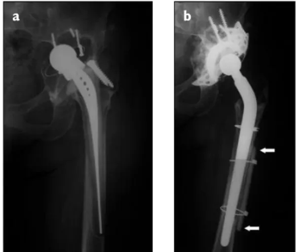
Figure 2 a) and b).Surgical treatment of an aseptic
loosening of a total prosthesis using a cementless femoral stem revision and a cortical onlay strut allografting (arrows), with 5 years of follow-up. Reconstruction of the acetabulum with a metallic cage and particulate cancellous bone allograft.
better when stabilized with metallic cables in close proximity to vascularized host bone.
Cancellous bone allografts were placed between the ends of the struts and the host bone, because they promote the bone healing process and en-hance strut-to-host bone union17,20,21. Strut union was seen within the first year after surgery. Studies of retrieved specimens have shown a close correla-tion between radiographic evidence of union and histologic observations22. Gradual callus formation occurs at the junction site, extending from the pe-riosteal surface of the native bone to the outer sur-face of the cortical bone allograft. There is some degree of creeping substitution at the allograft host junction, but the bulk of the cortical strut remains dead but structurally intact. On the external surface of the allograft, mesenchymal proliferation from the adjacent host cells leads to a thin layer of bone formation that becomes incorporated into the al-lograft cortex. In fact, the initial host response to the allograft bone strut is rapid mobilization of me -senchymal tissue, initiating intense osteogenesis. The healing process of cortical allograft to host bone is prolonged, following the steps of he ma -toma formation, inflammatory process, resorption of graft bone and revascularization, and finally re-placement of graft with new host bone. Neverthe-less, the graft is never entirely replaced with new host bone4,5.
Processed and preserved bone allografts are
favoured in clinical practice. Processing involves the removal of antigenic cells and proteins; preser-vation techniques include deep-freezing or freeze-drying. Deep frozen cortical struts retain their me-chanical properties and may be implanted after thawing, however, freeze-dried cortical struts are vulnerable in torsion and bending, because freeze-drying may alter the mechanical properties of the bone23-25. We therefore used struts stored in liquid nitrogen (cryopreserved) in order to achieve im-mediate structural support.
In the case of treatment of the atrophic no -nunion two very important requisites for success-ful bone formation were achieved: vascularity and mechanical stability. These factors are largely sur-geon-dependent and emphasise the importance of the surgical approach and the preparation of the site to be grafted.
Conclusion
Cryopreserved cortical onlay strut allografts act as biological bone plates, serving both a mechanical and a biological function. The results obtained in the present study show that the use of cortical struts, either alone or in conjunction with a metal-lic plate or with cancellous bone allografts, is a use-ful adjunct for revision hip arthroplasty of the femoral side, stabilization of femoral periprosthe -tic fractures, reinforcement of poor cor-tical bone and for the treatment of femoral nonunion.
Correspondence to
Fernando Judas
Orthopedics Department of Coimbra University Hospitals (HUC)
Praceta Prof. Mota Pinto, Bloco de Celas 3000 Coimbra, Portugal.
E-mail: fernandojudas@gmail.com
References
1. American Association of Tissue Banks: Accredited tis-sue bank annual survey. Available at: http://www. aatb.org/files/tissuesafety.pdf. Accessed July 24, 2008. 2. Delloye C, Cornu O, Druez V, Barbier O. Bone allo-grafts what they can offer and what they cannot. J Bone Joint Surg Br 2007;89: 574-579.
3. Mroz TE, Joyce MJ, Steinmetz MP et al. Musculoskele-tal allograft risks and recalls in the United States. J Am Acad Orthop Surg 2008;16:559-565.
4. Aronson J, Cornell CN. Bone healing and grafting. In Orthopaedic Knowledge Update, 1999; Chapter 2: 25-35, Edited by Beaty JH, American Academy of
Or-Figure 3.Incorporation of a cortical onlay strut allografting (arrows), with 8 years of follow-up, used to bridging a femoral structural defect in a revision hip arthroplasty.
thopaedic Surgeons.
5. Williams Amy, Szabo RM. Bone transplantation. Or-thopaedics 2004;Vol 27, Nº 5:488-495.
6. Gocke DJ. Tissue donor selection and safty. Clin Or-thop 2005;435:17-21.
7. Khan SN, Cammisa FP, Sandhu HS, and al. The biolo-gy of bone grafting. J Am Acad Orthop Surg 2005;Vol 13: 77-86.
8. De Long WG, Einhorn TA, Koval K et al. Bone grafts and bone grafts substitutes in orthopaedic trauma surgery. J Bone Joint Surg Am 2003;83: 649-658. 9. Gross AE, Wong PKC, Hutchison CR et al. Onlay
Cor-tical Strut Grafting in Revision Arthroplasty of the Hip. J Arthroplasty 2003;18; 104-106.
10. Head WC, Malinin TI. Results of onlay allografts. Clin Orthop 2000;371: 108-112.
11. Connolly J, Guse R, Lippiello L, Dehne R. Develop-ment of an osteogenic bone-marrow preparation. J Bone Joint Surg Am 1989;71:684-691.
12. Hernigou P, Poignard A, Manicom O et al. The use of percutaneous autologous bone marrow transplanta-tion in nonunio and avascular necrosis of bone. J Bone Joint Surg Br 2005;87:896-902.
13. Judas F, Teixeira L, Proença A. Coimbra University Hospitals’ bone and tissue bank: twenty-two years of experience. Transplant Proc 2005;37:2799-2801. 14. Judas F. Contribution to the study of Morselized Bone
Allografts and Biomaterials. PhD Thesis. 2002, Coim-bra University.
15. Kim Y-H, Kim J-S. Revision hip arthroplasty using strut allografts and fully porous-coated stems. J Arthroplasty 2005; 20: 454-459.
16. Blackley HRL, Davis AM, Gross AE. Proximal femoral allografts for reconstruction of bone stock in revision
arthroplasty of the hip. A nine to fifteeen-year follow-up. J Bone Joint Surg Am 2001;83: 346-354.
17. Pugh DMW, McKee MD. Advances in the manage-ment of humeral non-union. J Am Acad Orthop Surg 2003;11:48–59.
18. Van Houwelingen AP, McKee MD. Treatment of os-teopenic humeral shaft nonunion with compression plating, humeral cortical allograft struts, and bone grafting. J Orthop Trauma 2005;19:36–42.
19. Wang JW, Weng LH. Treatment of distal femoral nonunion with internal fixation, cortical allograft struts, and autogenous bone-grafting. J Bone Joint Surg Am 2003;85:436–440.
20. Judas F, Figueiredo MH, Cabrita AM, Proença A. In-corporation of impacted morselized bone allografts in rabbits. Transplant Proc 2005;37:2802-2804. 21. Stevenson S. Biology of bone grafts. Orthop Clin N
Am 1999;30:543-552.
22. Enneking WF, Campanacci DA. Retrieved human al-lografts: a clinicalpathological study. J Bone Joint Surg Am 2001;83: 971-986.
23. Bauer TW, Muschler GF. Bone graft materials. An overview of the basic science. Clin Orthop 2000; 371:10-27.
24. Boyce T, Edwards J, Scarborough N. Allograft bone. The influence of processing on safety and perfor-mance. Orthop Clin N Am 1999;30: 571-5581. 25. Jinno T, Miric A, Feighan J et al. The effects of
pro-cessing and low irradiation on cortical bone grafts. Clin Orthop 2000;375: 275-285.
