m u s c u l a r k i n e t i c s a n d f a t i g u e e v a l u a t i o n
o f k n e e u s i n g b y i s o k i n e t i c d y n a m o m e t e r
i n p a t i e n t s w i t h a n k y l o s i n g s p o n d y l i t i s
Nilay Sahin
*, Emel Ozcan
**, Akin Baskent
***, Ayse Karan
****, Erdem Kasikcioglu
******Assistant Professor, Selcuk University, Meram Faculty of Medicine, Physical Medicine and Rehabilitation Department, Meram/Konya **Professor, Istanbul University, Istanbul Faculty of Medicine, Physical Medicine and Rehabilitation Department, Capa/Istanbul ***Istanbul University, Istanbul Faculty of Medicine, Physical Medicine and Rehabilitation Department, Capa/Istanbul ****Professor, Istanbul University, Istanbul Faculty of Medicine, Physical Medicine and Rehabilitation Department, Capa/Istanbul *****Associate Professor, Istanbul University, Istanbul Faculty of Medicine, Sports Medicine Department, Capa/Istanbul
sease of unknown etiology. Inflammation prima rily affects the joints and causes secondary changes in these regions. The spine is the fundamentally af-fected region in AS1. In most of the patients, the pe-ripheral joints are mildly affected without showing any deformity. In time, impaired spinal mobility may cause articular instability and force the pa-tients to use their knee muscles more for postural control and for activities of daily living2,3. Further-more, changes in spine give rise to deformities in peripheral joints. Peripheral joint involvement may also affect posture and thus cause disability3. Pa-tients suffering from hip joint involvement rarely develop mild knee flexion pattern in knees during walking in order to make the walk more comfor -table2. Peripheral joint involvement can be present in about 25% of patients as an asymmetrical oligoarthritis predominantly in lower extremities, particularly affecting the knees4. On the other hand, systemic inflammation may also affect the knee muscles. Marcora et al., found reduced appendi -cular muscle mass in patients with long-standing AS compared to healthy controls. This muscle was-ting is significantly associated with reduced knee extensors muscle strength and grip strength of the dominant hand5. Local inflammation (achilles ten-don enthesitis), frequently seen in seronegative spon dyloarthritis patients, may affect the knee muscle6. Consequently, strength of knee muscles may be affected due to some reasons in AS patients who have a long-standing disease and impaired posture of spine. However, it is not yet clear whether strength of knee muscles may have an effect on AS patients who have no postural disorders.
In addition, for the chance of success it can be important to know which muscle group is mostly affected during the rehabilitation of AS patient. When weakness is suspected in a muscle group, it is useful to evaluate the isokinetic performance in increasing speed in every angle of that muscle7. Al-though isokinetic testing was used to different
Abstract
Objective: Ankylosing Spondylitis (AS) is an in-flammatory disease that is observed with arthritis, sacroiliitis and disability. The aim of the study was to compare the strength and fatigue of knee exten-sor/flexor muscle group usage by isokinetic in pa-tients with AS with controls.
Methods: Twenty-six AS diagnosed patients and twenty-six healthy volunteers with similar age, height, body weight and gender were included in this study. In both groups the isokinetic tests are conducted by isokinetic dynamometer for every subject. Knee extension/flexion patterns;peak torque, agonist/antagonist ratio and work fatigue isokinetic parameters were evaluated during the knee 60º/s,180º/s and 240º/s angular velocities. Results: Knee extension/flexion muscle strength in patient group was significantly lower compared to the control group in all angular velocities (p< 0.05). Conclusions: The study showed knee muscle weak-ness and fatigue in patients with AS compared to the control group.
Keywords: Muscle strength; Dynamometer; Knee; Ankylosing spondylitis; fatigue
Introduction
-joints in rheumatoid arthritis, fibromyalgia syn-drome and in some other rheumatologic based diseases as well, there is few knowledge about pe-ripheral weakness in patients with AS8-10. A study detected muscle weakness and fatigue in ankle plantarflexor muscle groups in AS patients com-pared to the control group11.
The objective of this study was to measure the strength and fatigue of the knee extensor/flexor muscle group using by isokinetic in AS patients, who do not have postural disorders or peripheral joint involvement; to compare with healthy con-trols, and to determine the relation of these values with the functional situation.
Material and Methods
Twenty-six male patients between 18-54 years of age diagnosed with AS and referred to Physical Medicine and Rehabilitation division AS unit, and a control group consisting of 26 healthy males be-tween 20-56 ages were enrolled for this study. The patient group was chosen according to Modified New York diagnosing criteria, diagnosed with AS but not in active period. Patients having serious knee injury, having serious lumbar pain, hip pain or knee pain, having some other systemic diseases, limitations in hip and knee joints, and having surgery in lower extremities were not included in this study group. The control group was selected from the hospital staff with similar age and gender. The control group with serious knee trauma, hip, knee and hip osteoarthritis demonstrated by X--rays, other comorbidities and ligament injury were excluded from the study. Informed consent of the subjects was sought and the ethical committee approval was obtained prior to the initiation of the study.
Evaluation parameters
Knee extensor/flexor muscle group isokinetic muscle strength (peak torque) of both groups was eva -luated by Biodex System 3PRO Multijoint System isokinetic dynamometer. Before testing AS patients the following evaluations were performed: Body weight-height, visual analog scale (VAS), modified lumbar Schober (MLS), lower extremity range of motion (ROM) as measured by goniometry, pre -sence of enthesitis as determined by Berlin Enthe-sitis Index (BEI), the activity of the disease as measured by Creactive protein (CRP), Bath Ankylo
-sing Spondylitis Disease Activity Index (BASDAI) score and functional status as determined by Bath Ankyolosing Spondylitis Functional Index (BAS-FI)12.
Pain
Pain was evaluated by VAS score between 0-10. No pain corresponded to (0), whereas intolerable pain was expressed with (10) points. The severity of the pain was investigated separately if it occurred at night and during resting. The higher points show the severity of the pain13.
Enthesitis
According to BEI, the patient is asked to evaluate presence of pain during palpation to 12 enthesis areas in the lower extremities. The patient replies with “yes” or “no” and score is determined between 0-12. The score gives an idea about the activity of the disease12.
Activity of the Disease
BASDAI gives information about the activity of the disease. The evaluated activity is mostly about the presence of inflammation in peripheral joints. Fa-tigue, axial pain, peripheral pain, morning stiffness and the presence of enthesopathy is evalua -ted by VAS between 0-10 points. It is accep-ted as the activity period of the disease, when BASDAI is >412,14.
Functional Status
In BASFI scale, 10 daily activities are evaluated. The patient is asked to evaluate each activity by VAS between 0 and 10 according to the difficulty expe-rienced during each activity. The scores show 0= no difficulty, 5= moderate difficulty, 10= maximum difficulty, the total maximum score is determined as 1015.
Muscle Testing
Isokinetic tests with Biodex System 3PRO Multi-joint System isokinetic dynamometer were applied to both groups The reliability of the dynamometer was determined both in healthy group and the AS patient group16-18. The tests were performed ac-cording to standardizations developed by Wilk et al10. Warm-up was accomplished on ergonomic bi-cycle for 10 min. at 60 rpm.
For knee extension/flexion pattern measurement the arms of the dynamometer were held pa -ral lel to the leg of the patient having pads fixed
dis-tally. Distal resistance pad was fixed. The stability of the patient on the dynamometer chair was achieved by putting a belt covering the thorax, hip and thigh regions, and the procedure was ex-plained to the patient to ensure good coopera-tion10.
Muscle strength is measured better with tests performed with low angular velocities, while high angular velocities are useful for the detection of functional status and endurance of the muscle7,18,19. For this reason, slow, moderate and high angular velocities such as 60º/s,180º/s, 240º/s were pre-ferred for the knee extension/flexion pattern19,21. The test was performed bilaterally, starting with the dominant side first. Four repetitions were per-formed at the first two angular velocities in exten-sion/flexion and at the third angular velocity, 20 repetitions were performed. Peak torque (New-tonmetre-Nm) (PT), peak torque/body weight (%) (PT/BW), maximal repetition total work (Joule-J) (MRTW ), work/body weight (%) (W/BW ), ago-nist/antagonist ratio (%) (Ag/An) and work fatigue (%) (WF) isokinetic parameters were evaluated at all angular velocities. In order to decrease the oc-currence of strain in the muscles, 60 sec. resting period was maintained between each angular veloci -ty22. In order to motivate the patients during the test maximal, strength was maintained by verbal instructions. The test was carried out in a quiet and appropriate physical environment with air condi-tioner.
PT is the highest torque value measured with all velocities in one angular velocity and is expressed in terms of newtonmeter. PT is the most conve-nient and the most used parameter in isometric test parameters20. PT/BW ratio is used to persona -lize, standardize and interpret isokinetic scores23. MRTW is one of the parameters where the rela-tionship between flexion and extension is inter-preted and is expressed as Joules21,24,25. W/BW is the maximum work (force x distance) produced in a single repetition. This could be a better represen-tation of the functional ability (over PT), because the muscle must maintain the force throughout the range of motion, as opposed to the force at one instant24. Ag/An ratio evaluates the balance be-tween the knee extensor/flexor muscles. With this ratio, the weakest muscle in the muscle group can be determined. The ag/an ratio is calculated as the ratio between the peak values of the concentric torque of the flexor muscles, and the concentric peak torque of the extensor of the knee. The
hams-tring action as antagonist is directly proportional to its ability to generate concentric strength26-28. WF test measures the weariness of the muscle after an excess number of repetitions. This is calculated as the percentage of the difference between the pro-duction of work between the first 1/3 and the last 1/3 repetitions at the 240º/sec. velocity. There is no standardized test to evaluate fatigue. The number of trials to evaluate fatigue is between 20-100. In this study, we used 20 trials. This parameter acts as a dependent variable used to evaluate the strength of the muscle and shows the endurance capacity of the muscle29,30.
Statistical Analysis
The comparison demographic data of both groups were assessed using Mann–Whitney U tests. Two way ANOVA was used in the group evaluations for comparisons between the groups for PT, PT/BW, MRTW, W/BW, AG/AN and dominant versus non--dominant leg isokinetic parameters. The compa-rison WF of both groups was assessed using Mann–Whitney U tests. The correlation between PT parameter and BASFI, VAS and ROM was as-sessed by Pearson correlation test. p<0.05 values were accepted as statistically significant.
Results
Age, gender, height, weight, MLS, VAS, CRP, BASFI, BASDAI, and BEI values are presented in Table I. There was no statistically significant difference be-tween the two groups for age, gender, height and weight. The test group was not in the active stage; BASDAI values were <4, BEI 0-2 and CRP was <511. Resting and night VAS values were below 5. Hip flexion and knee extension and flexion ROM mea-surements for isokinetic tests performed with the goniometer’s dynamometer revealed no statisti-cally significant difference between the two groups. In both groups, the right side was the dominant side.
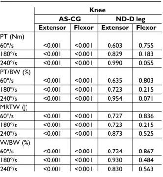
Significantly lower values than the healthy con-trol group were observed in AS patients for the knee PT, PT/BW, MRTW and W/BW parameters evalua -ted with bilateral extension and flexion performed at the angular velocities of 60º/s, 180º/s and 240º/s (p<0.001) (Tables II, III). There was no significant difference between the groups in dominant versus non-dominant leg for bilateral knee movement tested at 60º/s, 180º/s and 240º/s. Statistical eva
luation for the Ag/An parameter revealed a signifi -cant difference in AS patients compared to the healthy controls for bilateral knee movements tes -ted at 60º/s (p<0.05). There was no statistically signi ficant difference in Ag/An scores for bilateral knee movement tested at 180º/s and 240º/s. There was no significant difference for Ag/An values be-tween the dominant and non-dominant side in AS patients (Table IV).
A significant decrease of work fatigue in AS pa-tients was determined for knee extensors at 240º/s after 20 repetitions (P<0.05) (Table V).
There was no statistical correlation between muscle strength measurements and BASFI, VAS and ROM (p>0.05).
Discussion
Isokinetic dynamometer is an instrument which helps us to measure the joint movements in all an-gles, in constant angle speed, allowing maximal muscle contraction along with the measurement of the contraction and muscle capacity objective-ly8. In isokinetic measurements for painful chro nic diseases like osteoarthritis, rheumatoid arthritis, fi-bromyalgia syndrome and chronic low back pain angular velocities such as 60º/s 180º/s, 240º/s and 300º/s angular velocities were used and many rep-etitions were employed8,9,18-22,25,31. There is no stan-dardized model for isokinetic measurement in AS patients, so we have used 60º/s 180º/s, 240º/s an-gular velocities in our study. The most valid pa-rameter in isokinetic evaluation is the PT which may be affected by body mass index and the PT/BW value is important in this issue as well as the MRTW value that reflects the balance between
flexor and extensor muscle groups. The W/BW as one of the best indicator of PT values is also im-portant20,21,23-25. The parameters above at all angu-lar velocities showed significant lower scores for knee extensor and flexor muscles on both sides in AS patients as compared to the controls in this study. Although 60º/s Ag/An ratio was significantly decreased on both sides in the AS group, there was no significant difference between the Ag/An ratio for bilateral knee obtained at the angular velocities of 180º/s and 240º/s in the AS and the healthy con-trol groups. Also, higher velocities reflect the Ag/An ratio better than lower velocities8. Since the exten-sion and flexion losses for the knee joint are seen together in AS patients, there was no statistically significant difference in Ag/An scores. The low le -vel of the work fatigue showed that the work of knee extensor is decreased in the first third and the last third period of the work thus indica ting a de-crease in endurance capacity of the muscles. The main finding of this study indicates that in AS pa-tients the tested knee muscles were significantly weaker and the muscle endurance capacity de-creases compared to apparently healthy controls. Interestingly, the results of this study show that the forces at different angles and endurance of the tes -ted muscles rela-ted to non-involvement joints in patients with AS are lower than those of control subjects.
The reason of the decrease in muscle strength in AS patients is complicated32. The decreased muscle strength in inflammatory diseases is rela -ted to inflammation, pain, stiffness, inactivity, de-generation at the joints, fatigue and the primary symptoms of AS patients are also pain, enthesis and stiffness1,33,34. Inactivity that is related to pain, inflammation and stiffness has a great role in the weakness of muscles in AS patients. Muscle wea k -ness develops in the first week of inactivation. Af-ter that, weakness increases rapidly. Decreased physical activity or inactivation results with atrophy in the muscles, which further leads to wea k -ness in the muscles, causes a decrease in neuro-muscular performance and thus a decrease in the functional capacity ensues. But, this process does not have to be similar for all muscle groups1,32,35. In a study on inflammatory disease, the decrease in dynamic and isometric muscle strength was shown in early stages36. Inflammation raises catabolic stimulations including IL-6, IL-1 and TNF alpha cytokine, each case causes muscle protein cata -bolism. Consequently, inflammatory conditions Table I. Characteristics of patients with ankyolosing
spondylitis (AS) and the control group (CG)
AS CG (n:26) (n:26) p Age/mean 37.04±8.85 38.46±10.35 0.464 Height 172 174.31 0.139 Weight 75.19 73.42 0.288 Modified lumbar 18.72±2.62 Schober BASFI 3.12±2.20 BASDAI 2.28±1.41
may affect muscle mass and cause loss of stren -gth37,38. The chronic inflammatory response is li-kely to be a major cause of muscle wasting in AS patients. Marcoro et al., showed that patients with long-standing AS have significant losses of lean mass in arms and legs. This muscle wasting is signi -ficantly associated with reduced knee extensor muscle strength and grip strength of the dominant
hand5. The other reason of the de-crease in muscle strength is fatigue in AS patients. There are a lot of mechanisms res ponsible for the development of muscle fatigue19. An exceeding fatigue in AS patients is acquainted with activity of the disease, functional disability, and global wellness34. The fatigue in muscles is res ponsible in motor control deficit and in posture and balance chan ges1,12,24,34. Conside -ring the relationship between the postural changes and fatigue, fa-tigue may play an important role in postural changes in AS patients. In this study, we determined that the muscle endurance capaci ty de-creases in the patient group, even though we used the least number of trials recommen ded in the litera-ture. According to this result, the muscle weakness can be the cause of fatigue in AS patients, or fatigue seen in most of the AS patients can be one of the cau ses of muscle weakness.
The other possible reasons for muscle strength decrease mecha-nism are local inflammation (enthesitis) and proprioception dete -rio ration, which is related to it. Enthesitis, which is the basic me cha -nism of pathogenesis in AS pa tien ts, is an inflammation of en thesis, the location where the point at which a tendon or ligament or muscle in-serts into bone. Enthesitis leads to instable joint structure and these changes apparently cause muscle weakness in AS patients. Further-more, the attachment sites of the ligamentous formations harbor the afferent nerve endings, which regulate the information about posture and are res -ponsible for joint motion; therefore, a pathology at this site results with changes in proprioception in AS patients. Insufficiency of proprioce ption may a cause to decrease in muscle stren gth32,35,39-41. Con-sequently, the muscle weakness detected in our study may have correlation with the disorders in proprioceptors that is rela ted to enthesitis.
T a b le I I. M e a n s o f p a r a m e te r s e v a lu a te d b y t h e i s o k in e ti c t e s t fo r t h e k n e e G ro u p A S C G M E x te n so rs F le x o rs E x te n so rs F le x o rs S R L R L R L R L PT ( N m ) 60 º/ s 1 39 .5 6± 28 .2 1 13 9. 67 ± 24 .9 9 61 .6 3± 13 .5 2 60 .2 0± 10 .7 4 17 4. 29 ± 36 .0 9 17 1. 82 ± 33 .9 7 83 .9 2± 22 .7 8 83 .2 7± 18 .1 8 18 0º /s 10 5. 43 ± 21 .6 4 10 4. 90 ± 16 .9 1 58 .1 9± 12 .7 2 63 .0 0± 14 .8 7 12 2. 78 ± 25 .3 7 12 5. 29 ± 27 .3 6 75 .5 5± 19 .5 9 79 .1 6± 16 .0 6 24 0º /s 96 .6 1± 16 .5 2 95 .5 2± 12 .4 0 67 .2 5± 13 .1 1 74 .7 9± 15 .4 6 11 0. 62 ± 23 .2 1 11 1. 62 ± 25 .3 1 82 .2 1± 18 .6 8 89 .3 5± 17 .7 3 PT /B W ( % ) 60 º/ s 18 7. 48 ± 42 .1 0 18 7. 96 ± 38 .3 7 82 .5 0± 18 .2 7 81 .0 5± 16 .9 7 23 5. 89 ± 36 .5 3 23 3. 25 ± 36 .0 7 11 4. 30 ± 29 .6 7 11 3. 55 ± 24 .3 3 18 0º /s 14 1. 29 ± 29 .4 5 14 1. 06 ± 25 .3 8 78 .3 3± 19 .0 8 85 .0 0± 22 .3 4 16 6. 09 ± 24 .1 9 17 0. 13 ± 29 .1 2 10 3. 32 ± 27 .4 4 10 8. 43 ± 23 .2 3 24 0º /s 12 9. 58 ± 23 .9 7 12 7. 96 ± 17 .1 6 90 .7 0± 19 .9 7 10 1. 08 ± 25 .2 7 14 7. 80 ± 22 .4 5 14 9. 88 ± 30 .4 9 11 0. 54 ± 24 .9 8 12 0. 35 ± 23 .5 6 M RT W ( J) 60 º/ s 14 7. 21 ± 30 .9 2 14 7. 34 ± 25 .5 3 65 .2 9± 18 .0 9 64 .7 9± 15 .4 8 18 7. 88 ± 40 .0 5 18 3. 04 ± 38 .4 5 98 .9 4± 29 .3 0 97 .6 1± 24 .4 4 18 0º /s 11 7. 61 ± 27 .4 8 11 7. 19 ± 17 .7 0 49 .4 2± 18 .4 2 47 .2 2± 15 .2 1 14 1. 06 ± 30 .0 9 14 1. 47 ± 32 .1 7 73 .9 5± 23 .2 9 70 .1 5± 19 .9 9 24 0º /s 10 0. 84 ± 21 .6 7 10 0. 61 ± 15 .3 9 40 .2 0± 15 .9 0 38 .7 8± 14 .0 9 11 9. 89 ± 24 .6 8 12 1. 56 ± 28 .1 3 59 .7 8± 18 .7 8 56 .8 8± 19 .7 6 W /B W (% ) 60 º/ s 19 1. 60 ± 57 .1 7 19 8. 19 ± 38 .3 2 91 .7 7± 30 .0 8 94 .0 0± 43 .1 7 25 4. 58 ± 43 .0 6 24 8. 01 ± 37 .1 2 13 4. 72 ± 38 .0 6 13 2. 98 ± 31 .1 0 18 0º /s 15 7. 56 ± 36 .7 1 15 7. 45 ± 26 .6 2 66 .2 2± 24 .2 3 63 .7 5± 21 .7 4 19 0. 51 ± 26 .6 0 19 1. 65 ± 30 .9 0 10 0. 63 ± 29 .8 3 95 .6 9± 25 .0 1 24 0º /s 13 5. 28 ± 30 .1 7 13 5. 90 ± 22 .3 0 54 .2 7± 21 .3 1 52 .4 3± 20 .0 9 16 2. 02 ± 21 .6 6 16 4. 65 ± 28 .7 1 81 .0 2± 22 .5 1 78 .1 5± 23 .4 9 R : r ig ht , L : l ef t. A S: a nk yo ls in g sp on dy lit is , C G : c on tr ol g ro up . M : m us cl e, S : s id e.
Conclusions
In this study, we detected fatigue and muscle weakness in knee extensor and flexor muscle groups in AS patients compared to the control group. Rehabilitation of the muscle weakness and fatigue is important to delay the develop-ment of the posture disorder and thus prevent the development of ba lance problems in AS patients. More studies are needed to be done on this subject, in order to detect the effect of exercises especially on the lower extremities, on the activity of the disease, on the posture of the patient and on the functional status in ear-ly stages of the disease before any postural change occurs in AS patients. The results of this study showed us how important the effect of muscle weakness in maintaining posture in AS patients is. As a conclusion, functional di sability in patients with AS is not only develo -ped by axial deformities, but may also by mus-cular weakness and fatigue affect. Based on this knowledge, it should be reminded that
Table IV. Means of agonist/antagonist parameters evaluated by isokinetic test (%)
AS CG
Group R (F/E) L (F/E) R (F/E) L (F/E) p
Knee-AV 45.30±12.32 43.50± 6.56 48.28±8.77 48.68± 7.43 0.001
60°/s
180°/s 56.55±14.32 61.00±16.84 61.84±11.35 80.40±16.22 0.116
240°/s 72.37±18.91 80.05±17.49 75.31±14.65 82.01±17.28 0.312
R: right, L: left. AS: ankyolsing spondylitis, CG: control group. E:extensor, F: flexor.
Table V. Means of work fatique parameters evaluated by the isokinetic test (%) Group AS CG p Knee Extensor R 35.35 (2.7-64.0) 27.50 (-9.0-47.8) 0.034 240º/s L 35.90 (3.2-65.2) 23.50 (-7.6-45.3) 0.030 Flexor R 38.75 (10.2-69.4) 24.30 (-8.3-79.7) 0.253 L 41.10 (16.6-68.8) 30.70 (-6.0-71.0) 0.249
R: right, L: left. AS: ankyolsing spondylitis, CG: control group. p<0.05
Table III. p values of isokinetic testing in AS versus CG
Knee
AS-CG ND-D leg
Extensor Flexor Extensor Flexor
PT (Nm) 60º/s <0.001 <0.001 0.603 0.755 180º/s <0.001 <0.001 0.829 0.183 240º/s <0.001 <0.001 0.990 0.055 PT/BW (%) 60º/s <0.001 <0.001 0.635 0.803 180º/s <0.001 <0.001 0.723 0.215 240º/s <0.001 <0.001 0.954 0.071 MRTW (J) 60º/s <0.001 <0.001 0.727 0.836 180º/s <0.001 <0.001 0.723 0.215 240º/s <0.001 <0.001 0.873 0.525 W/BW (%) 60º/s <0.001 <0.001 0.724 0.867 180º/s <0.001 <0.001 0.930 0.484 240º/s <0.001 <0.001 0.830 0.563
AS: ankylosing spondylitis, CG: control group. ND: nondominant, D: dominant.
isokinetic evaluation is also important in the fol-low up of the efficacy of the scheduled effective re-habilitation in patients with AS.
Correspondence to
Nilay Sahin
Selcuk University, Meram Faculty of Medicine, Physical Medicine and Rehabilitation Department, Meram/Konya, Turkey
Phone: +90 5552332535 E-mail: [email protected]
References
1. Mengshoel AM, Jokstad K, Bjerkhoel F. Associations between walking time, quadriceps muscle strength and cardiovascular capacity in patients with rheuma-toid arthritis and ankylosing spondylitis. Clin Rheumatol 2004;23:299-305.
2. Khan MA. Clinical features of ankylosing spondylitis. In: Hochberg MC, Silman AJ, Smolen JS, Weinblatt ME, Weisman MH, eds. Rheumatology, Vol 2. Pitts-burg: Mosby; 2003: 1161-78.
3. Gran JT, Skomsvoll JF. The outcome of ankylosing spondylitis: a study of 100 patients. Br J Rheumatol 1997;36:766-771.
4. Carette S, Graham D, Little H, Rubenstein J, Rosen P. The natural disease course of ankylosing spondylitis. Arthritis Rheum 1983;26:186-190.
5. Marcoro S, Casanova F, Williams E, Jones J, Elaman -chi R, Lemmey A. Preliminary evidence for cachexia in patients with wellestablihed ankylosing spon -dylitis. Rheumatology 2006;45:1385-1388.
6. Emad Y, Ragab Y, Bassyouni I, et al. Enthesitis and re-lated changes in the knees in seronegative spondy-loarthropathies and skin psoriasis: magnetic reso-nance imaging case-control study. J Rheumatol 2010;37:1709-1717.
7. Kannus P, Beynnon B. Peak torque occurrence in the range of motion during isokinetic extension and fle -xion of the knee. Int J Sport Med 1993;14:422-426. 8. Meireles SM, Oliveira LM, Andrade MS, Silva AC,
Na-tour J. Isokinetic evaluation of the knee in patients with rheumatoid arthritis. Joint Bone Spine 2002; 69:566-573.
9. Maqeut D, Croisier JL, Renard C, Crielaard JM. Mus-cle performance in patients with fibromyalgia. Joint Bone Spine 2002;69:293-299.
10. Wilk KE. Isokinetik testing and exercise for the knee. In: Mangine RE, ed. Physical therapy of the knee. New York: Churchill Livingstone; 1995: 263-88. 11. Sahin N, Ozcan E, Baskent A, Karan A, Ekmekci O,
Kasikcioglu E. Isokinetic evaluation of muscle strenght and fatigue in patients with ankylosing spondylitis. Eur J Phys Rehabil Med 2011;2:Epub ahead of print.
12. Zochling J, Braun J, Heijde D. Assessments in ankilos-ing spondylitis. Best Prac Research 2006;20:521-537. 13. Frank JM, Moll MH, Hort JF. A comparison of three
ways of measuring of pain. Rheum Rehab 1982; 21:211-217.
14. Akkoc Y, Karatepe AG, Akar S, Kirazli Y, Akkoc N. A Turkish version of the Bath Ankylosing Spondylitis Disease Activity Index: reliability and validity. Rheumatol Int 2005;25:280-284.
15. Yanik B, Gursel YK, Kutlay S, Ay S, Elhan AH. Adapta-tion of the Bath Ankylosing Spondylitis funcAdapta-tional In-dex to the Turkish population, its reliability and validi ty: functional assessment in AS. Clin Rheuma-tol 2005;24:41-47.
16. Drouin JM, Valovich-mcLeod TC, Shultz SJ, Gansned-er BM, PGansned-errin DH. Reliability and validity of the Biodex system 3 pro isokinetic dynamometer veloci-ty, torque and position measurements. Eur J Appl Physiol 2004;91:22-29.
17. Baskent A, Ozcan E, Dincer N, Karan A. Test-retest re-liability of isokinetic muscle strength of the knee in patients ankylosing spondilitis. Ann Rheum Dis 2004;63:409.
18. Holmbäck AM, Porter MM, Downham D, Lexell J. Re-liability of isokinetic ankle dorsiflexor strength mea-surements in healthy young men and women. Scand J Rehabil Med 1999;31:229-239.
19. Hall C, Brody LT. Therapeutic Exercise Moving To-ward Function. Philadelphia: Williams and Wilkins; 1999.
20. Kannus P. Isokinetic evaluation of muscular perfor-mance: implications for muscle testing and rehabili-tation. Int J Sports Med 1994;15:11-18.
21. Siqueira CM, Pelegrini FR, Fontana MF, Greve JM. Isokinetic dynamometry of knee flexors and exten-sors: comparative study among non-athletes, jumper athletes and runner athletes. Rev Hosp Clin Fac Med Sao Paulo 2002;57:19-24.
22. Osternig LR. Isokinetic dynamometry: implications for muscle testing and rehabilitation. Exerc Sport Sci Rev 1986;14:45-80.
23. Borges O. Isometric and isokinetic knee extension and flexion torque in men and women aged 20-70. Scand J Rehabil Med 1989;21:45-53.
24. Prentice WE, Voight MI. Techniques in Musculoskele-tal Rehabilitation. New York: McGraw-Hill; 2001. 25. Pincivero DM, Lephart SM, Karunakara RG. Relation
between open and closed kinematic chain assess-ment of knee strength and functional performance. Clin J Sport Med 1997;7:11-16.
26. Aagaard P, Simonsen EB, Magnusson SP, Larsson B, Dyhre-Poulsen P. A new concept for isokinetic ham-string: quadriceps muscle strength ratio. Am J Sports Med 1998;26:231-237.
27. Baratta R, Solomonow M, Zhou BH, Letson D, Chuinard R, D’ Ambrosia R. Muscular coactivation. The role of the antagonist musculature in maintai -ning knee stability. Am J Sports Med 1998;16:113-122. 28. Oberg B, Moller M, Gillquist J, Ekstrand J. Isokinetic torque levels for knee extensors and knee flexors in soccer players. Int J Sports Med 1986;7:50-53. 29. Cools AM, Geerooms E, Van den Berghe DFM,
Cam-bier DC, Witvrouw EE. Isokinetic scapular muscle performance in young elite gymnasts. J Athl Train 2007;42:458-463.
30. Ellenbecker TS, Davies GJ. The application of isoki-netics in testing and rehabilitation of the shoulder complex. J Athl Train 2000;35:338-350.
31. Lesmes GR, Costill DL, Coyle EF, Fink WJ. Muscle strength and power changes during maximal isoki-netic training. Med Sci Sports 1978;10 266-269. 32. Carter R, Riantawan P, Banham SW, Sturrock RD. An
investigation of factors limiting aerobic capacity in patients with ankylosing spondylitis. Respir Med 1999;93:700-708.
33. Plasqui G. The role of physical activity in rheumatoid arthritis. Physiol Behav 2008;95:270-275.
34. Costa D, Dritsa M, Ring A, Fitzcharles MA. Mental health status and leisure-time physical activity con-tribute to fatigue intensity in patients with spondy-larthropathy. Arthritis Rheum 2004;51:1004-1008. 35. Hakkinen A, Malkia E, Hakkinen K, Jappinen I,
Laiti-nen L, HannoLaiti-nen P. Effects of detraining subsequent to strength trainig on neuromuscular function in
pa-tients with inflammatory arthritis. British J Rheum 1997;36:1075-1081.
36. Häkkinen A, Hannonen P, Häkkinen K. Muscle strength in healthy people and in patients suffering from recent-onset inflammatory arthritis. Br J Rheumatol 1995;34:355-360.
37. Stenholm S, Harris TB, Rantanen T, Visser M, Kritchevsky SB, Ferrucci L. Sarcopenic obesit-defini-tion, etiology and consequnces. Curr Opin Clin Nutr Metabc Care 2008;11:693-700.
38. Adamo ML, Farrar RP. Resistance training, and IGF involvement in the maintenance of muscle mass during the aging process. Ageing Res Rev 2006;5:310--331.
39. Aydog E, Depedipi R, Bal A, Eksioglu E, Ünlü E, Çakci A. Dynamic postural balance in ankylosing spondyli-tis patients. Rheumatology 2006;45:445-448. 40. Swinkels A, Dolan P. Spinal position sense in
ankylos-ing spondylitis. Spine 2004;413-420.
41. Sharma L. Proprioceptive impairment in knee os-teoarthritis. Rheum Dis Clin North Am 1999;25:299--314.