Luís Miguel da Fonseca Carvalho
Application of Gas Chromatography-Mass Spectrometry
for targeted and non-targeted analysis in toxic and edible
mushrooms
Dissertação do 2º Ciclo de Estudos Conducente ao Grau de Mestre em Toxicologia Analítica, Clínica e Forense
Trabalho realizado sob a orientação da Doutora Paula Guedes de Pinho – REQUIMTE, Laboratório de Toxicologia, Departamento de Ciências Biológicas da Faculdade de Farmácia da Universidade do Porto; e co-orientação do Professor Doutor Félix Carvalho – Laboratório de Toxicologia, Departamento de Ciências Biológicas da Faculdade de Farmácia da Universidade do Porto; e da Professora Doutora Paula Baptista – CIMO/Escola Superior Agrária do Instituto Politécnico de Bragança.
É autorizada a reprodução integral deste trabalho apenas para efeitos de investigação, mediante declaração escrita do interessado, que a tal se compromete.
iii
ACKNOWLEDGMENTS
Agradecimentos
Em primeiro lugar quero agradecer à Doutora Paula Guedes de Pinho, orientadora deste trabalho, pelos conhecimentos que me transmitiu ao longo deste ano, os quais foram fundamentais para a realização desta investigação, bem como pelo seu constante apoio, preocupação e incentivo.
De igual modo tenho a agradecer aos meus co-orientadores. Ao Professor Doutor Félix Carvalho pelos conselhos e saber transmitidos e pelo seu constante entusiasmo e otimismo. O meu agradecimento também à Professora Doutora Paula Baptista pela disponibilidade, simpatia e grande ajuda na vertente “micológica” deste trabalho.
Tenho também de agradecer à Professora Doutora Maria de Lourdes Bastos pelo seu empenho e esforço para o desenvolvimento, primando sempre pela excelência e qualidade, do Laboratório de Toxicologia da Faculdade de Farmácia da Universidade do Porto. É devido ao seu trabalho ao longo de vários anos que é, foi e será possível que os estudantes aprendam Toxicologia.
Quero também agradecer a todos os membros do Laboratório de Toxicologia por tornarem a integração mais fácil, a jornada mais descontraída e os momentos mais divertidos.
O meu obrigado especial ao Professor Doutor António César Ferreira e à Rita Monforte pela imensa ajuda no tratamento “non-targeted” dos dados, que em muito enriqueceu este trabalho. Também agradeço à Nathalie Moreira pela ajuda no tratamento estatístico dos dados e respetiva interpretação.
Aos meus colegas do MTACF, tanto aos “resistentes” como àqueles que seguiram por outros rumos, obrigado pela amizade, companheirismo e bons momentos passados. Desejo que todos encontrem aquilo que procuram!
Aos meus colegas da Residência Universitária da Bandeirinha que ao longo dos últimos dois anos foram os “lá de casa” e fizeram parte integrante da minha vivência nesta Cidade. A todos um obrigado do “Zé”!
Aos meus pais, ao meu irmão, ao meu tio e à minha madrinha, aos meus avós e, em particular, à minha Avó Natércia, pelo apoio, amor e incentivo. Sei que estão orgulhosos!
Aos meus amigos de sempre, a “família que se escolhe”, o Dário e a Paula, o Tiago, o Rui e a Nicole e todos os outros que não me lembro neste momento, o meu enorme obrigado pela amizade e apoio de há tanto tempo! É recíproco…
E porque os últimos são sempre os primeiros: À minha Márcia!...
Obrigado!
v
ABSTRACT
Macrofungi is an artificial group of fungi based on the size of reproductive structures, the carpophores or mushrooms, which are visible to the naked eye. Nowadays the mushrooms are important in many contexts, particularly, in the areas of gastronomy, economy, medicine and biotechnology. Mushrooms are, in general, best known for their culinary value, given the diversity and richness of flavours, textures and odours that characterize them. They are also nutritionally relevant, as they have high levels of proteins, amino acids, dietary fibers, vitamins and minerals and low calorie and fat levels. Besides all those nutrients, mushrooms are also rich in volatile compounds that are responsible for their smell. However, many species of mushrooms are toxic and therefore not edible, leading, in several cases, to death by accidental poisoning. The high morphological similarity between certain species and the fact that accurate expertise is needed, makes identification through morphological, macro- and microscopic characters difficult and therefore other alternatives are required. Currently, chemotaxonomy is used in these situations, resorting to molecular analysis, essentially, DNA sequencing, but also to chemical analysis, including the study of AA and FA profiles, as well as secondary metabolites such as volatile compounds. Furthermore, non-targeted approaches can be used, and it seems promising to identify chemotaxomical markers.
In the present work, two GC-MS methodologies were applied in the chemical analysis of 22 mushrooms species (12 edible, 3 toxic and 7 possibly toxic). The first one was a multi-target procedure to extract and derivatize AA, FA and sterols. The result was the identification of 25 compounds, of which 21 were quantified. Moreover, the resulting GC-MS data was also submitted to non-targeted analysis through PCA and PLS-DA, allowing the identification of a compound (5-carbon sugar alcohol) which is candidate to be a species-marker as it was present in a much higher amount in one edible species (Suillus bovinus). The second methodology applied was a HS-SPME/GC-MS procedure to volatile profiling of the species. Targeted analysis of data resulted in the identification of the main volatiles in mushrooms, i.e.8-carbon skeleton molecules, in almost all species. On the other hand, non-targeted data analysis (PCA and PLS-DA) allowed the identification of 6 molecules that can be species- or genus-specific: an ester of hexanoic acid, which was only identified in one edible species - Lycoperdon perlatum; and five sesquiterpene-like molecules that have not been formally identified, these molecules were only present in Lactarius
aurantiacus, a mushroom species whose edibility/toxic remains unknown.
Keywords: AA, FA, sterols and volatile compounds; GC-MS; mushroom; targeted and
RESUMO
Os macrofungos constituem um grupo artificial de fungos baseado no tamanho das estruturas reprodutoras, os carpóforos ou cogumelos, que são visíveis a olho nu. Atualmente os cogumelos são importantes em diversos contextos, nomeadamente, nas áreas da gastronomia, economia, medicina e biotecnologia. Os cogumelos são mais reconhecidos pelo seu valor culinário, dada a diversidade e riqueza de sabores, texturas e odores que os caracterizam. São também alimentos nutricionalmente relevantes, uma vez que apresentam elevados níveis de proteínas, aminoácidos, fibras, vitaminas e minerais e baixos níveis calóricos e lipídicos. Além de todos esses nutrientes, os cogumelos são também ricos em compostos voláteis que são responsáveis pelo seu aroma. Contudo, existem muitas espécies de cogumelos que são tóxicas e por isso não edíveis, provocando intoxicações, em certos casos, fatais. A elevada semelhança morfológica entre determinadas espécies e o facto de serem precisos conhecimentos especializados, torna difícil a identificação através de características morfológicas, macro- e microscópicas sendo, por isso, necessário encontrar outras alternativas. Atualmente, a quimiotaxonomia é utilizada nestas situações, recorrendo-se à análise molecular, essencialmente, a sequenciação de DNA, mas também à análise química, nomeadamente o estudo do perfil de aminoácidos (AA), ácidos gordos (AG), bem como de metabolitos secundários como os compostos voláteis. Além disto, os estudos
metabólicos, principalmente quando são utilizadas estratégias “non-targeted”, constituem
metodologias promissoras para a identificação de marcadores quimiotaxonómicos.
No presente trabalho, dois métodos de análise por GC-MS foram aplicados na análise química de 22 espécies de cogumelos (12 comestíveis, 3 tóxicas e 7 possivelmente tóxicas). O primeiro consistiu num procedimento analítico para extração e derivatização de AA, AG e esteróis. O resultado foi a identificação de 25 compostos, dos quais 21 foram quantificados. Além disso, os dados de GC-MS foram também submetidos a um tratamento “non-targeted”, cujo principal resultado foi a identificação de um composto (poliálcool com 5 átomos de carbono) que é um candidato a marcador químico de espécie, uma vez que estava presente em quantidades muito superiores numa espécie comestível (Suillus bovinus). O segundo método aplicado foi um procedimento de HS-SPME/GC-MS para obter os perfis de compostos voláteis das espécies. A análise de compostos-alvo resultou na identificação dos principais compostos voláteis dos cogumelos, i.e. moléculas com 8 átomos de carbono, em quase todas as espécies. Por outro lado, a análise “non-targeted” dos dados permitiu a identificação de 6 moléculas que poderão ser específicas da espécie ou do género: um éster do ácido hexanóico, que apenas foi identificado numa espécie comestível - Lycoperdon perlatum; e cinco compostos da família química dos sesquiterpenos, que não foi possível identificar formalmente, mas que apenas estavam presentes
nas amostras de Lactarius aurantiacus, uma espécie de cogumelos cuja
comestibilidade/toxicidade ainda é desconhecida.
Palavras-Chave: AA, AG, esteróis e compostos voláteis; análise “targeted” e “non-targeted”;
vii
INDEX
A
CKNOWLEDGMENTS... iii
A
BSTRACT... v
R
ESUMO... vi
I
NDEX... vii
F
IGURESI
NDEX... ix
T
ABLESI
NDEX... x
A
BBREVIATIONSL
IST... xi
P
ARTI
-
I
NTRODUCTION... 1
1.
M
USHROOMS... 3
1.1 Definition and taxonomical classification
... 3
1.2 The importance of mushrooms
... 5
1.3 Mushroom identification
... 6
1.4 Misidentification problems
... 8
2.
M
USHROOMA
UTHENTICITY... 12
2.1 DNA analysis
... 12
2.2 Analysis of other molecules
... 13
Chemical markers
... 13
2.2.1 Chemical analysis techniques
... 15
2.2.2
3.
M
ETABOLOMICS... 18
3.1 Terminology
... 18
3.2 Role of metabolomics in chemotaxonomy
... 19
3.3 Metabolomics workflow
... 20
P
ARTII
–
O
BJECTIVES... 23
4.
O
BJECTIVES ANDE
XPERIMENTALA
PPROACHES... 25
P
ARTIII
–
E
XPERIMENTAL PART... 27
5.1 Mushroom samples
... 29
5.2 Standards
... 31
5.3 Preparation of standard solutions
... 31
5.4 Methodology
... 32
Multi-target experiment
... 32
5.4.1 5.4.1.1 Metabolites extraction
... 32
5.4.1.2 Gas Chromatography-Ion Trap-Mass Spectrometry Analysis
... 32
Volatile profiling experiment
... 33
5.4.2 5.4.2.1 SPME fibers
... 33
5.4.2.2 HS-SPME extraction and Gas Chromatography-Ion Trap-Mass Spectrometry Analysis
... 33
5.5 Data analysis
... 34
Multi-target experiment
... 34
5.5.1 Volatile profiling experiment
... 35
5.5.2
6.
R
ESULTS ANDD
ISCUSSION... 36
6.1 Multi-target experiment
... 36
Targeted approach: AA, FA and Sterols profiling
... 36
6.1.1 Non-targeted approach: multivariate analysis
... 42
6.1.2 6.2 Volatile profiling experiment
... 48
Targeted approach: Main volatiles qualitative analysis
... 48
6.2.1 Non-targeted approach: multivariate analysis
... 50
6.2.2
C
ONCLUSIONS... 58
R
EFERENCES... 60
ix
FIGURES INDEX
Figure 1.1. Basidiocarp structure ... 4
Figure 1.2. Spore print ... 7
Figure 1.3. Result of reaction with 5% KOH ... 8
Figure 1.4. Examples of some mushroom species that may be confounded
because their morphological similarities ... 10
Figure 5.1. Some of the mushrooms collected and analysed in this work ... 30
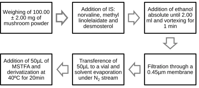
Figure 5.2. Schematic representation of the extraction and derivatization
procedure used in multi-target experiment. ... 32
Figure 5.3. Some differences in retention times of alanine in some mushroom
species ... 35
Figure 6.1. Scores plot resulting from PCA of mushrooms‘ AA, FA and sterols
quantification data ... 41
Figure 6.2. GC-MS chromatogram resulting from the AA, FA, and sterols
extraction procedure in Clitocybe dealbata (Sowerby) P. Kumm.species ... 42
Figure 6.3. Scores plot resulting from PCA of mushrooms‘ data resulting from the
multi-target experiment ... Erro! Marcador não definido.
Figure 6.4. Loadings plots corresponding to PC1 (A) and PC2 (B) resulting from
PCA of multi-target experiment data ... 45
Figure 6.5. Main loadings of PC1 and PC2 distribution among mushroom species
subjected to multi-target experiment ... 47
Figure 6.6. GC-MS chromatogram resulting from the volatile profiling procedure in
Agaricus sylvicola (Vittad.) Perck. species. ... 50
Figure 6.7. GC-MS chromatograms resulting from the volatile profiling procedure
in CB,CD and SI species ... 51
Figure 6.8. Scores plot resulting from PCA of mushrooms’ volatile composition
data ... Erro! Marcador não definido.
Figure 6.9. Loadings plots corresponding to PC1 (A) and PC2 (B) resulting from
PCA of volatile profiling experiment data ... 53
Figure 6.10. Main loadings of PC1 and PC2 distribution among mushroom species
subjected to volatile profiling ... 54
Figure 6.11. Scores plot resulting from PLS-DA of mushrooms’ volatile
composition data ... Erro! Marcador não definido.
Figure 6.12. Loadings plots corresponding to Factor 1resulting from PLS-DA of
volatile profiling experiment data ... 57
TABLES INDEX
Table 1.1. Some characteristics of the principal phyla where the macrofungi
belong ... 3
Table 1.2. Main taxonomical groups of macrofungal species. ... 4
Table 1.3. Some examples of edible and toxic mushrooms usually confounded. . 11
Table 2.1. Main techniques used in chemotaxonomical analysis of mushrooms .. 16
Table 5.1. Characterization of mushroom species. ... 29
Table 6.1. Quantification of amino acids in mushroom samples (mg/100g, wet
basis). ... Erro! Marcador não definido.
Table 6.2. Quantification of fatty acids in mushroom samples (mg/100g, wet
basis). ... Erro! Marcador não definido.
Table 6.3. Quantification of sterols in mushroom samples (mg/100g, wet basis). 40
Table 6.4. Tentative identification of the main loadings of PC1 and PC2 resulting
from the principal component analysis of multi-target experiment data. ... 46
Table 6.5. Volatile composition of mushrooms species. ... 48
Table 6.6. Tentative identification of the main loadings of PC1 and PC2 resulting
from the principal component analysis of volatile profiling experiment data... 54
xi
ABBREVIATIONS LIST
AA – Amino acid(s)
ARDRA – Amplified Ribosomal DNA
Restriction Analysis
CBOL – Consortium for the Barcode of Life
DNA – Deoxyribonucleic Acid EI – Electron impact
FA – Fatty acid(s)
FID – Flame Ionization Detector FTIR – Fourier Transform Infrared fw – Fresh weight
FWG – Fungal Working Group GC – Gas Chromatography
GC-FTIR –Gas Chromatography-Fourier Transform Infrared
GC-MS – Gas Chromatography-Mass
Spectrometry
HCA – Hierarchical cluster analysis
HPLC – High Performance Liquid
Chromatography
HS – Headspace
HS–SPME – Headspace–Solid Phase Microextraction
IR – Infrared
IRD – Infrared Detector IS – Internal standard
ITS – Internal Transcribed Spacer
LC-MS – Liquid chromatography–Mass Spectrometry MECK – Micellar Electrokinetic Chromatography MS – Mass spectrometry MSTFA – N-methyl-N-(trimethylsilyl) trifluoroacetamide
NMR – Nuclear Magnetic Resonance PC1 – Principal component 1
PC2 – Principal component 2
PCA – Principal component analysis PCR – Polymerase Chain Reaction
PLS–DA – Partial least-square
discriminant analysis
RAPD – Randomly Amplified Polymorphic DNA
RFLP – Restriction Fragment Length Polymorphism
rRNA – Ribosomal Ribonucleic Acid
SDE – Simultaneous Distillation
Extraction
SFE – Supercritical Fluid Extraction SM – Secondary metabolite(s) SPME –Solid Phase Microextraction TLC – Thin Layer Chromatography TMS – Trimethylsilyl
3
1. MUSHROOMS
Nowadays the concept of mycology refers the science that studies fungi. However, etymologically, mycology means “the study of mushrooms”, as this word comes from the greek mykes (Gr. μύκης), which means mushrooms, and logos (Gr. λόγος), which conveys the idea of study. The mycology started, indeed, with the study of mushrooms, as these fungal species were the most accessible to the Antiquity naturalists.[1]
1.1 Definition and taxonomical classification
Mushrooms, also designated as fruiting-bodies, are the macroscopic reproduction structure (bigger than 1 mm) of the some fungi. The fungal species that have this type of structure are designated as macrofungi. Although, this is consider an artificial group, the use of this term is useful to mycologists once it allows grouping all these fungi on a single group.[2]
Macrofungi are, in their majority, species that belong to two of five[3] phyla of the Fungi kingdom: Ascomycota Caval.-Sm. and Basidiomycota R.T. Moore (Table 1.1).
Table 1.1. Some characteristics of the principal phyla where the macrofungi belong.[1-3]
Ascomycota Caval.-Sm Basidiomycota R.T. Moore
Number of species (approximately) 32 739 species 29 914 species
Name / Shape of the spore
producing structure Ascus (plural Asci) / Bag shape Basidium (plural Basidia) / Paddle-shaped
Name / Number of spores Ascospores / Usually 8 Basidiospores / Usually 4
Name of the fruiting body Ascocarp Basidiocarp
Besides the lower number of species, the phyla Basidiomycota is the one that integrates the macrofungi whose fruiting bodies (basidiocarp) exhibit the shape more associated to mushrooms (Figure 1.1).
Figure 1.1. Basidiocarp structure: A – cap (or pileus); B – scales; C – hymenium; D – annulus (or
ring); E – stipe (or stalk) ; F – volva (or cup) (Adapted from [1])
The main taxonomical groups where the macrofungal species belong have been hierarchically organized as showed in Table 1.2.
Table 1.2. Main taxonomical groups of macrofungal species.[2]
Phylum Ascomycota Caval.-Sm. Basidiomycota R.T. Moore
Class Ascomycetes G. Winter Basidiomycetes G. Winter
Subclass Agaricomycetidae Parmasto (non-septated basidia) Tremellomycetidae Locq. (septated basidia) Order Pezizales J. Schrӧt Agaricales Underw. Boletales E.-J. Gilbert Cantharellales Gӓum. Phallales E. Fisch. Polyporales Gӓum. Russulales Kreisel ex
P.M. Kirk, P.F. Cannon & J.C. David
Auriculariales J. Schrӧt. (transversal septum) Tremellales Fr. (longitudinal septum)
5
1.2 The importance of mushrooms
Fungi are extremely important organisms, despite the fact that sometimes they are undervalued. Ecologically these species are essential, as they are crucial in the regulation of the ecosystems processes, but they also show a high anthropic value in gastronomy, medicine and biotechnology.
Fungi, being chemoheterotrophic organisms, need to obtain their nutrients from the environment.[2] Depending on the strategy used to obtain these compounds, the fungi may be classified in three groups[2, 4]: decomposer, which are important in the decomposition of the dead organic and in nutrient recycling matter through the release of enzymes to the soil[2, 4]; mycorrhizal, which, with the establishment of a symbiotic association with plant roots (mycorrhizal association), improve the plant intake of water and nutrients and bestow resistance/tolerance to the biotic and abiotic stresses[4, 5]; and finally the parasite, which establish an harmful association with hosts (usually plants), being able to create habitat to other organisms in case of hosts’ death.[6]
Beyond participating in all these processes, macrofungi, specifically their fruiting bodies, integrate the food chains of some animals and are part of the human diet (micophagy)[2]. Furthermore, mushrooms of some species are highly appreciated by humans due to their organoleptic properties (taste, smell and texture)[7, 8]. Furthermore, they are high nutritional value food, as they are rich in proteins, amino acids (AA), vitamins and minerals and have low lipid and caloric contents.[8-10] Hence, these macrofungal species have high gastronomic and economic values. Among these species, the mycorrhizal ones are more valorised because they are difficult to produce in culture. In this context, truffles (fruiting bodies of the species belonging to genus Tuber P. Micheli ex F.H. Wigg.) are the most appreciated and, consequently, they have expensive prices. The most expensive price of the “white truffle”, also known as “Alba Madonna” (Tuber magnatum Pico & Vitt.), may reach 17.500€/kg. In Portugal, Boletus edulis Bull., Cantharellus cibarius Fr. and Amanita caesarea (Scop.) are, economically, the most important species, with prices in the range of 15-30€/kg.[2, 11]
Besides their nutritional value, mushrooms are also important in medicine, since they have bioactive compounds that confer anti-atherosclerotic, anti-microbial, anti-neoplastic, anti-oxidant, immunomodulatory and hypoglycaemic properties.[12] Some of the species with recognized medicinal value are: Armillaria mellea (Vahl) P. Kumm., Auricularia auricula (L.) Underw, B. edulis, Flammulina velutipes (Curtis) Singer, Lactarius deliciosus
(L.) Gray, Lepista nuda (Bull.) Cooke, Marasmius oreades (Bolton) Fr., Pleurotus ostreatus (Jacq.) P. Kumm. and Trametes versicolor (L.) Lloyd.[2, 10]
1.3 Mushroom identification
Mushroom identification is always a challenge! Even if a species was previously identified, doubts about the identity of a mushroom can always arise when it is seen for the first time. Nevertheless, any mushroom is, probably, identifiable, if the time and resources (bibliographic and experimental) are available.[13]
Mushroom identification is performed through the analysis of macro and microscopic features. The macroscopic characteristics are essential to the identification of species in the field, and, in many cases, are enough to recognize the genus which the mushroom belongs.[14]
At macroscopic level, the identification is based on ecological, morphological and organoleptic features and also by using chemical reactions with specific reagents. The most relevant ecological information is the habitat, involving the type of soil and vegetation, the colonized substrate (soil, wood, etc.) and the growth patterns (alone, gregarious, etc.). Then, the ephemeral features must be registered, like the remains of veil and viscosity and also the organoleptic properties (colour, smell and taste).
After the ecological identification, the next step is the morphological analysis which should be thorough and organized. To achieve a complete morphological analysis of the mushroom, it is important to note, from the cap to the stipe, all the important details, named a “downward observation”. For each part of the mushroom, some specific details should be noticed, for example: the colour and shape of the cap; the presence/absence of gills or tubes in hymenium; and the presence/absence of annulus and/or volva in the stipe.
Despite the importance of the external analysis of the mushroom, it is also important to observe the internal structure of the specimen and some of its microscopic details. Concerning the microscopic features, the technique used by mycologists is called “spore print” – it is a procedure that results in the deposition of the spores on a paper sheet (Figure 1.2). The colour of the “spore print” must be registered once this is important to the mushroom identification. Furthermore, the obtained spores are usually tested with the Melzer’s reagent and observed in a microscope, to analyse their shape and size.
7
The morphological analysis is accomplished by observing the mushroom interior. Normally the specimen is longitudinally sliced, allowing the observation of the flesh’s anatomy and its colour – it is also important to pay attention to colour changes due to contact with air.[13] This procedure is also important to evidence some organoleptic properties, concerning taste and aroma, since they are not perceptible when the mushroom is intact. The analysis of these properties can be imperative to the identification of certain mushroom species. Despite the fact that the majority of the mushrooms have similar odours[15], certain species/genus have very specific smells which can help to the specimen identification – e.g. some species belonging to Mariasmus Fr., Agaricus Murrill and Clitocybe (Fr.) Staude genera smell like garlic, almonds and anise, respectively.[16-18] The taste, being one of the more specific features of mushrooms, could also be important in the species/genus identification. However, this practice can be dangerous, even deadly, and should only be done when there is already some certainty in the prior identification of the mushroom.[16] Moreover, when tasting the specimen some “safety measures” should also be taken – chew a small piece of mushroom for one or two seconds and then spit it, never swallow it.
Figure 1.2. Spore print: A – hymenium placed on the paper sheet; B – spore deposition (Adapted from the original available at:http://www.anbg.gov.au/fungi/images-captions/spore-print-0018.html [05/06/12])
Some chemical reactions can be performed by mycologists to improve mushroom identification. The goal of these reactions is search if there are specific colour changes in fruiting bodies (Figure 1.3). The reagents most commonly used are: potassium hydroxide
(30-40% KOH), iron sulfate (10% FeSO4)[19], ammonium hydroxide (25% NaOH) and Melzer’s reagent, aforementioned.[13]
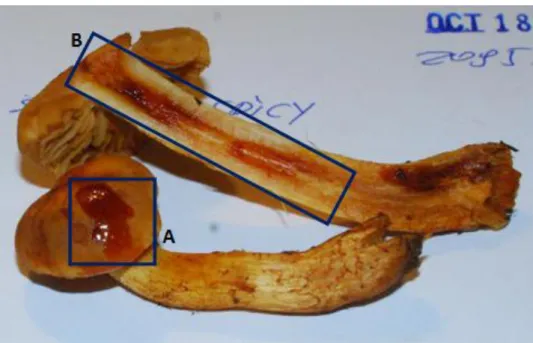
Figure 1.3. Result of reaction with 5% KOH in the cap (A) and flesh region (B) of a mushroom
belonging to the species Cortinarius callisteus (Fr.) Fr. (Adapted from the original available at:
http://www.anbg.gov.au/fungi/images-captions/spore-print-0018.html [06/09/12])
Finally, when there is the need to preserve a mushroom to further studies, the specimen is submitted to a dehydration process, and kept dry, which is called exsiccation (from Latin exsiccatus – to dry).
1.4 Misidentification problems
The correct identification of a wild mushroom, especially when the specimen is intended to human consumption, is extremely important, even if it is difficult and time consuming.
If identification is careless and undervalued, the wild mushroom consumption is always risky, once the ingestion could result in a severe intoxication, whose worst consequence may be death. Despite the fact that most cases of mushroom intoxications result from misidentification, there are also many cases where these intoxications results from
9
suicide/homicide attempts and as consequence of “intentional magic mushrooms” ingestion – those with hallucinogenic properties [Amanita muscaria (L.) Lam. and species from Psilocybe (Fr.) P. Kumm., Panaeolus (Fr.) Quél. and Copelandia Bres. genera].[20, 21]
Concerning the misidentification cases, most of them are due to the morphological similarity between some edible and toxic species (Figure 1.4 Table 1.3).[20, 22]
Epidemiologic studies reveal that mushrooms poisonings have been increasing.[21, 22] The most severe cases, those resulting from ingestion of mushrooms containing the toxins amanitine [species belonging to Amanita Pers., Galerina Earle and Lepiota (Pers.) Gray genera] and orellanine [species belonging to Cortinarius (Pers.) Gray genus], although follow the trend, remain uncommon.[22] Nowadays, several reports of wild mushroom poisoning are described in the literature.[23, 24]
Finally, it should be noted that the increasing number of poisonings is a consequence of the popularity of wild mushroom harvesting and consumption, and also the growing number of individuals who consume “magic mushrooms” in order to experience its hallucinogenic effects.[21, 22]
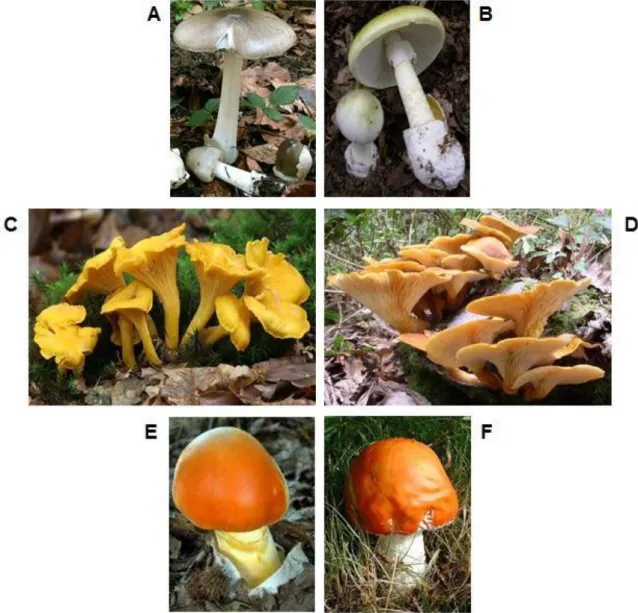
Figure 1.4. Examples of some mushroom species that may be confounded because their
morphological similarities: (A) Volvariella gloiocephala (DC.) Boekhout & Enderle (edible) confounded with (B) Amanita phalloides (Vaill. ex Fr.) Link (deathly), (C) Cantharellus cibarius Fr.
(edible) confounded with (D) Omphalotus olearius (DC.) Singer (toxic) and (E) Amanita caesarea (Scop.) Pers. (edible) confounded with (F) Amanita muscaria (L.) Lam. (toxic and hallucinogenic) when this, because of rain, loses is characteristic white scales. (Adapted from the originals available at:
(A) http://www.koleopterologie.de/arbeitsgemeinschaft/beitraege/esser/volvariella-gloiocephala-foto-keller-640x480.jpg [06/09/12], (B) http://upload.wikimedia.org/wikipedia/commons/9/99/Amanita_phalloides_1.JPG [06/09/12], (C) http://www.naturephoto-cz.com/photos/others/cantherellus-31880.jpg [06/09/12], (D) http://www.naturamediterraneo.com/Public/data/polypo/20041017181445_Omphalotus%20olearius.JPG [06/09/12], (E) http://t3.gstatic.com/images?q=tbn:ANd9GcRePlo5HgXbpVUBTMiUlAzu1QeKylifLI5D0XCE_X6XYMiccz4lWw&t=1 [06/09/12] e (F) http://farm1.static.flickr.com/124/319800093_b55e2cbba9.jpg [06/09/12])
11
Table 1.3. Some examples of edible and toxic mushrooms usually confounded. (Adapted from [22])Edible mushroom species Toxic mushroom species
Agaricus arvenis Schaeff Agaricus xanthodermus Genev.
Amanita ovoidae (Bull.) Link Amanita proxima Dumée
Armillaria mellea (Vahl) P. Kumm.
Clitocybe acromelalga Singer Galerina unicolor (Vahl) Singer
Calocybe gambosa (Fr.) Donk Inocybe erubescens A. Blytt
Clitocybe gibba (Pers.) P. Kumm. Clitocybe amoenolens Malençon
Coprinus comatus (O.F. Müll.) Pers. Coprinus atramentarius (Bull.) Fr.
Marasmius oreades (Bolton) Fr. Clitocybe dealbata (Sowerby) Gillet
Lepista inversa (Scop.) Pat. Clitocybe amoenolens Malençon
Agaricus xanthodermus Genev.
Morchella esculenta (L.) Pers. Gyromitra esculenta (Pers.) Fr.
2. MUSHROOM AUTHENTICITY
The unequivocal identification of a mushroom species through its phenotypical features (morphology and physiology) is extremely difficult, and in certain cases even impossible, due to subjectivity of these kind of analysis and also as a consequence of scarcity and ambiguity of these characteristics.[25, 26] Thus, it is essential to find and use other properties that allow a correct identification of the specimens. Therefore, the chemical/molecular composition of species is a very important taxonomical tool, being complementary to the phenotypical features in identification and classification of such organisms.
Chemotaxonomy, also designated molecular taxonomy, uses the chemical composition and/or molecular analysis of species to its classification and identification. The molecules which are the most used in this context are deoxyribonucleic acid (DNA), fatty acids (FA), proteins, carbohydrates and secondary metabolites (SM).[27]
2.1 DNA analysis
Interest and investigation in molecular biology lead to development of new technics that allows species identification[28], namely, through the study of the DNA molecule. Thus, the DNA analysis, also called molecular analysis, is, nowadays, the most common technique in fungal chemotaxonomy. The importance of DNA in this context is due to the fact that the genetic composition of each species is unique and specific, not being affected by age, physiological conditions or environmental factors.[29]
Molecular analysis techniques require, in first place, the extraction of genetic material. In the case of mushrooms, there are described in the literature different “basic” methods to extract DNA, both for fresh and dried mushrooms (exsiccated), ranging from simple commercial extraction kits to laboratory protocols[29, 30] a little more complex. After the extraction of DNA, the genetic material is amplified through the polymerase chain reaction (PCR). Then, there are two methodologies that are the most utilized: DNA sequencing[31] or the analysis of DNA fragments length[28]. To obtain and analyse DNA fragments, two techniques can be used: RAPD – randomly amplified polymorphic DNA[32]; and ARDRA – amplified ribossomal DNA restriction analysis[33].
Concerning, DNA sequencing and ARDRA technique, as its name indicates, the recommended and most commonly used DNA region in these studies is rDNA[28, 33], i.e. ribossomal DNA – the DNA region that contains the group of genes that codifies the ribossomal ribonucleic acid (rRNA). The Fungal Work Group (FWG) of the Consortium for
13
the Barcode of Life (CBOL), after several studies, concluded that the ITS1-5.8S-ITS2 region of rDNA (also known simply by ITS region – Internal Transcribed Spacer region), should be chosen as the molecular marker for the identification of fungal species.[34] The ITS region was chosen because it has several advantages, e.g. ease of amplification and the fact that it’s the most universal option[31, 34].
2.2 Analysis of other molecules
Chemotaxonomy, as mentioned above, is based on the study of different molecules of organisms in order to identify them. Thus, besides the aforementioned DNA molecule study, the analysis of other molecules, using, obviously, other kind of techniques and methodologies, is important and useful in the chemotaxonomical context.
Chemical markers
2.2.1
Chemotaxonomy, in its most global definition, can use any molecule (or molecule groups) of an organism to the species identification. However, depending on the organism to identify, it is common that certain groups of compounds are preferred. Concerning macrofungi, SM are the most used molecules for chemotaxonomical purposes.[35] Apart from the study of these compounds, in literature, there are also chemotaxonomical studies that are based on the analysis of FA, proteins, AA and carbohydrates, despite the fact that the use of these molecules is much smaller compared to SM.
SM have been extremely used in macrofungal species identification, although, they have been interpreted as “morphological/organoleptic markers”, once they are responsible for characteristics like colour, odour and taste.[35] When compared to primary metabolites (PM), SM are preferable in these kind of studies once most of them show higher ability to species differentiation.[35] PM, being vital to the maintenance and survival processes of organisms, such as growth and reproduction, are the “base molecules of life” and so they are common to the majority of species, even if these are phylogenetically distinct. Otherwise, the SM, not being essential to survival, are more differentiated among species as consequence of mutations and evolutionary processes. Yet, not all SM, when used individually, are potential chemotaxonomical markers, once phylogenetically different species can, through synthesis processes that are evolutionarily distinct, produce the same SM.[27] Thus, the “choice” of a molecule as chemotaxonomical marker should be
based on its differentiation efficiency[27], i.e. its specificity for a species and/or genus. Obviously, species identification based on a single molecule it is almost impossible. Therefore it is necessary to identify a group of molecules, which, in most of the cases, should contain compounds originated in different biosynthetic paths, to set a pattern of SM which can identify that species.[27, 35]
Species belonging to Basidiomycota phylum produce a large number of SM.[27] Although a large number of SM have been identified and characterized, such as pigments[36], toxins[37, 38] and volatile compounds responsible for aroma and taste[7, 39], few of these studies regarded the chemotaxonomic importance/utility of these molecules. Even so, there are in the literature some studies concerning the chemotaxonomical value of such molecules. A study developed in 2002 allowed the identification of some pigments named retipolides (spiromacrolactones) which are chemotaxonomical markers of the species belonging to Retiboletus Manfr. Binder & Berinsky genus.[40] Other work revealed that atromentin, thelephoric acid and some pulvinic acid derivatives were pigments common to several species belonging to Omphalotus Fayod genus and so they can be considered chemical markers for those species.[41] As an example of application of volatiles in chemotaxonomical investigations, there is a published work in which the volatile profiles of three species was achieved and the authors concluded that those patterns were distinctive among each specimen although the compounds present were similar in all samples – aliphatic and terpenoid alcohols, 4- and 5-olides (i.e. γ- and δ-lactones) and alicyclic and aromatic compounds.[42]
Despite the importance of SM in this context, it is important to note that the biosynthesis of these compounds depends on the stage of growth, abiotic factors and, in the case of mushrooms, can, sometimes, depend on the fruiting body region that is analysed.[27, 43]
With regard to macrofungi, besides the SM, other molecules have also been analysed for chemotaxonomical purposes, such as AA and FA. As an example, the study of FA profile in basidiospores of several species of mushrooms allowed the identification of molecules that are specific of certain species, e.g. heptadecanoic acid [(17:0) and docosanoic acid (C22:0) were only present in the Agaricus sylvicola (Vittad.) Peck and Armillaria borealis Marxm. & Korhonen species, respectively.[43] Another example is a study about AA composition in six species of Amanita Pers. genus where the main conclusion was that although there was some differences among AA contents it is not likely that those differences can be used for identification/classification purposes.[44]
15
Although there are several studies about the chemical composition of mushrooms, most of them does not focus species identification. Thus, due to the high number of mushroom species and to the severity of some mushroom intoxications, macrofungi chemotaxonomical analysis remain very important in order to discover patterns of compounds that can improve the correct identification of species.
Chemical analysis techniques
2.2.2
The chemical analysis, in the chemotaxonomical context, is done through several techniques, but the work methodology is, usually, the same: a sequential three-step process. The first step is the molecules extraction from the matrix, followed by the separation and detection of analytes through chromatographic techniques and, finally, their identification and/or characterization (Table 2.1).
The choice of extraction techniques to use depends on the chemical properties of analytes in study. However, all the techniques rely on the same principle: distribution of analyte(s) between two immiscible phases.[45] In the case of volatiles (i.e. inherently volatile compounds or derivatized compounds), the most common method is the extraction of these compounds from headspace (HS) through the adsorption of analytes into different kinds of adsorbents. Among the different HS extraction techniques, the HS-SPME (headspace solid phase microextraction) is undoubtedly the most used. In this technique, the adsorbents are fibers coated with various stationary phases with different thicknesses. So, the volatiles present in HS are adsorbed in SPME fibers and subsequently are thermally desorbed in the injector of the chromatographic apparatus. Besides HS techniques, volatile molecules can also be extracted through simultaneous distillation and extraction (SDE) and supercritical fluid extraction (SFE).[35] For non-volatile compounds (or semi-volatile compounds that cannot be extracted by HS techniques) solid-liquid extraction is usually used.[46, 47]
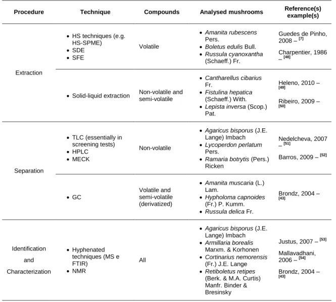
Table 2.1. Main techniques used in chemotaxonomical analysis of mushrooms, procedure and kind
of compounds to which they are associated, as well as examples of species analysed by these techniques.
Procedure Technique Compounds Analysed mushrooms Reference(s)
example(s) Extraction HS techniques (e.g. HS-SPME) SDE SFE Volatile Amanita rubescens Pers.
Boletus edulis Bull.
Russula cyanoxantha (Schaeff.) Fr. Guedes de Pinho, 2008 – [7] Charpentier, 1986 – [48]
Solid-liquid extraction Non-volatile and semi-volatile
Cantharellus cibarius Fr.
Fistulina hepatica (Schaeff.) With.
Lepista inversa (Scop.) Pat. Heleno, 2010 – [49] Ribeiro, 2009 – [50] Separation TLC (essentially in screening tests) HPLC MECK Non-volatile
Agaricus bisporus (J.E. Lange) Imbach
Lycoperdon perlatum Pers.
Ramaria botrytis (Pers.) Ricken Nedelcheva, 2007 – [51] Barros, 2009 – [52] GC Volatile and semi-volatile (derivatized) Amanita muscaria (L.) Lam. Hypholoma capnoides (Fr.) P. Kumm. Russula delica Fr. Brondz, 2004 – [43] Identification and Characterization Hyphenated techniques (MS e FTIR) NMR All
Agaricus bisporus (J.E. Lange) Imbach
Armillaria borealis Marxm. & Korhonen
Cortinarius nemorensis (Fr.) J.E. Lange
Retiboletus retipes (Berk. & M.A. Curtis) Manfr. Binder & Bresinsky Justus, 2007 – [53] Mallavadhani, 2006 – [54] Brondz, 2004 – [43]
FTIR – Fourier transform infrared spectroscopy; GC – gas chromatography; HPLC – high performance liquid chromatography; HS – headspace; HS-SPME – headspace solid phase microextraction; MECK - micellar electrokinetic chromatography; MS – mass spectrometry; NMR – nuclear magnetic resonance; SDE – simultaneous distillation extraction; SFE – supercritical fluid extraction; TLC – thin layer chromatography.
As extraction procedures, different chromatographic methods are used to separation of volatile and non-volatile molecules.
In the non-volatile context, planar chromatography techniques, namely thin layer chromatography (TLC) and paper chromatography, were the first to be utilized in separation of fungal molecules.[27] However, nowadays there are separation techniques more efficient than planar chromatography, and so these techniques, namely TLC, are only used for screening.[35, 54] Indeed, the non-volatile separation method most commonly used nowadays is high performance liquid chromatography (HPLC).[27] Micellar
17
electrokinetic chromatography (MECK) also allows the separation of non-volatile molecules[35], but is much less used than HPLC.
The separation of volatile compounds, or semi-volatile compounds after derivatization (e.g. AA, carbohydrates and FA/lipids), is performed by gas chromatography (GC).[27] In this technique, the stationary phase is chemically bonded to a fused silica capillary column. The type (polar or non-polar) and thickness of stationary phase used depends on the polarity and volatility of the compounds that are being analysed. In the other hand, the mobile phase is an inert gas, e.g. helium (He), hydrogen (H2) or nitrogen (N2). For the detection, the flame ionization detector (FID) remains the most widely used detector, once it allows detecting a large number of compounds. However, FID does not give any structural informal about the detected molecules, and thus the identification of unknown compounds is almost impossible.[27] In this context, infrared (IR) spectroscopy can also be used to detect and identify fungal molecules. Detection by IR spectroscopy is performed through an IRD (infrared detector) or a hyphenated technique called gas chromatography-Fourier transform infrared spectroscopy (GC-FTIR). A disadvantage of detection by IR spectroscopy is its low sensitivity when compared to other detection methods.[27]
Besides GC-FTIR, other hyphenated techniques are used to analyse fungal species, namely liquid mass spectrometry (LC-MS) and gas chromatography-mass spectrometry (GC-MS). GC-MS is nowadays the most important technique for detection and characterization of volatile molecules[35], which are responsible for the smell and taste of mushrooms. Some studies using GC-MS technique are reported in literature to the analysis of macrofungal species[7, 55, 56]. Although LC-MS is not so commonly used as GC-MS, there are some scientific publications in which this methodology is applied to macrofungi[52, 57, 58]. Electron impact (EI) is the ionization method used in GC- and LC-MS to promote ionization and, consequently, fragmentation of analytes. The mass spectrum resulting from fragmentation by EI allows the identification of molecules through comparison with mass spectra in a database. EI fragmentation patterns are also important for the characterization of unknown compounds, once through mass fragments and fragmentation mechanisms their chemical structures can be understood.
Finally, another important technique to the identification/characterization of unknown molecules is nuclear magnetic resonance (NMR). Concerning macrofungal species, NMR has been important in structural elucidation of several polysaccharides.[59-61]
3. METABOLOMICS
Nowadays, there are several “omics” technologies which are applied in many scientific areas, ranging from medicine to environmental studies. The goal of these “omics” technologies is the study and mapping of a group of biomolecules – the “omes”. For example, the study of all genes (genome) is called genomics, in the same way, proteomics is the study of all proteins and the aim of metabolomics is the study of metabolites produced by an organism (metabolome).[62] These “omics” approaches are considered important tools to understand the biology of an organism and to interpret the complexity of its biological processes.[63-65]
Metabolomics is the comprehensive study of metabolome – the set of all low molecular weight molecules (metabolites) produced by an organism, which can be organic compounds such as AA, FA, carbohydrates and vitamins, but also inorganic molecules and elemental species.[66] The study of metabolome consists on the identification and analysis of metabolite composition, which is itself a huge challenge, but also the understanding of their dynamics, interactions and responses to changes in their environment.[67, 68] The achievement of those purposes is possible due to the use of sophisticated analytical methods and multivariate statistical analysis.[69]
Results obtained from metabolomic studies can be used to biomarker identification, drug discovery/development, clinical toxicology, nutritional studies and quantitative phenotyping.[64]
3.1 Terminology
Since the development of metabolomics in the 1990’s, different approaches have been used to study metabolome. Actually there are five conceptual approaches that can be classified in two complementary groups: 1) Targeted analysis – which comprises A: metabolomic target analysis and B: metabolite profiling; and 2) Non-targeted analysis – which includes C: metabolic fingerprinting, D: metabolomics and E: metabonomics.
1) Targeted analysis, which have been applied before the arise of the term ‘metabolomics’, focuses on the analysis and study of specific group of metabolites:
19
A. Metabolomic target analysis refers to the identification and quantification of a small group of known metabolites (‘targets’) related to a specific metabolic reaction.[65, 66, 70]B. Metabolite profiling aims to identify a larger set of compounds than metabolic target analysis. Herein, the study focuses not only in known but also in unknown compounds of a specific metabolic pathway.[65, 66, 70, 71] 2) Non-targeted analysis is a global study of all metabolites and their changes as
response to environmental variations:
C. Metabolic fingerprinting is a rapid and global analysis of samples to generate a ‘metabolic signature’, which is compared to others with the aim of finding differences between those samples.[65, 66] Usually, quantification is not employed and only metabolites that allow samples discrimination are identified and studied to understand their biological roles.[65, 71]
D. Metabolomics is, as mentioned before, the non-biased identification and study of all metabolites (metabolome) produced in organism, and that are present in biological samples such as cells, tissues or biofluids.[65-67, 71] E. Metabonomics is the analysis of perturbations in endogenous metabolites
levels, i.e. the metabolic response of living systems, due to diseases, drugs and toxins intake and genetic modifications.[66, 70, 72] Metabonomics is considered a subset of metabolomics, a broader concept[73], but sometimes the terms are used interchangeably.[71, 73]
When compared to targeted approaches, non-targeted metabolomics is best suitable for chemotaxonomical purposes and species-specific markers discovery, as this kind of metabolomic experiment is designed to simultaneously analyse the largest number of metabolites possible and, consequently, to study unexplored metabolic pathways – such as secondary metabolites synthesis pathways. Thus, the term ‘metabolomics’ will be used herein to refer to non-targeted metabolomics.
3.2 Role of metabolomics in chemotaxonomy
A cross-interpretation of terms ‘chemotaxonomy’ (Section 2.2) and ‘metabolomics’ easily allows to conclude that metabolomics is a powerful and important strategy to discover chemotaxonomical relevant molecules. Indeed, metabolomics, and specifically its non-targeted approaches, aiming to identify all molecules produce by an organism, is the
ultimate tool to find those compounds/metabolite profiles that are species-specific or, at least, species-indicative.
As previously referred, molecules resulting from secondary metabolism are very important in this context, since a “fungal secondary metabolite” is a chemical compound produced by a limited number of species in a genus, an order, or even phylum, and has a high differentiation power”[27]. Nowadays there are some reviews on the use of metabolite profiling in fungal species (e.g. Frisvad et al. (2008)[27], Nielsen et al. (2004)[45] and Smedsgaard & Nielsen (2005)[74]), but in this context, Basidiomycota phylum is still widely unexplored[75].
3.3 Metabolomics workflow
Metabolomics investigations results from the combination of analytical methodologies, the tool to ‘reach’ metabolome, and bioinformatics (multivariate statistical analysis or chemometrics), which allows data processing and interpretation.[63, 65] Thus, a metabolomic experiment is a sequential multi-step procedure that can be divided into four parts: sample collection and preparation; metabolites separation and identification; data mining and extraction; and data analysis and interpretation.[76]
Sample collection refers to the samples harvesting and storage. Herein, it is important
to collect an appropriate number of samples to match the study requirements and reduce the influence of biological variance.[77] This is not always possible, namely when wild species are under analysis, such as wild mushrooms. Sample sampling and storage should ensure that there is no formation or degradation of metabolites due to enzymatic activity, oxidation processes or bacterial growth. So, after sample collection, metabolism should be stopped (sample quenching) as quickly as possible, which can be done by different processes (e.g. freezing in liquid nitrogen).[76] Sample preparation is a procedure step that serves several purposes: metabolite extraction from matrix (Section 2.2.2) – i.e. removal of matrix components that can interfere in the analysis (e.g. macromolecules); transference of metabolites to a medium compatible to the analytical technique – e.g. extract metabolites from biological tissues to an appropriate solvent to inject it in a gas chromatograph; metabolite enrichment – i.e. a pre-concentration step to increase the concentration of low-abundance metabolites; and, specifically in GC, derivatization to improve metabolites volatility.[76, 77] Ideally, a metabolomics analysis
21
would be performed without sample preparation, as this procedure necessary leads to metabolites loss and perhaps to contaminations.[76, 77]
Metabolite separation is achieved using chromatographic techniques such as liquid
and gas chromatographies (Section 2.2.2) and capillary electrophoresis. There are a large collection of separation methodologies that can be used in metabolomic experiments, and so the advantages and limitations, sensibility and costs of those techniques but also the nature of samples and metabolites under analysis should be taken in account to choose the most suitable technique to be applied.[70, 78] Nowadays the two analytical techniques most applied to metabolite identification in metabolomics’ studies are NMR spectroscopy and MS (Section 2.2.2).[63, 76] Both are high-throughput techniques, which allow the analysis of a large spectrum of metabolites in a fast and reproducible way. The main advantages of NMR are that it provides structural information on molecular structure, which facilitates unknown metabolite identification, and requires minimal sample preparation, but otherwise its low sensitivity is a considerable drawback.[63, 70, 76] In contrast to NMR, MS is more sensitive, but in the other hand it is destructive to samples and usually requires sample preparation.[63, 76] Besides these, other techniques can be used such as FTIR and Raman spectroscopies.[70]
Once the goal of metabolomics is the study a wide group of compounds, the application of separation/identification techniques to samples generates a large amount of data.[67, 76] Thus, data pre-processing, i.e. transformation of raw data into a standard and uniform format, is an essential part of data mining and extraction, once it allows the correction of data differences caused by experimental variables.[67, 77] Due to the high volume of data, this is done through specific software and this step includes: peak alignment (matching peaks across multiple samples), peak normalization (adjust peak intensities and reduce analytical drift), peak deconvolution (separate overlapped peaks to distinguish co-eluted metabolites) and baseline correction (remove background noise).[67, 76, 77] Nowadays there are several commercial and free software programs to perform these operations[67, 77], each one with its own advantages and limitations, but still none of those is universal.[76]
After pre-processing, data analysis is performed through several multivariate statistical analysis methods, which allows to “see” spectral patterns, i.e. metabolic signature of samples.[76] Herein there are two main approaches: supervised analysis and unsupervised analysis. Unsupervised methods (e.g. principal component analysis – PCA, probably the most commonly multivariate statistical analysis method used in metabolomics[79]; or
hierarchical cluster analysis – HCA) discriminate samples through their metabolite composition in an unbiased way, i.e. without any prior knowledge about samples.[78, 79] Thus, these methods enable the identification of differences between samples and also the presence of outliers in data.[79] In contrast, supervised methods, such as partial least-square discriminant analysis (PLS-DA) – one of the most currently used[79], requires the supply of information about samples (e.g. diseased and healthy subject, toxic and non-toxic specimen, etc.) to create cluster of patterns, i.e. reveal the metabolic features which are better to differentiate those groups .[77, 78] Once found the compounds responsible for differences, metabolite identification must be done through the techniques previously referred – i.e. comparison of NMR and/or MS spectrum(a) with those present in spectral databases or with the standard compound spectrum or, when it is a compound identified for the first time and its standard is not available in suppliers, through molecular structural information provided by those techniques.[78] In order to establish that a metabolite (or a group of them) is indeed a marker, either a biomarker or a chemotaxonomical marker, it is obviously necessary to perform more extensive studies to validate that conclusion.
Despite the improvements in separation, analytical and statistical methods, there are still some limitations in metabolomics, which can be a result of the complexity of metabolome, the large amount of data generated and the intra- and inter-individuality of metabolites composition among samples.
25
4. OBJECTIVES AND EXPERIMENTAL APPROACHES
The main purpose of this research work was to analyse wild edible and toxic mushrooms through GC-MS technique, aiming samples discrimination through metabolic profiles. Thus, to achieve this goal two approaches were chosen:
A. The application of a multi-target method for the identification and quantification of three classes of metabolites – amino acids, fatty acids and sterols.
Although these compounds were previously studied in different mushroom species and found not to be the most suitable to identification purposes, as they are primary metabolites, there were several reasons to perform this study:
Test the applicability of this multi-target method in mushrooms, once this methodology was never been applied in macrofungal species.
Identify and quantify metabolites of the mentioned classes (AA, FA and sterols) in several mushrooms species, some of which have been analysed in the present work for the first time.
Apply the “non-targeted analysis”, through multivariate statistical methods, to explore the metabolic pattern achieved with this technique, as it contains more compounds than those belonging to the referred classes and some of which can have chemotaxonomical potential.
B. The application of a HS-SPME/GC-MS methodology to study volatile profiles of mushrooms.
According to the purpose of this investigation, volatiles are important candidate compounds to distinguish edible from toxic mushrooms as they are fulcral SM participating to the specific aroma of each mushroom species. In agreement with that, there must be some volatiles, even present in low contents that may allow discrimination between edible and toxic species. Animals, whose olfaction is usually more developed than that of the humans, “use” this characteristic of volatiles (aroma) to choose which mushrooms may or may not be eaten.
Although the volatile composition of mushrooms is unquestionably widely studied, its analysis through a non-targeted approach, i.e. metabolic fingerprinting, has never been
used and this seems to be a good strategy to achieve the goal of this research. Herein, the two objectives were:
To analyse data through an unsupervised statistical methodology in order to identify the most suitable metabolites to differentiate mushroom species.
To perform a supervised data analysis (edible mushrooms vs. toxic mushrooms), aiming to understand if there are significant similarities within each group and, if so, to identify the volatile molecules which are the most appropriated to differentiate those groups.
29
5. MATERIAL AND METHODS
5.1 Mushroom samples
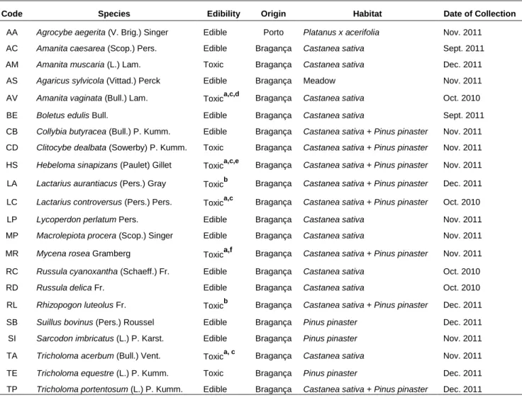
Samples of 22 different wild mushroom species were collected in Trás-os-Montes and Douro Litoral regions – Table 5.1 and Figure 5.1. After being harvested, species were taxonomically identified according to several authors[80-83] and a representative voucher of each specimen was deposited at the herbarium of Escola Superior Agrária of Instituto Politécnico de Bragança.
Table 5.1. Characterization of mushroom species.
Code Species Edibility Origin Habitat Date of Collection
AA Agrocybe aegerita (V. Brig.) Singer Edible Porto Platanus x acerifolia Nov. 2011
AC Amanita caesarea (Scop.) Pers. Edible Bragança Castanea sativa Sept. 2011
AM Amanita muscaria (L.) Lam. Toxic Bragança Castanea sativa Dec. 2011
AS Agaricus sylvicola (Vittad.) Perck Edible Bragança Meadow Nov. 2011
AV Amanita vaginata (Bull.) Lam. Toxica,c,d Bragança Castanea sativa Oct. 2010
BE Boletus edulis Bull. Edible Bragança Castanea sativa Sept. 2011
CB Collybia butyracea (Bull.) P. Kumm. Edible Bragança Castanea sativa + Pinus pinaster Nov. 2011
CD Clitocybe dealbata (Sowerby) P. Kumm. Toxic Bragança Castanea sativa + Pinus pinaster Nov. 2011
HS Hebeloma sinapizans (Paulet) Gillet Toxica,c,e Bragança Castanea sativa + Pinus pinaster Nov. 2011
LA Lactarius aurantiacus (Pers.) Gray Toxicb Bragança Castanea sativa + Pinus pinaster Dec. 2011
LC Lactarius controversus (Pers.) Pers. Toxica,c Bragança Castanea sativa + Pinus pinaster Oct. 2010
LP Lycoperdon perlatum Pers. Edible Bragança Castanea sativa Nov. 2011
MP Macrolepiota procera (Scop.) Singer Edible Bragança Castanea sativa Nov. 2011
MR Mycena rosea Gramberg Toxica,f Bragança Castanea sativa + Pinus pinaster Nov. 2011
RC Russula cyanoxantha (Schaeff.) Fr. Edible Bragança Castanea sativa Oct. 2010
RD Russula delica Fr. Edible Bragança Castanea sativa Oct. 2010
RL Rhizopogon luteolus Fr. Toxicb Bragança Castanea sativa + Pinus pinaster Dec. 2011
SB Suillus bovinus (Pers.) Roussel Edible Bragança Pinus pinaster Dec. 2011
SI Sarcodon imbricatus (L.) P. Karst. Edible Bragança Pinus pinaster Nov. 2011
TA Tricholoma acerbum (Bull.) Vent. Toxica, c Bragança Castanea sativa Nov. 2011
TE Tricholoma equestre (L.) P. Kumm. Toxic Bragança Pinus pinaster Dec. 2011
TP Tricholoma portentosum (L.) P. Kumm. Edible Bragança Castanea sativa + Pinus pinaster Dec. 2011
a Suspected to be toxic – Considered toxic to comparison purposes; b Edibility/Toxicity unknown – Considered toxic to comparison
purposes; c It is suspected that causes gastrointestinal disorders; dIt is suspected that causes haemolytic disorders; e May have cytotoxic cucurbitacins: f It is suspected to contain the toxin muscarine
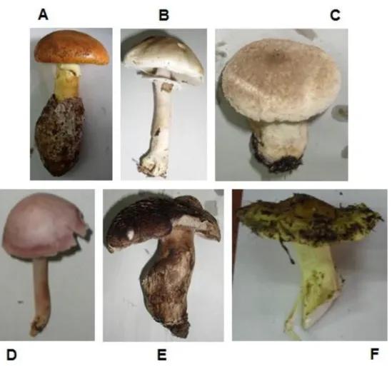
Figure 5.1. Some of the mushrooms collected and analysed in this work: (A) Amanita caesarea
(Scop.) Pers.; (B) Agaricus sylvicola (Vittad.) Perck; (C) Lycoperdon perlatum Pers.; (D) Mycena rosea Gramberg; (E) Sarcodon imbricatus (L.) P. Karst.; and (F) Tricholoma equestre (L.) P.
Kumm.. (Source: Author’s own file; Images are not to scale)
The fresh species were cleaned and fragmented into small pieces. Then, 5g of each sample were put into a 20 ml vial kept which was cap-sealed. Prepared mushroom samples were frozen at -20 ±1ºC until HS-SPME/GC-MS analysis. Some species collected in 2010 were already frozen when this work started, for that reason their volatile profiles were not studied.
The remaining of each mushroom was frozen at – 20ºC until lyophilisation. Lyophilized samples were powdered and screened through a 910 µm fine sieve before being stored in hermetically sealed bags. These samples were used for the AA, FA and sterols GC-MS analysis.
![Table 1.1. Some characteristics of the principal phyla where the macrofungi belong. [1-3]](https://thumb-eu.123doks.com/thumbv2/123dok_br/19214277.959707/15.892.147.812.748.918/table-characteristics-principal-phyla-macrofungi-belong.webp)
![Figure 1.1. Basidiocarp structure: A – cap (or pileus); B – scales; C – hymenium; D – annulus (or ring); E – stipe (or stalk) ; F – volva (or cup) (Adapted from [1] )](https://thumb-eu.123doks.com/thumbv2/123dok_br/19214277.959707/16.892.257.568.126.467/figure-basidiocarp-structure-pileus-scales-hymenium-annulus-adapted.webp)
![Figure 1.2. Spore print: A – hymenium placed on the paper sheet; B – spore deposition (Adapted from the original available at: http://www.anbg.gov.au/fungi/images-captions/spore-print-0018.html [05/06/12])](https://thumb-eu.123doks.com/thumbv2/123dok_br/19214277.959707/19.892.265.692.644.903/figure-spore-hymenium-deposition-adapted-original-available-captions.webp)
![Table 1.3. Some examples of edible and toxic mushrooms usually confounded. (Adapted from [22] )](https://thumb-eu.123doks.com/thumbv2/123dok_br/19214277.959707/23.892.168.787.157.556/table-examples-edible-toxic-mushrooms-usually-confounded-adapted.webp)