Braz. J. of Develop.,Curitiba, v. 6, n. 11, p.91343-91359 nov. 2020. ISSN 2525-8761
Analysis of skin microbiota alteration in patients after chemotherapy treatment
Análise da alteração da microbiota epidermal de pacientes após tratamento
quimioterápico
DOI:10.34117/bjdv6n11-517
Recebimento dos originais:08/10/2020 Aceitação para publicação:24/11/2020
Pedro Augusto Ramos Vanzele
Mestrando em Farmácia (Fisiopatologia e Toxicologia) pela Universidade de São Paulo Instituição: Faculdade de Ciências Farmacêuticas da USP – FCF-USP
Endereço: Avenida Prof. Lineu Prestes, 2565, Butantã, São Paulo – SP, Brasil E-mail: pedrovanzele@gmail.com
Rillery Calixto Dias
Graduanda em Biomedicina pelo Centro Universitário do Sul de Minas – UNIS-MG Instituição: Centro Universitário do Sul de Minas – UNIS-MG
Endereço: Avenida Alzira Barra Gazzola, 650, Aeroporto, Varginha – MG, Brasil E-mail: rillery_calixto@hotmail.com
Priscila Moraes Henrique Paiva
Doutora em Medicina Tropical e Doenças Infecciosas pela Universidade Federal do Triângulo Mineiro - UFTM
Instituição: Centro Universitário do Sul de Minas – UNIS-MG
Endereço: Avenida Alzira Barra Gazzola, 650, Aeroporto, Varginha – MG, Brasil E-mail: priscila.paiva@unis.edu.br
Giulia Maria de Alencar Castro Bani
Mestre em Biociências Aplicadas à Saúde pela Universidade Federal de Alfenas – UNIFAL Instituição: Centro Universitário do Sul de Minas – UNIS-MG
Endereço: Avenida Alzira Barra Gazzola, 650, Aeroporto, Varginha – MG, Brasil E-mail: giulia.bani22@gmail.com
Lívia da Silva Ciacci
Mestre em Engenharia Biomédica pela Universidade Federal de São João del-Rei - UFSJ Instituição: Centro Universitário do Sul de Minas – UNIS-MG
Endereço: Avenida Alzira Barra Gazzola, 650, Aeroporto, Varginha – MG, Brasil E-mail: livia.ciacci@unis.edu.br
Hadassa Cristhina de Azevedo Soares dos Santos
Doutora em Ciências pela Universidade de São Paulo Instituição: Centro Universitário do Sul de Minas – UNIS-MG
Endereço: Avenida Alzira Barra Gazzola, 650, Aeroporto, Varginha – MG, Brasil E-mail: hadassa.santos@unis.edu.br
Braz. J. of Develop.,Curitiba, v. 6, n. 11, p.91343-91359 nov. 2020. ISSN 2525-8761
ABSTRACT
The human body is inhabited by millions of micro-organisms, which play an auxiliary role in the digestion and absorption of nutrients, as well as in the immune protection of the host. The human skin contains an immense number of micro-organisms, which vary with the local interferences and appear different according to the area where they inhabit. However, such micro-organisms are in constant modification due to some disturbances. Knowing that chemotherapy is a very aggressive treatment for human cells and based on other related researches, the work in question is an evaluation of the changes caused by the epidermal microbiota in cancer patients after the chemotherapy treatment. The methods consisted in the collection of samples by sterile swab rubbed in the patients' skin pre- and post-chemotherapy treatment, and in the analysis of the samples, which were sown in three culture media with different properties. Subsequently, the main bacteria that grew in the medium were isolated and identified by means of Gram staining and biochemical tests. Some qualitative changes were found at the species level; however, we detected the preservation of the micro-organisms Staphylococcus
epidermidis, the main colonizers of the skin and that present beneficial role for the host. Therefore,
the results are positive when evaluated from a collaboration point of view to improve patients' quality of life. A more complete analysis of the microbiota is now required through molecular techniques, so that quantitative results can be verified.
Keywords: Microbiota, Skin, Chemotherapy. RESUMO
O corpo humano é habitado por milhões de microrganismos, os quais desempenham um papel auxiliar na digestão e absorção de nutrientes, assim como na defesa imunológica do hospedeiro. A pele humana contém uma imensa quantidade de microrganismos, que variam com as interferências locais e se apresentam diferentes de acordo com a área onde habitam. Porém, tais microrganismos estão em constante modificação devido alguns distúrbios. Sabendo que a quimioterapia é um tratamento bastante agressivo para as células humanas e tendo como base outros trabalhos relacionados, o trabalho em questão trata-se de uma avaliação das alterações ocasionadas na microbiota epidermal em pacientes com câncer após realizado o tratamento quimioterápico. O método consistiu na coleta de amostras por swab estéril friccionado na pele de pacientes pré e pós tratamento quimioterápico, e na análise das amostras, as quais foram semeadas em três meios de cultura de propriedades diferentes. Posteriormente, foram isoladas as principais bactérias que cresceram no meio e procedeu-se a identificação por meio de coloração de Gram e provas bioquímicas. Foram encontradas algumas alterações qualitativas a nível de espécie, porém houve a preservação dos microrganismos
Staphylococcus epidermidis, os principais colonizadores da pele e que apresentam papel benéfico para
o hospedeiro. Sendo assim, os resultados se apresentam positivos quando avaliados pelo ponto de vista da colaboração para melhorar a qualidade de vida dos pacientes. Agora se faz necessária uma análise mais completa da microbiota por meio de técnicas moleculares, de forma que seja possível verificar esses resultados a nível quantitativo.
Palavras-chave: Microbiota, Pele, Quimioterapia.
1 INTRODUCTION
The human body is inhabited by millions of micro-organisms. It is estimated that the man has approximately three times more prokaryotic cells than human cells (WILSON, 2008). The micro-organisms are necessary in the daily, either to assist immunologically, either in the digestion or absorption of nutrients (COSTELLO et al., 2014). Even if the gut microbiome be the most studied, the gut is not the only part of human organism inhabited by bacteria. It is known that the human skin
Braz. J. of Develop.,Curitiba, v. 6, n. 11, p.91343-91359 nov. 2020. ISSN 2525-8761 has several micro-organisms that extends from the surface to the subcutaneous areas, being distinct due local interferences as pH, temperature and oxygen content (GRICE; SEGRE, 2011). There are also differences according to the area that they inhabit, as sebaceous areas, moist locals or dry locals. Among these areas, Grice et al. (2009) verify that physiologically similar locals shown similar compositions of colonizer micro-organisms.
As well as in other body parts, some bacterial species can help in the prevention of diseases by the limitation of others bacteria growth, by means of production of determinates proteins (COGEN et al., 2010). According Nakatsuji et al. (2018), some strains of the micro-organism Staphylococcus
epidermidis produce a substance called 6-N-hydroxyaminopurine (6-HAP) able to inhibit the DNA
synthesis. When performing and analyzes the gene sequencing of individuals skin micro-organisms, it was identified similar strains to the 6-HAP producers in different body parts and in different frequencies and, in this study, it was observed that when administered this molecule by intravenous or topic via in mice, tumors as melanoma, one of the main types ok skin cancer, that grown in vivo, they were suppressed, giving the idea that the skin microbial community is able to help in the host defense giving resistance to the tumor growth. The 6-HAP is a molecule that has a structure very similar to adenine, thus, it is a kind of a “substitute” of the purine that will act against the tumor growth specifically by interfering in the base pairs adenosine-timidine, blocking the DNA-polymerase action. The human microbiota is in constant modification, due to disturbances that can reduce the number of some constituent species, providing a chance to the growth of others remaining micro-organisms or new settlers. The causer disturbances of microbial community alteration range from the host diet to the main subversives, the antibiotics. The major antimicrobials’ problem is the capacity of destroy, beside the desired micro-organisms, the habitual human microbiota components. Studies denotes that even though has alteration in the individuals microbiota, it is possible, in weeks, the micro-organisms’ recovery, unless that the antibiotics' effect remain in the organism longer than the community takes to recompose, what prevents its restauration and makes that is necessary a extern help to the host microbiota reassembly (COSTELLO et al., 2014).
Other disturbance that can bring on modifies in the human microbiota is the chemotherapy, one of the main treatments used against cancer, which, duo to its systemic effect, is too aggressive for many human organism cells. Despite having more effects in the neoplastic cells due to the difference between the generate metabolic processes quantity, the chemotherapy is not fully selective and, therefore, affect health cells, mainly those who are in constant renovation, like the bone marrow, hair and the digestive tract mucosa (AMERICAN CANCER SOCIETY, 2015). Due that aggression, is common the appearance of several side effects, highlighting weakness, diarrhea, loss or weight gain, mouth sores, hair loss and other body hairs, nausea, vomiting and vertigo (INCA, 2004).
Braz. J. of Develop.,Curitiba, v. 6, n. 11, p.91343-91359 nov. 2020. ISSN 2525-8761 In a study realized by Urbaniak et al. (2014), it was cited that the chemotherapy can cause changes in the human microbiota. When analyzes the maternal milk, it was possible verify a loss in the bacterial diversity found during the treatment, whereas that a collect before the treatment, or from women that not submitted to it, demonstrated major abundance in the presents micro-organism quantity. This difference was observed since the second week treatment. Montassier et al. (2015) revealed that has a significant change in the fecal microbiota, in samples collected before and after the chemotherapy treatment. And it was observed that high chemotherapy doses in patients caused a fast and drastic fall in fecal microbial community diversity.
Knowing that is possible to detect the micro-organisms’ presence in the human body, especially in the skin, and that chemotherapy can cause several side effects to the man, this work objective was to evaluate the capacity of this treatment to provoke significant alterations in the microbial composition of human epithelium.
2 MATERIAL AND METHODS
Conforming to the National Health Council exigencies, the study was authorized to the research by the technique director of Bom Pastor Hospital – Varginha-MG, duly signed and featured in Appendix A. As submitted to the Ethics Committee on Human Research (CEP) of the University Center do Sul de Minas - with CAAE number 00707418.9.0000.5111. After the legal process, it was realized a selection of six patients of the hospital oncological area, which were diagnosed with cancer and would start the treatment with chemotherapy use. As sampling technique, it was used the method by convenience, therefore, the selected patients were four women and two men, with age between 51 and 72 years old, which signed the Informed Consent Form (Appendix B) to confirm the participation as research volunteers. The neoplasias varied among breast, ovary, pancreas and rectum cancers and sigmoid colon and cecum adenocarcinoma.
The clinical material collection was realized by technical described by Brazilian Health Regulatory Agency (ANVISA, 2013) and in the dependences of the oncology sector of Bom Pastor Hospital. The choose sites to the collection of patients’ skin micro-organism was three locals with anatomical-physiological distinct: volar forearm (dry), retroauricular crease (sebaceous) and antecubital fossa (moist).
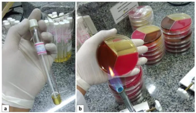
Before the patient’s chemotherapy treatment initiation, the material was collected using sterile swab, which was wet with 0,9% saline solution to help the collect. The samples was transported in test-tubes, properly identified, containing 2 ml of BHI broth (HiMedia) (Figure 1a) to the Campus II’s microbiology laboratory of University Center of Sul de Minas – UNIS/MG, where they were sown qualitative, in duplicate, in the culture mediums BHI Agar (HiMedia), Blood Agar (HiMedia) and
Braz. J. of Develop.,Curitiba, v. 6, n. 11, p.91343-91359 nov. 2020. ISSN 2525-8761 EMB Agar (Kasvi), respectively, in Petri dishes tripartite (Figure 1b). These culture mediums were produced by the authors, with properly's quality certification.
After 24 hours of cultivation, when verified the bacterial growth, the colonies multiplied in the medium was isolated, being sown in the same agars according with the previously presented growth. Each isolated was analyzed by biochemical tests of identification, as catalase, coagulase, sowing in identification medium Rugai (RenyLab) and TSI Agar (Laborclin), and resistance/sensibility tests to the novobiocin antimicrobial (Cecon), in addition of the conventional test of Gram's staining (NewProv). Concomitantly to the patients’ collection, it was realized a control group collection, by the same procedures, being the samples analyzed by the same methods. The control group was composed by healthy people of diversely ages, with similar hygiene habits and that shared the same environment.
Figure 1 – (a) Test tube containing 2 ml of BHI broth, identified and containing the swab used for sampling. (b) Three-part Petri dish containing the BHI, Blood Agar and EMB Agar culture media, used for sowing the samples.
The patients underwent different treatment protocols, of which the Protocols AC DH, Gemzar, Carboplatin + taxol, FLOX D1 and D2 and 5FU D1-D5 may be cited. According to the stage of the cancer and the treatment used, it was determined the interval between the cycles and their respective amount. Therefore, after five months, new samples from both the patients and the control group were collected and analyzed, using the techniques and conditions described above. This time, it was verified the occurrence of changes in the microbial skin community by comparison with the previous results.
Braz. J. of Develop.,Curitiba, v. 6, n. 11, p.91343-91359 nov. 2020. ISSN 2525-8761
3 RESULTS
3.1 PRE-CHEMOTHERAPY ANALYSIS
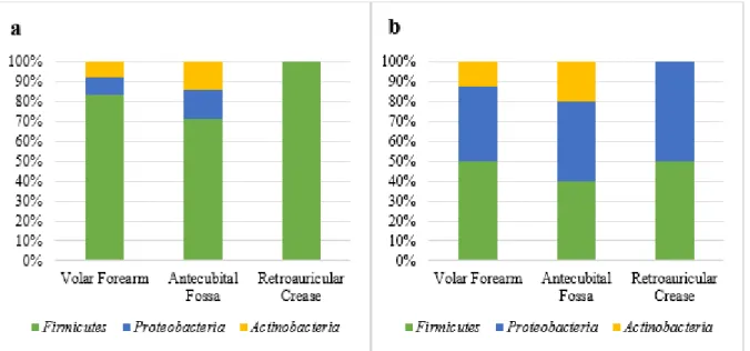
In the first patients’ microbiota analysis, it was verify the bacterial frequency by phylum and anatomical site (Figure 2a), highlighting the presence of bacteria from Firmicutes phylum (83,4%) followed by the Proteobacteria phylum (8,3%) and Actinobacteria (8,3%) in the volar forearm; in the antecubital fossa they were 71,4% from Firmicutes phylum, 14,3% from Proteobacteria phylum and 14,3% Actinobacteria; and, in the retroauricular crease, it was verify that 100% from the found micro-organism belonged to the Firmicutes phylum. In the control group analysis, it was observed the bacterial frequency by phylum and anatomical site too (Figure 2b), highlighting the percentage presented in the volar forearm as 50%, 37,5% and 12,5% from the Firmicutes, Proteobacteria and
Actinobacteria phylum, respectively. In the antecubital fossa, the frequency of the presented phylum
was 40% Firmicutes, 40% Proteobacteria and 20% Actinobacteria; and, finally, a frequency of 50% both Firmicutes phylum, and the Proteobacteria phylum, in the retroauricular crease.
Figure 2 – (a) Ratio of bacterial frequency per phylum and anatomical site from the patients before chemotherapy. (b) Ratio of bacterial frequency per phylum and anatomical site from control group in the initial collect.
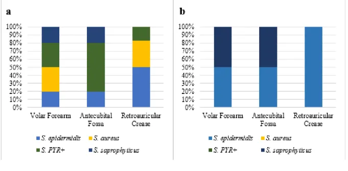
Still in the first analysis, it was verified the ratio of micro-organism frequency from the
Staphylococcus genus by patients’ anatomical site (Figure 3a), being in the volar forearm: S. aureus
(30%), S. PYR + (30%), S. saprophyticus (20%) and S. epidermidis (20%); in the antecubital fossa: S. PYR+ (60%), S. epidermidis (20%) and S. saprophyticus (20%); and, finally, in the retroauricular crease: S. epidermidis (50%), S. aureus (33,3%) and S. PYR+ (16,7%). In the control group, at the site volar forearm, 50% of the micro-organism were S. epidermidis and the others 50% were S.
saprophyticus; in the antecubital fossa, it has the same profile; and, in the retroauricular crease, 100%
Braz. J. of Develop.,Curitiba, v. 6, n. 11, p.91343-91359 nov. 2020. ISSN 2525-8761
Figure 3 – (a) Ratio of Staphylococcus frequency per species and anatomical site from the patients before chemotherapy. (b) Ratio of Staphylococcus frequency per species and anatomical site from control group in the initial collect
3.2 POST-CHEMOTHERAPY ANALYSIS
Figure 4 shown the ratio of bacterial frequency by phylum and anatomical site from the patients after chemotherapy (Figure 4a) and from the control group, after five months from the initial collect (Figure 4b). For patients’ samples, were found 81,8% of Firmicutes and 18,2% of Proteobacteria to the volar forearm; 60% of Firmicutes, 26,7% of Proteobacteria and 13,3% of Actinobacteria to the antecubital fossa; and, to the retroauricular crease, 69,2% of Firmicutes and 15,4% of Proteobacteria and Actinobacteria phyla. In the control group samples, the result was 75% Firmicutes and 25%
Proteobacteria to the volar forearm; 60% Firmicutes, 20% Proteobacteria and Actinobacteria to the
antecubital fossa; and 100% of the retroauricular crease micro-organisms from the control group were from the Firmicutes phylum.
Figure 4 – (a) Ratio of bacterial frequency per phylum and anatomical site from the patients after chemotherapy. (b) Ratio of bacterial frequency per phylum and anatomical site from control group, five months after the initial collect.
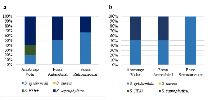
Braz. J. of Develop.,Curitiba, v. 6, n. 11, p.91343-91359 nov. 2020. ISSN 2525-8761 The interspecifically analysis of micro-organisms from Staphylococcus genus demonstrated, in the patients after chemotherapy (Figure 5a), a profile of 60% S. saprophyticus, 20% S. epidermidis and 20% S. PYR+ in the volar forearm; 50% S. epidermidis and 50% S. saprophyticus in the antecubital fossa; 66,7% S. epidermidis and 33,3% S. saprophyticus in the retroauricular crease. Meanwhile, in the control group, five months after the initial collect (Figure 5b), it was possible to evidence that, in the volar forearm it was found 50% S. epidermidis and 50% S. saprophyticus, as well as in the antecubital fossa; and, in the retroauricular crease, 100% of the Staphylococcus found were from the S. epidermidis species.
Figure 5 – (a) Ratio of Staphylococcus frequency per species and anatomical site from the patients after chemotherapy. (b) Ratio of Staphylococcus frequency per species and anatomical site from control group, five months after the initial collect
4 DISCUSSION
The microbiota has a lot of micro-organisms that can realize distinct functions in the human body. In the skin, the microbial communities protect the host of the prejudicial micro-organisms invasion and prepare the billions of T cells presents in the skin to respond against similar organisms, but pathogenic, because even being the same species, some strains of determinates micro-organisms can be pathogenic to the human organism (GRICE; SEGRE, 2011; OH et al., 2016). Whereas the skin as an ecosystem, is natural that disturbances of exogenous or endogenous origin can disturb the microbiota-host relation, what causes a loss of commensal diversity, leading to the onset or worsening of determinate diseases (GRICE; SEGRE, 2011; MYLES et al., 2016).
In order to establish a relation between chemotherapy and epidermal microbiota it is necessary to know if chemotherapy can affect the several skin layers. Fabbrocini et al. (2012) Among the main changes occurred in the skin due chemotherapy treatment, the presence of papule-pustular reactions, dry skin and nail and hairy changes are described (FABBROCINI et al., 2012; TAVARES et al., 2020). Erythematous follicular papules that can evolve to pustules were presents too, mainly in the
Braz. J. of Develop.,Curitiba, v. 6, n. 11, p.91343-91359 nov. 2020. ISSN 2525-8761 face, neck and retroauricular area, scalp and upper trunk. Also, it is noted high dehydration of skin and signs of damage to skin barrier function.
Still on the change in microbiota-host relationship, it is described that the pathogenicity of skin microbiota's bacteria suffers influence by immunologic factors from host, what makes that when analyzed diseases provoked by bacterial agents, it is necessary to investigate all context between microbiota, immunity and intrinsic factors from host (BELKAID; SEGRE, 2014). It is must to make a search about possible situations that can generate modifications in the skin microbiota, so that to do a comparative analysis about alterations, it can be as reliable as possible. An antibiotic treatment, for example, can provoke a drastic microbiota change. This study did not care about the patients’ selection according to the antibiotic treatment. Giving that the antibiotic use can impact in the chemotherapy efficacy (ZHOU et al., 2017), to these patients it is prescript the chemotherapy treatment interruption when it is suspected an infection with the necessity of antimicrobials use.
According Morvan and Valle (2018), some physiological situations from individual can, also, modify the microbiota. Stress situations may decrease the skin pH, what can be noticed by the presence of lactic acid producing agents, besides causing a skin microbial diversity reduction. These authors also verify that has a higher number of Actinobacteria and Firmicutes and a lowest number of
Bacteroidetes and Proteobacteria in the stressed groups when compared to the control group. With
this information, when analyzes this work results, it was realized that the first analyzes demonstrated the same pattern that what Morvan and Vallee (2018) found, what can be associated with the emotional fragility in which the patients were in the moment of first collection.
The importance in elucidate the differences in the topographic skin composition is to facility studies and to compare results. Grice et al. (2009) divided the human skin into three physiologically distinct locals: sebaceous, dry and moist areas. To this research, it was chosen three anatomical sites of distinct physiology, so that it can be possible to find the most microbial diversity as possible. According the results, the micro-organisms from the Firmicutes phylum were the one who presented higher percentage between the three sites, both before and after the chemotherapy, what it was different to Grice and Segre’s (2011) study, in which the Firmicutes phylum only was prevalent in antecubital fossa. While what, to Zhai et al. (2018), the most dominant phylum was Actinobacteria (34,5%), Proteobacteria (30,7%) and Firmicutes (25,6%) Such results’ discrepancy is because several factors, mainly the individual’s physiology, environment, occupation, life’s style and individuals genotype, besides the analysis method.
However, this research results presented like the proposed by Tortora, Funke and Case (2012), whose cited that Staphylococcus epidermidis, from Firmicutes phylum, constitute the majority of habitual skin microbiota. This can be justified by the S. epidermidis capacity of subsist in different
Braz. J. of Develop.,Curitiba, v. 6, n. 11, p.91343-91359 nov. 2020. ISSN 2525-8761 niches of the host, characteristic acquired thanks to great variation between found strains in different niches, what suggest a great pan-genome to the specie (OH et al., 2016).
The Gram-negative bacteria represents 10% to 50% of skin microbiome. A study realized by Myles et al. (2016) demonstrate that a half of atopic dermatitis patients did not presented any Gram-negative bacteria, associating this condition to the disease. Despite the Gram-Gram-negative micro-organisms’ scarcity in the research, none of the patients presented the condition of atopic dermatitis. When analyzing the temporal diversity of micro-organisms, Grice et al. (2009) verified that there are more consistent locals and others with an appreciatively change, giving emphasis to the volar forearm as a local that presented that, what would explain the variation found in the control group’s microbiota, mainly in the cited local.
Zhai et al. (2018) solicited, in their study, that in the skin locals which the samples would be collected, that was avoided the wash and the use of personal hygiene products (like cosmetics, hydrating creams and antiperspirant deodorants), by a 48 hours period, to the upper back and the volar forearms, and 12 hours to the cheeks. However, Two et al. (2016) when verifying the level of antimicrobial compounds benzalkonium chloride and triclocarban after the skin wash 10 minutes, 6 hours and 24 hours after, they do not found any difference that would determine an abundance drop in the skin commensal Staphylococcus epidermidis. Being these micro-organisms the most frequently found in the skin, it did that the volunteers of this work not be instructed to avoid the wash of the collect's local, also considering the immunologic debility of these patients and the comfort in the research collaboration. Still it is described that a rigorous wash can decrease their number without, however, eliminating them. The remaining micro-organisms in the hair follicles and in the sweat-glands can reestablish the normal population quickly (TORTORA; FUNKE; CASE, 2012).
Corroborating with this research, Myles et al. (2016) used the technique of swab wetted in sterile saline solution, which was rubbed in the patients' skin, more precisely in the antecubital fossa and in the volar forearm. However, it is emphasized that they were searching for Gram negative bacteria, after sown the samples in a R2A Agar plate.
A complete skin microbiota evaluation it is happened by combination of several collection techniques. The swab collection, even though the most used method, can give only data about the microbiota resident in the stratum corneum, while the scraping method, despite providing stratum corneum cells, granular layers and the top of the follicles, as well as the swab technique, is a non-invasive method and, also, does not provide microbiota total aspects. The most indicate way to do a complete analysis of the skin microbiota’s composition would be by the using of an invasive collect by biopsy’s method (DRÉNO et al., 2016). However, the collect method most appropriated to this research would be the mediated by swab, once that do not had justified to the use of other techniques too invasive, as the skin scrapping or biopsy.
Braz. J. of Develop.,Curitiba, v. 6, n. 11, p.91343-91359 nov. 2020. ISSN 2525-8761 Dréno et al. (2016) further argue that, the culture traditional methods are limited because each species necessity certain nutrients, doing that only a restrict number of bacterial micro-organisms develop in laboratories. This eventually causes an exacerbated growth in some less rigorous bacteria, overlapping those more exigent, what implies a difficult in the species isolation and identification. This information justifies the identification of few genus and species in relation to similar works, which uses of molecular techniques for the determination of these micro-organisms. Due to this reason, it is necessary to affirm that the micro-organisms’ identification, in this study, gave up by suggestive way, according with the presented morphology and biochemical characteristics and, to reliable confirmations, posteriorly, molecular techniques should be carried out.
Despite the limited methods in relation to other works, it was still possible identify a change in the individuals' microbiota after the chemotherapy, when analyzed by species level. The
Staphylococcus were the Firmicutes most found in the research, but, when identify the species, it was
verified that had a variation in the identified species in relation to the control group, which did not present no change.
It is notable that has a broad's reduction in the Staphylococcus species positives to PYR test, which were presents in all sites in the first analysis, and, in the post-chemotherapy analysis, were observed only in the volar forearm. Also, did not was identified any Staphylococcus aureus colony in the second analysis. It is emphasized the S. epidermidis capacity to propagate in different sites, excluding the volar forearm, in which there was lower frequency when compared to S. saprophyticus.
As cited by Zhai et al. (2018), micro-organisms with relatively small abundance are classified as transitory rather than resident. Therefore, it is worth emphasizing that the analysis of this work had qualitative purposes, only to verify if would have or not the micro-organisms’ presence, not being possible to quantify them, like Grice and Segre (2011) did in their studies, and neither to determine if they were microbiota's habitant or temporary.
According Belkaid and Segre (2014), S. epidermidis demonstrate to play an excellent immunologic role against pathogens, mainly in their capacity to inhibit the biofilm formation by S.
aureus. In addition, both this commensal and others, can increase the system's complement
components expression. With this information, the results are presented as positives to the chemotherapy's patients, because even though they be in weak situation, both emotional and physical, especially due the treatment side effects, did not had significant change in the S. epidermidis presence in these patients’ skin. It is wanted to know if these micro-organisms are functionally able to play their immunological role against pathogens, and, even, to produce the 6-HAP molecule, antitumoral treatment helper.
The authors Belkaid and Segre (2014), cite about the creation of probiotic or prebiotic therapies to mold the communities situated in skin. Products like hydrating creams, can play, beside your normal
Braz. J. of Develop.,Curitiba, v. 6, n. 11, p.91343-91359 nov. 2020. ISSN 2525-8761 function, the stimulant role to microbiological growth. Also, it is possible to cite the therapies use like the microbiota autologous transplant, of the areas that did not suffers the chemotherapy impact to these that were most affected, which would make the individuals not suffer by the loss of the commensal micro-organisms, guaranteeing then an improvement to the life’s quality of these patients by the preservation of the microbiota and their function.
5 CONCLUSION
In the analyzed individuals’ microbiota, with relation to the cultivable micro-organism, it was not possible to evidence significant changes when analyzed by phylum, being these changes most associated to species. However, having the knowing of the benefic role played by the skin colonizer micro-organisms’ resistant to this treatment, mainly the Staphylococcus epidermidis, the results presented positives when evaluated by the point of view in the collaboration to improve the patients’ quality life.
Knowing that the present study has an experimental character, it is necessary a more complete microbiota analyzes by molecular techniques, to be possible to determinate accurately the micro-organisms and confirm the chemotherapy resistance, so that it can to verify these results in a quantitative level.
Braz. J. of Develop.,Curitiba, v. 6, n. 11, p.91343-91359 nov. 2020. ISSN 2525-8761
REFERENCES
AMERICAN CANCER SOCIETY. Chemotherapy, 2015. Disponible in:
<https://www.cancer.org/treatment/treatments-and-side-effects/treatment-types/chemotherapy.html>. Access in: set. 18, 2018.
BECATTINI, S.; TAUR, Y.; PAMER, E. G. Antibiotic-Induced Changes in the Intestinal Microbiota and Disease. Trends Mol. Med., New York: CellPress, v. 22, n. 6, p. 458-478, jun. 2016.
BRASIL. Agência Nacional de Vigilância Sanitária. Microbiologia Clínica para o Controle de Infecção Relacionada à Assistência à Saúde. Módulo 4: Procedimentos Laboratoriais: da requisição do exame à análise microbiológica e laudo final. Brasília: Anvisa, 2013. 95p.
BELKAID, Y.; SEGRE, J. A. Dialogue between skin microbiota and immunity. Science, [s.l.], v. 346. n. 6212, p. 954-959, nov. 21, 2014.
BERMON, S. et al. The microbiota: an exercise immunology perspective. Exerc. Immunol. Rev., v. 21, p. 70-79, 2015.
COGEN, A. L. et al. Staphylococcus epidermidis Antimicrobial δ-Toxin (Phenol-Soluble Modulin-c) Cooperates with Host Antimicrobial Peptides to Kill Group A Streptococcus. PLoS ONE, v. 5. n. 1, p. 1-7, jan. 2010.
COSTELLO, E. K. et al. Microbiome Assembly across Multiple Body Sites in Low-Birthweight Infants. MBio, v. 4, n. 6, 12p, 2013.
COSTELLO, E. K. et al. The application of ecological theory towards an understanding of the human microbiome. Science, v. 336, n. 6086, p. 1255–1262, 2014.
DOMINGUEZ-BELLO, M. G. et al. Delivery mode shapes the acquisition and structure of the initial microbiota across multiple body habitats in newborns. Proc. Natl. Acad. Sci. U.S.A., v. 107, n. 26, p. 11971–11975, 2010.
DRÉNO, B. et al. Microbiome in healthy skin, update for dermatologists. J. Eur. Acad. Dermatol. Venereol., [s.l.], v. 30, p. 2038–2047, 2016.
FABBROCINI, G. et al. Chemotherapy and skin reactions. J. Exp. Clin. Cancer Res., [s.l], v. 31, n. 1, p. 50-55, may 28, 2012.
GRICE, E. A. et al. Topographical and Temporal Diversity of the Human Skin Microbiome. Science, [s.l.], v. 324, n. 5931, p. 1190–1192, may 2009.
GRICE, E. A.; SEGRE, J. A. The skin microbiome. Nat. Rev. Microbiol., v. 9, n. 4, p. 244–253, apr. 2011.
HOOPER, L. V.; LITTMAN, D. R.; MACPHERSON, A. J. Interactions between the microbiota and the immune system. Science, v. 336, n. 6086, p. 1268–1273, 2012.
Instituto Nacional do Câncer José Alencar Gomes da Silva (INCA). 2004. Disponible in: <http://www2.inca.gov.br/wps/wcm/connect/8e973c004eb686f794f896f11fae00ee/perguntas_qt.pdf ?MOD=AJPERES&CACHEID=8e973c004eb686f794f896f11fae00ee>. Access in: April 28, 2018.
Braz. J. of Develop.,Curitiba, v. 6, n. 11, p.91343-91359 nov. 2020. ISSN 2525-8761 KEENEY, K. M. et al. Effects of Antibiotics on Human Microbiota and Subsequent Disease. Annu. Rev. Microbiol., v. 68, p. 217-235, 2014.
MONTASSIER, E. et al. Chemotherapy-driven dysbiosis in the intestinal microbiome. Aliment. Pharmacol. Ther., v. 42, p. 515–528, 2015.
MORVAN, P. Y.; VALLEE, R. Evaluation of the Effects of Stressful Life on Human Skin Microbiota. Appli. Microbiol. Open Access, [s.l.], v. 4, n. 1, p. 1-8, 3 jan. 2018.
MYLES, I. A. et al. Transplantation of human skin microbiota in models of atopic dermatites. JCI Insight, [s.l.] v. 1, n. 10, 7 jul. 2016.
NAKATSUJI, T. et al. A commensal strain of Staphylococcus epidermidis protects against skin neoplasia. Sci. Adv., v. 4, n. 2, 9p, 2018.
OH, J. et al. Temporal Stability of the Human Skin Microbiome. Cell, [s.l.], v. 165, n. 4, p. 854–866, may 5, 2016.
TAVARES, M. B. et al. Caracterização das reações adversas a quimioterápicos em um hospital filantrópico. Braz. J. Hea. Rev., Curitiba, v. 3, n. 2, p. 2317-2326, mar./apr. 2020.
TORTORA, G. J.; FUNKE, B. R.; CASE, C. L. Microbiologia. 10. ed. Porto Alegre: Artmed, 2012. TWO, A. M. et al. The Cutaneous Microbiome and Aspects of Skin Antimicrobial Defense System Resist Acute Treatment with Topical Skin Cleansers. J. Invest. Dermatol., [s. l.], v. 136, p. 1950-1954, 2016.
URBANIAK, C. et al. Effect of chemotherapy on the microbiota and metabolome of human milk, a case report. Microbiome, v. 2, n. 24, p. 1-11, 2014.
WEYRICH, L. S. et al. The skin microbiome: Associations between altered microbial communities and disease. Australas. J. Dermatol., v. 56, n.4, p. 268-274, 2015.
WILSON, Michael. Bacteriology of Humans: an Ecological Perspective. 1. ed. Londres: Blackwell Publishing, 2008.
ZHAI, W. et al. Profile of the skin microbiota in a healthy Chinese population. J. Dermatol., Japanese Dermatological Association, v. 45, n. 11, p. 1289-1300, 2018.
ZHANG, M. et al. Oral Antibiotic Treatment Induces Skin Microbiota Dysbiosis and Influences Wound Healing. Microb. Ecol., Nova Iorque, v. 69, n. 2, p. 415-421, 10 out. 2014.
ZHOU, G. et al. The impact of antibiotic usage on the efficacy of chemoimmunotherapy is contingent on the source of tumor-reactive T cells. Oncotarget, v. 8, n. 67, pp. 111931-111942, 2017.
Braz. J. of Develop.,Curitiba, v. 6, n. 11, p.91343-91359 nov. 2020. ISSN 2525-8761
APPENDIX A
COPARTICIPANT INSTITUTION AGREEMENT
Varginha, October 17, 2018. To
Ethics Committee on Research of Centro Universitário do Sul de Minas – UNIS T/C: Coordinator of CEP/UNIS
AUTHORIZATION TO CARRY OUT RESEARCH
Me, Dr. Ítalo Denelle Venturelli, Technical Director of the Bom Pastor Hospital - FHOMUV, hereby inform you that the student Pedro Augusto Ramos Vanzele, from the Biomedicine course of the University of Southern Minas - UNIS, is authorized to carry out the research entitled "Analysis of skin microbiota alteration in patients after chemotherapy treatment ", under the guidance of Prof. Dr. Hadassa Cristhina de Azevedo Soares dos Santos.
I declare to know and comply with the Brazilian Ethical Resolutions, in particular Resolution CNS 196/96. This institution is aware of its co-responsibilities as a co-participant institution of this research project, and of its commitment to safeguarding the safety and well-being of the research subjects recruited in it, having the necessary infrastructure to guarantee such safety and well-being. Prioritize that this research will only be done when there is presentation of the Consubstantiated Opinion of the CEP/UNIS.
Braz. J. of Develop.,Curitiba, v. 6, n. 11, p.91343-91359 nov. 2020. ISSN 2525-8761
APPENDIX B
INFORMED CONSENT FORM
Project’s title: Analysis of skin microbiota alteration in patients after chemotherapy treatment. Researchers’ Data
Responsible researcher: Profa. Dra. Hadassa Cristhina de Azevedo Soares dos Santos Phone: (35) 99749-2525
E-mail: hadassa.santos@unis.edu.br
Research’s assistant: Pedro Augusto Ramos Vanzele Phone for contact: (35) 99867-1080
E-mail: pedrovanzele@gmail.com
Institution to which researchers belong: Centro Universitário do Sul de Minas-UNIS/MG
Volunteer’s Data
Volunteer’s name:
Age: ___ years old Legal responsible:
RG:
Phone:
E-mail:
You are being invited to participate in the research project "Analysis of skin microbiota alteration in patients after chemotherapy treatment", under the responsibility of the researcher Professor Dr. Hadassa Cristhina de Azevedo Soares dos Santos, whose objective is to evaluate if there are modifications in the skin microbiota of patients with cancer after accomplishment of the treatment with chemotherapeutics.
The methodology consists of collecting patients' skin samples by means of a sterile swab so that the effects caused by chemo in the normal microbiota can be checked and discarded.
Risks: This work presents a minimal risk to the physical or moral integrity of the volunteers, who may feel uncomfortable at the time of collection, performed with swab, or contribute to the research and have their results published, even with the absence of identification.
Benefits: Collaborating with this work development, it will also be helping to disseminate new scientific knowledge to the general population, in addition to obtaining the knowledge about the constitution of the epidermal microbiota itself.
Braz. J. of Develop.,Curitiba, v. 6, n. 11, p.91343-91359 nov. 2020. ISSN 2525-8761 This work is justified by the increase in the number of people who undergo chemotherapy and the concern with the effects that it can cause in the microbiota of the skin, which can play a beneficial role and even help against certain pathogens.
All data obtained will be used for the purpose of research and will be kept confidential, any information you provide about you will be preserved your identity.
In case of doubts the participants can have any guidance with the teacher as mentioned above, to remedy any kind of doubt that may not have been clarified. You can stop participating at any time if this is your will. Your participation is voluntary.
Me, _, RG number
___________________, declare that I have been informed and agree to participate as a volunteer in the research project described above.
Varginha, , 2018.