SERVIÇO PÚBLICO FEDERAL
MINISTÉRIO DA EDUCAÇÃO
UNIVERSIDADE FEDERAL DE UBERLÂNDIA
PROGRAMA DE PÓS-GRADUAÇÃO EM GENÉTICA E BIOQUÍMICAEfeito do tratamento com Vochysia rufa Mart. e glibenclamida sobre o estresse oxidativo e a expressão de proteínas motoras e de ancoragem em cérebro de ratos diabéticos
Aluna: Alice Vieira da Costa
Orientador: Dr. Foued Salmen Espindola Co-Orientadora: Dra. Luciana Karen Calábria
ii
SERVIÇO PÚBLICO FEDERAL
MINISTÉRIO DA EDUCAÇÃO
UNIVERSIDADE FEDERAL DE UBERLÂNDIA
PROGRAMA DE PÓS-GRADUAÇÃO EM GENÉTICA E BIOQUÍMICAEfeito do tratamento com Vochysia rufa Mart. e glibenclamida sobre o estresse oxidativo e a expressão de proteínas motoras e de ancoragem em cérebro de ratos diabéticos
Dissertação apresentada à Universidade Federal de Uberlândia como parte dos requisitos para obtenção do Título de Mestre em Genética e Bioquímica (Área Bioquímica).
Aluna: Alice Vieira da Costa
Orientador: Dr. Foued Salmen Espindola Co-Orientadora: Dra. Luciana Karen Calábria
iii Dados Internacionais de Catalogação na Publicação (CIP)
Sistema de Bibliotecas da UFU, MG, Brasil.
S587a
2013 Costa, Alice Vieira da, 1989- Efeito do tratamento com Vochysia rufa Mart. e glibenclamida
sobre o estresse oxidativo e a expressão de proteínas motoras e de ancoragem em cérebro de ratos diabéticos / Alice Vieira da Costa. -- 2013.
75 f.
Orientadora: Foued Salmen Espindola. Coorientador: Luciana Karen Calábria.
Tese (doutorado) - Universidade Federal de Uberlândia, Pro- grama de Pós-Graduação em Genética e Bioquímica.
Inclui bibliografia.
1. 1. Bioquímica - Teses. 2. Diabetes - Teses. 3. Miosinas – esTeses. I. Espindola, Foued Salmen. II. Calábria, Luciana Karen. IIIIIII. Universidade Federal de Uberlândia. Programa de Pós-Gra- duaduação em Genética e Bioquímica. IV. Título.
iv
SERVIÇO PÚBLICO FEDERAL
MINISTÉRIO DA EDUCAÇÃO
UNIVERSIDADE FEDERAL DE UBERLÂNDIA
PROGRAMA DE PÓS-GRADUAÇÃO EM GENÉTICA E BIOQUÍMICAEfeito do tratamento com Vochysia rufa Mart. e glibenclamida sobre o estresse oxidativo e a expressão de proteínas motoras e de ancoragem em cérebro de ratos diabéticos
ALUNO: Alice Vieira da Costa
COMISSÃO EXAMINADORA
Presidente: Dr. Foued Salmen Espindola
Examinadores:
Dr. Pablo Marco Veras Peixoto (NYU) Dra. Reggiani Vilela Gonçalves (UFJF)
Data da Defesa: 29/07/2013
As sugestões da Comissão Examinadora e as Normas PGGB para o formato da Dissertação/Tese foram contempladas
v
DEDICATÓRIA
vi
AGRADECIMENTOS
Ao Pai, ao Filho e ao Espírito Santo, por sempre estarem ao meu lado me dando forças e coragem para continuar nesta caminhada.
Aos meus pais, Antônio Soares da Costa e Maria Aparecida Vieira da Costa, por não medirem esforços em prol dos meus sonhos. Eu amo muito vocês!
À toda minha família, que de alguma forma esteve me apoiando em orações, em especial à minha madrinha Maria Divina Alves (“Dindinha”) que suportou a minha ausência e me apoiou mesmo distante.
À minha irmã Letícia Vieira da Costa, que alegra meus dias e sempre me ajudou em orações.
Ao meu namorado Marciel José Sampaio, que chegou em minha vida novamente só para acrescentar. Obrigada pelo apoio, amor e ajuda nesta reta final do meu mestrado.
Á minha amigona Bárbara Oliveira, que sempre torceu pelo meu sucesso e me ensinou a seguir sempre em frente, sem medo dos obstáculos que estivessem por vir.
Ao Prof. Dr. Foued Salmen Espindola, pela oportunidade, me proporcionando formação como pesquisadora e me permitindo trabalhar com pessoas tão excepcionais. Aprendi muito neste dois anos. Muito obrigada por abrir as portas do seu laboratório e por me orientar neste trabalho.
À Dr. Luciana Karen Calábria (Lu), muito obrigada por TUDO. Não existem palavras que expressam o quanto você é importante para mim. Tenho certeza que foi DEUS que permitiu que trabalhássemos juntas, e a partir disto nascesse uma linda amizade. Além de minha co-orientadora, você é uma grande amiga, irmã e “mãe cientifica”. Obrigada por ser este exemplo de determinação, fazendo tudo sempre com muito amor. TE AMO MUITO e desejo o melhor desta terra para sua vida.
vii Ao Dr. Alexandre Azenha Rezende (Ale), que foi conquistando seu espaço no meu coração. Grande amigo, professor e pesquisador. Obrigada por sempre ter me ajudado em momentos de dúvidas e questionamentos. Agradeço pela sua amizade.
À Patrícia Terra Alves pelo carinho e amizade nestes 2 anos de mestrado. Ao grupo Vochysia, Douglas, Helen, Neire, Luciana, Francyelle, Izabela, Camilla, Alexandre, Aline e Rosilene que muito me ensinaram sobre como é importante trabalhar em equipe.
Aos professores do Instituto de Genética e Bioquímica, aprendi muito com grandes pesquisadores.
Ao CNPq, pela concessão da bolsa de mestrado.
A Universidade Federal de Uberlândia e aos seus professores. Obrigada pelos ensinamentos e apoio na minha formação acadêmico cientifica.
Ao Instituto de Genética e Bioquímica e a todos os colegas do Laboratório de Bioquímica e Biologia Molecular da Universidade Federal de Uberlândia.
À FAPEMIG, pelo fomento ao projeto aprovado.
Obrigada!
viii
APRESENTAÇÃO
O formato desta tese obedece as normas do Programa de Pós-graduação em Genética e Bioquímica. Ela e composta de três capítulos, sendo o capitulo 1 referente a fundamentação teórica, que embasa os outros dois capítulos.
Capítulo 1 - Fundamentação Teórica.
Capítulo 2 - Tratamento com glibenclamida modula a expressão e localização de miosina-IIB no cérebro de ratos diabéticos.
Capítulo 3 - Extrato aquoso de Vochysia rufa promove expressão diferencial
de proteínas envolvidas na formação e tráfico de vesículas no cérebro de ratos diabéticos.
ix
RESUMO
x
LISTADEFIGURAS
Capítulo 1
Fig. 1 Visão esquemática das vias de sinalização da insulina que facilitam a captação de glicose._______________________________________________03 Fig. 2 Principais defesas antioxidantes.________________________________05 Fig. 3 Estruturas básicas das miosinas –II e –V e a distinção pelo domínio cauda.__________________________________________________________07 Fig. 4 Esquema da interação entre vesícula sináptica, sinapsina-I e CaMKII.___09 Fig. 5 Fosfoproteínas implicadas na exocitose sináptica.___________________10 Fig. 6 Espécie Vochysia rufa fotografada em Abadia dos Dourados, Minas Gerais.__________________________________________________________13
Capítulo 2
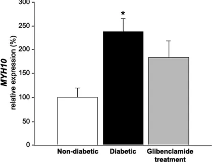
Fig. 1 MYH10 expression in the brains of non-diabetic, diabetic and diabetic rats treated with glibenclamide.__________________________________________41 Fig. 2 Western blot and quantification of myosin-IIB expression from supernatant brain samples.____________________________________________________42 Fig. 3 Myosin-IIB hematoxylin and immunolocalization and eosin stain in sections of the frontal cortex of rat brains: non-diabetic.____________________________43 Fig. 4 Myosin-IIB distribution and hematoxylin and eosin staining in the hippocampus of non-diabetic.________________________________________44
Capítulo 3
Fig. 1 Effect of Vochysia rufa extract on glutathione peroxidase (GPx), superoxide
dismutase (SOD) and glutathione S-transferase (GST) activity and reduced glutathione (GSH), total sulfhydryl and malondialdehyde (TBARS) concentrations in rat brains._______________________________________________________60
Fig. 2Effect of diabetes mellitus on myosin-Va, α-CaMKII, synapsin-1, SNAP-25
xi
Fig. 3 Myosin-Va (A-D) and α-CaMKII (E-H) distribution in the frontal cortex of non-diabetic (A; A’; E), non-diabetic (B; F), non-diabetic treated with glibenclamide (C; G), and diabetic treated with Vochysia rufa extract (D; H) rat brains._________________62 Fig. 4 Synapsin-1 (A-D), SNAP-25 (E-H) and GLUT4 (I-L) distribution in the frontal cortex of non-diabetic (A; A’; E; I), diabetic (B; F; J), diabetic treated with glibenclamide (C; G; K), and diabetic treated with Vochysia rufa extract (D; H; L) rat brains.__________________________________________________________63 Fig. 5 Myosin-Va (A-D) and α-CaMKII (E-H) distribution in the occipital cortex of non-diabetic (A; A’; E), diabetic (B; F), diabetic treated with glibenclamide (C; G), and diabetic treated with Vochysia rufa extract (D; H) rat brains._____________64 Fig. 6 Synapsin-1 (A-D), SNAP 25 (E-H) and GLUT4 (I-L) distribution in the occipital cortex of the non-diabetic (A; A’; E; I), diabetic (B; F; J), diabetic treated with glibenclamide (C; G; K), and diabetic treated with Vochysia rufa extract (D; H; L) rat brains.__________________________________________________________65
Fig. 7 Immunolocalization of α-CaMKII (A-D), synapsin-1 (E-H), SNAP 25 (I-L) and
xii
LISTADEABREVIATURAS
ATP – Adenosina tri-fosfato
CaMKII- proteína quinase-II dependente de Ca2+/calmodulina
CEUA – Comitê de Ética na Utilização de Animais
CuSOD – Superóxido dismutase citosólica
D – Diabético
DG – Diabético tratado com glibenclamida
DV- Diabético tratado com Vochysia rufa
dl – Decilitro
DNA – Ácido Desoxirribonucleico
DTNB – Ácido dinitrobenzóico
g – Gramas
GLUT- proteínas transportadoras de glicose
GPx – Glutationa peroxidase
GR – Glutationa redutase
GSH – Glutationa reduzida
GSSG – Glutationa oxidada
GST – Glutationa S-transferase
h - Horas
H.E. – Hematoxilina e Eosina
L - Litros
MDA – Malondialdeído
mRNA - ácido ribonucleico mensageiro
mg - Miligramas
mL - Mililitros
xiii NADH – Nicotinamida adenina dinucleotídio
NADPH - Nicotinamida adenina dinucleotídio fosfato
ND – Não diabético
pH – Potencial hidrogeniônico
RPM – Rotações por minuto
s - Segundos
SOD – Superóxido dismutase
SNAP 25- proteína associada ao sinaptossomo
SNARE- proteínas de fusão sensível ao receptor de ligação ao N-etilmaleimido solúvel
TBARS – Método para avaliação de peroxidação lipídica
TNF- - Factores de necrose tumoral alfa
ug - Microgramas
uL - Microlitros
uM - Micromolar
umol – Micromol
xiv
SUMÁRIO
Capítulo 1 1
1. Fundamentação Teórica 2
1.1 Diabetes mellitus 2
1.2 Diabetes mellitus, cérebro e estresse oxidativo 3
1.3 Defesas antioxidantes 4
1.4 Miosinas 6
1.5 Proteínas que se associam a vesículas sinápticas 8
1.6 Transportador de glicose 11
1.7 Vochysia rufa e as plantas no tratamento do diabetes mellitus 11
2. Referências 15
Capítulo 2 25
1. Artigo Científico: “Glibenclamide treatment modulates expression and localization of myosin-iib in diabetic rat brain” 26
Capítulo 3 45
1. Artigo Científico: “Aqueous extract of Vochysia rufa promotes differential expression of proteins implicated in vesicular trafficking in diabetic rat brain”
1
2
1. Fundamentação teórica
1.1 Diabetes mellitus
Diabetes mellitus é uma síndrome metabólica que afeta cerca de 171 milhões de pessoas no mundo (Mccrimmon, Ryan et al., 2012) e está associada com alterações de secreção e/ou ação da insulina, o processo patológico é agravado por danos acumulados ao longo do tempo, aumentando os riscos macro e microvasculares, e trazendo complicações para vários órgãos, como rins, coração e cérebro (Gispen e Biessels, 2000; Association, 2012; Mccrimmon, Ryan et al., 2012). No sistema nervoso central, o quadro hiperglicêmico característico do diabetes mellitus altera os níveis de neurotransmissores, causando morte celular e reduzindo a capacidade de aprendizagem e memória (Mooradian, 1988; Mccall, 1992; Biessels, Kappelle et al., 1994; Di Mario, Morano et al., 1995; Helkala, Niskanen et al., 1995; Mccrimmon, Ryan et al., 2012; Jing, Chen et al., 2013).
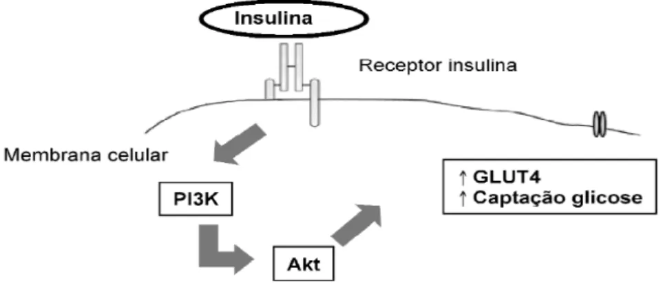
A insulina é um hormônio anabólico que desempenha funções de controle de glicemia, síntese proteica e controle do peso corporal (Haber, Curi et al., 2001; Wendt, 2005), além de atuar na modulação cognitiva do sistema nervoso central, na memória, aprendizado e plasticidade sináptica (Zhao, Koslovsky et al., 1996; Park, 2001). Este hormônio é responsável pela ativação de uma cascata de sinalização que regula a translocação dos transportadores de glicose para a membrana da célula (Schaan, Rabelo et al., 2004) (Figura 01). Alterando está sinalização, a captação de glicose fica prejudicada e a hiperglicemia é instalada desencadeando distúrbios no metabolismo e alterando o balanço entre pró- e antioxidantes (Halliwell; Gutterridge, 2003).
3 diagnosticado na gestação, podendo ou não persistir após o parto (Association, 2012; 2013).
Figura 01: Visão esquemática das vias de sinalização da insulina que facilitam a captação de glicose (Adaptado de Schaan, Rabelo et al., 2004).
1.2 Diabetes mellitus, cérebroe estresse oxidativo
A hiperglicemia no estado diabético aumenta as desordens fisiológicas, evidenciando as lesões celulares causadas pelos radicais livres e aumentando as complicações decorrentes da cronicidade da doença (Reis, Veloso et al., 2008), bem como é responsável por alterações celulares decorrentes dos processos de glicação não enzimática e de glico-oxidação, que está relacionada com a produção de espécies reativas do oxigênio e a formação de produtos finais de glicação avançada. Estas alterações contribuem para o aumento da peroxidação lipídica, modificação irreversível de proteínas, DNA e lipídios (Jennings, Jones et al., 1987; Baynes e Thorpe, 1999; Rosen, Nawroth et al., 2001; Genet, Kale et al., 2002; Siddiqui, Taha et al., 2005).
O estresse oxidativo é uma condição biológica de desequilíbrio entre a produção de espécies reativas de oxigênio e a capacidade do organismo em neutralizar estas espécies, criando um ambiente neurotóxico (Taniyama e Griendling, 2003; Gerecke, Kolobova et al., 2013).
4 O balanço redox tem mostrado ser um fator importante na atividade neuronal do sistema nervoso central e este reflete nos outros sistemas que estão sob seu controle, como o cardiovascular (Cardoso, Colombari et al., 2006). Um exemplo é o peróxido de hidrogênio, uma espécie reativa do oxigênio, que está relacionada com a modulação da transmissão (Chen, Avshalumov et al., 2001) e da plasticidade sináptica (Auerbach e Segal, 1997). Em decorrência deste desequilíbrio e da relação entre a atividade neuronal e os agentes oxidantes, algumas complicações são observadas em pacientes diabéticos, como demências, convulções, comprometimento cognitivo e alterações no aprendizado e memória. Porém, os mecanismos bioquímicos responsáveis por estas alterações ainda são desconhecidos (Coleman, Judd et al., 2004).
1.3 Defesas antioxidantes
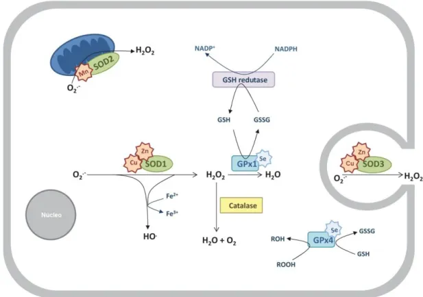
A célula desenvolveu uma série de mecanismos de defesa contra a exposição a radicais livres que vai desde mecanismos de reparo até defesas antioxidantes enzimáticas e não enzimáticas (Cadenas, 1997a; b). As defesas antioxidantes enzimáticas incluem a glutationa peroxidase, a superóxido dismutase e a catalase (Figura 02); enquanto as defesas antioxidantes não enzimáticas são representadas pelo ácido ascórbico (vitamina C), glutationa, α-tocoferol (vitamina E), flavonóides e carotenóides (Valko, Leibfritz et al., 2007). Em condições normais, existe um equilíbrio entre a produção de espécies reativas do oxigênio e a defesa antioxidante. Como o diabetes está associado com o aumento do estresse oxidativo, este balanço fica comprometido e induz modificações em lipídeos, carboidratos, proteínas e DNA (Du, Edelstein et al., 2000; Wen, Skidmore et al., 2002; Ceriello e Motz, 2004).
5 (CuZnSOD) no citosol e mitocôndria, SOD2 (MnSOD) na matriz mitocondrial e SOD3 (CuZnSOD) na matriz extracelular (Fukai e Ushio-Fukai, 2011; Kawamata e Manfredi, 2010).
Figura 02: Principais defesas antioxidantes. Abreviações: SOD, superóxido dismutase; GSH, glutationa reduzida; GSSG, glutationa oxidada; GPx, glutationa peroxidase (Adaptado de Sousa,
Afonso et al., 2012).
Outro antioxidante enzimático é a catalase, uma enzima encontrada no interior dos peroxissomos, que possui duas isoformas (independente e selênio-dependente) e exerce a função de decompor o peróxido de hidrogênio em água e oxigênio (Mates, Perez-Gomez et al., 1999).
6 Estudos comprovam que o maior número de espécies reativas do oxigênio durante a hiperglicemia contribui para o aumento de dano neuronal e alterações na transmissão sináptica. Calábria (2010) avaliou os efeitos do estresse oxidativo e os níveis de expressão de motores moleculares no cérebro de ratos diabetizados com estreptozotocina, e revelou que as miosinas das classes –II e –V estão associadas ao diabetes e que a suplementação antioxidante não altera a atividade das enzimas antioxidantes, apesar de restaurar os níveis proteicos de miosina-Va.
1.4 Miosinas
7 As miosinas da classe II estão presentes em células musculares e não musculares, e desempenham papel importante na migração de neurônios e nos cones de crescimento (Vallee, Seale et al., 2009), plasticidade sinaptica (Rex, Gavin et al., 2010) e libertação do neurotransmissor nas zonas ativas (Mochida, Kobayashi et al., 1994), podendo ser detectadas nos dendritos pós-sinápticos (Miyazaki, Watanabe et al., 2000; Cheng, Hoogenraad et al., 2006) no desenvolvimento de espinhos dendríticos (Ryu, Liu et al., 2006). A miosina-II apresenta mais de quinze isoformas de cadeia pesada em vertebrados (Conti e Adelstein, 2008), sendo três isoformas não muscular presentes em mamíferos: -IIA, -IIB e –IIC. A miosina-IIB é a mais frequentemente encontrada em cérebro (Murakami, Mehta et al., 1991), com localização cortical no corpo e axônio de neurônios (Cheng, Murakami et al., 1992; Rochlin, Itoh et al., 1995).
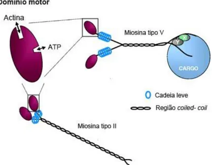
Figura 03: Estruturas básicas das miosinas –II e –V e a distinção pelo domínio cauda: tipo II, com cauda em espiral que medeia a dimerização e montagem em estruturas filamentosas, e tipo V com cadeias pesadas que dimerizam por meio de serpentinas enroladas, mas não em filamentos. As proteínas X e Y representam proteínas de ligação que podem mediar interações com carga (Adaptado de Hutagalung, Landsverk et al., 2002).
8 miosina-Vb, presente principalmente no hipocampo, giro denteado, amigdala e córtex cerebral, e no tecido epitelial (Zhao, Koslovsky et al., 1996; Lapierre, Kumar et al., 2001); e miosina-Vc, presente em tecidos epiteliais, glandulares e cerebelo (Bridgman e Elkin, 2000; Rodriguez e Cheney, 2002; Marchelletta, Jacobs et al., 2008; Jacobs, Weigert et al., 2009).
A miosina-Va está localizada na densidade pós-sináptica e em vesículas, e tem sido relacionada com função neuronal e de cognição (Hirokawa, Niwa et al., 2010), apresentando a característica de se ligar diretamente a várias proteínas citoplasmáticas, incluindo sinaptobrevina, sinaptofisina, sintaxina e CaMKII (Prekeris e Terrian, 1997; Evans, Lee et al., 1998; Costa, Mani et al., 1999; Ohyama, Komiya et al., 2001).
O cérebro possui grande quantidade de miosina-Va e dados de imunodetecção em cérebro e cerebelo de ratos, bem como análises de imunocitoquímica, mostram intensa marcação nas extensões dendríticas das células de Purkinje e na região perinuclear (Espindola, Espreafico et al., 1992; Tilelli, Martins et al., 2003), sendo as vesículas e as organelas as principais cargas transportadas por esta miosina nos neurônios e em outros tipos celulares (Langford e Molyneaux, 1998; Depina e Langford, 1999).
Além de todas as patologias envolvendo as miosinas –II e –V, estudos mostram que o diabetes altera a expressão destas miosinas no cérebro de ratos, apresentando uma diminuição na expressão de miosina-Va (Da Costa, Calabria et al., 2011; Da Costa, Calabria et al., 2013) e um aumento na expressão de miosina-IIB (Calabria, Da Cruz et al., 2011). Entretanto, os mecanismos de regulação da glicemia e os seus efeitos sobre a expressão de motores moleculares ainda são desconhecidos e aguardam maiores investigações.
1.5Proteínas que se associam a vesículas sinápticas
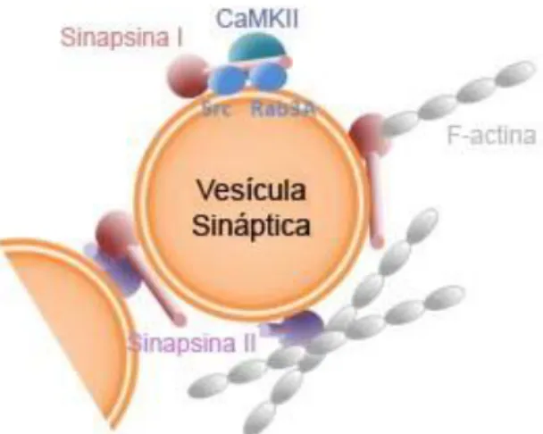
9 sinapsina-I desempenha um papel chave na libertação de neurotransmissores, regulando a disponibilidade de vesículas sinápticas (Llinas, Mcguinness et al., 1985; Ceccaldi, Grohovaz et al., 1995). Além disso, a sinapsina-I é um substrato importante para a proteína quinase-II dependente de cálcio e calmodulina (CaMKII) (Figura 04), sendo que o seu estado de fosforilação regula a liberação de neurotransmissores pela disponibilidade de vesículas sinápticas (Huttner, Schiebler et al., 1983; Nestler e Greengard, 1983; Llinas, Mcguinness et al., 1985; Schiebler, Jahn et al., 1986).
Figura 04: Esquema da interação entre vesícula sináptica, sinapsina-I e CaMKII (Adaptado de Humeau, Candiani et al., 2011).
10
Figura 05: Fosfoproteínas implicadas na exocitose sináptica. Abreviações: CaMKII, proteína quinase-II dependente de Ca2+/calmodulina; Cdk5, ciclina-dependente proteína quinase 5; CK2, caseína quinase II; NSF, proteína de fusão N-etilmaleimida sensível; PKA, proteína quinase A; PKC, proteína quinase C; SNAP, proteína solúvel NSF; SNAP25, proteína associada ao sinaptossomo; VAMP, proteína de membrana associada a vesícula (ou sinaptobrevina) (Adaptado de Turner, Burgoyne et al., 1999).
11 deste estado patológico, pode alterar os níveis de expressão das proteínas pré-sináptica, como sinapsina I, sinaptofisina e SNAP-25 (Van Guilder, Brucklacher et al., 2008).
1.6 Transportador de glicose
A captação da glicose no sistema nervoso dos mamíferos é mediada pela família de proteínas transportadoras de glicose (GLUT). Treze membros fazem parte da família GLUT, sendo que vários deles estão presentes no cérebro (Wood e Trayhurn, 2003; Uldry e Thorens, 2004), como o GLUT1, também expresso na barreira hematoencefalica (Wood e Trayhurn, 2003); o GLUT3, distribuído no corpo celular de neurônios por todo o sistema nervoso central (Nagamatsu, Sawa et al., 1993; Maher, Vannucci et al., 1994; Choeiri, Staines et al., 2002); o GLUT4, expresso numa localização somatodendrítico citoplasmática (El Messari, Leloup et al., 1998; Choeiri, Staines et al., 2002; El Messari, Ait-Ikhlef et al., 2002); e o GLUT8, presente nos compartimentos intracelulares de neurônios (Piroli, Grillo et al., 2002). O GLUT4, um transportador de glicose insulino-sensível, pode estar distribuído no cérebro (Rayner, Thomas et al., 1994), mas também no tecido adiposo, coração e músculo esquelético, onde é responsável pela redução dos níveis de glicose no plasma na condição pós-prandial. Neste caso, a insulina age estimulando a translocação de vesículas contendo GLUT4 para a membrana plasmática (Bryant, Govers et al., 2002; Wood e Trayhurn, 2003).
Alguns estudos, investigando a expressão de GLUTs no diabetes mostram uma discrepância na compreensão dos efeitos da concentração de glicose no cérebro (Shah, Desilva et al., 2012). Um exemplo disso é a detecção de níveis aumentados de GLUT4 no cerebelo de ratos db/db e diminuídos em ratos com diabetes induzido por estreptozotocina (Vannucci, Koehler-Stec et al., 1998).
1. 7 Vochysia rufa e as plantas no tratamento do diabetes mellitus
12 os efeitos da administração de extrato bruto ou princípio ativo de diferentes plantas no controle da glicemia, como também analisar o seu efeito no cérebro. Nesse sentido, tem sido mostrada a ação antioxidante do extrato de chá branco (Camellia sinensis) (Espinosa, Lopez-Jimenez et al., 2013); os efeitos sobre a disfunção cognitiva do extrato de raiz de ginseng (Panax ginseng) (Al-Hazmi, Rawi et al., 2013); a inibição de radicais livres e aumento da atividade de enzimas antioxidantes do ácido cafeíco de Abacopteris penangiana (Fu, Du et al., 2013); a redução na liberação de espécies reativas do oxigênio, TNF- e óxido nítrico como um efeito protetor de Rehmannia glutinosa (Tian, An et al., 2006); a atenuação do aumento da expressão de produtos finais de glicação avançada e NFk após o uso de semente de uva (Vitis vinifera) (Xu, Li et al., 2008); como também, o efeito contra danos oxidativos, alteração da expressão de miosina-Va e a perda neuronal no hipocampo com a administração de Pouteria ramiflora (Da Costa, Calabria et al., 2013).
As plantas têm um papel importante no tratamento do diabetes, particularmente em países em desenvolvimento, onde o acesso a medicamentos tradicionais é geralmente restrito à populacoes de maior poder aquisitivo. Portanto, estudo de plantas no desenvolvimento de tratamentos alternativos para o controle do diabetes mellitus e da obesidade é crucial (Manzan, Carneiro et al., 2002; Ali, Houghton et al., 2006).
13
Figura 06: Espécie Vochysia rufa fotografada em Abadia dos Dourados, Minas Gerais (Gouveia,
2012).
A família Vochysiaceae abrange seis gêneros e cerca de 200 espécies neotropicais, com poucos representantes em regiões subtropicais. Entretanto, o gênero Vochysia compreende cerca de 100 espécies características por serem grandes árvores e arbustos que ocorrem ao longo da América Tropical, do México ao Peru. Várias destas espécies são utilizadas por comunidades indígenas na América do Sul para uma variedade de aplicações terapêuticas relacionadas com a inflamação (Schultes e Raffauf, 1990), devido à presença do ácido betulínico (Weniger, Lobstein et al., 2005). As espécies desse gênero também são utilizadas no tratamento de feridas da pele e para aliviar doenças respiratórias, como asma e congestão pulmonar (Schultes e Raffauf, 1990); apresentam atividade antiulcerogênica gástrica devido à presença de triterpenos pentacíclicos, como o ácido betulínico, ácido betulínico epi, eritrodiol e misturas de derivados ursólicos e oleanólicos (Gomes Rde, Bonamin et al., 2009); além do tratamento de resfriados e como vermífugos (Silva, 2009).
14 o extrato de Vochysia rufa teve um efeito antioxidante, diminuindo os níveis de peroxidação lipídica e aumentando os níveis de algumas enzimas, além de conferir resistência à apoptose. Em estudos com células somáticas de Drosophila melanogaster, Moraes (2010) avaliou diferentes doses do extrato aquoso de Vochysia rufa quanto ao seu potencial mutagênico, sua ação anti-mutagênica e anti-recombinogênica contra os efeitos recombinogênicos do cloridrato de doxorrubicina. Na glândula parótida, análises morfométricas mostraram que o diabetes altera o diâmetro de células acinares, porém o tratamento com o extrato de Vochysia rufa não modifica este quadro (Caixeta, Moura et al., 2012); resultado observado também em análises feitas em ilhotas de Langerhans no pâncreas (Rodovalho, 2013).
15
2. Referências
Al-Hazmi, M. A., S. M. Rawi, et al. The potent effects of ginseng root extract and memantine on cognitive dysfunction in male albino rats. Toxicol Ind Health, Feb 13. 2013.
Ali, H., P. J. Houghton, et al. alpha-Amylase inhibitory activity of some Malaysian plants used to treat diabetes; with particular reference to Phyllanthus amarus. J Ethnopharmacol, v.107, n.3, Oct 11, p.449-55. 2006.
Association, A. D. Diagnosis and classification of diabetes mellitus. Diabetes Care, v.35 Suppl 1, Jan, p.S64-71. 2012.
______. Diagnosis and classification of diabetes mellitus. Diabetes Care, v.36 Suppl 1, Jan, p.S67-74. 2013.
Auerbach, J. M. e M. Segal. Peroxide modulation of slow onset potentiation in rat hippocampus. J Neurosci, v.17, n.22, Nov 15, p.8695-701. 1997.
Baynes, J. W. e S. R. Thorpe. Role of oxidative stress in diabetic complications: a new perspective on an old paradigm. Diabetes, v.48, n.1, Jan, p.1-9. 1999.
Benashski, S. E., A. Harrison, et al. Dimerization of the highly conserved light chain shared by dynein and myosin V. J Biol Chem, v.272, n.33, Aug 15, p.20929-35. 1997.
Bennett, M. K. e R. H. Scheller. A molecular description of synaptic vesicle membrane trafficking. Annu Rev Biochem, v.63, p.63-100. 1994.
Berg, J. S., B. C. Powell, et al. A millennial myosin census. Mol Biol Cell, v.12, n.4, Apr, p.780-94. 2001.
Biessels, G. J., A. C. Kappelle, et al. Cerebral function in diabetes mellitus. Diabetologia, v.37, n.7, Jul, p.643-50. 1994.
Bitar, M., M. Koulu, et al. Diabetes-induced alteration in brain monoamine metabolism in rats. J Pharmacol Exp Ther, v.236, n.2, Feb, p.432-7. 1986.
Bridgman, P. C. e L. L. Elkin. Axonal myosins. J Neurocytol, v.29, n.11-12, Nov-Dec, p.831-41. 2000.
Bryant, N. J., R. Govers, et al. Regulated transport of the glucose transporter GLUT4. Nat Rev Mol Cell Biol, v.3, n.4, Apr, p.267-77. 2002.
Buss, F., J. P. Luzio, et al. Myosin VI, a new force in clathrin mediated endocytosis. FEBS Lett, v.508, n.3, Nov 23, p.295-9. 2001.
16 ______. Basic mechanisms of antioxidant activity. Biofactors, v.6, n.4, p.391-7. 1997b.
Caixeta, D. C., F. B. R. Moura, et al. Effect of Vochysia sp. extract on the morphometry of the parotid gland in streptozotocin-induced diabetic rats. 10th International Congress on Cell Biology and the XVI Meeting of the Brazilian Society for Cell Biology, 2012, Rio de Janeiro. 2012.
Calábria, L. K. Efeitos do diabetes mellitus e da suplementação com antioxidantes no estresse oxidativo e na expressão de miosinas no cérebro de ratos. Instituto de Genetica e Bioquímica, Universidade Federal de Uberlândia, Uberlândia, 2010.
Calabria, L. K., G. C. Da Cruz, et al. Overexpression of myosin-IIB in the brain of a rat model of streptozotocin-induced diabetes. J Neurol Sci, v.303, n.1-2, Apr 15, p.43-9. 2011.
Cardoso, L. M., D. S. A. Colombari, et al. Espécies reativas de oxigênio no controle neurovegetativo da pressão arterial. Medicina (Ribeirão Preto), v.39, n.1, p.77-88. 2006.
Ceccaldi, P. E., F. Grohovaz, et al. Dephosphorylated synapsin I anchors synaptic vesicles to actin cytoskeleton: an analysis by videomicroscopy. J Cell Biol, v.128, n.5, Mar, p.905-12. 1995.
Ceriello, A. e E. Motz. Is oxidative stress the pathogenic mechanism underlying insulin resistance, diabetes, and cardiovascular disease? The common soil hypothesis revisited. Arterioscler Thromb Vasc Biol, v.24, n.5, May, p.816-23. 2004.
Chen, B. T., M. V. Avshalumov, et al. H(2)O(2) is a novel, endogenous modulator of synaptic dopamine release. J Neurophysiol, v.85, n.6, Jun, p.2468-76. 2001.
Cheney, R. E., M. K. O'shea, et al. Brain myosin-V is a two-headed unconventional myosin with motor activity. Cell, v.75, n.1, Oct 8, p.13-23. 1993.
Cheng, D., C. C. Hoogenraad, et al. Relative and absolute quantification of postsynaptic density proteome isolated from rat forebrain and cerebellum. Mol Cell Proteomics, v.5, n.6, Jun, p.1158-70. 2006.
Cheng, T. P., N. Murakami, et al. Localization of myosin IIB at the leading edge of growth cones from rat dorsal root ganglionic cells. FEBS Lett, v.311, n.2, Oct 19, p.91-4. 1992.
Choeiri, C., W. Staines, et al. Immunohistochemical localization and quantification of glucose transporters in the mouse brain. Neuroscience, v.111, n.1, p.19-34. 2002.
17 Conti, M. A. e R. S. Adelstein. Nonmuscle myosin II moves in new directions. J Cell Sci, v.121, n.Pt 1, Jan 1, p.11-8. 2008.
Costa, M. C., F. Mani, et al. Brain myosin-V, a calmodulin-carrying myosin, binds to calmodulin-dependent protein kinase II and activates its kinase activity. J Biol Chem, v.274, n.22, May 28, p.15811-9. 1999.
Da Costa, A. V., L. K. Calabria, et al. Neuroprotective effects of Pouteria ramiflora (Mart.) Radlk (Sapotaceae) extract on the brains of rats with streptozotocin-induced diabetes. Metab Brain Dis, Mar 7. 2013.
______. The streptozotocin-induced rat model of diabetes mellitus evidences significant reduction of myosin-Va expression in the brain. Metab Brain Dis, v.26, n.4, Dec, p.247-51. 2011.
Depina, A. S. e G. M. Langford. Vesicle transport: the role of actin filaments and myosin motors. Microsc Res Tech, v.47, n.2, Oct 15, p.93-106. 1999.
Di Mario, U., S. Morano, et al. Electrophysiological alterations of the central nervous system in diabetes mellitus. Diabetes Metab Rev, v.11, n.3, Oct, p.259-77. 1995.
Du, X. L., D. Edelstein, et al. Hyperglycemia-induced mitochondrial superoxide overproduction activates the hexosamine pathway and induces plasminogen activator inhibitor-1 expression by increasing Sp1 glycosylation. Proc Natl Acad Sci U S A, v.97, n.22, Oct 24, p.12222-6. 2000.
El Messari, S., A. Ait-Ikhlef, et al. Expression of insulin-responsive glucose transporter GLUT4 mRNA in the rat brain and spinal cord: an in situ hybridization study. J Chem Neuroanat, v.24, n.4, Nov, p.225-42. 2002.
El Messari, S., C. Leloup, et al. Immunocytochemical localization of the insulin-responsive glucose transporter 4 (Glut4) in the rat central nervous system. J Comp Neurol, v.399, n.4, Oct 5, p.492-512. 1998.
Espindola, F. S., S. R. Banzi, et al. Localization of myosin-Va in subpopulations of cells in rat endocrine organs. Cell Tissue Res, v.333, n.2, Aug, p.263-79. 2008.
Espindola, F. S., E. M. Espreafico, et al. Biochemical and immunological characterization of p190-calmodulin complex from vertebrate brain: a novel calmodulin-binding myosin. J Cell Biol, v.118, n.2, Jul, p.359-68. 1992.
Espinosa, C., J. A. Lopez-Jimenez, et al. Protective effect of white tea extract against acute oxidative injury caused by adriamycin in different tissues. Food Chem, v.134, n.4, Oct 15, p.1780-5. 2013.
18 Foth, B. J., M. C. Goedecke, et al. New insights into myosin evolution and classification. Proc Natl Acad Sci U S A, v.103, n.10, Mar 7, p.3681-6. 2006.
Fu, W., G. Du, et al. Neuroprotective effect of a caffeic acid derivative from Abacopteris penangiana. Pharm Biol, v.51, n.3, Mar, p.376-82. 2013.
Fukai, T. e M. Ushio-Fukai. Superoxide dismutases: role in redox signaling, vascular function, and diseases. Antioxid Redox Signal, v.15, n.6, Sep 15, p.1583-606. 2011.
Gaspar, J. M., A. Castilho, et al. Long-term exposure to high glucose induces changes in the content and distribution of some exocytotic proteins in cultured hippocampal neurons. Neuroscience, v.171, n.4, Dec 29, p.981-92. 2010.
Genet, S., R. K. Kale, et al. Alterations in antioxidant enzymes and oxidative damage in experimental diabetic rat tissues: effect of vanadate and fenugreek (Trigonellafoenum graecum). Mol Cell Biochem, v.236, n.1-2, Jul, p.7-12. 2002.
Gerecke, K. M., A. Kolobova, et al. Exercise protects against chronic restraint stress-induced oxidative stress in the cortex and hippocampus. Brain Res, v.1509, May 6, p.66-78. 2013.
Gispen, W. H. e G. J. Biessels. Cognition and synaptic plasticity in diabetes mellitus. Trends Neurosci, v.23, n.11, Nov, p.542-9. 2000.
Gomes Rde, C., F. Bonamin, et al. Antioxidative action of methanolic extract and buthanolic fraction of Vochysia tucanorum Mart. in the gastroprotection. J Ethnopharmacol, v.121, n.3, Jan 30, p.466-71. 2009.
Gouveia, N. M. Avaliação do controle glicêmico e do estresse oxidativo em pâncreas de animais diabéticos induzidos e não diabéticos tratados com extrato de Vochysia rufa e faseolamina. Instituto de Genética e Bioquímica, Universidade Federal de Uberlândia, Uberlândia, 2012.
Haber, E. P., R. Curi, et al. Secrecao da insulina: efeito autocrino da insulina e modulacao por acidos graxos. Arquivos Brasileiros de Endocrinologia & Metabolismo, v.45, n.3, p.219-27. 2001.
Helkala, E. L., L. Niskanen, et al. Short-term and long-term memory in elderly patients with NIDDM. Diabetes Care, v.18, n.5, May, p.681-5. 1995.
Hirokawa, N., S. Niwa, et al. Molecular motors in neurons: transport mechanisms and roles in brain function, development, and disease. Neuron, v.68, n.4, Nov 18, p.610-38. 2010.
Hodge, T. e M. J. Cope. A myosin family tree. J Cell Sci, v.113 Pt 19, Oct, p.3353-4. 2000.
19 Hudmon, A. e H. Schulman. Neuronal CA2+/calmodulin-dependent protein kinase II: the role of structure and autoregulation in cellular function. Annu Rev Biochem, v.71, p.473-510. 2002.
Humeau, Y., S. Candiani, et al. Functional roles of synapsin: lessons from invertebrates. Semin Cell Dev Biol, v.22, n.4, Jun, p.425-33. 2011.
Hutagalung, A. H., M. L. Landsverk, et al. The UCS family of myosin chaperones. J Cell Sci, v.115, n.Pt 21, Nov 1, p.3983-90. 2002.
Huttner, W. B., W. Schiebler, et al. Synapsin I (protein I), a nerve terminal-specific phosphoprotein. III. Its association with synaptic vesicles studied in a highly purified synaptic vesicle preparation. J Cell Biol, v.96, n.5, May, p.1374-88. 1983.
Jacobs, D. T., R. Weigert, et al. Myosin Vc is a molecular motor that functions in secretory granule trafficking. Mol Biol Cell, v.20, n.21, Nov, p.4471-88. 2009.
Jahn, R. e P. I. Hanson. Membrane fusion. SNAREs line up in new environment. Nature, v.393, n.6680, May 7, p.14-5. 1998.
Jennings, P. E., A. F. Jones, et al. Increased diene conjugates in diabetic subjects with microangiopathy. Diabet Med, v.4, n.5, Sep-Oct, p.452-6. 1987.
Jing, Y. H., K. H. Chen, et al. Neurodegeneration in Streptozotocin-Induced Diabetic Rats is Attenuated by Treatment with Resveratrol. Neuroendocrinology, Mar 9. 2013.
Kohen, R. e A. Nyska. Oxidation of biological systems: oxidative stress phenomena, antioxidants, redox reactions, and methods for their quantification. Toxicol Pathol, v.30, n.6, Nov-Dec, p.620-50. 2002.
Krendel, M. e M. S. Mooseker. Myosins: tails (and heads) of functional diversity. Physiology (Bethesda), v.20, Aug, p.239-51. 2005.
Lane, S. R. e Y. Liu. Characterization of the palmitoylation domain of SNAP-25. J Neurochem, v.69, n.5, Nov, p.1864-9. 1997.
Langford, G. M. e B. J. Molyneaux. Myosin V in the brain: mutations lead to neurological defects. Brain Res Brain Res Rev, v.28, n.1-2, Nov, p.1-8. 1998.
Lapierre, L. A., R. Kumar, et al. Myosin vb is associated with plasma membrane recycling systems. Mol Biol Cell, v.12, n.6, Jun, p.1843-57. 2001.
20 Lisman, J., H. Schulman, et al. The molecular basis of CaMKII function in synaptic and behavioural memory. Nat Rev Neurosci, v.3, n.3, Mar, p.175-90. 2002.
Llinas, R., T. L. Mcguinness, et al. Intraterminal injection of synapsin I or calcium/calmodulin-dependent protein kinase II alters neurotransmitter release at the squid giant synapse. Proc Natl Acad Sci U S A, v.82, n.9, May, p.3035-9. 1985.
Loubery, S. e E. Coudrier. Myosins in the secretory pathway: tethers or transporters? Cell Mol Life Sci, v.65, n.18, Sep, p.2790-800. 2008.
Maher, F., S. J. Vannucci, et al. Glucose transporter proteins in brain. FASEB J, v.8, n.13, Oct, p.1003-11. 1994.
Manzan, R. M., W. P. Carneiro, et al. Análise da ação hemaglutinante de plantas em eritrócitos humanos. . Bioscience Journal, v.18, p.67. 2002.
Marchelletta, R. R., D. T. Jacobs, et al. The class V myosin motor, myosin 5c, localizes to mature secretory vesicles and facilitates exocytosis in lacrimal acini. Am J Physiol Cell Physiol, v.295, n.1, Jul, p.C13-28. 2008.
Marshall, J. F. Further analysis of the resistance of the diabetic rat to d-amphetamine. Pharmacol Biochem Behav, v.8, n.3, Mar, p.281-6. 1978.
Marshall, J. F., M. I. Friedman, et al. Reduced anorexic and locomotor-stimulant action of D-amphetamine in alloxan-diabetic rats. Brain Res, v.111, n.2, Jul 30, p.428-32. 1976.
Mates, J. M., C. Perez-Gomez, et al. Antioxidant enzymes and human diseases. Clin Biochem, v.32, n.8, Nov, p.595-603. 1999.
Mccall, A. L. The impact of diabetes on the CNS. Diabetes, v.41, n.5, May, p.557-70. 1992.
Mccrimmon, R. J., C. M. Ryan, et al. Diabetes and cognitive dysfunction. Lancet, v.379, n.9833, Jun 16, p.2291-9. 2012.
Miyazaki, T., M. Watanabe, et al. B2 exon splicing of nonmuscle myosin heavy chain IIB is differently regulated in developing and adult rat brain. Neurosci Res, v.37, n.4, Aug, p.299-306. 2000.
Mochida, S., H. Kobayashi, et al. Myosin II is involved in transmitter release at synapses formed between rat sympathetic neurons in culture. Neuron, v.13, n.5, Nov, p.1131-42. 1994.
Mooradian, A. D. Diabetic complications of the central nervous system. Endocr Rev, v.9, n.3, Aug, p.346-56. 1988.
21 Graduação em Ciências Biológicas, Universidade Federal de Uberlândia, Uberlândia, 2010.
Moraes, I. B. Alterações histopatológicas, índice de apoptose e estresse oxidativo hepático em ratos diabéticos induzidos por estreptozotocina tratados com extrato aquoso de Vochysia rufa. Programa de Pós-Graduação em Biologia Celular e Estrutural Aplicadas, Universidade Federal de Uberlândia, Uberlândia, 2013.
Murakami, N., P. Mehta, et al. Studies on the distribution of cellular myosin with antibodies to isoform-specific synthetic peptides. FEBS Lett, v.288, n.1-2, Aug 19, p.247. 1991.
Nagamatsu, S., H. Sawa, et al. Neuron-specific glucose transporter (NSGT): CNS distribution of GLUT3 rat glucose transporter (RGT3) in rat central neurons. FEBS Lett, v.334, n.3, Nov 22, p.289-95. 1993.
Nascimento, A. A., R. E. Cheney, et al. Enzymatic characterization and functional domain mapping of brain myosin-V. J Biol Chem, v.271, n.29, Jul 19, p.17561-9. 1996.
Navone, F., G. Di Gioia, et al. Microvesicles of the neurohypophysis are biochemically related to small synaptic vesicles of presynaptic nerve terminals. J Cell Biol, v.109, n.6 Pt 2, Dec, p.3425-33. 1989.
Nayak, A. S., C. I. Moore, et al. Ca2+/calmodulin-dependent protein kinase II phosphorylation of the presynaptic protein synapsin I is persistently increased during long-term potentiation. Proc Natl Acad Sci U S A, v.93, n.26, Dec 24, p.15451-6. 1996.
Nestler, E. J. e P. Greengard. Protein phosphorylation in the brain. Nature, v.305, n.5935, Oct 13-19, p.583-8. 1983.
Ohyama, A., Y. Komiya, et al. Globular tail of myosin-V is bound to vamp/synaptobrevin. Biochem Biophys Res Commun, v.280, n.4, Feb 2, p.988-91. 2001.
Oyler, G. A., G. A. Higgins, et al. The identification of a novel synaptosomal-associated protein, SNAP-25, differentially expressed by neuronal subpopulations. J Cell Biol, v.109, n.6 Pt 1, Dec, p.3039-52. 1989.
Parfitt, K. D., B. J. Hoffer, et al. Norepinephrine and isoproterenol increase the phosphorylation of synapsin I and synapsin II in dentate slices of young but not aged Fisher 344 rats. Proc Natl Acad Sci U S A, v.88, n.6, Mar 15, p.2361-5. 1991.
22 Piroli, G. G., C. A. Grillo, et al. Peripheral glucose administration stimulates the translocation of GLUT8 glucose transporter to the endoplasmic reticulum in the rat hippocampus. J Comp Neurol, v.452, n.2, Oct 14, p.103-14. 2002.
Prekeris, R. e D. M. Terrian. Brain myosin V is a synaptic vesicle-associated motor protein: evidence for a Ca2+-dependent interaction with the synaptobrevin-synaptophysin complex. J Cell Biol, v.137, n.7, Jun 30, p.1589-601. 1997.
Rayner, D. V., M. E. Thomas, et al. Glucose transporters (GLUTs 1-4) and their mRNAs in regions of the rat brain: insulin-sensitive transporter expression in the cerebellum. Can J Physiol Pharmacol, v.72, n.5, May, p.476-9. 1994.
Reck-Peterson, S. L., D. W. Provance, Jr., et al. Class V myosins. Biochim Biophys Acta, v.1496, n.1, Mar 17, p.36-51. 2000.
Reis, J. S., C. A. Veloso, et al. Estresse Oxidativo: Revisão da Sinalização Metabólica no Diabetes Tipo 1. Arquivos Brasileiros de Endocrinologia & Metabologia, v.52, n.7, p.1096-1105. 2008.
Rex, C. S., C. F. Gavin, et al. Myosin IIb regulates actin dynamics during synaptic plasticity and memory formation. Neuron, v.67, n.4, Aug 26, p.603-17. 2010.
Rochlin, M. W., K. Itoh, et al. Localization of myosin II A and B isoforms in cultured neurons. J Cell Sci, v.108 ( Pt 12), Dec, p.3661-70. 1995.
Rodovalho, A. B. Avaliação bioquímica e morfológica de Ilhotas de Langerhans de ratos não diabéticos e diabéticos induzidos por estreptozotocina tratados com Vochysia rufa Curso de Ciências Biológicas, Universidade Federal de Uberlândia, Uberlândia, 2013.
Rodriguez, O. C. e R. E. Cheney. Human myosin-Vc is a novel class V myosin expressed in epithelial cells. J Cell Sci, v.115, n.Pt 5, Mar 1, p.991-1004. 2002.
Rosen, P., P. P. Nawroth, et al. The role of oxidative stress in the onset and progression of diabetes and its complications: a summary of a Congress Series sponsored by UNESCO-MCBN, the American Diabetes Association and the German Diabetes Society. Diabetes Metab Res Rev, v.17, n.3, May-Jun, p.189-212. 2001.
Ryu, J., L. Liu, et al. A critical role for myosin IIb in dendritic spine morphology and synaptic function. Neuron, v.49, n.2, Jan 19, p.175-82. 2006.
Saller, C. F. Dopaminergic activity is reduced in diabetic rats. Neurosci Lett, v.49, n.3, Aug 31, p.301-6. 1984.
23 Schiebler, W., R. Jahn, et al. Characterization of synapsin I binding to small synaptic vesicles. J Biol Chem, v.261, n.18, Jun 25, p.8383-90. 1986.
Schultes, R. E. e R. F. Raffauf. The Healing Forest: Medicinal and Toxic Plants of the Northwest Amazonia. Portland: Dioscorides Press, Oregon., p.484. 1990.
Shah, K., S. Desilva, et al. The Role of Glucose Transporters in Brain Disease: Diabetes and Alzheimer’s Disease. Int J Mol Sci, v.13, n.10, p.12629-55. 2012.
Siddiqui, M. R., A. Taha, et al. Amelioration of altered antioxidant status and membrane linked functions by vanadium and Trigonella in alloxan diabetic rat brains. J Biosci, v.30, n.4, Sep, p.483-90. 2005.
Silva, M. A. B. Levantamento etnobotânico e triagem hipolipidêmica de Plantas Medicinais do Cerrado Mato-grossense: Avaliação da Atividade hipolipidêmica da Simaba ferruginea St. Hil. e Vochysia rufa Faculdade de Ciências Médicas, Universidade Federal do Mato Grosso, Cuiabá, 2009.
Sousa, T., J. Afonso, et al. Lipid Peroxidation and Antioxidants in Arterial Hypertension. Biochemistry, Genetics and Molecular Biology. 2012.
Taniyama, Y. e K. K. Griendling. Reactive oxygen species in the vasculature: molecular and cellular mechanisms. Hypertension, v.42, n.6, Dec, p.1075-81. 2003.
Tian, Y. Y., L. J. An, et al. Catalpol protects dopaminergic neurons from LPS-induced neurotoxicity in mesencephalic neuron-glia cultures. Life Sci, v.80, n.3, Dec 23, p.193-9. 2006.
Tilelli, C. Q., A. R. Martins, et al. Immunohistochemical localization of myosin Va in the adult rat brain. Neuroscience, v.121, n.3, p.573-86. 2003.
Trulson, M. E., J. H. Jacoby, et al. Streptozotocin-induced diabetes reduces brain serotonin synthesis in rats. J Neurochem, v.46, n.4, Apr, p.1068-72. 1986.
Turner, K. M., R. D. Burgoyne, et al. Protein phosphorylation and the regulation of synaptic membrane traffic. Trends Neurosci, v.22, n.10, Oct, p.459-64. 1999.
Uldry, M. e B. Thorens. The SLC2 family of facilitated hexose and polyol transporters. Pflugers Arch, v.447, n.5, Feb, p.480-9. 2004.
Valko, M., D. Leibfritz, et al. Free radicals and antioxidants in normal physiological functions and human disease. Int J Biochem Cell Biol, v.39, n.1, p.44-84. 2007.
24 Van Guilder, H. D., R. M. Brucklacher, et al. Diabetes downregulates presynaptic proteins and reduces basal synapsin I phosphorylation in rat retina. Eur J Neurosci, v.28, n.1, Jul, p.1-11. 2008.
Vannucci, S. J., E. M. Koehler-Stec, et al. GLUT4 glucose transporter expression in rodent brain: effect of diabetes. Brain Res, v.797, n.1, Jun 22, p.1-11. 1998.
Vasconcelos, S. M. L., M. O. F. Goulart, et al. Espécies reativas de oxigênio e de nitrogênio, antioxidantes e marcadores de dano oxidativo em sangue humano: Principais métodos analíticos para sua determinação. Química Nova, v.30, n.5. 2007.
Walch-Solimena, C., J. Blasi, et al. The t-SNAREs syntaxin 1 and SNAP-25 are present on organelles that participate in synaptic vesicle recycling. J Cell Biol, v.128, n.4, Feb, p.637-45. 1995.
Wen, Y., J. C. Skidmore, et al. Relationship of glycation, antioxidant status and oxidative stress to vascular endothelial damage in diabetes. Diabetes Obes Metab, v.4, n.5, Sep, p.305-8. 2002.
Wendt, M. C. The Role of Oxidative Stress in Streptozotocin induced Early and Late Stage Diabetes Mellitus in the Rat (Rattus norvegicus, Berkenhout 1769). . Dissertação Universität Hamburg, 2005.
Weniger, B., A. Lobstein, et al. Bioactive triterpenoids from Vochysia pacifica interact with cyclic nucleotide phosphodiesterase isozyme PDE4. Phytother Res, v.19, n.1, Jan, p.75-7. 2005.
Wood, I. S. e P. Trayhurn. Glucose transporters (GLUT and SGLT): expanded families of sugar transport proteins. Br J Nutr, v.89, n.1, Jan, p.3-9. 2003.
Xu, L., B. Li, et al. Oral administration of grape seed proanthocyanidin extracts downregulate RAGE dependant nuclear factor- kappa BP65 expression in the hippocampus of streptozotocin induced diabetic rats. Exp Clin Endocrinol Diabetes, v.116, n.4, Apr, p.215-24. 2008.
25
Capítulo 2
26
1. ARTIGO CIENTÍFICO
Journal of Neurochemistry (fator de impacto: 4,061)
GLIBENCLAMIDE TREATMENT MODULATES EXPRESSION AND
LOCALIZATION OF MYOSIN-IIB IN DIABETIC RAT BRAIN
Alice Vieira da Costa1, Luciana Karen Calábria1,2, Paula de Souza Santos1, Luiz Ricardo
Goulart1, Foued Salmen Espindola1
1Institute of Genetics and Biochemistry, Federal University of Uberlândia, Campus
Umuarama, 38400-902, Uberlândia-MG, Brazil.
2Basic Departament - Health Area, Federal University of Juiz de Fora, Campus Avançado
de Governador Valadares, 35010-173, Governador Valadares-MG, Brazil.
Corresponding author: Foued Salmen Espindola
Universidade Federal de Uberlândia, Instituto de Genética e Bioquímica Laboratório de Bioquímica e Biologia Molecular
Campus Umuarama, Bloco 2E, sala 235 CEP: 38400-902
Uberlândia, MG, Brazil
fouedespindola@gmail.com or foued@ufu.br
27
Abstract
Myosin-IIB is a non-muscle isoform in the brain with increased expression in the brains of diabetic rats. Chronic hyperglycemia caused by diabetes can impair learning and memory. Oral hypoglycemic agents such as glibenclamide have been used to control hyperglycemia. We report changes in the expression and distribution of myosin-IIB in the frontal cortex and hippocampus of diabetic rats treated with glibenclamide. To establish this, brains were harvested after 43 days of treatment with glibenclamide (6 mg/kg bw orally), homogenized and analyzed by Western blotting, qRT-PCR and immunohistochemistry. As expected, myosin-IIB expression increased in the brains of diabetic rats. However, protein levels returned to normalcy after treatment with glibenclamide. In addition, MYH10 gene expression decreased in diabetic rats treated with glibenclamide. Moreover, we found weak myosin-IIB labeling in the hippocampus and frontal cortex of rats treated with glibenclamide. Therefore, the expression of myosin-IIB is affected by diabetes mellitus and may be modulated by glibenclamide treatment in rats. Structural changes in the hippocampus and prefrontal cortex are reversible, and glibenclamide treatment may reduce patho-physiological changes in the brain. Our findings suggest a possible correlation between glibenclamide effects and myosin-IIB function in the brain of diabetised rats.
Keywords
28
Introduction
Myosins are a superfamily of diverse proteins that are localized in different cell types and specialized areas. These proteins are involved in various pathways such as motility, mechanoenzymology, interactions with loads and the endomembrane system (Hartman & Spudich 2012). Some myosins are linked to vesicle trafficking Va), endosome and AMPA receptor recruitment (myosin-Vb), endocytic trafficking in neurons (myosin-VI), GLUT4 traffic (myosin-Ic) and actin dynamics in neurons (myosin-IIB) (Hartman & Spudich 2012, Hirokawa et al. 2010, Rex et al., Rex et al. 2010).
Myosin-II exist in three non-muscle isoforms: IIA, IIB and IIC. The motor activity of these proteins is regulated by the phosphorylation of a common pair of regulatory myosin light chains (Vicente-Manzanares et al. 2009, Yu et al. 2012). Myosin-IIB is the predominant form in the brain and is involved in vesicle transport in ganglion neurons, retrograde flow of growth cones, the phosphorylation status of the dendritic spine, cell migration and cytokinesis during cell division (Lo et al. 2004, Yang et al. 2012, Hodges et al. 2011, Takagishi et al. 2005, Golomb et al. 2004).
Recently, we showed the differential expression of myosins such as myosin-IIB (Calabria et al. 2011) and myosin-Va (da Costa et al. 2011, da Costa et al. 2013) in the brains of rats with streptozotocin-induced diabetes. Studies have shown that adults with type 1 and type 2 diabetes have compromised memory and executive function, which are associated with an increased risk of cognitive dysfunction. This damage is associated with chronic hyperglycemia and the long duration of diabetes (for reviews, see McCrimmon et al. 2012)
29 orally over 43 days for the treatment of hypoglycemia, on the expression and localization of myosin-IIB in the brains of rats with streptozotocin-induced diabetes.
Materials and methods Animals
Male Wister rats (200 – 220 g) were housed under standard conditions (22±1°C, humidity 60±5% and 12 h light/12 h dark cycle) with food and water ad libitum. Procedures for animal handling and use followed the resolutions proposed by the Brazilian Society of Science on Laboratory Animals and by the Ethics Committee on Animal Research of the Federal University of Uberlândia, Brazil (CEUA/UFU 060/10).
Induction of diabetes mellitus
The rats were let to fast for 24-h, anesthetized by intraperitoneal injection of xylazine (10 mg/kg)/ ketamine (75 mg/kg) and injected with streptozotocin (40 mg/kg bw, 0.01 M citrate buffer, pH 4.5; Sigma-Aldrich, USA) via the penile vein (2 mL/kg bw). Non-diabetic rats were injected with the same volume of citrate buffer. Fasting was maintained for another 90 min postinjection. Fasting blood glucose levels labove 250 mg/dL were scored as diabetic. Glycemic levels were monitored for an additional 10 days with Biocheck Glucose Test Strips (Bioeasy, Brazil).
Groups and glibenclamide treatments
Animals were randomly separated into three groups (n = 10 rats/group): non-diabetic (ND), non-diabetic (D) and non-diabetic treated with glicenclamide (6 mg/kg bw orally) (DG). Water and glibenclamide were administered by oral gavage for 43 days. Body weight was recorded weekly. After this period, the animals were fasted for 12-h, anesthetized and then had their brains were surgically removed.
Western blotting
30 1976) assay. The expression of myosin-IIB relative to beta-actin was analyzed by western blot as described previously by Calábria et al. (2011). The intensity of the protein bands was analyzed and compared using Scion Image software, version Alpha 4.0.3.2 (Scion Corporation, USA) and the results were expressed as a percentage of total protein content.
Quantitative real-time PCR (qPCR)
Total RNA was extracted from the brains using TRIzol® reagent following the manufacturer’s instructions (Invitrogen, USA). RNA samples (2 µg) were reverse -transcribed using MMLV-RT (Amersham Biosciences, Sweden). Standards were prepared by cloning PCR products of MYH10 and housekeeping gene β -2-microglobulin (B2M) fragments using a TOPOTA Cloning Dual Promoter Kit (Invitrogen, USA). Plasmid concentration was measured and the copy numbers calculated according to Yin et al. [26]. A MYO2 and B2M construct at a concentration of 7 copies per μL was serially diluted to create a standard curve ranging from 1 to 10. The efficiency of reaction was calculated using E = (10 - 1/slope - 1) × 100, where the log of each dilution was plotted against ΔCT (see below) of the housekeeping and target genes. To determine MYO2 cellular expression, quantitative RT-PCR analysis was performed using a 7300 Real Time PCR System (PE Applied Biosystems, CA) and SybrGreen PCR Core Reagent (PE Applied Biosystems, CA). PCR Primers were designed using the Primer Express 3.0 Program (PE Applied Biosystems, CA). The set of primers used for the B2M fragment amplification was: 5′-CGTCGTGCTTGCCATTCA-3′ and 5′ -TCCTCAACTGCTACGTGTCTCAG-3′. The MYO2 forward and reverse primers
were: 5′-CCATGCCGGAGAACACAGT-3′ and 5′
-AAGCCCAGACCAAAGAGCAG-3′, respectively. The relative expression of each specific product was calculated by 2-ΔΔCT (CT = the fluorescence threshold value; ΔCT = CT of the target gene- CT of the reference gene (B2M); ΔΔCT = ΔCT of the target sample – ΔCT of the calibrator sample).
Immunohistochemistry
31 paraffin at room temperature. Five micrometer sections were pretreated in a microwave for 5 min with 4 mM citrate buffer (pH 6.0) containing 0.025% Tween 20. The sections were then incubated with anti-myosin-IIB primary antibody for 16 h followed by incubation with a Post Primary Block NovoLink™ Max Polymer Detection System (Novo castra Laboratories Ltd., United Kingdom). After three washes with TBS-T, the sections were incubated with the NovoLink polymer for 30 min at 37 °C. Chromogen was developed using 3, 3′-diaminobenzidine and then the material was counterstained with hematoxylin, dehydrated, mounted with ERV-Mount (EasyPath, Brazil) and analyzed with the 40x and 100x objective lenses of a Leica DM500 microscope (Microsystems, Germany). Negative controls were prepared by omitting the primary antibody from the reaction.
Statistical analysis
All values obtained were expressed as mean±SEM (standard error of the mean). Statistical analysis was performed with the Student t-test and data were analyzed using SigmaStat 3.5 software (Systat Software Inc., USA). A p-value <0.05 was considered significant.
Results
In this study, correlation between myosin-IIB expression and oral administration of glibenclamide in the brain of diabetic rats. Figure 2 shows differential expression of the myosin-IIB protein was analyzed by Western blottin. Immunoreactivity of myosin-IIB increased by ~20% in diabetic compared to non-diabetic rat brains. Interestingly, these levels reached normalcy after glibenclamide treatment.
Real time PCR was performed to measure changes in myosin-IIB gene (MYH10) levels (Fig. 1). These changes were calculated using beta-2-microglobulin (B2M) as an endogenous control. MYH10 mRNA levels in diabetic brains increased by ~140% compared to non-diabetic brains. However, MYH10 mRNA concomitant
glibenclamide treatment reduced MYH10 levels by ~65%.
32 than in non-diabetic rats. The same was not observed in the cortex of diabetic rats treated with glibenclamide (Fig. 3). Immunostaining in diabetic rats revealed intense myosin-IIB labeling in the frontal cortex, mainly in pyramidal neurons and their projections (Fig. 3). Although the distribution of myosin-IIB in the brain was similar for all groups, different staining patterns may be linked to the function of myosin-IIB. The distribution of myosin-IIB in other regions of the brain was the same for non-diabetic and non-diabetic rats. Myosin-IIB staining was more intense in the dentate gyrus of the hippocampus of diabetic rats than it was in non-diabetic rats (Fig. 4). Otherwise, immunolabeling was faint in the cortex and hippocampus of diabetic rats treated with glibenclamide. Moreover, immunolabeling was more intense in the granule cell layer and polymorphic layers the diabetic group compared to non-diabetic and glibenclamide-treated groups. In mossy cells of the dentate gyrus, myosin-IIB was distributed mainly in the cytoplasm.
Discussion
In this study, we demonstrated that diabetic rats treated with glibenclamide for 43 days had lower levels of myosin-IIB protein and mRNA expression in the brain and weaker immunolabeling in the frontal cortex and hipocamppus compared to a diabetic control group.
33 Glibenclamide is one of the most widely used antidiabetic drugs and the most popular sulfonylurea. It produces effects consistent with ATP-K+ channel inhibition (Schmid-Antomarchi et al. 1987, Sturgess et al. 1988, Zunkler et al. 1988, Zunkler et al. 1989) that stimulate insulin secretion (Serrano-Martin et al. 2006, Sturgess et al. 1988) and consequent hypoglycaemic activity. Sulfonylurea biochemically increased myosin ATPase activity (Mozaffari et al. 1988), which was discovered by studying the mechanochemical cycle of myosin-IIB (Rosenfeld et al. 2003). However, the precise molecular mechanism for drug-mediated activity modulation is still unknown.
Glibenclamide also regulates the expression of GLUT-1 in the kidneys, heart and liver of diabetic rats (Sokolovska et al. 2012). It has anti-mutagenic and antioxidant properties (Rabbani et al. 2010) and can interfere with mitochondrial bioenergetics in nonpancreatic cells by inducing changes in membrane ion permeability (Fernandes et al. 2004). Some classes of myosins, such as I (Bose et al. 2002), II (Fulcher et al. 2008, Steimle et al. 2005) and V (Yoshizaki et al. 2007) are involved in insulin-induced GLUT4-vesicle fusion/translocation and/or directly in diabetes.
Recently, we demonstrated that members of the myosin family such as myosin-IIB (Calabria et al. 2011) and myosin-Va (da Costa et al. 2013, da Costa et al. 2011) are differentially expressed in the brains of rats with streptozotocin-induced diabetes. Indeed, these molecular motors have been identified in pancreatic islets (Espindola et al. 2008, Wilson et al. 1998) and identified by their involvement in the migration of insulin secretory granules (Lajoix et al. 2006, Lida et al. 1997). Myosin-II is abundantly expressed in the brain by three distinct isoforms present in isolated post-synaptic densities of mature forebrain synapses (Cheng et al. 2006, Cheng et al. 2000, Miyazaki et al. 2000). Myosin-IIB is a necessary component of memory formation and synaptic plasticity in the mature nervous system (Rex et al. 2010).
34 increased metabolism and protein synthesis in response to increased neuronal activity (da Costa et al. 2013).
In our conditions, no change in protein distribution were observed either in the frontal cortex, in accordance with Calabria et al (2011), or in the hippocampus. Although the distribution of myosin-IIB was similar in the brains of diabetic, control and treated animals, differences in the staining pattern may be linked to myosin-IIB function. This difference may be associated with myosin-IIB function in these regions (Calabria et al. 2011, Rochlin et al. 1995).
Myosin-IIB plays important roles in migrating neurons and growth cones (Vallee et al. 2009) and has been detected in postsynaptic dendrites (Cheng et al. 2006, Miyazaki et al. 2000), where it modulates development of these structures (Ryu et al. 2006). Myosin-IIB is widely distributed within the neurons where it provides a structural mechanism for synaptic plasticity (Cheng et al. 2000). We showed that myosin-IIB is mainly distributed in the projections of pyramidal neurons in the cerebral cortex of diabetic rats and that staining decreases after glibenclamide treatment.
Myosin-IIB was localized in the hippocampus, mainly in mossy cells, and in neurons of the granule and polymorphic cell layers. Immunolabeling was more intense in diabetic rat brains than in non-diabetic ones. Many studies have found changes in the hippocampus of diabetics, such as decreased spine density on pyramidal neurons (Martinez-Tellez et al. 2005), synaptic reorganization (Grillo et al. 2005), increased neuronal vulnerability (Revsin et al. 2005, Saravia et al. 2002), reduced cell proliferation and neurogenesis (Alvarez et al. 2009, Beauquis et al. 2006), neuronal apoptosis (Li et al. 2002), and increased nuclear diameters of neurons (da Costa et al. 2013). A known function of myosin-II in CA1 pyramidal neurons is to contribute to an actin-dependent process that underlies memory consolidation in response to synaptic stimulation (Rex et al. 2010). It is possible that myosin-II has unknown systems level functions that account for disruption in hippocampus-dependent memory consolidation (Rex et al. 2010).
high-35 affinity glibenclamide binding sites were found in the brain, especially in the cerebral cortex and hippocampus (Treherne & Ashford 1991). These discoveries are important because they indicate a possible regulation mechanism specific to myosin-IIB. This is also significant because of a recent study showing that acarbose treatment, an inhibitor of α-glycosidase used for glycemia control (Van de Laar et al. 2006), is incapable of restoring myosin-Va levels in the hippocampus (da Costa et al. 2013).
In summary, myosin-IIB expression is affected by diabetes mellitus and may be modulated by glibenclamide treatment in rats. Structural changes in the hippocampus (Luine et al. 1994) and prefrontal cortex (Radley et al. 2005) are reversible. Additionally, glibenclamide can reverse damage caused by diabetes on myosin-IIB expression and reduce patho-physiological changes in the brain. Finally, these results contribute to the knowledge base on myosin regulation in the brains of diabetic rats.
Acknowledgments
The authors would like recognize the Laboratory of Clinical Analysis of the School of Veterinary Medicine (Federal University of Uberlândia) and especially Antônio Vicente Mundim and Felipe Cesar Gonçalves for their help in processing the biochemical data. We would also like to thank Aline Borges Rodovalho, Camilla Manzan Martins, Douglas Carvalho Caixeta, Hélen Lara Machado and Izabela Barbosa Moraes (Federal University of Uberlândia) for contributions to experimental procedures, and Neire Moura de Gouveia for technical support. This study was supported by grants from FAPEMIG to FSE and from CNPq to LRG, and a fellowship from CNPq to AVC and PSS.
Conflict of interest
The authors are not having any conflict of interest related to this paper.
References