DOI: 10.5935/2359-4802.20170099
Abstract
Background: By observing the high prevalence of failures in the surgical treatment of myocardial revascularization (MR), with the use of the Left Internal Thoracic Artery (LITA) as a graft, evidenced by the international literature, it was sought to demonstrate the prevalence of lesions that would not allow the use of LITA as a graft in myocardial revascularization surgery, with possible alteration in the surgical management performed by the cardiac surgeon, and reduction of the morbimortality of these patients.
Objectives: To evaluate the prevalence of atherosclerotic lesions of the LITA, through selective preoperative angiography, in patients submitted to coronary angiography and indicated for myocardial revascularization. We also analyzed other lesions that made the use of LITA unfeasible as a main graft in cases of myocardial revascularization surgery (MRS).
Methods: This was a cross-sectional, prevalence study that evaluated, through selective angiography, the LITA of 39 patients with a median age of 63 years, submitted to coronary angiography, with indication of Coronary Artery Bypass Graft (CABG). Categorical variables were compared by chi-square test and Fisher's exact test. The single continuous variable, age, was tested for normality by the Kolmogorov-Smirnov test, described in median (P25; P75) and the groups compared with the Mann-Whitney test. The level of statistical significance adopted was p < 0.05.
The analyzes were performed in SPSS® software version 20.
Results: It was identified the presence of 7.7% of disorders in the LITA that made it unfeasible to be used. In all of the patients there was no specific symptomatology evidencing the lesion. No variable was shown as a predictor for the occurrence of the outcomes.
Conclusion: The prevalence of the lesions found in the study was significant, indicating that a preoperative evaluation of LITA could bring future benefits to the patients submitted to CABG. (Int J Cardiovasc Sci. 2018;31(2)97-106)
Keywords: Atherosclerosis; Myocardial Revascularization; Mammary Arteries; Coronary Angiography.
ORIGINAL ARTICLE
Mailing Address: Hadrien Felipe Meira Balzan
Avenida Guedner, s/n. Postal Code: 87050-390, Jardim Aclimação, Maringá, PR – Brazil. E-mail: hadrien.fel2@hotmail.com
Prevalence of Atherosclerotic Lesions in the Left Internal Thoracic Artery, Evidenced by
Selective Angiographic Findings
Hadrien Felipe Meira Balzan,
1Rafael Vinicius Lube Battilani,
1Otávio Celeste Mangili,
1Marcos Franchetti,
2Leonardo Celeste Mangili,
3Julio de Paiva Maia,
2Dorane Dias de Moura,
1Bruna Felipe de Melo Lage
4Centro Universitário de Maringá (CESUMAR),1 Centro de Diagnósticos Paraná – Cedipar,2 Cardioclínica Maringá,3 PR, Centro Universitário de Várzea Grande (UNIVAG),4 Várzea Grande, MT – Brazil
Manuscript received November 23, 2016; revised manuscript June 09, 2017; accepted July 07, 2017.
Introduction
Among the principal means available for the diagnosis of atherosclerotic coronary disease, coronary angiography, an invasive imaging method, is the choice to examinate the evaluation of patients with high cardiovascular risk, calculated through non-invasive scores. Coronary angiography is also indicated in patients who present angina and symptoms of
heart failure, and these recommendations are based on a high
degree of evidence.1
Following the establishment of the criteria for surgical indication, one should proceed to choose the type of graft to be used by the surgeon. Among the arterial options, the
best choice is the internal thoracic artery.2
In 1986, a study performed at the Cleveland Clinic demonstrated superiority in the use of the LITA, or left internal thoracic artery, compared to the use of the saphenous vein when anastomosed to the left anterior descending branch of the left coronary artery, with patency
indices (90%) in 10 years.3 This study was confirmed by
Boylan MJ et al,4 with a 20-year follow-up and maintenance
of patency rates of around 90% of patients undergoing LITA
graft surgery.Studies indicate that only 4% of LITA present
atherosclerosis, and only 1% are considered major stenoses.5
The possible disadvantages are the presence of spasms, possibility of atrophy when used to revascularize an artery without significant stenosis, and in case of bilateral use (LITA and right internal thoracic artery), a possible increase in the
incidence of sternal infections in obese and diabetic patients.2
On the other hand, the PREVENT IV study observed 1539
patients undergoing myocardial revascularization, with LITA grafting for 12-18 months after surgery, evidencing
a considerable rate of graft failure in LITA of about 8.6%.6
Recently, Shavadia et al.7 (2015), followed 5276 patients
who underwent coronary artery bypass grafting, where 281 patients had graft failure after 12 months of follow-up, demonstrating the presence of lesions that made the use of the LITA unfeasible.
This is an analytical, cross-sectional, prevalence study performed through the analysis of images by interventional cardiologists obtained through angiography, which quantified the prevalence of left internal thoracic artery stenosis in patients submitted to coronary artery bypass grafting (CABG). The images analyzed were from patients who were admitted to the hemodynamic service between January 2012 and August 2016.
Methods
The study was carried out with patients who underwent coronary angiography at the hemodynamics department of the Centro de Diagnósticos Paraná - CEDIPAR - Hospital Paraná, in the city of Maringá, PR.
Patients were selected independently of age, sex and comorbidities, and were indicated by interventional cardiologists of the CEDIPAR hemodynamic service to surgical correction of the lesions, CABG, based on the severity of the coronary artery lesions found.
After completion of the cardiac catheterization examination by radial route, and the need for coronary artery bypass grafting was confirmed, a Simmons 1 or 2 catheter was inserted, depending on the conformation of the aortic arch of the patient, and a selective catheterization of the artery left subclavian and internal thoracic with manual injection or through injection pump, of approximately 10 mL of contrast, iodixanol.
Data were collected from all patients in the study, including comorbidities and life habits, such as smoking, sedentarism, diabetes mellitus type 1 and 2, systemic arterial hypertension, hypercholesterolemia, previous history of AMI with supra-ST-segment elevation, without ST-segment elevation, and previous ischemic and hemorrhagic stroke. The degree of stenosis of the LITA was not evaluated, considering only the presence or absence of lesion. This data was organized and tabulated using the
Microsoft Excel 2010® program.
The primary objective of this study was to identify the presence of atherosclerotic lesions in the LITA, analyzed by means of angiography, in patients with indication for coronary artery bypass grafting, and the quantification of lesions that would not allow LITA to be used as a graft for the anterior descending branch of the left coronary artery.
The following study respected ethical standards, since it was submitted to the ethics and research committee, through
the Plataforma Brasil® applying the free and informed
consent form for all patients, and its approval was registered by opinion 1,651,761 (CAAE: 57529416.0.0000.5539).
Statistical analysis
Patients were divided into groups with and without changes in the LITA. Categorical variables were described in percentages and groups compared with chi-square test and Fisher's exact test. The only continuous variable, age, was tested for normality by the Kolmogorov-Smirnov test and, because it had no normal distribution, was described in the median (P25; P75) and the groups compared with the Mann-Whitney test. The level of statistical significance
was p < 0.05. The analyzes were performed in SPSS®
software version 20.
Results
40 35 30 25 20 15 10 5 0
Present Absent
Injuries Injuries
Graph 1 – Prevalence of injuries
Table 1 – Values of p
Values for p
Gender 0.508
Sedentarism 0.480
Family history for CHD 0.711
Smoker 0.101
Ex-smoker 0.674
High blood pressure 0.457
Type 2 diabetes mellitus 0.637
AMI without supra ST 0.597
Unstable angina 0.457
Dyslipidemia 0.444
patients was 63 years, being 79.5% male and 20.5% female. The prevalence of 7.7% of lesions in the LITA was identified (Graph 1). A case of LITA stenosis was observed, with > 70% of obstruction, and two lesions that made the LITA unfeasible as a graft, being collateral circulation to lower limbs through the LITA and epigastric, and a total occlusion of the subclavian artery in a portion proximal. The analysis of categorical variables using the chi-square test, and Fisher's exact test of predictors for outcomes, took into account patient's age, smoking, sedentary lifestyle, type 1 and type 2 diabetes mellitus, systemic arterial hypertension, hypercholesterolemia, previous history of AMI with supra-ST segment elevation, without ST-segment elevation, and previous ischemic and hemorrhagic encephalic vascular accident, where no variable was shown as a predictor factor for the occurrence of outcomes (Table 1). All patients were asymptomatic with regard to LITA alterations.
Discussion
After the determination of the degree of coronary lesions, evidenced by the cinecoronariography examination, and based on its severity, the conduct to be taken is determined by choosing among: clinical treatment, percutaneous coronary intervention or myocardial revascularization surgery.
The criteria for indication of myocardial revascularization surgery are based on two main objectives, being them the improvement of survival and improvement of symptoms. When we think of improved survival, the main indication (Class IB) is for patients with significant stenosis (> 50%
of the diameter) of the trunk of the left coronary artery.8-9
When analyzing other anatomical regions out of the trunk of the left coronary artery, there is a surgical indication for improvement of survival (Class IB) in cases of significant stenosis (> 70% of diameter) in three main coronary arteries, without involvement of the proximal region of the anterior descending artery, or if there is involvement of the proximal region of the anterior descending artery, the association with
a major coronary artery.10-11 Still related to the improvement
of survival, the surgical procedure is indicated for patients post cardiac arrest with presumed ischemia, mediated by ventricular tachycardias due to significant stenosis (> 70% of
the diameter) in coronary artery (Class IB).12 Related to the
when one or more significant stenosis of coronary arteries may be revascularized, or cases of unacceptable angina in
patients undergoing drug treatment.2
After the establishment of the criteria for surgical indication, one should proceed to choose the type of graft to be used by the surgeon. The arterial options include the internal thoracic, radial, gastroepiploic, and inferior epigastric, and among the veins, the saphenous vein is chosen. The efficacy of the procedure is directly related to graft viability. According to the American Heart Association's latest Guideline for coronary artery bypass grafting (CABG), the use of the left internal thoracic artery (LITA) is preferred to revascularize the left anterior descending artery (AD) when indicated (Class IB). In cases of non-viability of the LITA, it is recommended to use the
right internal thoracic artery (Class IC).2
The internal thoracic artery, described by the Jena Nomina Anatomica in 1936, originates in the subclavian artery, appearing antero-inferiorly in the first part of the subclavian, about 2 cm above the clavicle, medially to the
first rib.13 In 4-30% of patients may arise from a common
trunk along with other arteries that also originate in the subclavian, such as the thyrocervical trunk, suprascapular
and lower thyroid arteries.14 After its origin, it continues its
course posterior to the brachiocephalic vein and medially to the scalene muscle anteriorly, descending vertically near the sternal border, and later crossing the six upper costal cartilages, ending at a bifurcation at the level of the sixth rib, giving rise to the superior epigastric and
musculophrenic arteries.15
The PREVENT IV study analyzed 1539 patients through selective angiography, in order to describe the number of LITA grafts for the anterior descending (AD) graft, in a period of 12 to 18 months after being submitted to coronary artery bypass grafting. We found 132 patients with significant stenosis of LITA, being considered as significant, a stenosis greater than or equal to 75% of the vessel diameter. Among the patients under study, 61 had total occlusion of the LITA, three had a subtotal stenosis, between 95 and 99%, and a stenosis between 75-95%. The same study carried out a four-year follow-up of these patients, in order to evaluate the rate of major outcomes, such as death, AMI, and revascularization, and to compare it with the group without
significant stenosis.16 In cases of stenosis (32% vs. 16.5%),
clearly demonstrating the negative impact of graft patency on the prognosis of patients submitted to surgical treatment. The study considers as one of the main predictors of failure, non-significant left-sided stenosis, less than 75%; however, the study did not evaluate the presence of previous lesions
through a control angiography performed prior to surgery, and it was not possible to relate the non-viability after previous atherosclerotic disease, or even its contribution to
the long-term stenosis process.16
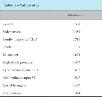
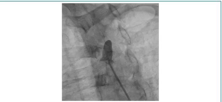
The results of our study show that in the selected population of patients who are candidates for CABG, the prevalence of atherosclerotic lesions and lesions that impair LITA is significant, and this artery is not routinely evaluated by interventional cardiology, and it is difficult to make a clinical diagnostic, since patients are usually asymptomatic, and the changes are only evidenced by selective angiography. In one patient in our study, who underwent selective angiography during catheterization, the presence of atherosclerotic lesion in LITA was evidenced, with stenosis >70% (Figure 1). In order to differentiate from a possible vasospasm, infusion of 200mcg intra-arterial nitroglycerin was performed, where the subocclusive lesion remained (Figure 2). Taking into account the small number of the sample, 39 patients underwent cardiac catheterization with indication of CABG, the result is relevant, since in this patient the graft alteration used for myocardial revascularization was chosen, excluding the use of the LITA , due to the great risk of treatment failure and increased mortality.
In 1993, Sons et al.17 demonstrated the high prevalence of
atherosclerotic lesions of LITA in patients with functional heart disease. The study analyzed 117 patients, all of whom had coronary artery disease (CAD), associated with valve abnormalities, or some other cardiac pathology. Atherosclerosis of the LITA was found in 11.1% of all patients investigated, indicating that risk factors such as the presence of peripheral arterial disease and hyperlipidemia deserve special attention in a patient with an indication for CABG, due to the high prevalence of the association of these factors with atherosclerosis of the main graft used for this surgery at the present time.
Chen et al.18 carried out a prospective study of LITA
Figure 1 – LITA with stenosis.
Figure 2 – LITA with stenosis after nitroglycerin infusion.
performed due to the high prevalence of lesions that may make it unfeasible, with possible future complications to the patient submitted to the surgical treatment.
In contrast, Perić et al.19 also analyzed by means of selective angiography the characteristics of LITA and its anatomical variations, in 80 randomly selected patients, and different from that found in the studies cited previously, no patient presented atherosclerosis in LITA. However, the degree of anatomical variations was greater, about 13.25% of
the LITAs evaluated, proving that the indication of selective angiography in all patients who would use the LITA graft for myocardial revascularization may be necessary.
The disagreement regarding the presence of atherosclerosis between the studies, and even in our study, can be attributed to the population used as the basis for
each author, since Sons et al.17 and Chen et al.18 evaluated
patients candidates for surgical correction of myocardium,
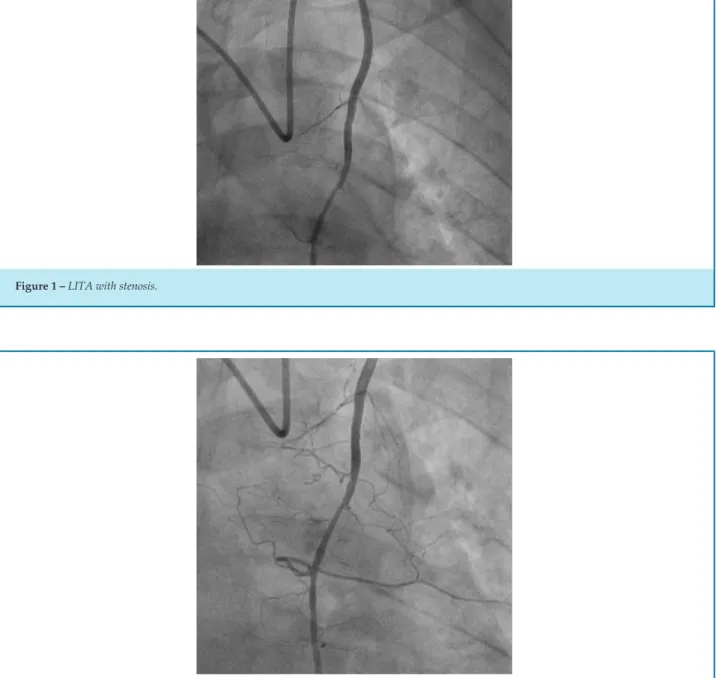
Figure 3 – LITA with presence of collateral circulation.
selected patients. All the studies present a reduced number of patients evaluated, which hinders both the homogeneity of the data and the agreement between the prevalence of the lesions. What is clear is the need to evaluate preoperative LITA in patients candidates for CABG, since in addition to diagnosing lesions that will impair the effectiveness of CABG, it is also possible to identify possible changes that cause consequences for patients , such as lower limb ischemia in cases of IAC acting as collateral circulation, and subclavian steal syndrome in patients with significant subclavian occlusion or stenosis.
The presence of chronic aortoiliac occlusive disease is considered an important predictor for the development of anatomical alterations involving LITA. Usually these patients develop collateral perfusions in order to reconstruct the arterial system of the pelvis and lower limbs, avoiding the ischemia of the same. The LITA, together with the superior and inferior epigastric arteries, work as the main parietal collateral pathway in the reconstruction of the external iliac artery, and if this graft is used for myocardial revascularization, the patient may have an acute ischemia
of the lower limbs in the post-surgery.20-25 The presence of
this collateral pathway is of such importance that the LITA becomes one of the main arteries responsible for irrigation of the lower limbs, accounting for 38% of the blood flow in the region, and doubling the volume of blood (LITA)
every minute.26 Studies show that the presence of LITA
and epigastric as a collateral route to the lower extremities is not an uncommon finding in patients with AOD, and its
prevalence is estimated in up to 12% of cases in that AOD
was greater than or equal to 75% of the vessel diameter.25
In our study, one patient presented LITA and epigastric as a collateral route for irrigation to the legs (Figure 3), alteration identified by preoperative selective angiography in patients with coronary artery bypass indication, without the presence of clinical changes which give evidence of the
existence of AOD.26 The identification of LITA as a collateral
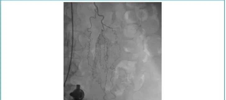
route occurred through the progression of contrast to lower vessels, upper and lower epigastric arteries (Figure 3), and continuity to the level of the pelvic vessels (Figure 4). We did not investigate alterations that might indicate the presence of AOD in the present study. However, authors
such as Kim et al.25 point out that the presence of weakened
femoral pulses in the affected extremity, with decreased amplitude and volume, and alterations in the ankle index doppler probe, less than 0.7, are important indicators of the presence of AOD. After identification of the presence of collateral circulation to the lower limbs by LITA and epigastric lesions, the presence of aortoiliac-atherosclerotic lesions at the site was demonstrated by selective aorto-iliac angiography (Figure 5).
Figure 4 – Collateral circulation of the LITA to the pelvic region.
Figure 5 – Aorta with suboclusive atherosclerotic lesions.
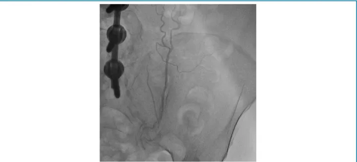
period, leading the patient to perform a myocardial revascularization surgery without effectiveness, since the blood flow of the LITA has its inverted direction, due to
subclavian occlusion.27 In addition, patients may have a
characteristic clinical presentation, due to vertebral artery flow also being directed to the subclavian, with symptoms such as dizziness, syncope and vertigo due to the ischemia generated by the deviation of the blood supply. The entire clinical picture generated by this pathophysiology is called
the subclavian steal syndrome, and these patients can be screened for both upper limb blood pressure difference, as well as for murmur survey at subclavian level, and pulse difference. Patients with total occlusion of the subclavian artery are at high risk of developing this syndrome
before and after CABG.28-30 The patient in this study was
asymptomatic when submitted to angiographic evaluation.
Figure 6 – Total occlusion of the left subclavian artery.
the predictors and the outcome. However, we consider this prevalence value to be significant, since a CABG with arterial graft failure can lead to serious complications, increasing mortality, and decreasing the effectiveness of the procedure.
Conclusion
In this study, the prevalence of atherosclerotic lesions and lesions that made the use of the LITA as an arterial graft unfeasible in patients candidates for CABG, evidenced by coronary angiography, were 7.7%. Thus, it is prudent to consider the preoperative assessment of the LITA in patients with indication for CABG, especially in the presence of clinical evidence of subclavian occlusion and AOD.
Author contributions
Conception and design of the research: Balzan HFM, Battilani RVL, Mangili OC, Franchetti M, Mangili LC, Maia JP. Acquisition of data: Balzan HFM, Battilani RVL, Moura DD, Lage BFM. Analysis and interpretation of the data: Balzan HFM, Battilani RVL, Mangili OC, Franchetti M, Mangili LC, Maia JP, Moura DD, Lage BFM. Statistical analysis: Balzan HFM, Battilani RVL, Mangili OC, Mangili LC, Maia JP. Obtaining financing:
Balzan HFM, Battilani RVL, Mangili OC, Franchetti M. Writing of the manuscript: Balzan HFM, Battilani RVL, Mangili OC, Franchetti M, Moura DD, Lage BFM. Critical revision of the manuscript for intellectual content: Balzan HFM, Battilani RVL, Mangili OC, Franchetti M, Mangili LC, Maia JP, Moura DD, Lage BFM. Supervision / as the major investigador: Balzan HFM, Battilani RVL, Mangili OC, Franchetti M.
Potential Conflict of Interest
No potential conflict of interest relevant to this article was reported.
Sources of Funding
There were no external funding sources for this study.
Study Association
This study is not associated with any thesis or dissertation work.
Ethics approval and consent to participate
1. Cesar LA, Ferreira JF, Armaganijan D, Gowdak LH, Mansur AP, Bodanese LC et al; Sociedade Brasileira de Cardiologia. Guideline for stable coronary artery disease. Arq Bras Cardiol. 2014;103(2 Suppl 2):1-56. doi: http://dx.doi.org/10.5935/abc.2014S004.
2. Hillis LD, Smith PK, Anderson JL, Bittl JA, Bridges CR, Byrne JG, et al; American College of Cardiology Foundation/American Heart Association Task Force on Practice Guidelines. 2011 ACCF/AHA guideline for coronary artery bypass graft surgery: executive summary. A report of the American College of Cardiology Foundation/American Heart Association Task Force on Practice Guidelines. J Thorac Cardiovasc Surg. 2012;143(1):4-34. doi: 10.1016/j.jtcvs.2011.10.015. Erratum in: J Thorac Cardiovasc Surg. 2012;143(5):1235.
3. The BARI Investigators. The final 10-year follow-up results from the BARI randomized trial. J Am Coll Cardiol. 2007;49(15):1600-6. doi: 10.1016/j. jacc.2006.11.048.
4. Boylan MJ, Lytle BW, Loop FD, Taylor PC, Borsh JA, Goormastic M, et al. Surgical treatment of isolated left anterior descending coronary stenosis: comparison of left internal mammary artery and venous autograft at 18 to 20 years of follow-up. J Thorac Cardiovasc Surg. 1994;107(3):657-62. PMID: 8127094.
5. Stephan WJ, O'Keefe JH Jr, Piehler JM, McCallister BD, Dahiya RS, Shimshak TM, et al. Coronary angioplasty versus repeat coronary artery bypass grafting for patients with previous bypass surgery. J Am Coll Cardiol. 1996;28(5):1140-6. doi: 10.1016/S0735-1097(96)00286-0.
6. Hess CN, Lopes RD, Gibson CM, Hager R, Wojdyla DM, Englum BR, et al. Saphenous vein graft failure after coronary artery bypass surgery: insights from PREVENT IV. Circulation. 2014;130(17):1445-51. doi: 10.1161/CIRCULATIONAHA.113.008193.
7. Shavadia J, Norris CM, Graham MM, Verma S, Ali I, Bainey KR. Symptomatic graft failure and impact on clinical outcome after coronary artery bypass grafting surgery: results from the Alberta Provincial Project for Outcome Assessment in Coronary Heart Disease registry. Am Heart J. 2015;169(6):833-40. doi: 10.1016/j.ahj.2015.02.022.
8. Caracciolo EA, Davis KB, Sopko G, Kaiser GC, Corley SD, Schaff H, et al. Comparison of surgical and medical group survival in patients with left main equivalent coronary artery disease. Long-term CASS experience. Circulation. 1995;91(9):2335-44. PMID: 7729019.
9. Dzavik V, Ghali WA, Norris C, Mitchell LB, Koshal A, Saunders LD, et al; Alberta Provincial Project for Outcome Assessment in Coronary Heart Disease (APPROACH) Investigators. Long-term survival in 11,661 patients with multivessel coronary artery disease in the era of stenting: a report from the Alberta Provincial Project for Outcome Assessment in Coronary Heart Disease (APPROACH) Investigators. Am Heart J. 2001;142(1):119-26. doi: 10.1067/mhj.2001.116072.
10. Jones RH, Kesler K, Phillips HR 3rd, Mark DB, Smith PK, Nelson CL, et al. Long-term survival benefits of coronary artery bypass grafting and percutaneous transluminal angioplasty in patients with coronary artery disease. J Thorac Cardiovasc Surg. 1996;111(5):1013-25. PMID: 8622299.
11. Smith PK, Califf RM, Tuttle RH, Shaw LK, Lee KL, Delong ER, et al. Selection of surgical or percutaneous coronary intervention provides differential longevity benefit. Ann Thorac Surg. 2006;82(4):1420-8. doi: 10.1016/j.athoracsur.2006.04.044.
12. Every NR, Fahrenbruch CE, Hallstrom AP, Weaver WD, Cobb LA. Influence of coronary bypass surgery on subsequent outcome of patients resuscitated from out of hospital cardiac arrest. J Am Coll Cardiol. 1992;19(7):1435-9. PMID: 1593036.
13. Sajja LR, Mannam G. Internal thoracic artery: anatomical and biological characteristics revisited. Asian Cardiovasc Thorac Ann. 2015;23(1):88-99. doi: 10.1177/0218492314523629.
14. Henriquez-Pino JA, Gomes WJ, Prates JC, Buffolo E. Surgical anatomy of the internal thoracic artery. Ann Thorac Surg. 1997;64(4):1041-5. PMID: 9354524.
15. Bruneton JN, Dalfin FY, Caramella E, Roux P, Héry M. Value of ultrasound in localizing the internal mammary vessels. Eur J Radiol. 1986;6(2):142-4. PMID: 3522233.
16. Harskamp, RE, Alexander JH, Ferguson TB Jr, Hager R, Mack MJ, Englum B, et al. Frequency and predictors of internal mammary artery graft failure and subsequent clinical outcomes: Insights From the Project of Ex-vivo Vein Graft Engineering via Transfection (PREVENT) IV Trial. Circulation. 2016;133(2):131-8. doi: 10.1161/CIRCULATIONAHA.115.015549.
17. Sons HJ, Godehardt E, Kunert J, Lösse B, Bircks W. Internal thoracic artery: prevalence of atherosclerotic changes. J Thorac Cardiovasc Surg. 1994;106(6):1192-5. PMID: 8246559.
18. Chen CW, Lin TK, Chen BC, Lin CT, Liu CJ, Lin CL. Preoperative semi-selective left internal mammary artery angiography: easy, safe, necessary and worthy. J Cardiovasc Surg (Torino). 2004;45(2):107-10. PMID: 15179343.
19. Perić M, Sagić D, Mirić M, Bojić M. [Angiographic characteristics of the internal thoracic artery–anatomic variations and their surgical importance]. Med Pregl. 2000;53(5-6):245-9. PMID: 11089364.
20. Melissano G, Di Credico G, Chiesa R, Grossi A. The use of internal thoracic arteries for myocardial revascularization may produce acute leg ischemia in patients with concomitant Leriche’s syndrome. J Vasc Surg 1996;24(4):698. PMID: 8911420.
21. Dietzek AM, Goldsmith J, Veith FJ, Sanchez LA, Gupta SK, Wengerter KR. Interruption of critical aortoiliac collateral circulation during nonvascular operations: a cause of acute limb-threatening ischemia. J Vasc Surg. 1990;12(6):645-51. doi: 10.1067/mva.1990.25254.
22. Kitamura S, Inoue K, Kawachi K, Morita R, Seki T, Taniguchi S, et al. Lower extremity ischemia secondary to internal thoracic-coronary artery bypass grafting. Ann Thorac Surg. 1993;56(1):157-9. PMID: 8328849.
23. Tsui SS, Parry AJ, Large SR. Leg ischaemia following bilateral internal thoracic artery and inferior epigastric artery harvesting. Eur J Cardiothorac Surg. 1995;9(4):218-20. PMID: 7605648.
24. Yapici F, Tuygun AG, Tarhan IA, Yilmaz M, Tuygun AK, Yapici N, et al. Limb ischemia due to use of internal thoracic artery in coronary bypass. Asian Cardiovasc Thorac Ann. 2002;10(3):254-5. doi: 10.1177/021849230201000315.
25. Kim J, Won JY, Park SI, Lee DY. Internal thoracic artery collateral to the external iliac artery in chronic aortoiliac occlusive disease. Korean J Radiol. 2003;4(3):179-83. doi: 10.3348/kjr.2003.4.3.179.
26. Yurdakul M, Tola M, Özdemir E, Bayazit M, Cumhur T. Internal thoracic artery-inferior epigastric artery as a collateral pathway in aortoiliac occlusive disease. J Vasc Surg. 2006;43(4):707-13. doi: 10.1016/j.jvs.2005.12.042.
27. Gomes V, Roman M, Barcellos C, Lasevitch R, Hickmann P, Alcalde R, et al. Prevalence of stenosis of the left subclavian artery in CABG candidates: a multicenter registry. Rev Bras Cardiol Invas. 2008;16(3):307-11. doi: http://dx.doi.org/10.1590/S2179-83972008000300011.
28. Schatzl S, Karnik R, Gattermeier M. Coronary subclavian steal syndrome: an extracoronary cause of acute coronary syndrome. Wien Klin Wochenschr. 2013;125(15-16):437-8. doi: 10.1007/s00508-013-0386-3.
29. Rogers JH, Calhoun RF 2nd. Diagnosis and management of subclavian artery stenosis prior to coronary artery bypass grafting in the current era. J Card Surg. 2007;22(1):20-5. doi: 10.1111/j.1540-8191.2007.00332.x.
30. Kargiotis O, Siahos S, Safouris A, Feleskouras A, Magoufis G, Tsivgoulis G. Subclavian steal syndrome with or without arterial stenosis: a review. J Neuroimaging. 2016;26(5):473-80. doi: doi: 10.1111/jon.12371.