DM
November | 2017
José Filipe Sousa de Olim
MASTER IN APPLIED BIOCHEMISTRYPreliminary Studies
on the Use of Exosomes
as Dendrimer Carriers
MASTER DISSERTATION
José Filipe Sousa de Olim
MASTER IN APPLIED BIOCHEMISTRYon the Use of Exosomes
as Dendrimer Carriers
MASTER DISSERTATION
ORIENTADORA
Helena Maria Pires Gaspar Tomás
CO-ORIENTADOR
Preliminary studies on the use
of exosomes as dendrimer
carriers
Dissertation submitted to the University of Madeira in fulfilment of the requirements for the degree of Master in Applied Biochemistry
By José Filipe Sousa de Olim
Work developed under the supervision of Professor Dr. Helena Maria Pires Gaspar Tomás and Co-supervised by Professor Dr. João Manuel Cunha Rodrigues
Faculdade de Ciências Exatas e de Engenharia, Centro de Química da Madeira,
Campus Universitário da Penteada,
Funchal – Portugal November 2017
Acknowledgments
First, I would like to thank the person who contributed the most to the success of this body of work, my supervisor Prof. Helena Tomás, for her advice, her words of support, her help and patience through the making of this thesis. Also, for being one of the best people I’ve met, personally and professionally. I would also like to thank my co-supervisor Prof. João Rodrigues and other professors who contributed to my academic development.
Thank you the LBCC members who contributed to the elaboration of this Master Thesis, specially to Ana Olival and Carla Alves, for the support, help and guidance in some experiments.
A special thank you to my colleague and good friend Débora Reis, for the help, the laughs, the irreplaceable friendship and all the support given throughout these years. I also want to thank all my CQM colleagues who became my dearest friends and who I will forever cherish: Ana Rute, Bruna Pereira, Carla Miguel, Catarina Silva, Gonçalo Martins, Mariana Vieira, Núria Fernandes, Rosa Perestrelo, Sonia Medina and Vítor Spínola.
I’m also deeply thankful to the laboratory technician Paula Andrade, for her support, her disponibility in providing the necessary tools for the elaboration of this work, and also for her friendship.
I’m extremely grateful for the support and love given by my family, specially my mother, for all the efforts and sacrifices she had to make to give me a better education and providing me with the tools for me to build a better future.
Last but not least, I’d like to thank my amazing boyfriend Alex Faria, for his endless love, support and for providing me with the best moments in my life. He inspires me to become a better person each and every day. Without him, things would be for sure, harder to overcome. I love you.
Finally, I want to thank CQM for providing me the tools and support in the development of this Master Project.
This work was supported by FCT-Fundação para a Ciência e a Tecnologia (project Pest-OE/QUI/UI0674/2013, CQM, Portuguese Government funds), and Associação Regional
para o Desenvolvimento da Investigação Tecnologia e Inovação (ARDITI) through the
project M1420-01-0145-FEDER-000005 - Centro de Química da Madeira - CQM+ (Madeira 14-20).
Preliminary notes
This Master Thesis was the first research work at CQM (Centro de Química da Madeira) exploring the use of extracellular vesicles as nanocarriers. Although many questions remain to be answered due to the complexity of this field and the limited time available for the experimental work, this work allowed the gain of experience in the isolation and characterization of extracellular vesicles and, certainly, will serve as a basis for the development of future studies in CQM. For the student, it was an opportunity to learn how to access and discuss scientific data, design a research project, interpret experimental results and communicate in a scientific manner. Regarding the laboratory skills, it was also a chance for him to learn basic animal cell culture techniques, how to label dendrimers with fluorescent dyes, and how to characterize nanomaterials with techniques such as NMR, FTIR-ATR, UV/Vis, DLS and fluorescence spectroscopy. The student also gained experience using microscopy techniques, such as fluorescence microscopy and TEM, as well as using biological/biochemical techniques, like cell viability evaluation, organelle staining, and enzyme activity determination.
Presentations in scientific meetings in the scope of this master project:
1) Filipe Olim, João Rodrigues, Helena Tomás. Nanomaterials inclusion in exosomes for drug delivery. Oral presentation in the 4th CQM Annual Meeting.
3-4 February 2017; Funchal, Madeira (Portugal).
Courses done in the scope of this master project:
1) Basics of Extracellular Vesicles online course provided by University of California, Irvine, and coordinated by Cecilia Lässer, Researcher at Krefting Research Center, University of Gothenburg. Approved with a final classification of 98.9 %. The respective certificate is in Annex C.
Abstract
Eukaryotic and prokaryotic cells can release vesicles into the biological fluids that differ in size and mechanism of biogenesis. In recent years, particular attention has been paid to exosomes which are naturally occurring nanoparticles (50 – 150 nm) that have been found in various biological fluids and are recognized as having an important role in intercellular communication and other biological processes. Human mesenchymal stem cells (hMSCs) are multipotent cells that can be found in different tissues in the adult. hMSCs have a large capacity for ex vivo expansion and show immunosuppressive properties, making them the ideal source of exosomes for biomedical applications.
Dendrimers are nanoscale molecules that have promising properties as drug/gene delivery vehicles and, as such, their inclusion inside hMSCs exosomes may be a way for their easier spreading and target reaching in the body. The present work was focused on the hypothesis that exosomes can be loaded before their isolation from cells by simple cell exposure to dendrimers. First, dendrimers were labelled with rhodamine and then characterized by 1H NMR, FTIR, UV/Vis spectroscopy, and
fluorescence spectroscopy. Then, the effect of the pH in the stability of the fluorescence emission of these conjugates was study, as well as their cytotoxicity behavior and kinetics of cellular internalization. After, a protocol for exosome isolation was established using a precipitation-based approach and the isolated exosomes were characterized by DLS, TEM and acetylcholinesterase activity. hMSCs were then exposed to a solution containing the labelled dendrimers and the released exosomes were collected to evaluate the presence of the dendrimers inside. Contrary to what was expected, the dendrimers were not excreted inside exosomes and, instead, were accumulated in the cellular perinuclear zone. By fluorescence microscopy and using specific biochemical markers, it was possible to co-localize the conjugated dendrimers with the Golgi complex and the endoplasmic reticulum.
Resumo
As células eucariotas e procarióticas podem libertar vesículas nos fluidos biológicos que diferem no tamanho e mecanismo de biogénese. Os exossomas, nanopartículas de ocorrência natural (50 - 150 nm), podem ser encontrados em diversos fluidos biológicos e possuem um papel importante na comunicação intercelular e noutros processos biológicos. As células estaminais mesenquimais humanas (hMSCs) são células multipotentes que podem ser encontradas em diferentes tecidos adultos, que possuem uma larga expansão ex vivo e propriedades imunossupressoras.
Os dendrímeros são moléculas com tamanhos nanométricos que possuem propriedades promissoras para a entrega de fármacos/genes e, como tal, a sua inclusão dentro de exossomas derivados de hMSC poderá representar uma maneira de facilitar a sua circulação no corpo de modo a atingirem os alvos biológicos. O presente trabalho focou-se na hipótese de que os exossomas podem ser carregados por simples exposição das células às soluções de dendrímero. Primeiro, os dendrímeros foram marcados com rodamina e caracterizados utilizando 1H RMN, FTIR, espectroscopia
UV/Vis e espectroscopia de fluorescência. Depois, estudou-se o efeito do pH na
estabilidade da emissão de fluorescência desses conjugados, bem como o seu comportamento de citotoxicidade e cinética de internalização celular. Seguidamente, os exossomas foram isolados por precipitação através da utilização de um reagente comercial e caracterizados por DLS, TEM e pela atividade da acetilcolinesterase. As hMSCs foram então expostas a uma solução contendo os dendrímeros e os exossomas libertados foram colhidos e avaliados de maneira a determinar a presença de dendrímeros no seu interior. Ao contrário do que era esperado, os dendrímeros não foram excretados dentro dos exossomas e, em vez disso, ficaram acumulados na região perinuclear das células. Por microscopia de fluorescência e utilizando marcadores bioquímicos específicos, foi possível co-localizar os dendrímeros conjugados com o complexo de Golgi e o retículo endoplasmático.
Index
1. Introduction ... 29
1.1. Extracellular vesicles (EVs) ... 29
1.1.1. Apoptotic bodies ... 30
1.1.2. Ectosomes or microvesicles ... 31
1.1.3. Exosomes ... 32
1.1.3.1. Origin and molecular composition of exosomes ... 33
1.1.3.2. Biological functions of exosomes ... 35
1.1.4. International Society for Extracellular Vesicles position in the definition of extracellular vesicles ... 36
1.2. Exosome isolation methods ... 38
1.2.1. Ultracentrifugation-based isolation techniques ... 38
1.2.2. Precipitation-based approaches ... 39
1.2.3. Immunoaffinity capture-based techniques ... 40
1.2.4. Microfluidics-based isolation techniques ... 40
1.3. Characterization of extracellular vesicles ... 42
1.3.1. Physical characterization of EVs ... 42
1.3.1.1. Dynamic light scattering ... 42
1.3.1.2. Nanoparticle tracking analysis ... 43
1.3.1.3. Tunable resistive pulse sensing ... 44
1.3.1.4. Flow cytometry ... 45
1.3.1.5. Atomic force microscopy ... 46
1.3.1.6. Electron microscopy ... 47
1.3.2. Chemical/Biochemical characterization of EVs ... 47
1.4. Mesenchymal stem cells (MSCs) as the source of exosomes ... 48
1.4.1. Background of mesenchymal stem cells ... 48
1.4.2. Clinical and therapeutic applications of MSCs ... 49
1.4.3. Advantages of using MSCs as exosome producers for drug delivery ... 50
1.5. Methods for the loading of therapeutic cargo into exosomes ... 51
1.5.1. Exosome loading methods after EV isolation ... 52
1.5.1.1. Electroporation ... 52
1.5.1.2. Simple incubation ... 53
1.5.1.4. Other methods ... 54
1.5.2. Exosome loading methods before EV isolation ... 55
1.5.2.1. Cell exposure to the therapeutic cargo ... 55
1.5.2.2. Transfection of exosome producing cells ... 55
1.6. Dendrimers as nanocarriers for drug delivery ... 56
1.6.1. Characteristics and molecular structure of PAMAM dendrimers... 56
1.6.2. Biomedical applications of PAMAM dendrimers ... 59
1.7. Thesis objectives ... 62
2. Materials and methods ... 67
2.1. Synthesis and characterization of PAMAM(NH2)-RITC conjugates ... 67
2.1.1. Materials ... 67
2.1.2. Preparation and characterization of PAMAM(NH2)-RITC conjugates ... 67
2.1.3. Effect of the pH in the photostability of PAMAM(NH2)-RITC conjugates ... 68
2.2. Cytotoxicity studies of PAMAM(NH2)-RITC conjugates ... 69
2.2.1. Cell culture conditions for cytotoxicity analysis ... 69
2.2.2. Effect on cell viability of PAMAM(NH2)-RITC conjugates ... 69
2.3. Kinetics of cell uptake of PAMAM(NH2)-RITC conjugates ... 70
2.4. Establishment of an exosome isolation protocol ... 71
2.4.1. Definition of the protocol ... 71
2.4.2. Effect of storage temperature on the stability of exosome solutions ... 72
2.4.3. Exosome characterization ... 73
2.4.3.1. Dynamic Light Scattering (DLS) ... 73
2.4.3.2. Transmission Electron Microscopy (TEM) ... 73
2.4.3.3. Acetylcholinesterase (AChE) activity ... 73
2.5. Exosome loading with PAMAM(NH2)-RITC conjugates ... 74
2.6. Intracellular distribution of PAMAM(NH2)-RITC conjugates ... 75
3. Results and discussion ... 79
3.1. Synthesis and characterization of PAMAM(NH2)-RITC conjugates ... 79
3.2. Effect of the pH in the fluorescence intensity of PAMAM(NH2)-RITC conjugates ... 84
3.3. Cytotoxicity studies of PAMAM(NH2)-RITC conjugates ... 86
3.5. Establishment of an exosome isolation protocol and exosome
characterization ... 92
3.6. Exosome loading with PAMAM(NH2)-RITC conjugates ... 98
3.7. Intracellular distribution of PAMAM(NH2)-RITC conjugates ... 101
Conclusions and Future Work ... 109
References ... 115
Figures index
Figure 1 – Origin of the different types of EVs. ... 29
Figure 2 – The formation of apoptotic bodies during programmed cell death (apoptosis) and their phagocythosis. ... 30
Figure 3 – The process of microvesicle/ectosome biogenesis. ... 32
Figure 4 – Biogenesis of exosomes. ... 33
Figure 5 – Biochemical composition of exosomes. ... 35
Figure 6 – The role of exosomes in different tissues and their possible therapeutic application.. ... 36
Figure 7 – Experimental design of the ExoChip. ... 41
Figure 8 – Representation of the nanoparticle tracking analysis (NTA) technique. ... 43
Figure 9 – Tunable resistive pulse sensing ... 44
Figure 10 – Atomic force microscopy imaging of extracellular vesicles... 46
Figure 11- The differentiation capacity of MSCs. ... 49
Figure 12 – Paracrine factors release by mesenchymal stem cells which may play an important role in mitogenesis, angiogenesis, apoptosis and scarring. ... 50
Figure 13 – Advantages of using mesenchymal stem cells as a source of exosomes. 51 Figure 14 – Scheme with the different strategies for exosome loading. ... 53
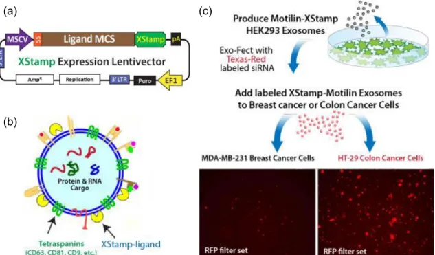
Figure 15 – XStampTM technology commercialized by System Biosciences company. ... 56
Figure 16 – Representation of the general structure of dendrimers. ... 57
Figure 17 – Molecular structure of PAMAM dendrimers. ... 58
Figure 18 – Gene delivery mediated by dendrimers. ... 60
Figure 19 – Conjugation conditions for the preparation of G4 PAMAM(NH2)-RITC conjugates (1:1.5 PAMAM:RITC molar ratio). ... 68
Figure 20 - Scheme for testing the influence of filtration and concentration steps in the isolation of exosomes. ... 72
Figure 21 – Representation of the reaction between the primary amines in the PAMAM dendrimer and the electrophilic carbon of the RITC isothiocyanate group. ... 80
Figure 22 – The 1H NMR spectrum of the PAMAM(NH 2)-RITC conjugate in D2O (400 MHz). ... 81
Figure 23 – FTIR-ATR spectra of (a) RITC molecule, (b) PAMAM(NH2) G4 dendrimer and (c) PAMAM(NH2)-RITC conjugate. ... 82
Figure 25 – Emission spectra for the PAMAM(NH2)-RITC conjugate and the free RITC.
... 84
Figure 26 – Normal acidification of endosomes during their maturation to late
endosomes. ... 84
Figure 27 – Molecular structures of the three Rhodamine B forms. ... 85 Figure 28 – Effect of pH change on the fluorescence intensity of the prepared
PAMAM(NH2)-RITC conjugates. ... 86
Figure 29– Cytotoxic behaviour of generation 4 PAMAM(NH2) dendrimers over hMSCs.
... 87
Figure 30 – Cytotoxic behaviour of PAMAM(NH2)-RITC conjugates over hMSCs. ... 87
Figure 31 – Kinetics of cell uptake of PAMAM(NH2)-RITC. ... 89
Figure 32 – Fluorescence microscopy images of hMSCs exposed to PAMAM(NH2)-RITC
conjugates during different times. ... 91
Figure 33 - Effect of the introduction of filtration and concentration steps in the protocol
for exosome isolation from NIH 3T3 cells. ... 94
Figure 34 – Schematized protocol of the established exosome isolation protocol. ... 95 Figure 35 – Effect of storage temperature in the hydrodynamic size (Z-average) of the
exosome solutions. ... 96
Figure 36 – Characterization of exosomes derived from hMSCs by DLS. ... 96 Figure 37 – Transmission electron microscopy images of a negative stained hMSCs
exosome solution. ... 97
Figure 38 – Acetylcholinesterase assay of the exosome suspension obtained from
hMSCs cells cell culture medium. ... 98
Figure 39 – Fluorescence microscopy images of the PAMAM(NH2)-RITC conjugates
inside microvesicles. ... 99
Figure 40 – Scheme showing the cellular excretion processes of nanoparticles along
with their intracellular trafficking and endocytosis mechanisms. ... 100
Figure 41 – Intracellular transport of nanoparticles. ... 101 Figure 42 – Illustration of the proton sponge effect, leading to endosome and lysosome
escape. ... 102
Figure 43 – Co-localization and intracellular distribution of the PAMAM(NH2)-RITC
Tables index
Table 1 – Different categories of proteins and their expected presence in EVs isolates,
Abbreviations
AA Antibiotic-Antimycotic AChE Acetylcholinesterase ADP Adenosine diphosphate AFM Atomic force microscopy ARF6 ADP-ribosylation factor 6 ATPase Adenylpyrophosphatase BSA Bovine serum albumin C3b complement component 3 c-Met Tyrosine-protein kinase Met CT Computed tomography
DAPI 4',6-diamidino-2-phenylindole DLS Dynamic light scattering
DMEM Dulbecco’s modified eagle medium DMSO Dimethyl sulfoxide
DNA Deoxyribonucleic acid EDA Ethylenediamine
EDS Energy dispersive spectroscopy EDTA Ethylenediaminetetraacetic acid EF-1 Elongation factor 1
ELISA Enzyme-linked immunosorbent assay EM Electron microscopy
ESCRT Endosomal sorting complexes required for transport EVs Extracellular vesicles
FBS Fetal bovine serum
FITC Fluorescein isothiocyanate
FTIR-ATR Attenuated total reflectance Fourier-transform infrared spectroscopy GTP Guanosine triphosphate
HDL High density lipoprotein
hMSCs Human mesenchymal stem cells HPA Helix pomatia agglutinin
HPLC High-performance liquid chromatography Hsp70 Heat shock protein 70
ILVs Intraluminal vesicles iRNA Interference RNA
ISEV International Society for Extracellular Vesicles KRAS Kirsten rat sarcoma viral oncogene homolog LAMP2 Lysosome-associated membrane protein 2 LDL Low density lipoprotein
MEM Minimum essential medium
MFG-E8 Milk fat globule-EGF factor 8 protein MHC Major histocompatibility complex
miRNA MicroRNA
MRI Magnetic resonance imaging mRNA Messenger RNA
MSCs Mesenchymal stem cells MVBs Multivesicular bodies MWCO Molecular weight cut-off NMR Nuclear magnetic resonance NTA Nanoparticle tracking analysis PAMAM Poly(amidoamine)
PBS Phosphate buffered saline PdI Polydispersity index
PEG Polyethylene glycol
PET Positron-emission tomography PLGA Poly(lactic-co-glycolic acid) PVDF Polyninylidene difluoride RFU Relative fluorescence units RIPA Radioimmunoprecipitation assay RITC Rhodamine B isothiocyanate RNA Ribonucleic acid
RSD Relative standard deviation
SDS-PAGE Sodium dodecyl sulfate polyacrylamide gel electrophoresis SEM Scanning electron microscopy
siRNA Small interfering RNA T-cell T lymphocyte
TEM Transmission electron microscopy TRPS Tunable resistive pulse sensing
TSG101 Tumor susceptibility gene 101 protein VAMP3 Vesicle-associated membrane protein 3 VPS4 Vacuolar protein sorting-associated protein 4
Part 1. Introduction
Contents
1. Introduction
1.1. Extracellular vesicles (EVs)
1.1.1. Apoptotic bodies
1.1.2. Ectosomes or microvesicles 1.1.3. Exosomes
1.1.3.1. Origin and molecular composition of exosomes
1.1.3.2. Biological functions of exosomes 1.1.4. International Society for Extracellular Vesicles position in the definition of extracellular vesicles
1.2. Exosome isolation methods
1.2.1. Ultracentrifugation-based isolation techniques 1.2.2. Precipitation-based approaches
1.2.3. Immunoaffinity capture-based techniques 1.2.4. Microfluidics-based isolation techniques
1.3. Characterization of extracellular vesicles
1.3.1. Physical characterization of EVs 1.3.1.1. Dynamic light scattering 1.3.1.2. Nanoparticle tracking analysis 1.3.1.3. Tunable resistive pulse sensing 1.3.1.4. Flow cytometry
1.3.1.5. Atomic force microscopy 1.3.1.6. Electron microscopy
1.3.2. Chemical/Biochemical characterization of EVs
1.4. Mesenchymal stem cells (MSCs) as the source of exosomes
1.4.1. Background of mesenchymal stem cells 1.4.2. Clinical and therapeutic applications of MSCs 1.4.3. Advantages of using MSCs as exosome producers for drug delivery
1.5. Methods for the loading of therapeutic cargo in exosomes
1.5.1. Exosome loading methods after EV isolation 1.5.1.1. Electroporation
1.5.1.2. Simple incubation
1.5.1.3. Use of transfection agents 1.5.1.4. Other methods
1.5.2. Exosome loading methods before EV isolation 1.5.2.1. Cell exposure to the therapeutic cargo 1.5.2.2. Transfection of exosome-producing cells
1.6. Dendrimers as nanocarriers for drug delivery
1.6.1. Characteristics and molecular structure of PAMAM dendrimers
1.6.2. Biomedical applications of PAMAM dendrimers
1. Introduction
1.1. Extracellular vesicles (EVs)
Intercellular communication is a crucial and important aspect in multicellular organisms. Communication among cells can occur by direct contact, through adhesion molecules and gap junctions, or by mediation of soluble factors, such as cytokines, growth factors and hormones [1]. In the last two decades, a third mechanism is being elucidated, consisting of the intercellular transfer of extracellular vesicles (EVs), at short and long distances [2,3]. These membrane-based structures serve as vehicles to carry different types of cellular cargo to recipient cells, like lipids, proteins and nucleic acids [4].
The term “extracellular vesicle” was recently introduced and is being used for the description of any type of membrane vesicle secreted into the extracellular milieu, independently of their biogenesis and composition. All cells produce EVs, including those of prokaryotic organisms [5]. EVs have been traditionally classified based on their cell or tissue of origin. For example, prostasomes are derived from prostate cells and oncosomes from tumour cells [1]. These EVs can be distinguished based on their size, mechanism of biogenesis, buoyant density, and so on. A recent classification of EVs based on their biogenesis pathway or intracellular origin led to three different subsets of these structures: apoptotic bodies, ectosomes (or microvesicles) and exosomes [6], which are represented in Figure 1.
Figure 1 – Origin of the different types of EVs. Apoptotic bodies are formed in the late stage of programmed
cell death (apoptosis). Ectosomes (or microvesicles) are formed through the outward shedding of the plasma membrane. Exosomes are released upon the fusion of multivesicular bodies with the cell membrane. Figure from reference [7]. Ectosomes (100-2000 nm) Apoptotic bodies (1-4 µm) Exosomes (50-150 nm)
1.1.1. Apoptotic bodies
Apoptosis is the process of cell death and may occur in both normal and cancer cells. This mechanism has different stages, starting by nuclear chromatin condensation, followed by membrane blebbing and finishing in the incorporation of cellular content into membrane vesicles called apoptotic bodies or apoptosomes [8]. Apoptotic bodies are generally the biggest EVs, with sizes around 500 – 4000 nm, and are characterized by having organelles inside. However, smaller vesicles (50 – 500 nm) can be released in this process too [9].
Normally, most of the apoptotic bodies are phagocytosed by macrophages and locally cleared upon their formation (Figure 2). This process is mediated by the establishment of specific interactions between receptors in the phagocyte membrane and ligands at the apoptotic body membrane. One of the best characterized events in the apoptosis mechanism is the translocation of phosphatidylserine to the outer part of the lipid membrane. When phosphatidylserines are translocated, they can bind to Annexin V, and therefore be recognized by phagocytes [10]. Other well characterized change in the apoptotic process is the oxidation of proteins localized at the membrane surface. These oxidation sites can bind to thrombospondin and C3b, and thus be equally recognized by phagocytes [11]
Figure 2 – The formation of apoptotic bodies during programmed cell death (apoptosis) and their
phagocythosis. Tsp: Trombospondin; C3b: Large element formed by the cleavage of complement component 3. Figure adapted from reference [12].
1.1.2. Ectosomes or microvesicles
Microvesicles, also known as ectosomes, result from direct shedding of the cell membrane (13). Relative to exosomes, microvesicles normally have a bigger size, around 50-2000 nm. Although there is an overlap between the size range of these two types of EVs, their main difference is the underlying biogenesis mechanism.[13].
The formation process of microvesicles results from different factors, such as cytoskeletal protein contraction and phospholipid redistribution. The cell membrane has a heterogenous distribution of proteins and phospholipids, forming micro-domains. This heterogeneity is regulated by the aminophospholipid translocase. This enzyme, also known as flippase, is responsible for the transportation of phospholipids from the outer to the inner side of the cell membrane, whereas floppases transfer phospholipids from the inner to the outer side of the cell membrane. It is reported that membrane shedding/microvesicle formation is induced by the translocation of phosphatidylserine to the outer side of the cell membrane [14] and that the shedding process is completed through contractions of cytoskeletal structures resulting from actin-myosin interactions [15].
The content of microvesicles seems to be enriched in a set of proteins. For example, these melanoma microvesicles were enriched in B1 integrin receptors and other membrane proteins, such as the vesicle-associated membrane protein 3 (VAMP3) [15]. However, the transferrin receptors, highly present in exosomes, do not seem to appear in microvesicles [16].
Figure 3 – The process of microvesicle/ectosome biogenesis. In a melanoma model, the overexpression of
the Guanosine triphosphate (GTP)-binding protein Adenosine diphosphate (ADP)-ribosylation factor 6 (ARF6), a Rho family member, resulted in microvesicle secretion increase. The activated ARF6 starts a signal cascade, first with phospholipase D (PLD) activation and finishing by phosphorylation and activation of the myosin light chain, like showed in Figure 3. These signalling cascade events do not alter the formation and secretion of smaller vesicles, like exosomes, which supports the fact that the biogenesis of microvesicles is distinct from that of exosomes; ERK: Extracellular signal-regulated signal; MLCK: Myosin light-chain kinase. Figure adapted from reference [12].
1.1.3. Exosomes
In 1983, Harding et al followed the cell uptake of gold-labelled transferrin through a clathrin-mediated endocytosis process in rat reticulocytes. They reported that the gold-labelled transferrin accumulated inside vesicles which existed inside non-lysosomal endosomes referred as multivesicular bodies. These multivesicular bodies then fused with the plasma membrane and released their inner vesicles by exocytosis [17]. The term exosome was introduced by Johnstone et al in 1987, to refer to these released vesicles [18]. They observed that during the process of reticulocyte maturation, there were large sacs inside the cells containing small vesicles (exosomes). Then, they also labelled the transferrin receptor with gold nanoparticles, and observed the fusion of the large sacs with the cell membrane and subsequent release of the small vesicles into the extracellular environment [19].
1.1.3.1. Origin and molecular composition of exosomes
Exosomes have been discovered in a variety of supernatants from different cell lines in culture, and are present in all the human fluids, which suggests that all cells have the potential to produce them [20]. Exosomes are nano-sized vesicles with a diameter of 50-150 nm, formed by the endocytic cellular pathway which can be separated into three stages: (i) the first stage corresponds to plasma membrane invagination to form the endocytic vesicles; (ii) in the second stage, the inward budding of endosomal membrane starts giving rise to multivesicular bodies (MVBs); (iii) in the last and third stage, MVBs fuse with the plasma membrane and release their vesicular content (exosomes) [21]. These steps are shown in Figure 4.
As mentioned before, endocytic vesicles are created from the invagination of the plasma membrane, creating what are called “endosomes”, which mature and become late endosomes. Then, through the inward budding of these late endosomes, vesicles are accumulated in the endosomal lumen. The accumulation of these vesicles inside late endosomes leads to the formation of multivesicular bodies (MVBs). The MVBs can either fuse with lysosomes for degradation of their content or fuse with the plasma membrane, releasing their intraluminal vesicles (ILVs) in the extracellular space, where they become known as exosomes The main difference between these pathways relies on the action of protein sorting complexes known as endosomal sorting complexes required for transport (ESCRT) [22].
The ESCRT machinery consists of four multi-protein complexes, namely ESCRT-0, ESCRT-I, ESCRT-II and ESCRT-III. These complexes are recruited to sort a set of specific proteins into ILVs. The process is known to require ubiquitination of the cytosolic tail of endocytosed receptors [23]. These cargos constitute proteins that will be incorporated into ILVs and later become part of the exosomes released. For example, the protein codified by the tumour susceptibility gene 101 (TSG101), a component of ESCRT-I, forms a complex with the ubiquitinated cargo protein, and helps in the activation of ESCRT-II, inducing endosomal membrane budding. This complex is then involved in the sequestration of MVB proteins and the recruitment of the deubiquitination enzyme to remove ubiquitin from the cargo proteins before sorting them into the ILVs. Then, at last, the ESCRT-III is disassembled by the vacuolar protein sorting-associated protein 4 (VPS4) adenosine triphosphatase (VPS4 is an ATPase) [24].
The nano-spherical membrane of exosomes is formed by a bilayer of lipids. The membrane contains a set of diverse types of lipids and proteins (Figure 5), which are related to the parent cells from which exosomes are released. According to ExoCarta, an exosome database, there are currently 8000 proteins and 194 lipids known to be associated with exosomes [25].
Since exosomes are originated from endosomes, they contain multiple families of proteins, such as tetraspanins (CD63, CD81, CD9), heat shock proteins (Hsp70), lysosomal proteins (Lamp2b), and fusion proteins (CD9, flotillin, Annexin), as shown in Figure 5. Tetraspanins have received a high attention, because of the use of the trinity CD63, CD81 and CD9 as exosome markers. However, as mentioned before, the existence of a single exosome-specific protein remains to be disclosed [26]. Besides proteins and lipids, exosomes also contain biologically active nucleic acids, especially ribonucleic acid (RNA), demonstrating their ability to mediate the horizontal transfer of genetic material. The existence of two types of RNA inside exosomes, specifically messenger RNA (mRNA) and micro RNA (miRNA), has also been demonstrated [27].
Figure 5 – Biochemical composition of exosomes. Figure adapted from reference [28].
1.1.3.2. Biological functions of exosomes
Exosomes circulate in the body through different biofluids, such as saliva, serum, urine, blood, synovial fluid and breast milk, which suggests that these EVs play an important role in the intercellular communication and biological responses. There are some examples of the biological roles that exosomes can play in the body, like the removal of unnecessary proteins from cell maturation processes. One factor that affects the biological response of exosomes is their cellular origin. Some studies demonstrated that exosomes secreted from antigen presenting cells can have the ability to express, and have at their surface, both the major histocompatibility complex (MHC) I and II. These complexes help in the activation of CD8+ and CD4+ T-cells to trigger specific
immune responses [29]. Besides the delivery and exchange of proteins and lipids among different cells, exosomes can transport some types of nucleic acids, as mentioned before. For example, there are studies reporting the secretion of mRNA and small RNA by mast cells in exosomes, which were then translated in the recipient cells [30].
Besides their role in normal biological responses and processes, exosomes seem to be involved in many diseases. For example, in cancer, it seems that exosomes can induce or facilitate the development of a tumoral microenvironment with the transfer of mRNA and some proteins to distant cells. One example is the mutant Kirsten rat sarcoma
(kRAS) viral oncogene protein and the c-Met oncoprotein that promote tumour cell proliferation, thrombosis and angiogenesis [31,32].
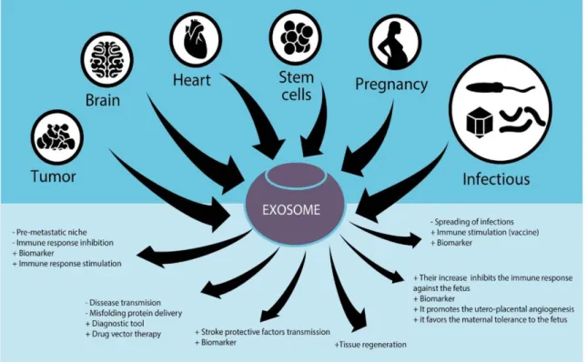
Like it was mentioned before, exosomes can be isolated from a variety of cells, and given the fact that these EVs participate in intercellular communication and in the maintenance of normal and pathological conditions, there are several studies regarding their use for diagnostic and therapeutic purposes. For example, exosomes were used to detect tumours in patients with ovarian, prostate and breast cancer [33–35]. Some of these applications and therapeutic effects are schematized in Figure 6. Regarding the use of exosomes in therapeutics, these vesicles can induce tissue regeneration with the delivery of lipids, proteins, growth factors and nucleic acids, for example [36].
Figure 6 – The role of exosomes in different tissues and their possible therapeutic applications. Figure
adapted from reference [29].
1.1.4. International Society for Extracellular Vesicles position in the definition of extracellular vesicles
The extracellular milieu, like serum and plasma, may be very complex and contain a diversity of non-vesicular materials such as extracellular RNA, protein complexes and lipoproteins. Separation of these non-vesicular materials from EVs is not fully achieved by common isolation protocols, like ultracentrifugation and commercial precipitation kits.
Consequently, there is a need to determine the distinct contributions of EVs in any experiment that describes the molecular content or the functional role of the isolated material. So, the ISEV (International Society for Extracellular Vesicles) has adopted a set of requirements that researchers can use to discriminate EV from non-EV components [37]. One of the first requirements to define EVs is that they should be isolated from extracellular fluids, like conditioned cell culture medium or body fluids. Here, an important aspect is that the collection of EV-containing fluid must be gentle, limiting cell disruption, which may contribute to the isolation of vesicles from the intracellular compartments. The second requirement regards to the characterization of these EVs is that a general overview of their protein composition must be provided, including description or quantification of necessarily expected proteins, like those shown in Table 1. ISEV suggested that investigators should report at least 3 or more proteins (and, at least, in a semi-quantitative manner) expected to be present in the EVs of interest, like transmembrane proteins and proteins from the cytosol that were linked to the membrane (Table 1, groups 1 and 2, respectively). The amount of proteins not expected to be present in EVs should also be determined, such those associated with cellular structures different from the cell or endosome membranes (Table 1, group 3). Furthermore, protein isolates should be compared with those described for other EVs, by searching within databases, like EVpedia and Vesiclepedia. The presence of extracellular proteins (Table 1, group 4) in EVs preparations should be regarded very carefully. For example, acetylcholinesterase (AChE) has been used as a EV marker but ISEV requires confirmation of the presence of this protein by other techniques such as Western blotting or by performing assays where inhibitors of AChE are used. The third and last requirement for the definition of EVs, is the characterization of single vesicles within a mixture to get information on the heterogeneity of the EV preparation. As a rule, at least 2 different technologies should be used to characterize individual EVs. When using electron microscopy (EM) or atomic force microscopy (AFM), images should show a wide field (so that several vesicles can be observed) and high-magnification images of individual vesicles. Size distribution measurement of EVs obtained, for example, by nanoparticle-tracking analysis, dynamic light scattering or resistive pulse sensing, may also provide valuable information, representative of the whole EV population. However, the results acquired using these techniques should be compared with data from transmission EM (TEM), AFM or other microscopy techniques, since they are not able to distinguish between vesicles and other particles of similar size [37].
Table 1 – Different proteins expected in EVs isolates, including some examples (non-exclusive). Adapted
from reference [37].
1.2. Exosome isolation methods
The techniques used in the isolation of exosomes should have a high efficiency, independently of the sample source. After, the quality of the exosome suspensions should be analysed by several physical and chemical/biochemical techniques [38]. With the fast evolution of science and technology, different techniques have been developed for the isolation of exosomes that are able to provide a high quantity of these vesicles with a elevated degree of purity. Each technique has a set of unique advantages and disadvantages for exosome isolation, and these will be discussed in the next sections.
1.2.1. Ultracentrifugation-based isolation techniques
When a suspension is submitted to a centrifugal force, the particles in the suspension will sediment according to their physical properties, such as the size, density and shape. The sedimentation process will also depend on the applied centrifugal force, as well as on solvent density and viscosity. Normally, in ultracentrifugation processes, one can reach high centrifugal forces, up to 1 000 000 x g. The ultracentrifugation-based approaches are the gold standard for exosome isolation, accounting for 56 % of the exosome isolation reports [39]. Exosomes can be isolated by preparative
ultracentrifugation. There are two types of preparative ultracentrifugation, the differential ultracentrifugation and the density gradient ultracentrifugation.
The differential ultracentrifugation is based on the use of different centrifugation cycles at different centrifugal forces and periods of time to isolate exosomes from other components in a sample. Before exosome isolation, normally a clean-up step is carried out to remove large particles from plasma or serum [40]. Between the centrifugation cycles, and depending on the centrifugal force used, the pellet or the supernatant is resuspended in an isotonic solution, such as Phosphate buffered saline (PBS). This method normally is referred as the pelleting or the simple ultracentrifugation method [41]. In the density gradient ultracentrifugation, separation of exosomes is accomplished according to their mass, density and size in a density gradient medium pre-constructed in a centrifuge tube. The sample is layered in the top of the tube containing the gradient and then submitted to an extensive ultracentrifugation. After applying a specific centrifugal force, the solutes, including the exosomes, move to different zones, according to their specific sedimentation rate. Then, the exosomes can be recovered by a simple fraction collection [41].
1.2.2. Precipitation-based approaches
Water-excluding polymers, such as polyethylene glycol (PEG), can be used to alter the solubility or dispersability of exosomes, and consequently, the precipitation of these vesicles in biological fluids [42]. This water-excluding polymers act by tying up the water molecules and forcing less soluble components to precipitate [42,43]. Normally, the samples are incubated with PEG having a molecular weight of 8 kDa [44]. Then, after incubation at 4ºC overnight, the samples are centrifuged at low speeds and the exosomes pelleted [42]. The precipitation is easy and does not require the use of expensive and specialized equipment [45].
There are several exosome precipitation kits in the market for different biofluids, such plasma, serum, urine, ascites, cerebrospinal fluid and culture medium. However, before precipitation, the samples need to be pre-cleaned from different interferents, such whole cells and cellular debris. Also, one disadvantage of the polymer-based precipitation is the co-precipitation of other contaminants, such as proteins and polymeric materials [39]. The use of pre- and post-isolation steps can reduce these problems. The pre-isolation steps can be used to remove particles, such as lipoproteins, and the
post-isolation steps to remove polymeric materials with the use, for example, of a Sephadex G-25 column, for example.
1.2.3. Immunoaffinity capture-based techniques
Due to their natural and cellular biogenesis, exosomes have a highly diverse set of proteins and receptors both in the lumen and membrane. This offers the possibility for the development of high selective isolation techniques, based on the interaction between those proteins (antigens) and their antibodies. Normally, the exosome biomarkers used in immunoisolation are localized in the exosome membranes.
For example, a microplate-based enzyme-linked immunosorbent assay (ELISA) was developed for the capture and quantification of exosomes from urine, plasma and serum [39]. Other variants based on immunoaffinity capture were developed. For example, Zarovni et al. [39] used submicron-size magnetic particles coated with specific antibodies for the isolation of exosomes from cell culture. The CD63 membrane protein, that is highly expressed in most of human exosomes, was the first characterized member of the tetraspanin family [43,46]. Having this is mind, this surface protein became of high interest in the development of immunoisolation techniques for exosomes in complex sample matrices. Some investigations suggested that immunoaffinity-based isolation techniques have a higher efficiency in the isolation of exosomes from human colon cancer cell culture media compared to ultracentrifugation techniques [47]. In comparison to the microplate-based technique, the use of magnetic beads for the immunoisolation of exosomes is more efficient and sensitive due to the a larger surface area available for interaction and a more homogenous capture process. Also, the starting sample volume in magnetic-based isolation approaches does not represent an imposition, and this kind of approach can be applied to a diverse set of sample volumes.
1.2.4. Microfluidics-based isolation techniques
The advances in the microfabrication technologies led to the development of
microfluidics-based devices for the efficient and quick isolation of exosomes. These devices mainly work based on the physical and biochemical properties of exosomes at the microscale, like their size, density and immunoaffinity. Other innovative mechanisms are also being proposed for microfluidics-based exosome isolation that involve
electrophoretic and electromagnetic manipulations [48], as well as acoustic processes [49].
To increase the specificity and the possibility of studying subpopulations of exosomes, Chen et al. [50] tried to integrate the use of immunoaffinity capture in a microfluidic chip for the isolation of exosomes. Like it was mentioned before, this immunocapture technique relies in the interaction between the exosome membrane proteins (antigens) and the antibodies immobilized on the chip. There are already some microchips commercialized for exosome capture, specifically the ExoChip. The ExoChip is a microfluidic device functionalized with tetraspanin CD63, a largely expressed protein in the human exosomes surface. Then, a fluorescent carbocyanine dye (DiO) stains the membrane of exosomes, and the quantification is performed with the use of a microplate reader. The experimental design of this chip is summarized in Figure 7.
Figure 7 – Experimental design of the ExoChip. (a) The first chip designed for the isolation of exosomes.
(b) A modified design of the same chip, the ExoChip with a single channel (W = 0.75 mm, L = 73 mm, P = 9 mm, H = 100 μm). (c) Experimental scheme of a 3 channel ExoChip. (d) Working scheme of the ExoChip, which involves three steps: (i) the capture of CD63-positive exosomes from a blood infusion. (ii) staining with the DiO fluorescent dye and (iii) Chip analysis with conventional methods. Figure adapted from reference [51].
1.3. Characterization of extracellular vesicles
Due to the complexity of EVs structure and composition, there is a need for the development of methods capable of a) characterizing their biochemical content and b) determining their physical properties. The biomolecular characterization of EVs involves the determination of their protein, nucleic acid and lipid content, while the physical characterization involves the determination of parameters such as morphology, size, charge, mechanical properties and density. Together, these characteristics can give us information about the biological function and origin of EVs.
1.3.1. Physical characterization of EVs
In the last decades, there was a great attention towards the physical characterization of small EVs, because there was an enormous development in techniques that can detect objects with a size below 200 nm. In this section, the physical techniques that are currently more often used in the physical characterization of EVs will be described.
1.3.1.1. Dynamic light scattering
Dynamic light scattering (DLS) technique is the most used optical technique for the determination of nanoparticles size in solution. This technique is based on the incidence of a laser beam in a colloidal solution, where there is dispersion of this light by nanoparticles in the solution, resulting in fluctuations of the scattered light intensity. These nanoparticles are under Brownian motion effect, which is related to their hydrodynamic size. So, it is possible to determine particle size distribution through a mathematical formula, known as the Stokes-Einstein equation. It is important to refer that this equation assumes that all the nanoparticles are spherical. This technique can detect particles and vesicles with nanoscale sizes, which makes this technique suitable for the characterization of EVs [52,53]. One of the limitations of DLS is that it does not work that well with polydisperse samples, because the larger objects can scatter more light and may hide the smaller particles [54]. For the characterization by DLS, EVs can be isolated from cell culture medium [54,55] or from plasma samples [56].
aggregates, like proteins or larger vesicles, making the technique more suitable for the EV population under study [57].
1.3.1.2. Nanoparticle tracking analysis
The nanoparticle tracking analysis (NTA) has recently been used as an alternative technique to DLS for the determination of EVs size, especially for the smallest ones, like exosomes. The NTA technique is similar to DLS since it also detects the scattered light of particles with the use of a laser beam. However, the NTA technique uses a conventional microscope, allowing the direct visualization of the light scattering particles (Figure 8). The tracking of the Brownian motion allows the determination of the hydrodynamic size of single particles [58].
Figure 8 – Representation of the nanoparticle tracking analysis (NTA) technique. Figure adapted from
reference [58].
One of the advantages of this technique in relation to DLS, is the fact that the concentration of particles can be measured. Also, NTA is more suitable for the analysis of polydisperse samples, because particles are analysed in an individual basis, instead of the ensemble-average signal used in DLS.
One of the limitations of NTA is the fact that small particles scatter few light, making this technique only suitable for particles with a size bigger than 50 nm [59,60]. Another limitation is the analysis of vesicles in complex biological samples, which cannot be
distinguished from other kind of particles, like protein aggregates [57,61]. Nevertheless, NTA remains a convenient and fast technique in the characterization of EVs [61,62].
This technique has been used in diverse studies, for example, in the determination of EVs sample stability and quality [63], and in the characterization of EVs found in blood samples of cancer [64] and Parkinson´s disease patients [65,66].
1.3.1.3. Tunable resistive pulse sensing
Electrical methods, based on the Coulter principle, have been proposed as an alternative to optical methods for the size determination of EVs. Normally, the setup of these electrical techniques is based in two chambers filled with an electrolyte solution that are separated by a pore. The passage of a nanoparticle through the pore results in the decrease of the ionic conductivity across the aperture, referred as blockade event (Figure 9). The amplitude of the blockade event is related to the particle volume.
Figure 9 – Tunable resistive pulse sensing (A) Sectional scheme of a pore. The sample is normally placed
in the upper fluid cell. (B) Example of a baseline and blockade event (current dips) that are caused by the passage of a particle through the pore. Each event is analysed for full width half maximum (FWHM) duration, related to the particle surface charge, and Δip, related to the particle volume. (C) The Izon qNano instrument, showing the fluid cell, teeth and the membrane with the aperture. Figure adapted from reference [67].
This technology, referred as tunable resistive pulse sensing (TRPS), can detect vesicles down to 50 nm, using, for example, the qNano device, commercialized by Izon [68–70]. One of the major advantages of this technique is the use of an elastic pore, which size can be tuned to increase the detection range for bigger particles, making this technique suitable for polydisperse samples. This technique has already been used in the characterization of EVs in many contexts. For example, Böing et al. [71] used TRPS to investigate the role of caspase-3 in the production of EVs from breast cancer cells.
1.3.1.4. Flow cytometry
Flow cytometry is a technique that detects both the fluorescence and scattering signal from individual particles through the incidence of a laser beam while these particles flow through a nozzle.
When the light’s wavelength is smaller than the particles, the forward scattering light can be used to determine the size of cells. For smaller particles, such EVs, the signal can be deconvoluted through their side scattering, if the structure and refractive index value are known. However, like it was mentioned before, normally the suspensions of EVs are very heterogeneous in size, making this strategy uncertain. So, the calibration through the use of beads of known refractive index and size is being suggested for a more precise determination of liposome [72] and EV [73] size. As an alternative, the membrane of EVs can be labeled with fluorescent probes to determine their size, since homogenous membrane staining results in a fluorescence intensity that is proportional to the surface area [74].
However, the detection of vesicles with sizes of few manometers remains a hard task in conventional flow cytometry [75,76]. Some optimized systems and protocols can detect particles with sizes down to 100 nm [75]. These limitations are due to the fact that small EVs or particles have a low scattering signal, and can be affected by the Swarm effect, where multiple objects are detected just as a single event [77].
1.3.1.5. Atomic force microscopy
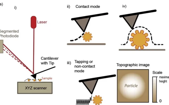
The atomic force microscopy (AFM) is a surface imaging technique, that can have a sub-nanometer resolution. One of the advantages of this technique is that the measurements can be directly done in aqueous environments, like most part of biological solutions. This technique is based on the use of a very sharp tip, mounted in the end of a cantilever. The interaction between the tip and the sample surface is monitored through the deflection of the cantilever. AFM can be used to study the sample topography, but also to study other sample characteristics, like its chemical and mechanical properties. The topographical images can be acquired by the contact (Figure 10-ii) and non-contact mode (Figure 10-iii) [78]. The tapping mode is normally used for the characterization of soft particles, such EVs, because the contact mode can result in vesicle deformation and rupture. However, when using AFM in the characterization of EVs, one should have attention on the sample preparation, specially sample immobilization in the surface [79]. Sharma et al. [80] used AFM tips coated with anti-CD63 to show that the increase in tip-vesicle binding force was related to the increase in the density of this receptor per tip-vesicle in patient samples.
Figure 10 – Atomic force microscopy imaging of extracellular vesicles. I) working principle of AFM. The AFM
can operate in ii) contact mode, iii) in tapping mode yielding a topographical image of the surface (iv). Figure from reference [81].
1.3.1.6. Electron microscopy
Due to their small size, EVs cannot be detected by standard optical microscopy. Instead, an electron microscopy (EM) can be used to visualize these vesicles. EM uses electrons instead of photons to create images with resolutions down to nanometers. Because of this capacity, EM has become the gold method for visualization of EVs, and to study their morphology and structure. There are two main EM techniques, the scanning electron microscopy (SEM) and the transmission electron microscopy (TEM). In the first one, the topographical image of the surface is acquired through the detection of secondary electrons originated from the incidence of an electron beam in the sample. However, one of the drawbacks of SEM, is that samples must have a conductive surface, being necessary, sometimes, to coat the surface with a thin layer of a conductive material, like gold. Despite the use of SEM and other variants, in the characterization of EVs [63,82], TEM is mostly used. In TEM the samples do not need to have a conductive surface, and therefore ultra-thin samples can be directly analysed in transmission mode. However, in standard TEM, samples need to be fixated and dehydrated due to the applied vacuum. Also, the samples are normally stained for a better contrast and visualization. One of the advantages of TEM is the possibility to do immuno-electron microscopy. This allows for the detection of specific biomolecules in the outer membrane of the EVs with the use of gold-conjugated antibodies. TEM in combination with immunostaining has been used to investigate diverse EVs populations [83].
1.3.2. Chemical/Biochemical characterization of EVs
One of the most commonly used methods for the identification of proteins in EVs is Western blotting. In this technique, a mixture of proteins is separated based on their molecular weight through a gel electrophoresis. Then, the proteins are transferred to a membrane of nitrocellulose, Polyvinylidene difluoride (PVDF) or nylon. Membrane sites with unbound protein are blocked with a blocking solution, such as Bovine serum albumin (BSA) or non-fat dry milk. Then the membrane is incubated with antibodies specific for the protein of interest. In this process, a primary antibody labelled with an enzyme can be used or, alternatively, a primary antibody will be used first followed by the addition of a secondary antibody labelled with an enzyme. The unbound antibody is washed, leaving only the antibody bound to the protein of interest. The bound antibody is then detected by exposure to a specific substrate, which gives a detectable signal [84]. For a more deeper analysis, proteomic approaches, like Sodium dodecyl sulphate-Polyacrylamide
gel electrophoresis (SDS-PAGE) or High-performance liquid chromatography (HPLC) followed by mass spectrometry can be used [85]. Conde-Vancells et al. [86] have done an extensive proteomic characterization of exosomes secreted by hepatocytes using both methods. The lipid content of exosomes can also be analysed by lipidomic approaches. Llorente et al. [87] did an extensive analysis of exosome lipid contents and of their cells of origin. There are diverse databases, such Vesiclepedia, in which the protein and lipid contents of EVs are described [88].
1.4. Mesenchymal stem cells (MSCs) as the source of exosomes
One important aspect when using exosomes for drug delivery applications is their origin, that is, the cell source used for obtaining them. For example, exosomes isolated from dendritic cells can be loaded with antigenic peptides for T cell proliferation, and thus be of great potential as a vaccine in cancer and infectious diseases [89]. However, exosomes derived from dendritic cells are immunogenic, which does not make them a suitable vehicle for drug delivery. Thus, the ideal cell source would be one capable of producing non-immunogenic exosomes in abundance. In fact, for drug delivery, it is described that the best option is to use mesenchymal stem cells derived exosomes [90].
1.4.1. Background of mesenchymal stem cells
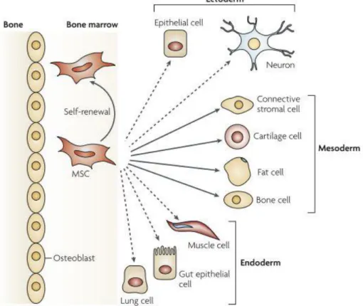
MSCs are multipotent stem cells that can be found in diverse adult tissues, such as adipose tissue, periosteum, liver, lung, spleen, muscle connective tissue, amniotic fluid, placenta, umbilical cord blood, dental pulp and aborted foetal tissues [90]. According to the International Society for Cellular Society, MSCs must be plastic adherent when maintained in standard culture conditions; they must express CD105, CD73 and CD90, and lack expression of CD45, CD34, CD14 or CD11b, CD79α or CD19 and HLA-DR surface molecules. At last, they must be able to differentiate into osteoblasts, adipocytes and chondroblasts in vitro [91]. The differentiation potential of MSCs is shown in Figure 11. Although several research papers report the differentiation of MSCs towards cells of ectoderm and endoderm origin, this is still controversial.
1.4.2. Clinical and therapeutic applications of MSCs
Mesenchymal stem cells are the most studied stem cells, with more than 49000 publications cited in Web of Science since 1900. This could be attributed to their application in a wide range of diseases and injuries [92], to their easy availability from different tissues (such as bone marrow and fat tissue) and to their capacity for large ex
vivo expansion [93]. Until 2015, the database of the US National Institutes of Health (NIH)
registered about 493 MSC-based clinical trials [94]. MSCs have been explored in four main fields: local implantation of MSCs to treat local diseases; systemic transplantation of MSCs; use of stem cell therapy together with gene therapy; and use of MSCs in tissue engineering procedures [92]. Regarding the local implantation of MSCs, these cells were shown to be efficient in the treatment of bone diseases [95]. For example, some clinical reports demonstrated
Figure 11- The differentiation capacity of MSCs. The figure shows the ability of MSCs to self-renew (curved
arrow) and to differentiate into mesodermal lineage (solid arrows). The reported capacity to transdifferentiate in cells of other lineages (ectoderm and endoderm) is showed by dashed arrows (even though this is still controversial). Figure adapted from reference [96].
the capacity of autologous MSCs in the treatment of large bone defects, when injected
ex vivo [97]. Also, some studies applied MSCs in the repair of cartilage tissue [98]. The
MSCs can also be used for the treatment of vascular diseases, like vascular ischemia, peripheral arterial disease [99] and coronary artery disease [100]. These studies showed very good results, but more trials are needed in a bigger number of randomized patients.
Beyond the possibility of direct differentiation into different cell types, MSCs are able to secret several biochemical factors that have local paracrine and autocrine actions (throphic factors), as shown in Figure 12 [101]. For example, the secretion of trophic factors led to a reduction in the infarct incidence and improved the cardiac functions in a pig model of chronic ischemia [102]. Later, it was shown that these therapeutic factors were released inside membrane vesicle, more specifically in the exosomes [103].
Figure 12 – Paracrine factors release by mesenchymal stem cells which may play an important role in
mitogenesis, angiogenesis, apoptosis and scarring. Figure adapted from reference [104].
1.4.3. Advantages of using MSCs as exosome producers for drug delivery
Mesenchymal stem cells present many features that make them ideal candidates as producers of exosomes. For example, these cells present a large ex vivo expansion capacity, and since they can be isolated from human adult tissues, their use is ethically non-controversial. Also, as there are already several reports that describe the safe clinical transplantation of MSCs, one can conclude that the use of exosomes derived from these cells will not lead to adverse effects. The administration of human MSC-derived exosomes in an immunocompetent mouse model for acute myocardial ischemia
One of the most important aspects in the clinical use of MSCs is their ability to make use of suppressive and regulatory effects over the innate immune cells and independently of their autologous or allogeneic origin [107]. This immunomodulatory ability can result in a longer exosome longevity and bioavailability of their therapeutic cargo. Another important aspect of the MSCs is their high ex vivo proliferative capacity. Although this expansion is finite, some studies were made regarding this question. For example, Chen et al. [108] created an immortalized MSC line with the myc oncogene. The immortalization compromised the differentiation capacity of these cells but did not affect the production and therapeutic effect of the exosomes. The immortalization of MSCs could reduce the need for constant new batches of these cells, also reducing the need for constant derivation, testing and validation and therefore making the production of MSC exosomes more commercially sustainable. One last topic that should be addressed related to the use of MSCs as candidates for the production of exosomes is that these cells are among the most prolific producers of exosomes [109]. All these properties are presented in Figure 13.
Figure 13 – Advantages of using mesenchymal stem cells as a source of exosomes. Figure adapted from
reference [4].
1.5. Methods for the loading of therapeutic cargo into exosomes
Exosomes have a lipid bilayer membrane that serves like a natural barrier for the protection of their cargo in order for it to not be degraded in the bloodstream. However, the presence of this membrane makes exosome loading a challenging task. Indeed, the successful delivery of the therapeutic cargo transported by exosomes depends directly
on the efficiency of the selected loading method [110,111]. Exosome loading methods can be classified in two groups: (1) after EV isolation; and (2) before EV isolation (like schematized in Figure 14). A brief description of the most important methods used is presented in the following sections.
1.5.1. Exosome loading methods after EV isolation
1.5.1.1. Electroporation
When exosomes (or cells) are submitted to an electrical field, pores can be created in the membranes, facilitating the sorting of cargo into the lumen of EVs [112]. For example, Alvarez-Erviti et al. [113] loaded small interfering RNA (siRNA) in exosomes with the use of the electroporation technique. Other studies also used this method for the incorporation of therapeutic cargo, with voltages in the range of 150-700 V. Interestingly, the applied voltage for efficient exosome loading by electroporation may vary with the type of donor cell (immature dendritic cells, monocytes and HeLa cells, for example) [114,115].
Electroporation may be a good option for exosome loading since it is easy to control the experimental parameters. However, some authors believe that it may induce adverse effects in the loaded cargo or in exosome integrity.
For example, Kooijmanns et al. [111] showed that the process of electroporation can induce the formation of siRNA aggregates, and that only 0.05 % of the siRNA was successfully incorporated in exosomes using this method. Other work showed that electroporation can also induce the aggregation of exosomes themselves,; however, when the parameters are optimized, the aggregation can be significantly reduced, still allowing the incorporation of iron particles [110]. This method was also used for the incorporation of doxorubicin in targeted exosomes, showing that it can represent an efficient way to load chemotherapeutics in exosomes. Moreover, the drug was able to maintain its biological function [114].
Figure 14 – Scheme with the different strategies for exosome loading. Left side: Incorporation of the
therapeutic cargo before exosome isolation. Transfection of cells with a vector encoding a protein and by simple exposure to a therapeutic molecule. Right side: Incorporation of the therapeutic cargo after exosome isolation. Electroporation, simple incubation, application of ultrasonic frequencies, repeated freeze-thaw cycles, treatment with detergent-like molecules, such as saponin, and extrusion. Figure adapted from reference [116].
1.5.1.2. Simple incubation
Simple incubation of the therapeutic cargo with exosomes is a common method for exosome loading. Only 5 minutes of incubation was needed to efficiently incorporate curcumin in exosomes at 22 ºC, and result in a significant anti-inflammatory effect in different disease models [117,118]. Curcumin can induce lipid rearrangement and changes in the membrane fluidity, and for this reason the incorporation of this molecule


![Figure 5 – Biochemical composition of exosomes. Figure adapted from reference [28].](https://thumb-eu.123doks.com/thumbv2/123dok_br/19277221.985799/39.892.212.680.101.494/figure-biochemical-composition-exosomes-figure-adapted-reference.webp)

![Figure 13 – Advantages of using mesenchymal stem cells as a source of exosomes. Figure adapted from reference [4]](https://thumb-eu.123doks.com/thumbv2/123dok_br/19277221.985799/55.892.174.733.555.844/figure-advantages-mesenchymal-source-exosomes-figure-adapted-reference.webp)