ACADEMIC DISSERTATION To be publicly discussed with the permission of the Medical Faculty
of the University of Helsinki
in Biomedicum Helsinki 1, Haartmaninkatu 8, Helsinki, at 1PM on the 14th of April 2023.
Clinical Neurosciences, University of Helsinki Department of Neurology, Helsinki University Hospital
ACUTE STROKE CARE:
STRATEGIES FOR IMPROVING DIAGNOSTICS
Olli S. Mattila
ISBN 978-951-51-9005-5 (paperback) ISBN 978-951-51-9006-2 (PDF)
http://ethesis.helsinki.fi Unigrafia, Helsinki 2023
Department of Neurology, Helsinki University Central Hospital Helsinki, Finland
Reviewers Professor Pekka Jäkälä Department of Neurology Institute of Clinical Medicine University of Eastern Finland Kuopio, Finland
Professor Per Wester
Department of Public Health and Clinical Medicine Norrland University Hospital
Umeå Universitet Umeå, Sweden
Opponent Professor Heinrich Audebert Center for Stroke Research Berlin Department of Neurology
Charité - Universitätsmedizin Berlin Berlin, Germany
Be kind, for everyone you meet is fighting a hard battle.
“…success in medicine has dimensions that cannot be found on a play- ing field. For one, lives are on the line. Our decisions and omissions are therefore moral in nature. We also face daunting expectations. In medicine, our task is to cope with illness and to enable every human being to lead a life as long and free of frailty as science will allow. The steps are often uncertain. The knowledge to be mastered is both vast and incomplete. Yet we are expected to act with swiftness and consist- ency, even when the task requires marshaling hundreds of people—
from laboratory technicians to the nurses on each change of shift to the engineers who keep the oxygen supply system working—for the care of a single person. We are also expected to do our work humanely, with gentleness and concern. It’s not only the stakes but also the complex- ity of performance in medicine that makes it so interesting and, at the same time, so unsettling.”
Atul Gawande – Better
To all of us who may someday face acute stroke symptoms – and to my family.
ABSTRACT
Stroke is one of the leading causes of death and disability, with a high incidence of over 11 million cases annually worldwide. Costs of treatment and rehabilitation, loss of work, and the hardships resulting from stroke are a major burden both at the individual and at the societal level. Importantly, stroke therapies need to be initiated early for them to be effective. Thrombolytic therapy and mechanical thrombectomy are early treatment options of ischemic stroke. In hemorrhagic stroke, optimization of hemodynamic and hemostatic parameters is central, and surgery is considered in a subset of patients.
Efficient treatment of stroke requires early and precise recognition of stroke at all stages of the treatment chain. This includes identification of patients with suspected acute stroke by emergency medical dispatchers and emergency medical services staff, and precise admission diagnostics by the receiving on- call stroke team. Success requires grasping the complexity of stroke symptoms that depend on the brain areas affected, and the plethora of medical conditions that can mimic stroke.
The Helsinki Ultra-acute Stroke Biomarker Study includes a cohort of 1015 patients transported to hospital due to suspected acute stroke, as candidates for revascularization therapies. Based on this cohort, this thesis work has explored new avenues to improve early stroke diagnostics in all stages of the treatment chain.
In a detailed investigation into the identification of stroke by emergency medical dispatchers, we analyzed emergency phone calls with missed stroke identification. We also combined data on dispatch and EMS and hospital records to identify causes for missing stroke during emergency calls. Most importantly, we found that a patient’s fall at onset and patient confusion were strongly associated with missed identification. Regarding the Face Arm Speech Test (FAST), the most likely symptom to be misidentified was acute speech disturbance.
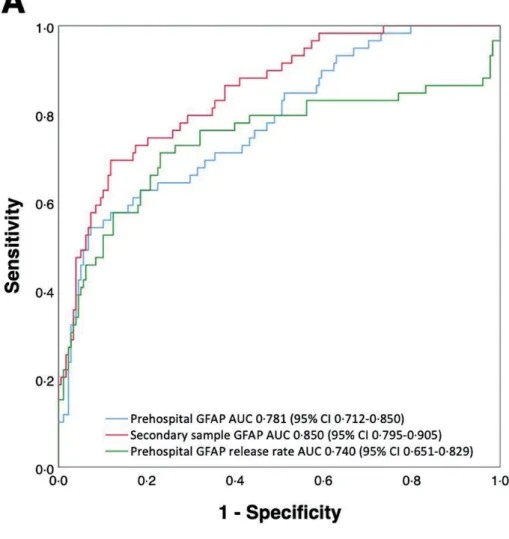
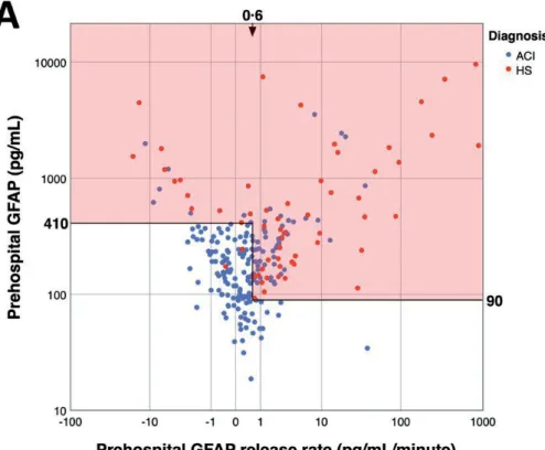
Using prehospital blood sampling of stroke patients, and ultrasensitive measurement, we investigated the early dynamics of the plasma biomarkers glial fibrillary acidic protein (GFAP) and total tau. Utilizing serial sampling, we demonstrate for the first time that monitoring the early release rate of GFAP can improve the diagnostic performance of this biomarker for early differentiation between ischemic and hemorrhagic stroke. In our analysis of early GFAP levels, we were able to differentiate with high accuracy two-thirds of all patients with acute cerebral ischemia from those with hemorrhagic stroke,
supporting further investigation of this biomarker as a promising point-of-care tool for prehospital stroke diagnostics.
We performed a detailed review of the admission diagnostics of our cohort of 1015 patients to explore causes and predictors of admission misdiagnosis.
We then investigated the consequences of misdiagnosis on outcomes. We demonstrate in this large cohort that the highly optimized and rapid admission evaluation in our hospital district (door-to-needle times below 20 minutes) did not compromise the accuracy and safety of admission evaluation. In addition, we discovered targets for improving future diagnostics.
Finally, our detailed neuropathological investigation of a case of cerebral amyloid angiopathy (CAA) -related hemorrhage after stroke thrombolysis provided unique tissue-level evidence for this common vasculopathy as a notable risk factor for intracranial hemorrhagic complications in the setting of stroke. These findings support research to improve the diagnostics of CAA, and the prediction of hemorrhagic complications associated with stroke thrombolysis.
In conclusion, these proposed targets and strategies will aid in the future improvement and development of this highly important field of diagnostics.
Our proof-of-concept discoveries on early GFAP kinetics help guide further study into this diagnostic approach just as highly sensitive point-of-care GFAP measurement instruments are becoming available. Finally, our results support the safety of worldwide efforts to optimize emergency department door-to- needle times when care is taken to ensure sufficient expertise is in place, highlighting the role of the on-call vascular neurologist as a central diagnostic asset.
TIIVISTELMÄ
Aivohalvaus on yksi yleisimpiä kuolinsyitä ja työkyvyttömyyden aiheuttajia, ja sen maailmanlaajuinen vuosittainen ilmaantuvuus on yli 11 miljoonaa tapausta.
Hoito- ja kuntoutuskustannukset, työkyvyn menetys ja aivohalvauksen aiheuttamat arkielämän vaikeudet ovat suuri taakka sekä yksilön että yhteiskunnan tasoilla. Vaikka hoitovaihtoehtoja on useita, kuten liuotushoito ja mekaaninen trombektomia iskeemisen aivohalvauksen hoitoon, sekä hemodynamiikan ja hemostaasin optimointi ja leikkaushoito aivoverenvuotojen hoitoon, nämä hoidot on aloitettava nopeasti, jotta ne olisivat tehokkaita.
Aivohalvauksen hoitojen vaatima nopeus edellyttää aivohalvauksen varhaista ja tarkkaa tunnistamista hoitoketjun kaikilla tasoilla. Tähän sisältyy aivohalvauksen tunnistaminen hätäkeskuksen ja ensihoidon toimesta, nopea triage ja kuljetus oikeaan sairaalaan, ja onnistunut diagnostiikka päivystyspoliklinikan vastaanottavalta aivohalvaustiimiltä. Haasteina ovat monimutkaisuus aivohalvauksen oireissa, jotka riippuvat sairastuneista aivoalueista, sekä lukuisat muut sairaudet, jotka voivat jäljitellä aivohalvauksen oireita.
Helsinki Ultra-acute Stroke Biomarker Study -tutkimus käsittää 1015 potilaan kohortin, jotka kuljetettiin sairaalaan epäillyn akuutin aivohalvauksen vuoksi arvioitavaksi ja diagnosoitavaksi rekanalisaatiohoitojen varhaista aloittamista varten. Tässä väitöskirjatyössä etsittiin uusia keinoja kehittää aivohalvauksen varhaisdiagnostiikkaa hoitoketjun kaikissa eri vaiheissa.
Yksityiskohtaisessa tutkimuksessa aivohalvauksen tunnistamisesta hätäkeskuspäivystäjien toimesta analysoimme hätäpuheluita, joissa aivohalvauksen tunnistaminen epäonnistui. Lisäksi yhdistimme hätäpuheluiden, ensihoitokertomusten ja sairaskertomusten tietoja löytääksemme syitä aivohalvauksen tunnistamisen epäonnistumiseen hätäpuhelujen aikana. Havaitsimme, että potilaan kaatuminen ja potilaan sekavuus olivat keskeisiä tekijöitä, jotka liittyivät epäonnistuneeseen tunnistamiseen. Face Arm Speech Test (FAST) -seulontaoireista puhehäiriö oli todennäköisimmin väärin tunnistettu.
Akuuttivaiheen verinäytteitä ja äärimmäisen herkkää määritysmenetelmää hyödyntäen tutkimme kahden verestä mitattavan merkkiaineen, tähtisolujen säikeisen happaman proteiinin (GFAP) ja taun varhaista dynamiikkaa aivohalvauspotilailla. Osoitamme ensimmäistä kertaa, että GFAP:n varhaisen vapautumisnopeuden seurantaa sarjanäytteistä voidaan hyödyntää parantamaan tämän merkkiaineen diagnostista suorituskykyä iskeemisen ja hemorragisen aivohalvauksen varhaisessa erottamisessa. Varhaisten GFAP-
tasojen analyysissämme pystyimme erottamaan suurella varmuudella kaksi kolmasosaa kaikista iskeemiseen aivohalvaukseen sairastuneista niistä potilaista, joilla oli aivoverenvuoto. Tulokset viittaavat siihen, että GFAP merkkiaine voisi olla jatkossa kehitettävissä ambulansseissa hyödynnettäväksi pikaverikokeeksi, joka auttaisi aivohalvauksen eri muotojen varhaisessa erottelussa.
Teimme yksityiskohtaisen katsauksen kaikkien kohorttimme potilaiden (n
= 1015) päivystysdiagnostiikkaan selvittääksemme diagnostisten vaikeuksien syitä ja ennustajia sekä tutkimme virheellisen tulovaiheen diagnoosin seurauksia potilaiden ennusteeseen. Osoitamme ensimmäistä kertaa suuressa aineistossa, että sairaanhoitopiirissämme käytetty aivohalvauksen rekanalisaatiokandidaattien erittäin nopea vastaanottoarviointi (liuotushoidon mediaaniviive alle 20 minuuttia) ei vaaranna diagnostiikan tarkkuutta ja turvallisuutta. Lisäksi kuvailemme diagnostiikan kehittämiskohteita tulevaisuudessa.
Väitöskirjan viimeisessä osatyössä laadimme yksityiskohtaisen neuropatologisen tapausselostuksen aivojen amyloidiangiopatiaan liittyvästä verenvuodosta aivohalvauksen liuotushoidon jälkeen saadaksemme ainutlaatuista kudostason näyttöä tämän yleisen verisuonisairauden aiheuttamasta komplikaatioriskistä aivohalvauksen rekanalisaatiohoidoissa.
Havaintomme tukevat tutkimusta aivojen amyloidiangiopatian diagnostiikan parantamiseksi ja verenvuotokomplikaatioiden ennustamisen kehittämiseksi.
Yhteenvetona voidaan todeta, että esitetyt tutkimukset havainnollistavat aivohalvauksen varhaisen diagnosoinnin monitahoisia haasteita.
Väitöskirjatyössä esitetyt kehityskohteet ja menetelmät auttavat tämän erittäin tärkeän diagnostisen alan tulevassa kehitystyössä. Mikä tärkeintä, nämä tulokset sisältävät uusia ja ainutlaatuisia konseptihavaintoja varhaisen GFAP-kinetiikan käytöstä aivohalvauksen diagnostiikassa ja erittäin nopean päivystysarvion turvallisuudesta käytettäessä sairaanhoitopiirimme nopeaa liuotushoitoprotokollaa.
CONTENTS
ABSTRACT ... 6
TIIVISTELMÄ ... 8
LIST OF ORIGINAL PUBLICATIONS ...12
ABBREVIATIONS ...13
1 INTRODUCTION ...15
2 REVIEW OF THE LITERATURE ...16
2.1 The societal burden of stroke 16 2.2 Acute stroke and stroke mimics: presentation and acute treatment 17 2.2.1 Acute cerebral ischemia: ischemic stroke and TIA 17 2.2.2 Hemorrhagic stroke: intracerebral and subarachnoid hemorrhage 21 2.2.3 Stroke mimics and chameleons 24 2.2.4 Distribution of diagnoses within stroke code patients 27 2.3 Diagnostic checkpoints in acute stroke care 28 2.3.1 Recognition of stroke symptoms by laypeople 28 2.3.2 The emergency call 29 2.3.3 Prehospital stroke diagnostics by EMS 30 2.3.4 Admission diagnostics 36 3 STUDY AIMS ...43
4 SUBJECTS AND METHODS...44 4.1 The Helsinki Ultra-acute Stroke Biomarker Study 44 4.2 Study setting and design 44 4.3 Blood sampling, sample processing, and biomarker measurement 46 4.4 Data collection and emergency call analysis 47 4.5 Statistical methods 48
5 RESULTS ...49
5.1 Targets for improving stroke detection during emergency phone calls (I) 49 5.2 Prehospital use of plasma GFAP to rule out hemorrhagic stroke (II) 50 5.3 Pitfalls in the admission evaluation of stroke code patients (III) 52 5.4 CAA as a risk factor for post-thrombolytic hemorrhage (IV) 53 6 DISCUSSION ...54
6.1.1 Improving dispatcher stroke identification (I) 54 6.1.2 Point-of-care biomarkers to support prehospital stroke therapy (II) 55 6.1.3 The stroke neurologist as a central diagnostic asset (III) 56 6.1.4 Improving the safety of stroke thrombolysis (IV) 56 6.2 Study limitations 57 6.3 Implications for practice and future research 59 7 CONCLUSIONS ...61
ACKNOWLEDGEMENTS ...63
REFERENCES ...67
ORIGINAL PUBLICATIONS ...99
LIST OF ORIGINAL PUBLICATIONS
This thesis is based on the following original publications, referred to in the text by their Roman numerals:
I Mattila OS, Puolakka T, Ritvonen J, Pihlasviita S, Harve H, Alanen A, Sibolt G, Curtze S, Strbian D, Pystynen M, Tatlisumak T, Kuisma M, Lindsberg PJ. Targets for improving dispatcher identification of acute stroke. Int J Stroke. 2019 Jun;14(4):409-416.
II Mattila OS, Ashton NJ, Blennow K, Zetterberg H, Harve-Rytsälä H, Pihlasviita S, Ritvonen J, Sibolt G, Nukarinen T, Curtze S, Strbian D, Pystynen M, Tatlisumak T, Kuisma M, Lindsberg PJ. Ultra-early differential diagnosis of acute cerebral ischemia and hemorrhagic stroke by measuring the prehospital release rate of GFAP. Clinical Chemistry.
2021 Oct;67(10):1361-1372.
III Pihlasviita S, Mattila OS, Ritvonen J, Sibolt G, Curtze S, Strbian D, Harve H, Pystynen M, Kuisma M, Tatlisumak T, Lindsberg PJ. Diagnosing cerebral ischemia with door-to-thrombolysis times below 20 minutes.
Neurology. 2018 Aug 7;91(6):e498-e508.
IV Mattila OS, Sairanen T, Laakso E, Paetau A, Tanskanen M, Lindsberg PJ. Cerebral amyloid angiopathy related hemorrhage after stroke thrombolysis: case report and literature review. Neuropathology. 2015 Feb;35(1):70-4.
The original publications are reproduced in the print version of this thesis with the permission of the copyright holders. Publication (III) will later be included also in the doctoral thesis of Dr. Pihlasviita.
ABBREVIATIONS
ACI Acute cerebral ischemia CAA Cerebral amyloid angiopathy
CI Confidence interval
CMB Cerebral microbleed CSF Cerebrospinal fluid
CT Computer tomography
CTA Computer tomography angiography CTP Computer tomography perfusion DALY Disability-adjusted life year
ED Emergency department
EMD Emergency medical dispatcher EMS Emergency medical services EMT Emergency medical technician EVT Endovascular thrombectomy FAST Face Arm Speech (Time) test
GCS Glasgow coma scale
GFAP Glial fibrillary acidic protein
HS Hemorrhagic stroke
HUH Helsinki University Hospital
HUS Helsinki and Uusimaa Hospital District ICH Intracerebral hemorrhage
ICP Intracranial pressure
INR International normalized ratio IQR Interquartile range
IVT Intravenous thrombolysis LVO Large-vessel occlusion MRI Magnetic resonance imaging MSU Mobile stroke unit
mRS Modified Rankin Scale
NCCT Non-contrast computer tomography NIHSS National Institutes of Health Stroke Scale
POC Point-of-care
rtPA Recombinant tissue plasminogen activator SAH Subarachnoid hemorrhage
SC Stroke code
SM Stroke mimic
TCD Transcranial Doppler TIA Transient ischemic attack
VIPS Volumetric impedance phase-shift spectroscopy YLL Years of life lost
1 INTRODUCTION
Acute stroke, a medical emergency requiring rapid initiation of treatment, is for emergency care, one of its most challenging diagnostic areas. Due to the wide spectrum of different symptoms and presentations, and because symptom onset is usually not accompanied by pain, stroke symptoms are difficult for laypeople to identify, and often do not trigger the necessary urgency in seeking immediate help.
The diagnostic difficulties are challenging also for emergency medical dispatchers and emergency medical services (EMS) personnel. Stroke-like symptoms can result from a plethora of other acute neurological and non- neurological conditions only differentiated by clinical expertise and complicated diagnostic equipment. Dispatchers and EMS in most parts of the world have very limited means to make triage decisions for these patients, and initiation of specific therapies before hospital arrival is rarely possible. Patients with acute stroke-like symptoms and sufficiently short delays from symptom onset are therefore transported as stroke code patients to the closest stroke center for diagnostics and treatment.
The challenges are greatest at the end of the line, the receiving emergency department, where the on-call physician must rapidly synthesize all available information. This includes medical and EMS records, information from the patient, next of kin, and EMS personnel, and findings from clinical examination, point-of-care blood tests, hemodynamics, and multimodal neuroimaging.
Further, all of this must be done immediately, skillfully identifying conditions mimicking stroke.
Although stroke medicine steadily progresses, challenging professionals to make timely changes and improvements in their stroke code pathway, acute diagnostic and therapeutic decisions have become more complicated, with new therapies and diagnostic capabilities, requiring decisions for which evidence- based guidelines are limited. In the Helsinki Ultra-acute Stroke Biomarker Study and in this thesis, the goal has been to examine the diagnostic performance of our stroke code care pathway to identify strategies for improving diagnostic accuracy and speed of treatment at all levels, from the emergency call to the emergency department.
2 REVIEW OF THE LITERATURE
2.1 THE SOCIETAL BURDEN OF STROKE
For epidemiological purposes, stroke is traditionally defined based on World Health Organization (WHO) descriptions, which define stroke through clinical findings and symptoms as rapidly developed signs of focal or global disturbance of cerebral function lasting more than 24 hours (unless interrupted by death), with no apparent origin other than vascular (Aho & Fogelholm 1974).
For the purposes of clinical work and research, where more exacting diagnostic methods are readily available, the definition of stroke has undergone refinement towards a tissue-based definition. According to the definition in an expert consensus document of the American Heart and Stroke Associations, central nervous system (CNS) infarction is brain, spinal cord, or retinal cell death attributable to ischemia, based on 1) pathological, imaging, or other objective evidence of cerebral, spinal cord, or retinal focal ischemic injury in a defined vascular distribution; or 2) clinical evidence of cerebral, spinal cord, or retinal focal ischemic injury based on symptoms persisting ≥24 hours or until death, and other etiologies excluded. In addition to CNS infarction, stroke specifically also includes non-traumatic intracerebral hemorrhage (ICH) and subarachnoid hemorrhage (SAH), and cerebral venous thrombosis (Sacco et al. 2013). The benefit of this tissue-based definition is the inclusion of asymptomatic CNS ischemia and hemorrhage not included in the traditional epidemiological definition.
Globally, stroke is the third leading cause of lost disability-adjusted life years (DALYs), an epidemiological measure of disease burden, when considering all age groups (GBD 2019 Diseases and Injuries Collaborators 2020). Further, stroke is the third leading cause, worldwide, of years of life lost (YLLs) (GBD 2017 Causes of Death Collaborators 2018). Based on the Global Burden of Disease Study, the crude total number of new stroke cases worldwide (incidence) increased between 1990 and 2017 by 76%, to a total estimate in 2017 of 11.9 million annual cases. The corresponding age-standardized global rate of new strokes in 2017 was estimated to be 150.5 cases per 100 000 (Avan et al. 2019).
The total crude global prevalence of stroke was estimated in 2017 to be 104.2 million (Avan et al. 2019).
Importantly, the incidence of stroke is highly dependent on age and traditional vascular risk factors such as hypertension, smoking, poor diet,
impaired glucose tolerance, obesity, and hypercholesterolemia (Lawes et al. 2004; Buttar et al. 2005; Prospective Studies Collaboration et al. 2007) (O’Donnell et al. 2010). On a global scale, 1.3% of stroke incidence is in people aged < 40 years, 10.9% in 40- to 64-year-olds, and 87.8% in those ≥65 years (Avan et al. 2019). It is particularly alarming that the incidence of stroke has increased in recent decades particularly in younger generations (<55 years) (Béjot et al. 2014; Tibæk et al. 2016; L. Li et al. 2022).
Stroke leaves survivors with varying degrees of long-lasting neurological deficits, including motor and sensory impairment, pain, disturbances in speech, apraxia, and cognitive issues (Kotila et al. 1999; Morley et al. 2005; G. Chen et al. 2005; Mukherjee et al. 2006; Snaphaan et al. 2009; Kyrozis et al. 2009;
Broomfield et al. 2014; Jokinen et al. 2015). These deficits often disrupt daily life, leading to loss of work, difficulties in carrying out daily activities, and sometimes requiring daily assistance and care. Finally, stroke has a myriad of emotional, mental, and social effects on survivors, further disrupting life.
Due to this complexity, stroke is a devastating disease, on both societal and individual levels.
2.2 ACUTE STROKE AND STROKE MIMICS: PRESENTATION AND ACUTE TREATMENT
2.2.1 ACUTE CEREBRAL ISCHEMIA: ISCHEMIC STROKE AND TIA
In its normal state, cerebral energy metabolism is highly dependent on the oxidative metabolism of glucose, its main energy source (Vannucci et al. 1997).
When a cerebral artery is suddenly occluded, a gradient of insufficient blood flow forms, with a central tissue area of very deep ischemia, surrounded by a larger area with less severe blood flow deficiencies. The severity of ischemia and oxygen deficiency ranges from benign oligemia with minor effects on cellular metabolism, to more severe ischemia in the so-called penumbral area, which leads to various disruptions in cellular physiology including shutdown of protein synthesis, disruption in the electrical activity of the brain tissue, peri-infarct spreading depression, increases in lactate production, glucose depletion, and finally severe membrane depolarization as cellular ion pumps fail (Heiss 1992;
Hossmann 1993b; Hossmann 1993a; Hossmann 1994; DeGracia et al. 2008).
The deepest zone of ischemia is in the infarct core where ischemia quickly leads to necrosis and permanent damage to brain tissue (Hossmann 1994;
Whereas neurons are the most sensitive cell type facing ischemia, the other components forming the basic structural and functional element of cerebral tissue – the neurovascular unit – including astrocytes, endothelial cells, pericytes, resident inflammatory cells, and oligodendrocytes, are also severely affected, leading to a cascade of disruptive events (del Zoppo 2009; Stanimirovic & Friedman 2012; Dalkara & Alarcon-Martinez 2015).
This includes loss of blood flow regulation, accumulation of waste products, microvascular failure with microvascular plugging, cellular and vascular tissue edema, an uncontrolled inflammatory response, and uncontrolled proteolysis (del Zoppo 2009; Iadecola & Anrather 2011). At later stages, recanalization of the occluded artery can lead to reperfusion injury, with severe oxidative injury and further inflammatory activation and infiltration of inflammatory cells (del Zoppo 2009; Bai & Lyden 2015). Inflammatory injury and later apoptotic cell death continue in the days after ischemia, sometimes together with more severe complications such as tissue hemorrhage and severe edema (Beghi et al. 1989; Bai & Lyden 2015).
Symptoms of acute cerebral ischemia (ACI) begin suddenly, and depend largely on which central nervous system (CNS) areas are undergoing ischemia.
Symptoms usually reach their maximum intensity in a few minutes or hours.
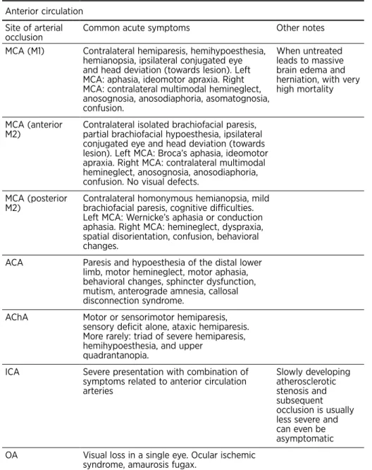
Presentations of ACI are typically classified according to the vascular territory affected (Table 1), with four out of every five occlusions occurring in the anterior carotid circulation, and one-fifth in the posterior vertebrobasilar circulation (Bogousslavsky 1994; Nouh et al. 2014; Yassi et al. 2015; C.-H. Park et al. 2016).
The five main branches arising from the internal cerebral artery include the middle cerebral artery, the anterior cerebral artery, the anterior choroidal artery, the posterior communicating artery, and the ophthalmic artery (Tatu et al. 1998). The posterior circulation consists of the two vertebral arteries that join to form the basilar artery. Smaller paramedian and circumferential branches and four cerebellar arteries arise from the basilar artery, which finally divides into two posterior cerebral arteries (Tatu et al. 1996). Cerebral ischemia in any of these specific vascular territories leads to a corresponding clinical syndrome (Table 1) (Grivé et al. 2005). Notably, a multitude of congenital anatomical variants of the cerebral vasculature exist (Dimmick & Faulder 2009; Varga et al. 2019). For example, a relatively common variant is decreased or absent patency of the posterior communicating artery, which may also influence the clinical manifestation and prognosis of posterior circulation strokes (Ahn et al.
2018; J. S. Park et al. 2022). Although many of these variants are uncommon, what is important is to remember their existence in clinical practice.
In addition to occlusion of major vascular arterial branches, two other lesion types are worth noting. Lacunar ischemic lesions are small subcortical infarcts with a diameter under 1.5 cm, which involve occlusion of deep perforant
arteries (FISHER 1965; Cannistraro et al. 2019). Watershed infarcts involve the junctional areas between the distal regions of two separate arteries, and are typically caused by systemic hemodynamic failure, either alone or in combination with tight stenosis of a larger proximal artery (Momjian-Mayor
& Baron 2005).
Table 1. Common stroke syndromes Anterior circulation
Site of arterial
occlusion Common acute symptoms Other notes
MCA (M1) Contralateral hemiparesis, hemihypoesthesia, hemianopsia, ipsilateral conjugated eye and head deviation (towards lesion). Left MCA: aphasia, ideomotor apraxia. Right MCA: contralateral multimodal hemineglect, anosognosia, anosodiaphoria, asomatognosia, confusion.
When untreated leads to massive brain edema and herniation, with very high mortality
MCA (anterior
M2) Contralateral isolated brachiofacial paresis, partial brachiofacial hypoesthesia, ipsilateral conjugated eye and head deviation (towards lesion). Left MCA: Broca’s aphasia, ideomotor apraxia. Right MCA: contralateral multimodal hemineglect, anosognosia, anosodiaphoria, confusion. No visual defects.
MCA (posterior
M2) Contralateral homonymous hemianopsia, mild brachiofacial paresis, cognitive difficulties.
Left MCA: Wernicke’s aphasia or conduction aphasia. Right MCA: hemineglect, dyspraxia, spatial disorientation, confusion, behavioral changes.
ACA Paresis and hypoesthesia of the distal lower limb, motor hemineglect, motor aphasia, behavioral changes, sphincter dysfunction, mutism, anterograde amnesia, callosal disconnection syndrome.
AChA Motor or sensorimotor hemiparesis, sensory deficit alone, ataxic hemiparesis.
More rarely: triad of severe hemiparesis, hemihypoesthesia, and upper
quadrantanopia.
ICA Severe presentation with combination of symptoms related to anterior circulation arteries
Slowly developing atherosclerotic stenosis and subsequent occlusion is usually less severe and can even be asymptomatic OA Visual loss in a single eye. Ocular ischemic
syndrome, amaurosis fugax.
Posterior circulation
VA Déjerine syndrome, Wallenberg syndrome, Babinski-Nageotte syndrome.
PICA Vertigo, nausea, vomiting, nystagmus, ipsilateral ataxia, gait ataxia.
AICA Vertigo, nausea, vomiting, nystagmus, ipsilateral deafness with tinnitus, ipsilateral peripheral type facial palsy, facial
hypoesthesia, Horner’s syndrome, ipsilateral ataxia, dysarthria, contralateral thermoalgesic sensory deficit.
SCA Ipsilateral limb and gait ataxia, dysarthria, nystagmus, Horner’s syndrome
BA Quadriplegia, bilateral facial palsy, horizontal gaze palsy, locked-in syndrome, coma. A multitude of specific brainstem syndromes have been described.
Often the most severe form of stroke with high mortality, although presentations vary depending on collateral circulation PCA Contralateral thermoalgesic sensory deficit,
contralateral hemianopsia, contralateral facial nerve, vagus nerve and hypoglossal nerve paresis, ipsilateral oculomotor nerve deficit
In 2-10% of patients the PCA is fed mainly from the anterior circulation via a large PCoA Thalamic Thalamic pain syndrome, neuropsychiatric
symptoms
MCA: middle cerebral artery, ACA: anterior cerebral artery, AChA: anterior choroidal artery, ICA: internal carotid artery, OA: ophthalmic artery, VA: vertebral artery, PICA:
posterior inferior cerebellar artery, AICA: anterior inferior cerebellar artery, SCA: superior cerebellar artery, BA: basilar artery, PCA: posterior cerebral artery, PCoA: posterior communicating artery.
Early treatment of acute ischemic stroke focuses on ensuring hemodynamic stability of the patient, recanalizing the occluded cerebral artery as swiftly as possible, and preventing complications (Powers et al. 2019). As prehospital differentiation of ACI and hemorrhagic stroke (HS) is currently in most cases impossible, early prehospital care focuses on ensuring an adequate airway, breathing, and oxygenation, and circulatory stability, including control of very low or high blood pressure, and initiation of basic fluid resuscitation with a crystalloid solution. Other symptoms such as severe nausea and vomiting, pain, confusion, or early seizures also require treatment early in the prehospital setting. In general, most ACI patients are hypertensive but otherwise hemodynamically stable, and the need for intubation is rare.
The main approaches to recanalization include thrombolytic therapy and mechanical thrombectomy, both of which require more detailed in-hospital work-up, including neuroimaging, to ensure correct patient selection (Powers
et al. 2019). Thrombolysis is based on intravenous infusion of a thrombolytic drug such as tissue-plasminogen activator (tPA) or tenecteplase, which bind to the fibrin-rich areas of the culprit cerebrovascular occlusion, and are thus activated to induce rapid fibrinolysis and dissolution of the clot through local production of plasmin from endogenous plasminogen (Medcalf 2017). Whereas thrombolysis shows high recanalization rates in milder strokes, occlusion of larger vessels and tandem occlusions of cervical arteries are often resistant to thrombolysis alone (Lees et al. 2010; Riedel et al. 2011). Mechanical thrombectomy is highly effective and valuable in these larger occlusions, and involves an intervention performed using intra-arterial catheters and x-ray illumination, usually by an interventional radiologist, to either aspirate or mechanically retrieve the occluding blood clot (M. Goyal et al. 2016; Powers et al. 2019). Finally, prevention of early complications of ACI such as aspiration pneumonia, deep-vein thrombosis, or recurrent stroke, requires monitoring and specialist care. This occurs ideally in a dedicated stroke unit, to optimize all physiological parameters including hematological factors, to actively treat early symptoms, and to provide the seamless multidisciplinary care that eases the way toward active rehabilitation (Davenport et al. 1996; Stroke Unit Trialists’
Collaboration 1997; Powers et al. 2019).
Transient ischemic attack (TIA) is a milder form of ACI, in which symptoms do not persist, but end spontaneously through endogenous recanalization of the culprit occlusion (J. Wang et al. 2020; Mendelson & Prabhakaran 2021).
Frequently, short-lived TIA symptoms, which follow the vascular territories as described above, precede more severe ACI (J. Yang et al. 2010). In the setting of symptomatic stroke code patients being transported to a hospital, ACI patients’ symptoms sometimes disappear during transport or quickly after hospital arrival, leading to a final classification of TIA. Nonetheless, TIA patients also require detailed examination and early etiological studies, ideally at an experienced stroke center, as these patients are at risk of more severe strokes, often prevented with correct interventions (Amarenco 2020;
Mendelson & Prabhakaran 2021).
2.2.2 HEMORRHAGIC STROKE: INTRACEREBRAL AND SUBARACHNOID HEMORRHAGE
Intracerebral hemorrhage (ICH), a severe and deadly form of stroke, is characterized by sudden spontaneous bleeding into brain tissue (Sacco et al.
1984; Qureshi et al. 2001). Its common causes include chronic degeneration of deep cerebral small vessels due to hypertension and other vascular risk
(CAA), a form of cerebrovascular degeneration characterized by lobar and meningeal accumulation of beta-amyloid protein around vessels (Viswanathan
& Greenberg 2011). ICH can also result from other underlying conditions, including vascular malformations, cerebral venous thrombosis, hematological disorders, Moyamoya disease, reversible cerebral vasoconstrictive syndrome, infective endocarditis, neoplasms, anticoagulant therapy, substance abuse, and vasculitis (Meretoja, Strbian, Putaala, et al. 2012; Koivunen et al. 2015; Martí- Fábregas et al. 2018).
When intracerebral bleeding begins, usually from a small artery, blood is rapidly and aggressively pushed into the cerebral tissue, resulting in immediate mechanical disruption, with the expanding hematoma compressing surrounding tissue and dissecting surrounding neural structures (Qureshi et al. 2001; Boltze et al. 2019). This is thought to lead to further rupture of new vessels surrounding the expanding hematoma, leading to sudden new sites of bleeding, and thus to a cascade of tissue disruption and enlargement of the hematoma within the first hours after ICH (Brott et al. 1997). As blood enters the intercellular space, the coagulation cascade is immediately activated due to tissue factor, and mixing of plasma and CSF activates the complement system (Lindsberg et al. 1996; Lee et al. 1996; Xi et al. 1998; Holste et al. 2021).
Thrombin, the major end product of the coagulation cascade, is also a highly proinflammatory molecule, inducing inflammatory effects in the surrounding tissue through dedicated PAR-receptors (Hua et al. 2007; Xue et al. 2009).
Further mechanisms also contributing to cerebral injury include hemolysis of red blood cells and release of heme-associated iron, uncontrolled inflammatory cell infiltration, immediate blood brain barrier disruption and vascular edema, cellular necrosis and apoptosis, intraventricular hemorrhage, and disruption of CSF flow (Lee et al. 1996; Xi et al. 1998; Tuhrim et al. 1999; Power et al.
2003; Hua et al. 2007; Xue et al. 2009; Dixon et al. 2012; Qian Li et al. 2018;
Boltze et al. 2019; W.-S. Yang et al. 2020; Holste et al. 2021).
Neurological symptoms of ICH are similar to those resulting from ACI, corresponding to the brain areas affected by the enlarging hematoma in the same manner as the vascular territories described above for ACI. In cases of severe ICH, symptoms can progress rapidly as the hematoma is enlarging, with progressive deterioration of consciousness (Lord et al. 2015). Rupture of the hemorrhage into the cerebral ventricles can lead to posturing and sudden coma (Lord et al. 2015). Most importantly, ICH and ACI cannot be safely distinguished by clinical symptoms alone (Weir et al. 1994). However, some symptoms do differ in their frequency between these two central diagnosis groups, and presentation with these symptoms, including coma, neck stiffness, seizures, diastolic BP >110 mmHg, vomiting, and headache, do, in undiagnosed patients, raise the likelihood of hemorrhagic stroke (Runchey & McGee 2010).
Conversely, a cervical bruit on auscultation and a prior TIA reduces the likelihood of a hemorrhagic stroke diagnosis (Runchey & McGee 2010).
Treatment options for ICH are still limited, focusing mainly on optimizing hemodynamic parameters to reduce the enlargement of the acute hematoma, stabilize the patient, and prevent complications (Hemphill et al. 2015). Early reversal of any anticoagulant medication is important. Vitamin-K antagonists are primarily reversed with prothrombin complex concentrate (PCC) and vitamin-K (Huttner et al. 2006; Kuramatsu et al. 2015; Steiner et al. 2016). Non- vitamin-K antagonist oral anticoagulants (NOAC) are generally best reversed with dedicated antidotes (Siegal et al. 2015; Purrucker et al. 2016; Pollack et al. 2017). Although the prior use of acetosalicylic acid and other antiplatelet medications may be associated with more fatal outcomes (Saloheimo et al. 2006;
Thompson et al. 2010), administration of platelets to patients previously on antiplatelet medication is, however, deleterious (Baharoglu et al. 2016). In ICH patients taking no anticoagulant medications, trials exploring administration of coagulation factors and fibrinolysis inhibitors have found no efficacy (Yuan et al. 2010; Hemphill et al. 2015; Sprigg et al. 2018; Meretoja et al. 2020).
For now, surgical treatment of ICH is still controversial, and is mainly reserved for selected cases with cortical, easily accessible, hematomas, or with posterior fossa bleeds that, if left untreated, show a high risk for rapid herniation and death (Mendelow et al. 2013; Hemphill et al. 2015; Mendelow 2015; Hanley et al. 2017; Staykov et al. 2017; Hanley et al. 2019). Blood-pressure reduction seems to be safe and may improve functional recovery, at least to a systolic level of 140 mmHg (C. S. Anderson et al. 2013). Although the ATACH-2 trial investigating aggressive systolic BP reduction further below 140 mmHg was negative (Qureshi et al. 2016), an important later post-hoc analysis suggested that the subgroup with early treatment within 2 hours of onset showed benefit (Qi Li et al. 2020).
Proposed future treatment options include elegant endoscopic microsurgical approaches to aspirate the developing hematoma and ensure that bleeding has ceased (Kellner et al. 2020; Troiani et al. 2021; M. Ali et al. 2021). Methods to diagnose ICH earlier, in the prehospital setting, may prove crucial, as a significant proportion of hematoma enlargement, an important therapeutic target, is thought to occur very early, withing the first hours after symptom onset (Brott et al. 1997; Demchuk et al. 2012).
Subarachnoid hemorrhage (SAH), the third most common form of stroke, is a result of sudden bleeding into the subarachnoid space, the area surrounding brain tissue that is normally filled with circulating CSF (Sacco et al. 1984). The most common SAH etiology is rupture of an intracranial aneurysm: a chronic bulbous enlargement of a cerebral vessel that grows slowly. Aneurysm formation
and high blood pressure (Feigin et al. 2005; Vlak et al. 2011). Additionally, a small portion of non-traumatic SAH cases result from spontaneous bleeding from a cortical vessel (Cuvinciuc et al. 2010).
Just as in ICH, the blood that is released into the CSF space in SAH initiates immediate activation of hemostatic and inflammatory pathways. In addition to this, because large cerebral arteries and veins extend across the surface of the brain adjacent to the CSF space, SAH can cause disruption in these vessels’
regulation and lead to unregulated vasoconstriction of arteries, which can further lead to what is called secondary, delayed cerebral ischemia (Dankbaar et al. 2009; Rostami et al. 2018). Finally, disruption of CSF flow can lead to communicative hydrocephalus, which causes increased intracranial pressure and can lead to herniation.
The typical symptoms and patient history of SAH patients differ from those of ACI and ICH patients to a degree that allows EMS in most hospital districts to recognize these patients in the prehospital setting, and transport them directly to a dedicated neurosurgical unit. Key symptoms of SAH include thunderclap headache immediately reaching peak intensity, vomiting, neck stiffness and loss of consciousness (Suwatcharangkoon et al. 2016; Mac Grory et al. 2018).
Difficulty in prehospital differential diagnosis arises especially in cases where SAH is so severe as to also cause intraparenchymal bleeding; in these cases, symptoms can resemble those of ICH. It is therefore usual for a small proportion of SAH patients and of patients with combined SAH and ICH to be among the stroke code patient population preselected by EMS.
Acute treatment of SAH includes optimization of hemodynamic parameters to prevent increase in the subarachnoid bleeding. After initial stabilization, the ruptured aneurysm is usually treated with either intravascular coiling or with microsurgical clipping of the artery, both highly challenging procedures performed in experienced neurosurgical units (Etminan & Macdonald 2017).
SAH patients often require intensive care and watchful follow-up to prevent further complications (Roquer et al. 2020; Virta et al. 2021).
2.2.3 STROKE MIMICS AND CHAMELEONS
Whereas the main forms of stroke, ACI and HS, may often be difficult to distinguish, a further challenge to diagnostic efforts arises from other disease states with symptoms similar to those of stroke. These disease states, neurological or non-neurological, are collectively called “stroke mimics” (SM).
For the common SM diagnoses see Table 2. Two reviews have recently described rates of the most common SM types (McClelland et al. 2019; Pohl et al. 2021).
Another source of misdiagnosis is the “stroke chameleon”; a stroke with symptoms resembling those of other diseases, being thus the opposite of a stroke mimic. This type of stroke includes bilateral thalamic infarcts (Lok et al. 2013), limb-shaking TIA (Alonso et al. 2015; Bartolini et al. 2018), capsular warning syndrome (He et al. 2019), and bilateral occipital strokes (Holt & S.
F. Anderson 2000).
Table 2.
Stroke mimics Neurological causes
Diagnosis Key diagnostic findings References Intracranial epidural
hemorrhage Preceding head trauma, traumatic findings on physical exam, CT/MRI findings
(Charcos et al. 2021)
Intracranial subdural
hemorrhage Preceding head trauma (in most cases), traumatic findings on physical exam, may also be chronic with transient neurological deficits, CT/MRI findings
(Blaauw, Meelis, et al.
2022) (Blaauw, Hertog, et al. 2022)
Traumatic brain
injury Preceding head trauma, traumatic findings on physical exam, CT/MRI findings
(A. R. Mayer et al. 2017) (Howley et al. 2021) CNS neoplasm Slow onset, symptoms of elevated
ICP, CT/MRI findings Encephalitis,
abscess Signs of infection, elevated inflammatory parameters (CRP), antibody determination (in autoimmune encephalitis), CT/
MRI findings, findings on lumbar puncture, EEG findings
(Bagga & Simons 2011)
Meningitis Signs of infection, elevated inflammatory parameters (CRP), neck stiffness, CT/MRI findings, findings on lumbar puncture
(Mathern & Calestino 2020)
Epileptic seizure and postictal state (Todd’s paralysis)
History of epilepsy, typical symptom progression, EEG- findings, spontaneous resolution of symptoms, CT-perfusion findings
(Manganotti et al. 2019)
Migraine History of migraine symptoms, typical symptom progression, hypoperfusion in CTP
(Granato et al. 2020)
Multiple sclerosis History of MS, subacute onset, CT/
MRI findings, CSF findings (Khedr et al. 2013) Spinal epidural
hematoma Occipital, neck, and/or back pain, and hemiparesis occurring simultaneously or within 1 h after onset of pain; spinal CT/MRI findings
(Inatomi et al. 2020) (Akimoto et al. 2014)
Posterior reversible encephalopathy syndrome (PRES)
MRI findings, underlying high BP, typical presentation (headache, visual symptoms, seizures, altered mental state)
(Malomo & Ntlholang 2016)
Transient global
amnesia Typical anterograde amnesia without other neurological symptoms, spontaneous resolution of symptoms
(Werner & Woehrle 2021)
Peripheral
neuropathies (Bell’s palsy)
Symptoms limited to area of peripheral nerve, ENMG, (typical facial paresis of Bell’s palsy also involving the upper branches of the facial nerve)
(Mackay et al. 2016)
Other causes Vestibulocochlear diseases (benign paroxysmal positional vertigo, vestibuloneuronitis)
Typical findings in tests of balance
and inner ear, HINTS test (Kohn 2014) (Newman- Toker et al. 2008)
Hypoglycemia Low blood glucose, history of
diabetes, insulin use (Ohshita et al. 2015) Electrolyte
disturbances Laboratory results, history of electrolyte disturbances; focal neurological findings rare
(Wareing et al. 2015)
Hepatic
encephalopathy History of liver disease/
insufficiency/failure, ultrasound findings in liver, levels of ammonia in blood
(Younes et al. 2019)
Systemic infection Elevated inflammatory parameters in blood, findings in blood cultures, fever, other symptoms of infection
(Zuurbier et al. 2021)
Functional
neurologic disorder, conversion disorder
History of conversion/functional symptoms, psychiatric history, findings in specific tests for these symptoms.
(Popkirov et al. 2020;
Jones et al. 2020)
Malingering Similar findings as in functional/
conversion symptoms, evident external benefit from hospital admission/tests
(Sequeira et al. 2018)
Delirium Recent history of significant alcohol use, history of other causes of delirium, altered mental state
(Jesse et al. 2017)
Limb trauma X-ray/CT-findings of fractures and other trauma, skin findings, recent history of falls or other trauma, limb pain
(Daly & Langhammer 2022)
Limb ischemia Cold limb with weak capillary reaction, ischemic limb pain, imaging findings in CT-angiography of limb
(Verma et al. 2020)
Ocular diseases Findings limited to eye/eyes, ophthalmoscopy findings, ultrasound/MRI imaging findings
(Khaleeli et al. 2018)
Alcohol intoxication Breathalyzer measurement, blood alcohol measurement, history of alcohol abuse, intoxicated behavior and findings on exam, odor on breath, alcohol use observed by EMS
(Hassing et al. 2019)
Substance abuse History of substance abuse, needle marks on skin, findings on drug tests, altered mental state, findings of intoxication (of note: some substances can cause stroke)
(Finch & Vilke 2020)
2.2.4 DISTRIBUTION OF DIAGNOSES WITHIN STROKE CODE PATIENTS
EMDs and EMS personnel assign a high-priority stroke code to all patients suspected of an acute stroke without evident contraindications for stroke treatment. This code leads to a prespecified rapid transport protocol, specific to each hospital district, triaging the patient to an appropriate stroke center.
The challenge of diagnosing acute SC patients is largely dependent on their initial selection, and on the resulting distribution of their main diagnoses:
ACI, HS, and SM.
By far the most common stroke sub-type is ischemic stroke. In a study from the United States, 87% of all strokes were ischemic, 10% were intracerebral hemorrhage, and 3% subarachnoid hemorrhage (Mozaffarian et al. 2015). These proportion differ slightly for the Finnish population, in which these figures are 79%, 14%, and 7% (Meretoja et al. 2011). Notably, the distribution of the stroke code patient population differs somewhat from these distributions, because of the inevitable inclusion of stroke mimics. In addition, the distribution of diagnoses among the SC patients of each hospital district depends on demographic factors and the selection criteria of both EMD and EMS for identifying SC patients.
Table 3. Distribution of main diagnosis classes in a small illustrative set of earlier study cohorts of SC patients. Any SAH patients found are included in the HS group.
Study n ACI % HS% SM%
(Requena et al. 2018) 2778 69.3 13 17.7
(Quenardelle et al. 2016) 1361 57 5 38
(Suzuki et al. 2018) 1482 44.7 31.8 23.5
(RIGHT-2 Investigators 2019) 1149 61 13 26
(Gioia et al. 2016) 960 51.5 5.3 43.2
2.3 DIAGNOSTIC CHECKPOINTS IN ACUTE STROKE CARE
2.3.1 RECOGNITION OF STROKE SYMPTOMS BY LAYPEOPLE
The sudden onset of new stroke symptoms is a shocking and unsuspected event for every stroke victim. As timely evaluation and transportation to hospital is key, it is highly important that stroke victims and those accompanying them be alerted to the immediate danger implied by their symptoms, and the need to call the emergency number. Unfortunately, stroke symptoms can be mild, can resemble other less dangerous diseases, and often include no pain. In addition, stroke symptoms themselves, ones such as neglect and confusion, may prevent the victim from recognizing their symptoms for themselves.
In a survey of 10,228 individuals from nine European countries, researchers estimated that only 51% would call an ambulance when someone suffers a stroke (Mata et al. 2014). In a US survey in 2014, of 35,862 queried, 68.3%
of respondents identified all five stroke symptoms listed (Patel et al. 2019). In comparison, a recent US survey of 9,844 young adults aged <45 years found that only 28.9% of respondents were not aware of all five stroke symptoms listed (Mszar et al. 2020). In addition, the study found less stroke awareness in ethnic minorities and in people with a lower educational level (Mszar et al. 2020), which is in line with some older findings (Greenlund et al. 2003;
Fussman et al. 2009). All in all, these large and most recent studies illustrate the lack of stroke knowledge in the general public. It is still unclear how this level of knowledge could be improved.
A body of literature exists on campaigns to improve recognition of stroke symptoms in widely differing populations (Silver et al. 2003; Hodgson et al.
2007; Williams & Noble 2008; Reeves et al. 2008; Kleindorfer et al. 2008;
Miyamatsu et al. 2012; Dombrowski et al. 2013; Mellon et al. 2014; Flynn et al.
2014). Although guidelines recommend educational stroke programs (Powers et al. 2019), as of now, no standardized or widely accepted approaches are available for achieving improved stroke recognition. The strategies of these campaigns have most often included acronyms describing stroke symptoms (such as FAST), and have utilized mass-media outlets (Advani et al. 2016; Tan et al.
2021). Unfortunately, achieving long-standing retention of this type of generic medical information may be difficult; in a recent Norwegian study, a mass- media intervention led to an increased number of stroke-related admissions for approximately 6 months before this increase tapered off (Advani et al. 2016).
2.3.2 THE EMERGENCY CALL
After stroke victims themselves or the individuals surrounding them recognize any symptoms as dangerous, the usual reaction is to call the local emergency number. Emergency medical dispatchers answering these calls must recognize those cases meriting a stroke code dispatch from among tens of thousands of other calls, and it is also common for other emergency calls related to police, fire, or other emergencies to go through the same call centers, further complicating matters.
Approaches to call processing differ between hospital districts, including strictly algorithmic approaches with carefully designed prespecified question- and-answer options (such as the Medical Priority Dispatch System). In comparison, other systems provide more freedom for dispatchers to carry out the call with only general notes regarding their approach to different presentations. Generally, telephone-based stroke recognition relies on executing typical stroke recognition scales, like the Face, Arm, Speech Test (FAST).
Sufficient awareness to call the emergency number, and the accuracy of stroke identification by emergency medical dispatchers (EMD) are central checkpoints in the stroke treatment chain; they determine whether the patient will be swiftly transported by EMS. In a US study of 204,591 stroke patients, EMS transport was independently associated with earlier hospital arrival, and faster ED evaluation and treatment, including shorter door-to-needle times, and a larger proportion of patients eligible for thrombolytic treatment (Ekundayo et al. 2013).
In a 2015 US study of 398,798 stroke patients, only 59% arrived at a hospital by EMS (Mochari-Greenberger et al. 2015). Use of EMS was lower for many ethnic minorities, with Hispanic men being least likely to use EMS (52%) (Mochari-Greenberger et al. 2015). In that study, a paresis, an altered level of consciousness, or aphasia were associated with EMS utilization.
EMD stroke identification is an area still insufficiently studied, with a limited evidence base to guide practice. Most importantly, guidelines recommend the use of a prehospital stroke identification tool, one like FAST or CPSS, to improve stroke recognition, although such tools’ positive predictive values and sensitivity are limited when used over the phone (De Luca et al. 2013; Dami et al. 2017; Powers et al. 2019).
2.3.3 PREHOSPITAL STROKE DIAGNOSTICS BY EMS
2.3.3.1 Initial evaluation
Upon arrival on scene, EMS protocols focus on evaluating and ensuring basic vital functions, most typically according to a ABCDE evaluation (airway, breathing, circulation, disability, exposure), with some differences between the specific tests and evaluations involved (Drenck et al. 2019; T. Li et al. 2021).
This type of initial evaluation is then quickly followed by–or it coincides with–
collection of a more detailed medical history. This is often largely guided by the initial dispatch code of the EMS run and initiated with open-ended questions.
The initial evaluation is a critical phase of the diagnostic process in cases in which a suspicion of an acute stroke has not arisen during the emergency call and dispatch phase, requiring EMS to be sufficiently exact and alert in their initial evaluation to raise this possibility, and to test for stroke symptoms.
Importantly, prehospital delays have been found to be longer in stroke cases in which the dispatch code was not stroke-related (Abbas et al. 2021). EMS stroke recognition might be improved by including more routine testing of stroke symptoms in situations where stroke may be a possibility, for example based on certain presentations or demographic factors. However, research in this area is still scarce, and is made difficult by the large differences in EMS procedures between hospital districts, with detailed EMS instructions and training often being confidential.
2.3.3.2 Prehospital stroke scales
Validated and thoroughly researched clinical scales remain the backbone of prehospital stroke recognition. These diagnostic instruments include prespecified symptoms and factors for determination on scene and are usually the main method by which EMS selects patients for a high-priority stroke code transport to hospital.
The best-known prehospital stroke recognition scales include the Face, Arm, Speech Test (FAST), the Cincinnati Prehospital Stroke Scale (CPSS), and the Los Angeles Prehospital Stroke Screen (LAPSS), which have only minor differences (Kothari et al. 1999; Kidwell et al. 2000; Harbison et al. 2003).
The FAST, used widely by Finnish EMS, includes evaluation for facial droop and asymmetry, unilateral arm weakness, and speech difficulty. In practice, Finnish EMS usually also evaluate for unilateral leg weakness, although this is not officially a part of the FAST. In comparison, the CPSS also evaluates
patient age (>45 years), and the LAPSS is significantly more complicated, also evaluating history of seizures, previous disposition (wheelchair-bound or bedridden), blood glucose, time since symptom onset, and handgrip; it does not screen for speech disturbances. Other more widely known stroke recognition instruments include the MASS, Med PACS, OPSS, and ROSIER, the detailed scoring of which falls outside the scope of this review (Bray et al. 2005; Nor et al. 2005; Chenkin et al. 2009; Studnek et al. 2013).
A large set of studies from 2000 to 2014 have evaluated and compared the accuracy of these stroke-recognition instruments, using differing study settings and sample sizes. Estimates of sensitivity and specificity have differed widely, with sensitivities between 74% and 99%, and specificities between 13% and 99% (Kidwell et al. 2000; Bray et al. 2005; Wojner-Alexandrov et al. 2005;
Chenkin et al. 2009; Bray et al. 2010; Studnek et al. 2013; S. Chen et al. 2013;
Fothergill et al. 2013; Asimos et al. 2014).
An important caveat concerning these standard stroke scales is their modest accuracy in identifying strokes occurring in the posterior cerebral circulation, often associated with symptoms differing from the spectrum captured by the scales (Gulli & Markus 2012).
The advent of mechanical thrombectomy in treatment of LVO stroke, and its significant time-dependent benefits, have introduced new challenges in the prehospital triage of stroke patients. Mechanical thrombectomy requires both advanced CT-angiography imaging and a suitable angiography suite, with dedicated nursing staff, expensive equipment, and trained operators, usually interventional radiologists, to perform the procedure (M. Goyal et al. 2016;
Mendelson & Prabhakaran 2021). Due to these requirements, hospital districts usually have only one or few such thrombectomy-capable stroke centers (in Finland, five university hospitals), with other hospitals equipped only to administer thrombolysis. Thus, EMS urgently require new methods to identify LVO patients in order to provide the thrombectomy team with prenotification, and to transport the appropriate patients to a thrombectomy-capable center (Kass-Hout et al. 2021).
One strategy aiding in prehospital LVO detection has been the development of new prehospital symptom scales for EMS screening of stroke code patients.
These scores have been based on the NIHSS, by selecting among its sub-tests to generate new scales with optimized accuracy for LVO detection (Lima et al.
2016). A large number of these scores quickly emerged, as their development was straightforward from old registries. Most of these scores have now been prospectively or retrospectively evaluated in a prehospital setting, with largely similar, but modest sensitivity and positive predictive values (Nguyen et al.
2021; Duvekot et al. 2021; Puolakka, Virtanen, Kuisma, et al. 2021). Naturally,
is limited in LVO detection to begin with, because as high a percentage as 15%
of acute LVO patients have low NIHSS scores in their acute phase, likely due to good collateral blood flow (Heldner et al. 2013). In addition, one must note when using these scores that cases of severe HS are an important false-positive group (Pérez de la Ossa et al. 2014).
2.3.3.3 Phone and video consultation
Although it is economically or practically unfeasible to have an experienced vascular neurologist on board each ambulance, most hospital districts, to improve EMS stroke recognition and to aid in triage decisions, have organized the possibility for physician consultation, usually by phone. These stroke- specific consultations are typically routed to the on-call stroke physician or emergency medicine physician.
United States stroke treatment guidelines recommend EMS prenotification for stroke code patients arriving at the emergency department, an action known to reduce in-hospital delays, but these guidelines omit specific recommendations as to the organization of diagnostic and decision support for EMS (Powers et al. 2019). In general, when compared to the often complex EMS systems in the USA, EMS systems are more centralized and streamlined in European countries.
Limited formal studies exist on the diagnostic benefit provided by EMS phone consultations (Alhanati et al. 2014; Mazya et al. 2020; Gude et al. 2022), but these seem to be diagnostically useful, a finding also supported by the ongoing use of phone consultations in many hospital districts. Larger formal studies would be beneficial to allow for the inclusion in guidelines of this important part of EMS diagnosis support.
Further improving EMS consultations with a video feed to support symptom evaluation has been shown to be feasible (Bergrath et al. 2012; Wu et al. 2014;
Lippman et al. 2016; Valenzuela Espinoza et al. 2016), but further data on the usefulness of such a feed is still required. One suggestion is that EMS video consultation and the additional information it provides could help the receiving stroke team reduce in-hospital delays (Koster et al. 2018; Kasab et al. 2021).
Interestingly, stroke neurologists consulted by phone may be superior in identifying LVO cases than are prehospital LVO scales alone (Puolakka, Virtanen, Kinnunen, et al. 2021). Notably, the Stockholm EMS has, in their hospital district, recently used physician phone consultation to improve LVO triage, thus supporting its wider use (Mazya et al. 2020).
2.3.3.4 Point-of-care blood biomarkers
Blood-based laboratory tests for diagnosis of acute myocardial infarction have been in clinical use for decades, but no such markers have emerged for the diagnosis of acute stroke patients, despite several decades of active research, and such are still not in clinical use today. This has been thought to be due to the blood-brain barrier (BBB) and its ability to reduce the release of CNS- specific biomarkers. This results in very low systemic circulation concentrations of these targets (Lindsberg et al. 2017).
Because levels of circulating biomarkers are unlikely to provide spatial information on possible CNS lesions, neurologists have generally been skeptical of their usefulness in clinical practice. Highly accurate neuroimaging with CT technology is already widely available across emergency departments around the world, but in rural areas and in prehospital settings, a diagnostic stroke biomarker, if measurable with rapid and easy point-of-care methodology, could prove useful and cost-effective in the early triage of stroke patients. Such a biomarker could also have the potential to guide suitable therapies, including optimization of hemodynamic and hemostatic parameters in acute HS (Foerch et al. 2009; Lindsberg et al. 2017).
Blood, perfusing through most of the tissues in the body, is a complex research target. Efforts to identify circulating diagnostic biomarkers have included measurement of all available molecular targets, including cells, microvesicles, exosomes, proteins, free DNA, peptides, metabolites, neurotransmitters, and lipid particles, to list those of highest importance. Protein biomarkers have generally been the most common research target. The wide-ranging literature on candidate diagnostic stroke biomarkers has been reviewed thoroughly elsewhere, and is outside the scope of this work (Foerch et al. 2009; Saenger
& Christenson 2010; Jickling & Sharp 2011; Whiteley et al. 2012; Monbailliu et al. 2017; Dolmans et al. 2019; Dagonnier et al. 2021). Unfortunately, a large part of that literature involves single-timepoint sampling, generally with collection occurring outside the very early time window (<3 hour from symptom onset), and measured with poorly validated and low-sensitivity methodology (Dolmans et al. 2019; Dagonnier et al. 2021).
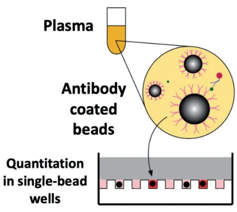
Two specific protein biomarkers, glial fibrillary acidic protein (GFAP) and tau, are relevant to this thesis work. GFAP, an intermediate-filament protein part of the astrocyte cytoskeleton, has the capability to form polymeric filaments (Z.
Yang & K. K. W. Wang 2015). Highly specific to CNS tissues, and abundantly expressed, GFAP is released into the circulation upon BBB disruption as a results of injury to astrocyte end-feet (Herrmann et al. 2000). Relevantly for acute stroke diagnostics, in HS, GFAP is rapidly elevated, but in ACI shows a significantly slower elevation (Dvorak et al. 2009). Further, levels in SM