KATIANE DE OLIVEIRA PINTO COELHO NOGUEIRA
ESTRUTURA E ULTRAESTRUTURA DO OVIDUTO DE FÊMEAS DE Hemidactylus mabouia (REPTILIA, GEKKONIDAE) NO PERÍODO VITELOGÊNICO, COM ÊNFASE NA ESTOCAGEM E FAGOCITOSE DE
ESPERMATOZÓIDES
KATIANE DE OLIVEIRA PINTO COELHO NOGUEIRA
ESTRUTURA E ULTRAESTRUTURA DO OVIDUTO DE FÊMEAS DE Hemidactylus mabouia (REPTILIA, GEKKONIDAE) NO PERÍODO VITELOGÊNICO, COM ÊNFASE NA ESTOCAGEM E FAGOCITOSE DE
ESPERMATOZÓIDES
Tese apresentada à Universidade Federal de Viçosa, como parte das exigências do Programa de Pós-Graduação em Biologia Celular e Estrutural, para obtenção do título de Doctor Scientiae.
APROVADA: 17 de setembro de 2010.
Profª. Mariana Machado Neves Prof. Robson Campos Silva
--
Profa. Sirlene Souza Rodrigues Sartori Prof. Vinícius Albano Araújo
(Coorientadora) (Coorientador)
AGRADECIMENTOS
A Universidade Federal de Viçosa e ao Programa de Pós-Graduação em Biologia Celular e Estrutural, por me proporcionarem o ensino público de qualidade.
A Deus, por sua imensa bondade em me prover a vida. Por me guiar pelos caminhos da vida me fazendo sentir, a cada instante, sua presença ao meu lado.
Ao meu esposo Leonardo, pelo amor, carinho e cumplicidade. Pela imensa dedicação à nossa família, que me permitiu ter tranqüilidade nos momentos em que precisei estar ausente.
Aos meus amados filhos Lucas e Isabela, por ser o incentivo de toda minha caminhada. Foi o pensamento em vocês que me trouxe forças nos momentos mais difíceis.
A minha mãe, que por trás de tamanha doçura esconde uma verdadeira fortaleza onde sempre encontro incentivo e disposição para lutar.
Ao meu pai, pelo orgulho e o brilho nos olhos a cada passo da minha caminhada, na qual sempre procurei seguir o exemplo de honestidade, dignidade e respeito.
Ao meu querido irmão, Amantino, pelo elo de cumplicidade e fraternidade que nos une.
Ao Sr. Edvaldo e D. June, por sempre acreditarem em mim. A toda minha família, fonte de apoio e carinho.
Ao meu mestre, professor Clóvis Andrade Neves pela orientação, pelos grandes ensinamentos e exemplo de ética profissional.
Aos meus queridos conselheiros e amigos Vinícius Albano Araújo e Sirlene Souza Rodrigues Sartori pela sabedoria, simplicidade e por estarem sempre à disposição para ajudar e ensinar.
A Universidade Federal de Ouro Preto, em especial ao chefe do Departamento de Ciências Biológicas, professor Deoclécio Alves Chianca Jr., pelo incentivo e confiança.
Aos professores do laboratório de Biologia Estrutural, em especial ao Sérgio, que em 2003 me recebeu no laboratório e me ensinou muito.
Ao meu querido amigo Vinícius, por estar presente nos momentos mais difíceis me auxiliando na microscopia eletrônica e na confecção da versão final deste trabalho. Bem humorado, otimista e sempre transmitindo palavras de apoio e carinho.
Aos professores Mariana Machado Neves e Robson Campos Silva, por terem aceitado o convite para participar deste trabalho.
Ao coordenador do Programa de Pós-graduação em Biologia Celular e Estrutural, professor José Eduardo Serrão sempre empenhado em aprimorar a qualidade do programa e também por sua infinita prestatividade.
Ao núcleo de Microscopia e Microanálise, pelo suporte da minha pesquisa. Ao Departamento de Biologia Geral pelo apoio logístico, em especial a Beth pelo empenho e disposição em ajudar.
A todos os meus contemporâneos da Biologia Celular e Estrutural.
BIOGRAFIA
Katiane de Oliveira Pinto Coelho Nogueira, filha de José Mauro Santana Pinto Coelho e Teresinha do Carmo de Oliveira Pinto Coelho. Nasceu em Viçosa, Minas Gerais, em 05 de outubro de 1981.
Em fevereiro de 2005 casou-se com Leonardo Brandão Nogueira e, em julho do mesmo ano recebeu um grande presente de Deus, seu filho Lucas Oliveira Brandão, principal incentivador de sua carreira profissional.
Em maio de 2006, graduou-se como Bacharel em Ciências Biológicas pela Universidade Federal de Viçosa (UFV), Viçosa-MG. Durante o período de graduação, de 2003 a 2006, foi estagiária do Laboratório de Biologia Estrutural do Departamento de Biologia Geral da UFV e monitora de histologia.
SUMÁRIO
RESUMO... viii ABSTRACT ... x 1. INTRODUÇÃO GERAL ...
1.1. Biologia de Hemidactylus mabouia ... 1.2. Morfofisiologia do aparelho reprodutor feminino ...… 1.3. Estocagem de espermatozóides no trato reprodutor feminino ……… 1.4. Fagocitose de espermatozóides no trato reprodutor feminino ……… 1.5. Estudos da biologia reprodutiva dos lagartos ………. 1.6. Referências Bibliográficas ...
01 01 02 03 03 04 05
2. OBJETIVOS ………. 08
3. ARTIGOS ………. 09
3.1 Capítulo 1: Oviductal Structure and Ultrastructure of the oviparous gecko, Hemidactylus mabouia (Moreau De Jonnès, 1818) ………...
3.2 Capítulo 2: Estocagem de espermatozóides na tuba uterina da lagartixa tropical, Hemidactylus mabouia (Moreau De Jonnès, 1818) ...
3.3 Capítulo 3: Phagocytosis of spermatozoa by epithelial cells in the vagina of the lizard Hemidactylus mabouia (Reptilia, Squamata) ……….
09
35
49 4. CONCLUSÕES ...
4.1. Conclusão Geral ...
RESUMO
NOGUEIRA, Katiane de Oliveira Pinto Coelho, D.Sc., Universidade Federal de Viçosa, setembro de 2010. Estrutura e ultraestrutura do oviduto de fêmeas de Hemidactylus mabouia (Reptilia, Gekkonidae) no período vitelogênico, com ênfase na estocagem e fagocitose de espermatozóides. Orientador: Clóvis Andrade Neves. Coorientadores: Vinícius Albano Araújo, Sirlene Souza Rodrigues Sartori, Sérgio Oliveira de Paula e Leandro Licursi de Oliveira.
ABSTRACT
NOGUEIRA, Katiane de Oliveira Pinto Coelho, D.Sc., Universidade Federal de Viçosa, september, 2010. Structure and ultrastructure of the oviduct of female Hemidactylus mabouia in the vitellogenic period, with emphasis on storage and phagocytosis of spermatozoa. Adviser: Clóvis Andrade Neves. Co- advisers: Vinícius Albano Araújo, Sirlene Souza Rodrigues Sartori, Sérgio Oliveira de Paula and Leandro Licursi de Oliveira.
1. INTRODUÇÃO GERAL
1.1 - Biologia de Hemidactylus mabouia
A família Gekkonidae pertence a ordem Squamata e compreende cerca de 870 espécies ovíparas e vivíparas que atualmente possuem distribuição cosmopolita (Pough et al., 1998). O geconídeo Hemidactylus mabouia é considerado uma lagartixa exótica no Brasil, possui hábitos noturnos e é encontrado principalmente em ambientes antrópicos (Pough et al., 1998). Acredita-se que as primeiras lagartixas do gênero Hemidactylus chegaram ao continente americano durante o período de colonização, trazidas da África (Vanzolini, 1978) e invadiram todos os estados brasileiros, sendo considerada uma espécie colonizadora de sucesso, devido principalmente a alta fecundidade (Meshaka et al. 1994; Bock, 1996). A alta fecundidade e a maturidade precoce são características de espécies colonizadoras que tendem a maximizar o rendimento reprodutivo (Lewontin 1965). Atualmente H. mabouia vêm colonizando também a América do Norte (Lawson et al., 1991; Butterfield et al., 1993; Meshaka et al. 1994 e Meshaka, 2000), ocupando e deslocando o nicho de duas outras espécies do gênero Hemidactylus, H. garnotti e H. turcicus (Meshaka, 1994, 2000, Eckstut et al., 2009).
grandes com grande quantidade de vitelo ou vários ovos menores (Pough et al., 1998). Geralmente, H. mabouia produz ninhadas de tamanho fixo, com dois ovos (Vitt, 1986; Meshaka et al., 1994; Anjos e Rocha, 2008). A oviparidade aumenta o rendimento reprodutivo, uma vez que a fêmea pode colocar mais de uma ninhada por estação, principalmente em climas tropicais. Dentre os Squamata, durante o período de incubação dos ovos o cuidado parental é um procedimento comum (Pough et al., 1998).
1.2 – Morfofisiologia do aparelho reprodutor feminino
Lloyd, 2010) e está relacionada com a eficiência da colonização e o estabelecimento de espécies invasoras (Conner e Crews, 1980).
Em animais ovíparos o útero geralmente é uma região glandular onde ocorre a deposição da casca do ovo. O ovo é posteriormente direcionado a vagina, porção final do oviduto que se abre na cloaca. Esta é uma região muscular que está relacionada a expulsão do ovo durante a ovoposição. A cloaca recebe os espermatozóides depositados pelo macho e os conduz ao oviduto para o recomeço do ciclo reprodutivo.
1.3 - Estocagem de espermatozóides no trato reprodutor feminino
A estocagem de espermatozóides é um fenômeno de ocorrência comum para a maioria dos répteis, pois a inseminação normalmente ocorre em período diferente da ovulação e fertilização (Schuett, 1992; Sever e Hamlett, 2002). O tempo de estocagem de espermatozóides varia dentre os Squamata. Em lagartos, foram relatados períodos de estocagem de trinta dias a um ano e seis meses, e em serpentes, de noventa dias a sete anos (Holt e Lloyd, 2010).
Na família Gekkonidae os espermatozóides normalmente ficam estocados na região da tuba uterina (Sever e Hamlett, 2002). A estocagem de espermatozóides pelas fêmeas de répteis é considerada um processo vantajoso já que diminui o risco de predação durante a cópula e aumenta a competição reprodutiva.
1.4 - Fagocitose de espermatozóides no trato reprodutor feminino
eliminados por fagocitose (Chakraborty e Nelson, 1975; Koyanagi e Nishiyama, 1981; Matthijs et al., 2000; Eisenbach, 2003; Suarez e Pacey, 2006).
Os sítios de fagocitose variam entre as espécies, já sendo relatado sua ocorrência em lagartos, nos locais de estocagem (Sever e Hopkins, 2004; Siegel e Sever, 2007).
A fagocitose de espermatozóides no trato reprodutor feminino é um processo que tem como principal funções a eliminação de espermatozóides supranumerários que não atingiram o sítio de fertilização e evitar processos inflamatórios (Eisenbach, 2003).
1.5 - Estudos da biologia reprodutiva dos lagartos
1.6 - Referências Bibliográficas
Anjos LA; Rocha CFD (2008) Hemidactylus mabouia, a fixed clutch size invader species: how do these gekkonid maximize their reproductive success? Iheringia, Sér. Zool. 98:1-5.
Birkhead TR; Mollar AP (1993) Sexual selection and the temporal separation of reproductive events: Sperm storage data from reptiles, birds and mammals. Biol. J. Linnean Soc. 50: 293-311.
Bock BC (1996) Interclutch interval and egg aggregation in the tropical gecko,
Hemidactylus mabouia. Herpetol. Rev., 27 (4): 181-183.
Butterfield BP; Hauge B; Meshaka, WE (1993) The occurrence of Hemidactylus mabouia on the United States mainland. Herp. Rev. 24(3): 111-112.
Chakraborty J; Nelson L (1975) Fate of surplus sperm in the fallopian tube of the white mouse. Biol. Reprod. 12: 455-463.
Conner J; Crews D (1980) Sperm transfer and storage in the lizard, Anolis carolinensis. J. Morphol. 163:331-348.
Cuellar O (1966) Oviductal anatomy and sperm storage structures in lizards. J. Morphol. 119: 7-20.
Eckstut ME, Lemons ER, Sever DM. 2009. Annual dynamics of sperm production and storage in the Mediterranean gecko, Hemidactylus turcicus, in the southeastern United States. Amphibia-Reptilia 30:45-56.
Eisenbach M (2003) Why are sperm cells phagocytosed by leukocytes in the female genital tract? Med. Hypotheses. 60(4): 590-592.
Fox H (1977) The urinogenital systemof reptiles. In: Gans C, Parsons TS, editors. Biology of the Reptilia. London: Academic Press. pp. 1-157.
Girling JE, Cree A, Guillette LJ Jr. (1997) Oviductal structure in a viviparous New Zealand gecko, Hoplodactylus maculatus. J. Morphol. 324:51-68.
Girling JE, Cree A, Guillette LJ Jr. (1998) Oviductal structure in four species of gekkonid lizard differing in parity mode and eggshell structure. Reprod. Fert. Develop. 10(2):139-154.
Girling JE, Guillette LJ Jr., Cree A. (2000) Ultrastructure of the uterus in an ovariectomized gecko (Hemidactylus turcicus) after administration of exogenous estradiol. J. Exp. Zool. 286(1): 76-89.
Gist DH; Fischer EN (1993) Fine structure of the sperm storage tubules in the box turtle oviduct. J. Reprod. Fertil. 97: 463-468.
Holt WV; Lloyd RE (2010) Sperm storage in the vertebrate female reproductive tract: How does it work so well? Theriogenology 73:713-722.
Koyanagi F; Nishiyama H (1981) Fate of spermatozoa that do not participate in fertilization in the domestic fowl. Cell Tissue Res. 214, 89-95.
Lawson R; Frank PG; Martin DL (1991) A gecko new to the United States herpetofauna, with notes on geckoes of the Florida Keys. Herp. Rev. 22(1): 11-12.
Lewontin RC (1965) Selection for colonizing ability, p.77-91. In: The genetics of colonizing species. HG Baker & GL Stebbins (eds.) Academic Press, New York, New York.
Matthijs A; Hakze R; Postma A et al., (2000) Leukocyte recruitment and phagocytosis of boar spermatozoa. In Johnson, L.A.; Guthrie, H.D. Boar Semen Preservation IV. pp.35-41.
Meshaka WE (2000) Colonization dynamics of two exotic geckos (Hemidactylus garnotii and H. mabouia) in Everglades National Park. J. Herpetol. 34(1): 163-168.
Meshaka WE; Butterfield BP; Hauge B. (1994) Hemidactylus mabouia as an established member of the Florida herpetofauna. Herp. Rev. 25(2): 80-81.
Murphy-Walker S; Haley SR (1996) Functional sperm storage duration in female
Hemidactylus frenatus (Family Gekkonidae). Herpetologica 52 (3): 365-373. Picariello O, Ciarcia G, Angelini F. 1989. The annual cycle of oviduct in Tarentola
m. mauritanica L. (Reptilia, Gekkonidae). Amphibia-Reptilia 10: 371-386. Pough FH; Janis CM; Heiser JB (1998) A vida dos vertebrados. São Paulo: Ateneu,
875p.
Sever DM; Hamlett WC (2002) Female sperm storage in reptiles. J. Exp. Zool. 292, 187-199.
Sever DM; Hopkins WA (2004) Oviductal Sperm storage in the ground skink
Scincella laterale Holbrook (Reptilia: Scincidae). J Exp Zool 301A:599-611. Siegel DS, Server DM. 2007. Sperm aggregations in female Agkistrodon piscivorus
(Reptilia:Squamata): a histological and ultrastructural investigation. J Morphol 269(2): 189-206.
Vanzolini PE (1978) On South American Hemidactylus (Sauria, Gekkonidae). Papéis Avulsos Zool. São Paulo. 31 (20): 307-343.
2. OBJETIVOS
2.1 Objetivos Gerais
• Gerar dados que possam ser utilizados em comparações interespecíficas de
geckonídeos;
• Contribuir para maximizar o entendimento da biologia reprodutiva de H.
mabouia.
2.2 Objetivos Específicos
• Descrever, identificar e associar, durante o período vitelogênico, a
estrutura e as peculiaridades ultraestruturais de cada segmento do oviduto de Hemidactylus mabouia;
• Caracterizar principalmente com parâmetros ultraestruturais, a estocagem
de espermatozóides na tuba uterina;
Capítulo 1
ABSTRACT. The reptiles of the Gekkonidae family display a variety of reproductive patterns, as evidenced by the presence of viviparous and oviparous representatives. The species Hemidactylus mabouia consists of oviparous individuals. In this work, the oviduct structure was examined by light microscopy after routine histological and histochemical techniques, as well as by scanning and transmission electron microscopy. The oviduct is composed of four different regions: the infundibulum, which opens into the coelomic cavity and receives the oocyte released at the time of ovulation; the uterine tube, where sperm storage takes place; the uterus, which is responsible for the eggshell production; and the vagina, the final portion of the oviduct that leads to the cloaca. The oviduct structure of H. mabouia is similar to that of other oviparous lizard species. However, some differences were observed, and the results of the present investigation can be useful to understand the reproductive biology of H. mabouia and other reptiles species.
1. Introduction
The female reproductive system of reptiles is formed by ovaries and oviducts. The term oviduct means the structures derived from the embryonic paramesonephric ducts (Wake, 1985). Oviducts are a pair of organs that are formed by morphologically distinct segments, which may have small variations between different species. Usually the reptile oviduct is divided into four or five regions called the infundibulum, uterine tube, isthmus, uterus and vagina (Blackburn, 1998). The oviducts have very important functions for reproduction. The Gekkonidae family includes both viviparous and oviparous representatives (Girling et al., 1998). In oviparous species, such as Hemidactylus mabouia, a prominent function of the oviducts is eggshell production. In viviparous species, oviducts act in the formation of the placenta (Blackburn, 1993a, 1993b, 1998; Girling, 2002; Stewart and Thompson, 1993, 1996; Yaron, 1985). In addition to the functions related to reproduction, the oviduct can also influence the reproductive success of geckos. Some species of the family Gekkonidae store sperm in specific oviduct regions, dissociating mating from fertilization.
analysis among reptile species. This study describes the histology and ultrastructure of each segment of the H. mabouia oviduct during the vitellogenic period.
2. Material and methods
Specimens of Hemidactylus mabouia were collected for this research under IBAMA (Brazilian Institute of Environment and Renewable Natural Resources) license number 10504-1. Their carcasses are lodged at the Museum of Zoology Joao Moojen, Federal University of Viçosa (UFV), Brazil. The whole experiment was conducted in accordance with the ethical principles for the laboratory use of animals published by the Brazilian College of Animal Experimentation – COBEA.
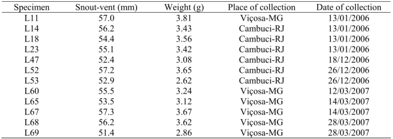
Twelve adult vitellogenic females of H. mabouia were captured from human habitations in the cities of Viçosa-MG and Cambuci-RJ, Brazil. As in the study of Cree and Guillette (1995), the follicles of vitellogenic females were firm and spherical (6-10 mm) and could be identified by gentle palpation of the abdomen and confirmed upon dissection. The snout-vent lengths of the H. mabouia specimens used in this study varied between 51.4 and 57.2 mm (Table 1), and, in accordance with the criteria established by Rocha et al. (2002), the specimens were characterized as adults.
(RM-2155, Leica) with glass knives. The sections were stained in 1% toluidine blue (TB) or with the following histochemical techniques: periodic acid-Schiff (PAS) and Alcian blue pH 2.5 (AB) for the detection of neutral and acid glycoconjugates, respectively; Nile blue sulfate (NB) (Bancroft and Steven, 1996) for acid and neutral lipid detection; and xylidine Ponceau (XP) for protein detection (Mello and Vidal, 1980). After treatments, the material was mounted on glass slides with Entellan® (Merck). The tissues were analyzed using an Olympus BX-60 microscope with a Q-Color 3 (Olympus) digital camera in the Insect Cytogenetics Laboratory - Federal University of Viçosa, Brazil.
3. Results
The oviducts are located in the coelomic cavity of females. Usually, the left oviduct’s anterior extremity is located posteriorly to the right. Each oviduct of H. mabouia can be divided anatomically into four regions: the infundibulum, the uterine tube, the uterus and the vagina (Fig. 1). The vagina is the final portion of the oviduct that opens into the cloaca. The cloaca is, in turn, an area composed of three portions where the digestive, genital and urinary systems are lead out.
Infundibulum: The infundibulum is the most anterior segment of the oviduct, which is organized in several longitudinal folds in the coelomic cavity. It possesses an ostium, through which oocytes penetrate after ovulation. The infundibulum’s mucosa is lined by a simple epithelium that contains predominantly columnar ciliated cells with few non-ciliated cells (Fig. 2A and 2B). The nuclei and apical cytoplasm of the non-ciliated cells seemed to protrude into the lumen (Fig. 2C). The lamina propria is thin, composed of loose connective tissue and lacking in glands. Underneath the mucosa, there is a thin layer of smooth muscle, one or two cells thick. The smooth muscle cells present irregular surfaces, and they have a no clearly defined orientation in the layer. The serosa, formed by simple squamous epithelium, includes the muscularis. Some non-ciliated cells possess apical granules that stained positively with AB for acid glycoconjugates but did not stain with PAS for neutral glycoconjugates.
glycoconjugates in the secretions. The coating epithelium penetrates into the lamina propria for originating crypts, which are elongate branched tubules that communicate with the lumen via ducts. These ducts contain both ciliated and non-ciliated secretory cells. Some crypts contained many stored sperm cells (Fig. 3B,E). Uterus: The uterine wall has numerous folds. From an anatomical point of view, it corresponds to the thickest area of the whole oviduct. The uterine epithelium contains both columnar ciliated and non-ciliated cells with elliptic nuclei (Fig. 4A,C). The non-ciliated cells are of two types; one type possesses an arched rough surface, while the other has narrow apices with flat surfaces (Fig. 4B). Semi-thin sections of the uterus late in the vitellogenic period illustrated that epithelium is simple, in spite of seeming stratified in most of the analyzed sections. In females of this species, the volume of the epithelial cells was significantly increased. The infra-nuclear area of the epithelial cells is rich in granular material when stained with TB. Beneath the epithelial layer, there is a profusion of small blood vessels forming an extensive plexus (Fig. 4A,D). The apical border of the non-ciliated cells stained positively for neutral and acid glycoconjugates. Granules positive for neutral and acid glycoconjugates can be seen in the connective tissue underlaying the epithelium (Fig 4E). The uterine epithelial cells did not show positive reactions with the histochemical techniques used for the detection of proteins and lipids.
the nucleus of some cells and in the lumen in the sample from late in the vitellogenic period (Fig. 5D).
Vagina: The vagina is the final region of the oviduct. The vaginal mucosa is organized into folds that increase in size as they approach the cloaca. The morphological characteristics of the vaginal wall facilitate its division into two segments: the anterior segment that lies adjacent to the uterus and the posterior segment that leads to the cloaca. Both portions of the vagina are surrounded by a muscular layer that thickens as it approaches the cloaca. The anterior segment of the vagina (anterior vagina) is lined by a simple columnar epithelium containing ciliated and non-ciliated cells (Fig. 6A-D). Ciliated cells predominate in the whole extension of the anterior vagina (Fig. 6A). Two types of non-ciliated cells were observed, one of which is typically secretory (Fig. 6D). Its secretion is metachromatic when stained with TB and stained positively for neutral and acid glycoconjugates (Fig. 7A). This type of cell prevails close to the transition between the anterior and the posterior vagina (Fig. 7B). The posterior vagina possesses stratified epithelium composed of non-ciliated cells (Fig. 7C). Its secretion gave a slightly positive reaction to the AB histochemical technique (for acid glycoconjugates) and did not stain with PAS (for glycoconjugates) or XP (for proteins).
4. Discussion
The terminology as well as the anatomical and histological division of reptilian oviducts vary among authors, mainly in interspecific comparisons. Among the geckos, in the species Hemidactylus turcicus, Saltuarius wyberba, Hoplodactylus maculatus, Hoplodactylus duvauvelii (Girling et al. 1998) and Tarentola mauritanica
infundibulum, uterine tube, isthmus, uterus and vagina. In Hemidactylus mabouia, oviducts were anatomically divided into four areas, and the region of the isthmus, which, according to Girling et al. (1998, 2002), is a small glandular region between the uterine tube and uterus, was not observed. A similar type of organization was observed by Guillette et al. (1989) in other families, such as the Crotaphytidae (Crotaphilus collaris) and Amphisbaenidae (Eumeces obsoletus). The region of the uterine tube in H. mabouia has been described as posterior infundibulum in other Squamata, such as in the lizard Calotes versicolor (Kumari et al., 1990) and the snake Seminatrix pygaea (Sever et al. 2000).
The infundibulum of H. mabouia presents non-ciliated cells that protrude into the lumen during the vitellogenic period. This type of cell was also observed in the infundibulum of Hemidactylus turcicus by Girling (2002). These observations could be explained in terms of hormonal influences on the oviduct. It is known that estradiol, a hormone secreted by the ovary, is responsible for stimulating the growth and hypertrophy of the oviduct, while progesterone induces the production and liberation of several proteins in the oviducts of mammals and reptiles (Abrams Motz and Callard, 1991; Roberts and Bazer, 1988). Among these proteins, avidin is of significant importance in reptiles (Callard et al., 1992; Guillette et al., 1991). This protein exhibits antimicrobial, nutritive, supportive and water-binding properties for the embryo in development (Palmer and Guillette, 1991).
1988). According to these authors, it seems that the eggs of reptiles of the Lepidosauria subclass have lost the albumen layer.
The observation of non-ciliated cells in the infundibulum of H. mabouia by scanning electronic microscopy was hindered by the presence of a great amount of cilia, as was also the case in Lampropholis guichenoti (Adams et al., 2004). However, non-ciliated cells are evident under light microscopy and transmission electron microscopy.
The uterine tube of H. mabouia is a discrete region; however, it cannot be considered only a transitional region due to the significant histological differences between the uterine tube, the infundibulum and the uterus. Moreover, the uterine tube is the region where sperm storage occurs, and this is perhaps one of the most important functions of the uterine tube in H. mabouia. The storage of sperm was previously observed and has been described in many species of lizards (Adams and Cooper, 1988; Blackburn, 1998; Eckstut et al., 2009; Girling et al., 1997; Murphy-Walker and Haley, 1996), snakes (Birkhead, 1993; Halpert et al., 1982; Sever and Hopkins, 2004; Sever and Ryan, 1999; Siegel and Sever, 2007), chelonians and crocodiles (Girling, 2002). The storage is essential in some species due to the asynchronous reproductive cycle of males and females. Moreover, the sperm storage allows sexual intercourse to be independent from the process of fertilization (Girling, 2002), which can explain the capacity of H. mabouia females to lay eggs throughout the year.
protection and lubrication of epithelial surfaces, which facilitates the passage of the oocyte and the egg through the oviduct (Botte 1973; Girling, 2000; Sever et al., 2000).
The uterine epithelium of H. mabouia is lined by columnar ciliated and non-ciliated cells, as it is in other oviparous lizards (Adams et al., 2004; Girling et al., 1997; 1998; 2000; Girling, 2002; Guillette et al., 1989; Palmer et al., 1993; Perkins and Palmer, 1996; Picariello et al., 1989).
Despite the increase in height observed in uterine epithelial cells late in the vitellogenic period, there is no evidence of secretion during the vitellogenic period. Girling et al. (2000) relate an increase in the size of epithelial cells in the presence of estradiol in the species Hemidactylus turcicus.
The presence of numerous uterine glands in H. mabouia, probably responsible for eggshell secretion, was also observed in others oviparous lizards of the Gekkonidae family (Girling et al., 1998; Guillette et al., 1989; Palmer et al., 1993). In viviparous species of Squamata, the number of glands is much reduced (Corso et al., 2000; Sever et al., 2000). In spite of the well-known secretory function of the uterine glands (Packard and DeMarco, 1991), they reacted negatively to the techniques for glycoconjugate and protein identification in the vitellogenic period of
H. mabouia, as observed in Hoplodactylus maculatus and Hemidactylus turcicus
(Girling et al., 1997; Girling et al., 1998). Neutral lipids were found in glands of one of samples analyzed in H. mabouia, and similar results were observed in viviparous
Seminatrix pygaea snake (Sever et al. 2000). The secretory nature of these glands is, probably, modified in function by small hormonal alterations during the sexual cycle.
and the differences are only present in response to sexual cycle phases. Adams and Cooper (1988) studied the vaginal morphology of the lizard Holbrookia propinqua
and identified three uniformly ciliated regions that they characterized as anterior, middle and posterior. In H. mabouia, the division into two regions is clearly demarcated by the epithelial transition between the anterior and posterior vagina; however, the posterior region does not present cilia.
An increase in the number and size of folds was observed in the vaginal mucosa of H. mabouia and was also observed in Sceloporus woodi (Palmer et al. 1993). A different arrangement, in which the mucosa folds become reduced in size as they approach the cloaca, was observed in other lizards (Bott 1973; Girling et al. 1997; 1998).
The vagina of H. mabouia does not possess crypts in the thin connective tissue constituting its lamina propria. This observation suggests that sperm storage does not occur in this region.
This work described, for the first time in H. mabouia, aspects of the morphology and histochemistry of the oviduct, providing data that when combined with other behavioral, ecological and morphological studies contributes to clarifying aspects of the reproductive biology of the group.
Acknowledgments
Literature Cited
Abrams-Motz V, Callard IP. 1991. Seasonal variations in oviductal morphology of the painted turtle, Chrysemys picta. J Morphol 207:59-71.
Adams CS, Cooper WE. 1988. Oviductal morphology and sperm storage in the keeled earless lizard, Holbrookia propinqua. Herpetologica, 44(2):190-197. Adams CS, Hosie MJ, Murphy CR, Thompson MB. 2004. Changes in oviductal
morphology of the skink, Lampropholis guichenoti, associated with egg production. J Morphol 262:536-544.
Bancroft JD, Stevens A. 1996. Theory and Pratice of Histological Techniques. 4th ed. Churchill Livingstone, New York.
Birkhead TR. 1993. Sexual selection and the temporal separation of reproductive events: sperm storage data from reptiles, birds and mammals. Biol J Linn Soc 50:296-311.
Blackburn DG. 1993a. Chorioallantoic placentation in squamate reptiles: Structure, function, development, and evolution. J Exp Zool 266:414-430.
Blackburn DG. 1993b. Histology of the late-stage placentae in the matrotrophic skink Chalcides chalcides (Lacertilia;Scincidae). J Morphol 216:179-195. Blackburn DG. 1998. Structure, function, and evolution of the oviducts of squamate
reptiles, with special reference to viviparity and placentation. J Exp Zool 282:560-617.
Botte V. 1973. Some aspects of oviduct development in the lizard Lacerta sicula in relation to the annual cycle. B Zool 40: 315-321.
Callard IP, Fiteti LA, Perez LE, Sorbera LA, Giannoukos G, Klosterma LL, Tsang P, Mccracken JA. 1992. Role of the corpus luteum and progesterone in the evolution of vertebrate viviparity. Am Zool 32:264-75.
Carson FL, Martin JH, Lynn JA. 1973. Formalin fixation for electron microscopy: a re-evaluation. Am J Clin Pathol 59:365-373.
Cordero-López N, Morales MH. 1995. Lack of proteins of oviductal origin in the eggs of a tropical anoline lizard. Physiol Zool 68:512-523.
Corso G, Delitala GM, Carcupino M. 2000. Uterine morphology during the annual cycle in Chalcides ocellatus tiligugu (Gmelin) (Squamata: Scincidae). J Morphol 243:153-165.
Eckstut ME, Lemons ER, Sever DM. 2009. Annual dynamics of sperm production and storage in the Mediterranean gecko, Hemidactylus turcicus, in the southeastern United States. Amphibia-Reptilia 30:45-56.
Girling JE, Cree A, Guillette LJJr. 1997. Oviductal structure in a viviparous New Zealand gecko, Hoplodactylus maculatus. J Morphol 324:51-68.
Girling JE, Cree A, Guillette LJJr. 1998. Oviductal structure in four species of gekkonid lizard differing in parity mode and eggshell structure. Reprod Fert Develop 10(2):139-154.
Girling JE, Guillette LJJr., Cree A. 2000. Ultrastructure of the uterus in an ovariectomized gecko (Hemidactylus turcicus) after administration of exogenous estradiol. J Exp Zool 286(1): 76-89.
Girling JE. 2002. The reptilian oviduct: a review of structure and function and directions for future research. J Exp Zool 293(2):141-170.
Guillette LJJr., Fox SL, Palmer BD. 1989. Oviductal morphology and egg shelling in the oviparous lizard Crotaphilus collaris and Eumeces obsoletus. J Morphol 201: 145-159.
Guillette LJJr., Demarco V, Palmer BD. 1991. Exogenous progesterone or indomethacin delays parturition in the viviparous lizard Sceloporus jarrovi. Gen Comp Endocr 81:105-112.
Halpert AP, Garstka WR, Crews D. 1982. Sperm transport and storage and its relation to the annual sexual cycle of the female red-sided garter snake,
Thamnophis sirtalis parietalis. J Morphol 174:149–159.
Kumari TRS, Sarkar HBD, Shivandappa T. 1990. Histology and histochemistry of the Oviductal sperm storage pockets of the agamid lizard Calotes versicolor. J Morphol 203:97–106.
Mello MLS, Vidal BC. 1980. Práticas de Biologia Celular. Edgard Blücher Ltda. São Paulo, Brasil.
Murphy-Walker S, Haley SR. 1996. Functional sperm storage duration in female
Hemidactylus frenatus (family Gekkonidae). Herpetologica 52:365–73.
Packard MJ, Thompson MB, Goldie KN, Vos M. 1988. Aspects of shell formation in eggs of the tuatara, Sphenedon punctatus. J Morphol 197:147-157.
Packard MJ, DeMarco VG. 1991. Eggshell structure and formation in eggs of oviparous reptiles. In ‘Egg Incubation. Its Effects on Embryonic Development in Birds and Reptiles’. (Eds D. C. Deeming and M. W. J. Ferguson.) Cambridge: Cambridge University Press.
DC; Editors. Environmental influences of avian and reptilian embryonic development. Cambridge: Cambridge University Press.
Palmer BD, Demarco VC, Guillette LJJr. 1993. Oviductal morphology and the eggshell formation in the lizard, Sceloporus woodi. J Morphol 217:205-217. Perkins JM, Palmer BD. 1996. Histology and functional morphology of the oviduct
of an oviparous snake, Diadophis punctatus. J Morphol 277:67-79.
Picariello O, Ciarcia G, Angelini F. 1989. The annual cycle of oviduct in Tarentola m. mauritanica L. (Reptilia, Gekkonidae). Amphibia-Reptilia 10:371-386. Roberts RM, Bazer FW. 1988. The functions of the uterine secretions. J Reprod
Fertil 82:875-92.
Rocha CFD, Dutra GF, Vcibradic CD, Menezes VA. 2002. The terrestrial reptile fauna of the Abrolhos archipelago: species list and ecological aspects. Braz J Biol 62(2):285-291.
Sánchez-Martínez PM, Ramírez-Pinilla MP, Miranda-Esquivel DR. 2007. Comparative histology of the vaginal-cloacal region in Squamata and its phylogenetic implications. Acta Zool (Stockholm) 88(4):289-307.
Sever DM, Hopkins WA. 2004. Oviductal Sperm storage in the ground skink
Scincella laterale Holbrook (Reptilia: Scincidae). . J Exp Zool 301A:599-611. Sever DM, Ryan TJ. 1999. Ultrastructure of the reproductive system of the black
swamp snake (Seminatrix pygaea). I. Evidence for oviductal sperm storage. J Morphol 241:1-18.
Sever DM, Ryan TJ, Morris T, Patton D, Swafford S. 2000. Ultrastructure of the reproductive system of the black swamp snake (Seminatrix pygaea). II. Annual oviductal cycle. J Morphol 245:146-160.
Siegel DS, Server DM. 2007. Sperm aggregations in female Agkistrodon piscivorus
(Reptilia:Squamata): a histological and ultrastructural investigation. J Morphol 269(2): 189-206.
Stewart JR, Thompson MB. 1993. A novel pattern of embryonic nutrition in a viviparous reptile. J Exp Biol 174:97-108.
Stewart JR, Thompson MB. 1996. Evolution of reptilian placentation: Development of extraembryonic membranes of the Australian scincid lizards, Bassiana duperreyi (oviparous) and Pseudomoia entrecasteauxii (viviparous). J Morphol 227:349-370.
Vitt LJ. 1986. Reproductive tactics of sympatric gekkonid lizards with a comment on the evolutionary and ecological consequences of invariant clutch size. Copeia 3:773-786.
Wake MH. 1985. Oviduct structure and function in non-mammalian vertebrates. Forts Zool 30:427-435.
Yaron Z. 1985. Reptilian placentation and gestation: Structure, function, and endocrine control. In: Gans C, Billet F, editors. Biology of the Reptilia. NewYork:Wiley and Sons.
Zamprogno C, Teixeira RL. 1998. Hábitos alimentares da lagartixa-de-parede
Table 1. Identification, length, weight, place of collection and date of collection. Specimen Snout-vent (mm) Weight (g) Place of collection Date of collection
L11 57.0 3.81 Viçosa-MG 13/01/2006
L14 56.2 3.43 Cambuci-RJ 13/01/2006
L18 54.4 3.56 Cambuci-RJ 13/01/2006
L23 55.1 3.42 Cambuci-RJ 13/01/2006
L47 52.4 3.08 Cambuci-RJ 18/12/2006
L52 57.2 3.65 Cambuci-RJ 26/12/2006
L53 52.9 2.62 Cambuci-RJ 26/12/2006
L60 55.5 3.24 Viçosa-MG 12/03/2007
L65 53.5 3.12 Viçosa-MG 14/03/2007
L67 57.3 3.67 Viçosa-MG 14/03/2007
L68 56.2 3.62 Viçosa-MG 28/03/2007
Legends of the figures
Figure 1. Scheme of the female reproductive tract and cloaca of the H. mabouia. a- infundibulum; b- uterine tube; c- uterus; d- vagina; e- ovaries. * - cloaca. Bar: 2 mm.
Figure 2. Light microscopy (A), scanning (B) and transmission (C) electron microscopy of the infundibulum of the H. mabouia. A and C- Ciliated (arrow) and non-ciliated cells (arrow heads). B - Ciliated (c) and non-ciliated cells (circle). Note the nucleus and apical cytoplasm of non-ciliated cells protruding into the lumen. Bars: A- 20 μm; B- 5 μm; C- 3 μm. c- cilia; L- lúmen; m- muscular tunic; n- nucleus; s- serosa.
Figure 3. Light microscopy (A), scanning (B) and transmission (C-E) electron microscopy of the uterine tube of the H. mabouia. A- Ciliated cell (arrow) in a crypt and secretory non-ciliated cells (arrow head). B– Sperm storage (arrow head) in the crypts (arrows). C- Ciliated (arrows) and secretory cells (arrow head). D- Secretory cell (arrow head). E- Sperm cell (arrow head) and secretion into the lumen. Bars: A and B- 10 μm; C-E- 2 μm. L- lumen; n- nucleus; sr- secretion.
connective tissue (arrow heads). Bars: A- 10 μm; B and E- 20 μm; C and D- 3 μm. ep- epithelium; L-lumen; sr- secretion.
Figure 5. Light microscopy (A, B and D), scanning (C) and transmission electron microscopy (E) of the uterine glands of the H. mabouia. Note in A, the duct (dc) and the secretory portion (sp) and in B, note the duct lumen (arrow). C- Base of the glands showing the external surface of the secretory portion (arrows). D- Positive reaction to the histochemical test NB, evidencing the presence of neutral fats in the base of the secretory cells. E- Section showing secretory granules (arrow heads) and the lumen (arrow). Bars: A and B- 20 μm; D- 10 μm; E- 5 μm. C- 40 μm. dc- duct; sp- secretion portion.
Figure 6. Light microscopy (A), scanning (B) and transmission electron microscopy (C and D) of the anterior vagina of the H. mabouia. A- Columnar simple epithelium covering several pleats (arrows). B- Ciliated (arrow head) and non-ciliated cells (arrow). C- Section showing the simple epithelium. D- Ciliated (arrow) and non-ciliated secretory cells (arrow head). n- nucleus; sr- secretion. Bars: A- 15 μm; B- 5 μm; C- 3 μm; D- 2 μm.
Capítulo 2
RESUMO. A estocagem de espermatozóides é um fenômeno de ocorrência comum para a maioria dos répteis. Os primeiros relatos de estocagem em fêmeas de lagartos surgiram na década de 50. Na família Gekkonidae os espermatozóides normalmente ficam estocados na região da tuba uterina, característica mais basal dentre os Squamata. Fêmeas da espécie ovípara, Hemidactylus mabouia, possuem o oviduto composto por infundíbulo, tuba uterina, útero e vagina. A tuba uterina é revestida por epitélio simples com células ciliadas e não ciliadas secretoras. A partir do revestimento interno da tuba uterina formam-se as criptas, onde os espermatozóides são estocados na forma de feixes compactos. O denso empacotamento dos espermatozóides possivelmente está relacionado a redução de atividade e manutenção da viabilidade durante o tempo de estocagem. A fertilização dos ovos por espermatozóides estocadosna tuba uterina de H. mabouia permite que as fêmeas coloquem ninhadas ao longo do ano. A estocagem diminui o risco de predação durante a cópula e aumenta a competição reprodutiva, garantido vantagens adaptativas que são inerentes a perpetuação da espécie.
1. Introdução
Na maioria das fêmeas de répteis a estocagem de espermatozóides no oviduto durante o ciclo reprodutivo é um fenômeno comum, já que a inseminação normalmente ocorre em período diferente da ovulação e fertilização (Schuett, 1992; Sever e Hamlett, 2002). No grupo dos Squamata, os primeiros relatos do processo de estocagem ocorreram nas serpentes Leptodeira annulata (Haines, 1940) e Crotalus viridis viridis (Ludwig e Hahn, 1943) e, posteriormente, no lagarto da família Chamaeleonidae, Microsaura pumila pumila (Atsatt, 1953).
Evidências de estocagem de espermatozóides baseiam-se em observações de aglomerados dessas células em regiões específicas do oviduto e na ocorrência de fertilização em fêmeas apartadas dos machos (Adams e Cooper, 1988). O tempo de estocagem de espermatozóides varia dentre os Squamata. Em lagartos, por exemplo, foram relatados períodos de estocagem de 30 dias a 1.5 anos e em serpentes, de 90 dias a 7 anos (Holt e Lloyd, 2010).
Lagartos são capazes de estocar espermatozóides na tuba uterina, na vagina ou em ambas as regiões (Sever e Hopkins, 2004; Siegel e Sever, 2007). Representantes da família Gekkonidae comumente estocam espermatozóides na tuba uterina (Cuellar, 1966; Murphy-Walker e Haley, 1996, Girling et al., 1997), sendo essa característica considerada mais basal para os Squamata (Sever e Hamlett, 2002).
Vários trabalhos descrevem aspectos estruturais e ultra-estruturais dos locais de estocagem em lagartos (Cuellar, 1966; Conner e Crews, 1980; Bou-Resli et al., 1981; Girling et al., 1997, 1998; Sever e Hamlett, 2002, Sever e Hopkins, 2004). Neste trabalho descreve-se o processo de estocagem de espermatozóides na tuba uterina de
H. mabouia, ampliando o conhecimento sobre a fisiologia reprodutiva dos Squamata.
2. Material e Métodos
2.1 Animais
Foram utilizadas dez fêmeas de Hemidactylus mabouia no período vitelogênico do ciclo reprodutivo (Licença IBAMA 10504-1). As carcaças foram depositadas no Museu de Zoologia João Moojen da Universidade Federal de Viçosa, Minas Gerais, Brasil. O experimento foi conduzido de acordo com os princípios éticos para utilização de animais em laboratório publicados no Colégio Brasileiro de Experimentação Animal – COBEA. A identificação e coleta da tuba uterina foram feitas in situ através de uma incisão longitudinal mediana na região ventral dos animais em solução salina.
2.2 Microscopia de Luz
Fragmentos da tuba uterina foram fixados em solução aquosa de formol tamponado a 10% (Carson et al., 1973) por 24 horas a temperatura ambiente e, em seguida, desidratados em concentrações crescentes de álcool. Os fragmentos foram
incluídos em glicol metacrilato (Historesin®, Leica) e seccionados na espessura de 2
μm. As secções obtidas foram coradas com azul de toluidina-borato de sódio 1%, as
lâminas montadas com Entellan® (Merk) e analisadas em microscópio Olympus
2.3 Microscopias Eletrônicas de Varredura e Transmissão
3. Resultados
O oviduto de Hemidactylus mabouia é composto por quatro regiões distintas. Da extremidade anterior para posterior, são denominadas: infundíbulo, tuba uterina, útero e vagina. Na análise morfológica a tuba uterina é revestida por epitélio simples com células ciliadas e não ciliadas secretoras (Fig.1A,C). A secreção produzida pelas células não ciliadas é metacromática quando corada com AT (Fig. 1A). A partir do revestimento interno da tuba uterina formam-se as criptas, invaginações epiteliais ramificadas. Ao longo da extensão de suas paredes as criptas são revestidas predominantemente por células ciliadas que não foram observadas na base. (Fig. 1A). A ocorrência de células não ciliadas dá um aspecto liso ao fundo das criptas quando observadas à microscopia eletrônica de varredura (Fig. 1B). O retículo endoplasmático rugoso das células não ciliadas é muito desenvolvido e o núcleo com cromatina descondensada possui uma indentação e geralmente nucléolo evidente. Algumas mitocôndrias e inúmeras vesículas também podem ser observadas dispersas no citoplasma (Fig. 2A). As células ciliadas possuem núcleo indentado com cromatina descondensada, muitas mitocôndrias, longos corpúsculos basais no citoplasma apical e quantidade moderada de retículo endoplasmático cerceando a região nuclear (Fig. 2B).
Os espermatozóides de H. mabouia são estocados no fundo das criptas, na forma de feixes compactos, com as cabeças voltadas para a base e as caudas posicionadas em direção a luz (Fig. 1B e Fig. 2C). Os espermatozóides não mantêm contato direto com as células epiteliais das criptas. Entre eles ocorre uma faixa estreita repleta de secreção. (Fig. 2D,E).
4. Discussão
A presença de espermatozóides estocados em criptas da tuba uterina de
Hemidactylus mabouia é mais uma evidência de que esta seja uma característica comum aos membros da família Gekkonidae, conforme proposto por Sever e Hamlett (2002).
O epitélio de revestimento da tuba uterina composto por células ciliadas e por células secretoras não ciliadas é muito semelhante ao descrito em outras espécies de lagartos (Bou-Resli et al., 1981; Murphy-Walker e Haley, 1996; Girling, 1998; Sever e Hamlett, 2002; Yamamoto e Ota, 2006; Eckstut et al., 2009). Não parece haver um padrão nas proporções das células ciliadas e não ciliadas entre as espécies de lagartos estudadas. Arranjo semelhante ao de H. mabouia com numerosas células ciliadas no colo e ausentes na base foi observado em Acanthodactylus scutellatus (Gekkonidae) (Sever e Hopkins, 2004) e Scincella laterale (Bou-Resli et al.1981). Em Anolis sagrei a parte basal das criptas não é especializada e a porção do colo possui uma proporção semelhante entre as células ciliadas e não ciliadas (Sever e Hamlett, 2002). A secreção produzida pelas células epiteliais não ciliadas presentes no fundo das criptas pode estar relacionada a diversas funções como nutrição (Cuellar, 1966; Bou-Resli et al., 1981), defesa, (Bou-Resli et al., 1981), quimiotaxia (Saint-Girons, 1973; Adams e Cooper, 1988) e manutenção de um ambiente aquoso propício a sobrevivência dos espermatozóides. Contudo, estudos que comprovem de forma inequívoca estas possíveis funções não foram encontrados na literatura.
não foram encontrados espermatozóides em outras regiões. Além disso, o denso empacotamento dos espermatozóides poderia ser uma maneira de diminuir sua atividade e manter a viabilidade durante o tempo de estocagem. Essa organização dos espermatozóides durante a estocagem também foi observada em outras espécies de lagartos (Fox, 1963; Conner e Crews, 1980; Sever e Hopkins, 2004; Eckstut et al., 2009) e serpentes (Hoffman e Wimsatt, 1972; Halpert et al., 1982; Perkins e Palmer, 1996). A orientação oposta dos espermatozóides, isto é, com as cabeças voltadas para a luz dos túbulos de estocagem, foi observada na serpente Seminatrix pygaea (Siegel e Sever, 2007).
Os espermatozóides de H. mabouia estocados nas criptas não estabelecem contato direto com as células epiteliais, como observado em Anolis carolinenses
(Fox 1963). No entanto, em outras espécies de lagartos, como Acanthodactylus scutellatus e Eumeces egregius espermatozóides foram observados nos espaços intercelulares e no citoplasma de células epiteliais (Bou-Resli et al., 1981; Schaefer e Roeding, 1973). Segundo Cuellar (1966) e Adams e Cooper (1988) em Holbrookia propinqua os espermatozóides também pareciam penetrar nas células epiteliais. Esse contato entre os espermatozóides e as células epiteliais também foi observado nas serpentes Thamnophis sirtalis, Thamnophis sirtalis parietalis e Agkistrodon piscivorus (Hoffman e Wimsatt, 1972; Halpert et al., 1982; Siegel e Sever, 2007). Na serpente Thamnophis sirtalis, os espermatozóides foram observados entre as células epiteliais dos túbulos de estocagem de espermatozóides e essa interação foi comparada a relação entre espermatozóide e célula de Sertoli (Hoffman e Wimsatt, 1972).
mabouia não indicam essa evacuação das criptas como um processo ativo. Possivelmente o mecanismo de liberação desses espermatozóides está relacionado à passagem do ovócito que exerce pressão mecânica contra a parede da tuba, provocando sua distensão e a liberação dos espermatozóides, conforme sugerido para outras espécies (Fox, 1956; Hoffman e Wimsatt, 1972; Siegel e Sever, 2007). A ausência de contato direto entre os espermatozóides estocados e as células epiteliais da base das criptas é uma forte evidência para corroborar tal hipótese.
O período de estocagem de espermatozóides em H. mabouia não foi monitorado, no entanto, na lagartixa tropical Hemidactylus frenatus
filogeneticamente próxima, observou-se um período mínimo de armazenamento de 36 semanas (Murphy-Walker e Haley, 1996). H. frenatus, assim como H. mabouia
possui ciclos reprodutivos que se estendem ao longo do ano, o que sugere períodos de estocagem semelhantes para essas espécies. Além disso, a possibilidade de colocar ninhadas ao longo do ano a partir da fertilização dos ovos por espermatozóides estocados diminui o risco de predação e aumenta a competição reprodutiva entre espécies que dividem o mesmo nicho. Na Flórida e no Texas, H. mabouia, H. frenatus e H. garnotti apresentam vantagem reprodutiva sobre seu congênere H. turcicus, que possui reprodução sazonal, e está sendo eliminado dessas regiões (Eckstut et al., 2009).
Referências Bibliográficas
Adams CS; Cooper WE (1988) Oviductal morphology and sperm storage in the keeled earless lizard, Holbrookia propinqua. Herpetologica 44(2):190-197. Atsatt, SR (1953) Storage of sperm in the female chameleon Microsaura pumila
pumila. Copeia 1953:59.
Bou-Resli MN; Bishay LF; Al-Zaid NS (1981) Observations on the one structure of the sperm storage crypts in the lizard Acanthodactylus scutellatus Hardyi. Arch. Biol. (Bruxelles) 92:287-298.
Carson FL, Martin JH, Lynn JA. 1973. Formalin fixation for electron microscopy: a re-evaluation. Am J Clin Pathol 59:365-373.
Conner J; Crews D (1980) Sperm transfer and storage in the lizard, Anolis carolinensis. J. Morphol. 163:331-348.
Cuellar O (1966) Oviductal anatomy and sperm storage structures in lizards. J. Morphol. 119: 7-20.
Eckstut ME; Lemons ER; Sever DM (2009) Annual dynamics of sperm production and storage in the Mediterranean gecko, Hemidactylus turcicus, in the southeastern United States. Amphibia-Reptilia 30:45-56.
Fox W (1963) Special tubules for sperm storage in female lizards. Nature 194:500-501.
Girling JE; Cree A; Guillette LJ Jr. (1997) Oviductal structure in a viviparous New Zealand gecko, Hoplodactylus maculatus. J. Morphol. 324:51-68.
Girling JE; Cree A; Guillette LJ Jr. (1998) Oviductal structure in four species of gekkonid lizard differing in parity mode and eggshell structure. Reprod. Fert. Develop. 10(2):139-154.
Haines, (1940) Delayed fertilization in Leptodeira annulata polysticta. Copeia 28, (2): 116-118.
Halpert AP; Garstka WR; Crews D (1982) Sperm transport and storage and its relation to the annual sexual cycle of the female red-sided garter snake,
Thamnophis sirtalis parietalis. J Morphol 174:149–159.
Hoffman LH; Wimsatt WA (1972) Histochemical and electron microscopic observations on the sperm receptacles in the garter snake oviduct. Am. J. Anat. 134:71-96.
Ludwig M; Rahn H (1943) Sperm Storage and Copulatory Adjustment in the Prairie Rattlesnake. Copeia 1:15-18.
Murphy-Walker S; Haley SR (1996) Functional sperm storage duration in female
Hemidactylus frenatus (Family Gekkonidae). Herpetologica 52 (3): 365-373. Nogueira KOPC (2008) Morfologia e ultra-estrutura do oviduto de Hemidactylus
mabouia (Moreau de Jonnès, 1818) (Reptilia, Squamata, Sauria, Gekkonidae) durante o ciclo reprodutivo. 42f. Tese.Universidade Federal de Viçosa, Viçosa, MG, 2008.
Saint Girons H (1962) Presence de receptacles seminaux chez les cameleons. Beaufortia 9:165–172.
Saint Girons H (1973) Sperm survival and transport in the female genital tract of reptiles. In: Hafez ESE, Thibault CG, editors. The Biology of Spermatozoa. Basel, Switzerland: Karger. pp. 105–113.
Schafer GC; Roeding CE (1973) Evidence for vaginal sperm storage in the mole skink, Eumeces egregius. Copeia 1973, 346-347.
Schuett GW (1992) Is long-term sperm storage an important component of the reproductive biology of temperate pitvipers? In: Campbell JA, Brodie ED Jr, editors. Biology of the pitvipers.Tyler,TX: Selva. pp. 169-184.
Sever DM; Hamlett WC (2002) Female sperm storage in reptiles. J. Exp. Zool. 292, 187-199.
Sever DM; Hopkins WA (2004) Oviductal Sperm storage in the ground skink
Scincella laterale Holbrook (Reptilia: Scincidae). . J Exp Zool 301A:599-611. Siegel DS, Server DM. 2007. Sperm aggregations in female Agkistrodon piscivorus
(Reptilia:Squamata): a histological and ultrastructural investigation. J Morphol 269(2): 189-206.
Legendas das Figuras
Figura 1. Micrografias de luz (A), eletrônica de varredura (B) e transmissão (C) da região da tuba uterina do oviduto de Hemidactylus mabouia. A-Secção longitudinal mostrando célula ciliada (seta), células não ciliadas (cabeça de seta) e uma cripta (asterisco). B-Note a superfície das células não ciliadas que confere um aspecto liso no fundo das criptas (cabeças de seta) e a região da luz das criptas repleta de espermatozóides (seta). C-Secção longitudinal mostrando detalhes de células ciliadas (seta) e não ciliadas (cabeça de seta). bl = lâmina basal; n = núcleo. Barras: A = 20 µm; B = 10 µm e C = 5 µm.
Capítulo 3
ABSTRACT: Hemidactylus mabouia is an oviparous lizard dispersed across almost all continents. In countries with tropical climates, such as Brazil, these lizards are capable of breeding during the entire year. Such reproductive capacity may be attributed to a peculiar reproductive biology factor in the majority of reptiles, possibly the ability of females to store spermatozoa in specialized regions of the genital tract. Considering that in H. mabouia thestorage of spermatozoa is restricted to the region of the uterine tube, in this study we utilized optical and transmission electron microscopy to investigate the processes related to the large number of spermatozoa in the vagina. Although it was possible to visualize spermatozoa in the vagina, an ultrastructural analysis of the region revealed that significant phagocytosis occurs, which is mediated by the epithelial cells. Such a process indicates that the anterior portion of the vagina is related to the elimination of supernumerary or deficient spermatozoa and not storage.
1. Introduction
In the majority of vertebrates, during mating, part of the spermatozoa deposited in the female reproductive tract does not reach the fertilization site. The residual spermatozoa are eliminated from the female reproductive tract by phagocytosis (Chakraborty and Nelson, 1975; Koyanagi and Nishiyama, 1981; Matthijs et al., 2000; Eisenbach, 2003; Suarez and Pacey, 2006). Phagocytosis of spermatozoa in the female reproductive tract is characterized as a slow process beginning with the formation of pseudopodia, followed by the consumption of entire spermatozoa or their fragments (Mori and Uchida, 1974; Murakami et al., 1985; Phillips and Mahler, 1977a, 1977b).
The sites of phagocytosis vary between species; in lizards, its occurrence has been reported in storage locations, such as those in the uterine tube (Sever and Hopkins, 2004) and vagina (Siegel and Sever, 2007). Spermatozoa were reported to be present in the vagina of the lizards Eumeces egregius and Hemidactylus frenatus
(Schafer and Roeding, 1973 and Eckstut et al. 2009); however, this region was not specified as the location of phagocytosis or storage due to the absence of specialized structures for this function, such as crypts.
characterized in this study the structure and ultrastructure of this region, searching for explanations for the large number of these cells outside the storage site.
2. Material and methods
2.1 Animals: Six female Hemidactylus mabouia lizards were collected under license number 10504-1 of IBAMA, and the carcasses were deposited at the João Moojen Animal Science Museum of the Universidade Federal de Viçosa (UFV), Brazil. The experiment was conducted in accordance with the ethical principles for utilization of laboratory animals published in the Colégio Brasileiro de Experimentação Animal – COBEA. The snout-vent length of the specimens utilized varied between 51 and 57 cm and is in accordance with the criteria established by Rocha et al. (2002) for their characterization as adults. Identification and collection of the reproductive organs were done in situ via a median longitudinal incision in the ventral region of the animals in a saline solution.
2.2 Optical Microscopy: Fragments of the vagina were fixed in a buffered formalin solution (Carson et al., 1973) for 24 hours at room temperature and were then dehydrated in increasing concentrations of ethanol. The fragments were embedded in
glycol methacrylate (Historesin®, Leica) and sectioned at a width of 2 μm in an automatic microtome (Reichert-Jung, Germany). The obtained sections were stained
with 1% toluidine blue/sodium borate and mounted on slides with Entellan®
(Merck) for analysis with an Olympus BX-60 microscope.
dehydrated in increasing series of acetone and embedded in Epon resin. Ultrathin sections were obtained in an ultramicrotome and contrasted with 2% uranyl acetate and 0.2% lead citrate. The material was examined and photographed in an EM 109- Zeiss transmission electron microscope.
3. Results
The female reproductive system of H. mabouia is composed of the ovaries and oviducts. Each oviduct is formed by four histologically distinct regions, known as the infundibulum, the uterine tube, the uterus and the vagina (Fig. 1A).
The anterior portion of the vagina is composed of a muscular tube internally lined with a simple prismatic epithelium containing ciliated cells and two types of ciliated cells (Fig. 1B, 2A). An ultrastructural analysis showed that the non-ciliated cells may be characterized as secretory and phagocytic (Fig. 2A).
The ciliated cells predominate and possess long cilia with a well-developed basal corpuscle. The nucleus has an oval shape and is located in the basal third of the cell. The numerous mitochondria are concentrated in the supranuclear region (Fig 2A).
Secretory cells present a rounded nucleus near their base and extensive rough endoplasmic reticulum. The apical third of the cell is filled by large secretory granules with different densities (Fig. 2A).
In the cytoplasm, near the phagosomes containing spermatozoa, small oval-shaped electron-lucent vesicles measuring roughly 0.4 μm along their greatest axis were observed (Fig. 3B). Each vesicle had one eccentrically located electron-dense granule in its interior (Fig. 3B).
Sometimes the material in the digestion process can be identified as fragments of the heads and tails of the spermatozoa, indicating that these cells are completely consumed (Fig. 3A, 3C). The majority of spermatozoa are consumed along their longitudinal axis; however, spermatozoa in other orientations were consumed inside the cells (Fig. 3C).
4. Discussion
Studies of the morphology of the oviduct in reptiles have described the ciliated and non-ciliated cells of the vaginal epithelium (Girling et al. 1997, 1998; Girling, 2002; Sever et al. 2000). However, these studies considered the non-ciliated cells to function solely as secretory cells. In this study, we showed that there are two non-ciliated cell types in H. mabouia, one secretory type commonly reported for reptiles and the other type characterized as an epithelial phagocytic cell. The epithelial phagocytic cells were first reported in the oviducts of mammals (Austin, 1960) and possess typical morphological characteristics different from those characterized for the professional phagocytes (Sever, 1992).
Many of the spermatozoa phagocytosed by the epithelial phagocytic cells of H. mabouia were enveloped by vesicles in the cytoplasm. It is possible that these vesicles containing electron-dense granules are lysosomes because they were observed grouped around material in the process of being digested. The phagocytosed spermatozoa may present defects that were not observed in morphological analyses; the anterior vagina is believed to be the location of selection, in which spermatozoa without fertilization capacity are eliminated before they can generate an inflammatory response, as observed in mammals (Eisenbach, 2003).
explains the large number of these cells observed in the anterior portion of the vagina.
Acknowledgments
We thank the Microscopy and Microanalysis Nucleus of the Federal University of Viçosa, as well as CAPES and the FAPEMIG for their financial support.
5. References
Austin CB (1960) Fate of spermatozoa in the female genital tract. J. Reprod. Fertil. 1, 151-156.
Chakraborty J; Nelson L (1975) Fate of surplus sperm in the fallopian tube of the white mouse. Biol. Reprod. 12, 455-463.
Eckstut ME; Lemons ER; Sever DM (2009) Annual dynamics of sperm production and storage in the Mediterranean Gecko, Hemidactylus turcicus, in the
southeastern United States. Amphibia–Reptilia 30, 45-56.
Eisenbach M (2003) Why are sperm cells phagocytosed by leukocytes in the female genital tract? Med. Hypotheses. 60(4), 590-592.
Girling JE; Cree A; Guillette LJ Jr. (1997) Oviductal structure in a viviparous New Zealand gecko, Hoplodactylus maculatus. J. Morphol. 324, 51-68.
Girling, JE; Cree A; Guillette LJ (1998) Oviductal structure in four species of gekkonid lizard differing in parity mode and eggshell structure. Reprod. Fertil. Dev. 10, 139-154.
Girling JE (2002) The reptilian oviduct: A review of structure and function and directions for future research. J. Exp. Zool. 293, 141-170.
Koyanagi F; Nishiyama H (1981) Fate of spermatozoa that do not participate in fertilization in the domestic fowl. Cell Tissue Res. 214, 89-95.
Matthijs A; Hakze R; Postma A et al. (2000) Leukocyte recruitment and phagocytosis of boar spermatozoa. In Johnson, L.A.; Guthrie, H.D. Boar Semen Preservation IV. pp.35-41.
Mori T; Uchida T (1974) Electron microscopic analysis of the mechanism of fertilization in Chiroptera 11. Engulfment of spermatozoa by epithelial cells of the fallopian tube in the Japanese house bat, Pipistrellus abramus. Zoo. Mag. 83, 163-170.
Murakami M; Nishida T; Shiromoto M; Iwanaga S (1985) Phagocytosis of spermatozoa and latex beads by the epithelial cell of the cat oviduct: combined SEM and TEM study. Arch. histol. jap. 48(5), 519-526.
Olsson M; Madsen T (1998) Sexual selection and sperm competition in reptiles. London: Academic Press. p 503-570.
Phillips DM; Mahler S (1977b) Leucocyte emigration and migration in the vagina following mating in the rabbit. Anat. Rec. 189, 45-59.
Rocha CFD; Dutra GF; Vcibradic CD; Menezes VA (2002) The terrestrial reptile fauna of the Abrolhos archipelago: species list and ecological aspects. Braz. J. Biol. 62(2), 285-291.
Schafer GC; Roeding CE (1973) Evidence for vaginal sperm storage in the mole skink, Eumeces egregius. Copeia 1973, 346-347.
Sever DM (1992) Spermiophagy by the spermathecal epithelium of the salamander
Eurycea cirrigera. J. Morphol. 212, 281-290.
Sever DM; Brunette; NS (1993) Regionalization of eccrine and spermiophagic activity in spermathecae of the salamander Eurycea cirrigera (Amphibia: Plethodontidae). J. Morphol. 217, 161-170.
Sever DM; Ryan TJ; Morris T; Patton D; Swafford S (2000) Ultrastructure of the reproductive system of the black swamp snake (Seminatrix pygaea). II. Annual oviductal cycle. J. Morphol. 245, 146-160.
Sever DM; Hamlett WC (2002) Female sperm storage in reptiles. J. Exp. Zool. 292, 187-199.
Sever DM; Hopkins WA (2004) Oviductal sperm storage in de Ground Skink
Scincella lateralae Holbrook (Reptilia; Scincidae) J. Exp. Zool. 301A, 509-611.
Sheehan D; Hrapchak B (1980) Theory and practice of Histotechnology, second ed. Battelle Press, Ohio.
Siegel DS; Server DM (2007) Sperm aggregations in female Agkistrodon piscivorus
(Reptilia:Squamata): a histological and ultrastructural investigation. J. Morphol. 269(2), 189-206.
Legends of the figures
Figure 1. A- Scheme of the right portion of the female reproductive system of H. mabouia.
a- infundibulum, b- uterine tube, c- uterus, d- vagina, o- ovary. Bar:1.5 mm. B- Light microscopy of the anterior portion of the vagina. Arrow- phagocytic cell, white arrow head- ciliated cell, black arrow head- secretory cell. ct = connective tissue. Bar: 20 μm.
Figure 2. A- Transmission electron microscopy of the anterior portion of the vagina. PC-phagocytic cell, CC-ciliated cell, SC- secretory cell. c = cilia, is = intercellular space, m = mitochondria, n = nucleus, sg = secretory granule. Bar: 5 μm.
Figure 1
4. CONCLUSÕES
As análises desenvolvidas no presente estudo permitiram concluir que em fêmeas de H. mabouia:
1- O oviduto é composto por quatro segmentos – infundíbulo, tuba uterina, útero e vagina – evidenciados por seus aspectos peculiares através da associação de técnicas histológicas e ultraestruturais que permitiram a caracterização e identificação de cada região;
2- Não ocorre a região do istmo entre a tuba uterina e o útero, como descrito para outras espécies de geckonídeos;
3- A estocagem de espermatozóides em criptas na tuba uterina é um processo integrante do ciclo reprodutivo de H. mabouia; e
4- A vagina anterior é local de intensa fagocitose e seleção de espermatozóides.
4.1. Conclusão Geral