Identification of Symptomatic Fetuses
Infected with Cytomegalovirus Using
Amniotic Fluid Peptide Biomarkers
Cyrille Desveaux1☯¤, Julie Klein2,3☯, Marianne Leruez-Ville4,5, Adela Ramirez-Torres6,
Chrystelle Lacroix3,7, Benjamin Breuil2,3, Carine Froment3,7, Jean-Loup Bascands2,3, Joost P. Schanstra2,3, Yves Ville1,5*
1Department of Obstetrics and Fetal Medicine, Hospital Necker-Enfants-Malade, APHP, Paris, France, 2Institut National de la Santé et de la Recherche Médicale (INSERM), U1048, Institute of Cardiovascular and Metabolic Disease, Toulouse, France,3Université Toulouse III Paul-Sabatier, Toulouse, France, 4Department of Virology, Hospital Necker-Enfants Malades, AP-HP, Paris, France,5University Paris Descartes, EA 7328, Paris, France,6Sanford Burnham Prebys Medical Discovery Institute, La Jolla, California, United States of America,7Centre National de la Recherche Scientifique, Institut de Pharmacologie et de Biologie Structurale, Toulouse, France
☯These authors contributed equally to this work.
¤ Current Address: Department of Obstetrics, La Reunion University Hospital, Saint Pierre, La Réunion
*ville.yves@gmail.com
Abstract
Cytomegalovirus (CMV) is the most common cause of congenital infection, and is a major cause of sensorineural hearing loss and neurological disabilities. Evaluating the risk for a CMV infected fetus to develop severe clinical symptoms after birth is crucial to provide appropriate guidance to pregnant women who might have to consider termination of preg-nancy or experimental prenatal medical therapies. However, establishing the prognosis before birth remains a challenge. This evaluation is currently based upon fetal imaging and fetal biological parameters, but the positive and negative predictive values of these parame-ters are not optimal, leaving room for the development of new prognostic factors. Here, we compared the amniotic fluid peptidome between asymptomatic fetuses who were born as asymptomatic neonates and symptomatic fetuses who were either terminated in view of severe cerebral lesions or born as severely symptomatic neonates. This comparison allowed us to identify a 34-peptide classifier in a discovery cohort of 13 symptomatic and 13 asymptomatic neonates. This classifier further yielded 89% sensitivity, 75% specificity and an area under the curve of 0.90 to segregate 9 severely symptomatic from 12 asymptomatic neonates in a validation cohort, showing an overall better performance than that of classical fetal laboratory parameters. Pathway analysis of the 34 peptides underlined the role of viral entry in fetuses with severe brain disease as well as the potential importance of both beta-2-microglobulin and adiponectin to protect the injured fetal brain infected with CMV. The results also suggested the mechanistic implication of the T calcium channel alpha-1G (CACNA1G) protein in the development of seizures in severely CMV infected children. These results open a new field for potential therapeutic options. In conclusion, this study demonstrates that amniotic fluid peptidome analysis can effectively predict the severity of OPEN ACCESS
Citation:Desveaux C, Klein J, Leruez-Ville M, Ramirez-Torres A, Lacroix C, Breuil B, et al. (2016) Identification of Symptomatic Fetuses Infected with Cytomegalovirus Using Amniotic Fluid Peptide Biomarkers. PLoS Pathog 12(1): e1005395. doi:10.1371/journal.ppat.1005395
Editor:Sallie R. Permar, Duke University, UNITED STATES
Received:August 24, 2015
Accepted:December 21, 2015
Published:January 25, 2016
Copyright:© 2016 Desveaux et al. This is an open access article distributed under the terms of the Creative Commons Attribution License, which permits unrestricted use, distribution, and reproduction in any medium, provided the original author and source are credited.
Data Availability Statement:All relevant data are within the paper and its Supporting Information files.
Funding:CE-MS equipment was funded by the Fondation pour la Recherche Médicale“Grands Equipements pour la Recherche Biomédicale”and the CPER2007-2013 program. The funders had no role in study design, data collection and analysis, decision to publish, or preparation of the manuscript.
congenital CMV infection. This peptidomic classifier may therefore be used in clinical set-tings during pregnancy to improve prenatal counseling.
Author Summary
CMV is the most common cause of congenital infection, and can result in significant neo-natal morbidity and neurological disabilities. The birth prevalence of congenital CMV is estimated at 0.7% worldwide, and 10 to 20% of these neonates develop severe symptoms. In such cases the outcome is generally poor. Therefore, identification of additional prog-nostic markers is crucial for prenatal counseling in cases with an infected fetus. This may influence the decision of continuing with the pregnancy or requesting its termination, but also the decision of starting experimental antiviral therapy. The pathophysiology of CMV brain injury is not completely understood, and the identification of new biomarkers of CMV infection might also pave the way towards the development of new therapeutic alter-natives. Here, we apply a recently developed and modern non-targeted peptidomics approach to amniotic fluid obtained from symptomatic and asymptomatic CMV-infected fetuses/neonates, followed by network analysis of the peptides of interest in the context of fetal infection and in relation with outcome. Our study identified 34 amniotic fluid pep-tides that form new prognostic biomarkers that could be used in clinical settings to improve prenatal counseling. In addition, this study provides novel mechanistic insight into the pathobiology of CMV congenital disease.
Introduction
Cytomegalovirus (CMV) is the most common cause of congenital infection with an incidence of 0.7% at birth [1]. Congenital CMV infection is the leading cause of non-genetic hearing loss and the most frequent viral cause of neurodevelopmental delay. Primary maternal CMV infec-tion in pregnancy carries a 30% to 40% risk of vertical transplacental transmission, and 10% of those infected fetuses will be born as infected infants with clinical symptoms and long-term disabilities including sensorineural hearing loss and cognitive deficits such as mental retarda-tion, cerebral palsy or seizures [2]. In addition, 5 to 10% of asymptomatic infants will develop milder forms of sensorineural hearing loss and of psychomotor delay later in life [2].
prognostic value of fetal platelet count, CMV DNA levels and beta-2-microglobulin serum lev-els [8,9]. However, this procedure is invasive and is associated with a 1–3% risk of fetal loss. Finally, the prognostic value of the levels of CMV DNA loads in amniotic fluid is controversial [10–13]. Altogether, the prenatal prognosis of fetal CMV infection remains difficult to estab-lish. In the past, the only option discussed when infected fetuses were suspected of severe dis-abilities was elective termination of pregnancy (TOP). Alternative medical strategies such as CMV hyperimmune globulins and other antiviral drugs are being evaluated with recent studies showing conflicting results on the incidence of sequelae among treated infected fetuses [14] [15]. Therefore, the identification of new effective prognostic markers is critical to help counseling pregnant women carrying an infected fetus through the dilemma of continuing or not with their pregnancy but also to decide upon starting medical therapyin utero.
Amniotic fluid has been subjected to proteome analysis (i.e. analysis of the global protein
content of a sample) for the identification of biomarkers of several prenatal conditions [16]. Specific amniotic fluid proteins have been related to various conditions affecting the fetus including Down-syndrome [17][18], intrauterine inflammation leading to preterm birth [19] [20], preterm premature rupture of the membranes [21] and intrauterine growth restriction [22]. Moreover, we and others have shown that capillary electrophoresis coupled to mass spec-trometry (CE-MS) analysis of body fluids can help identifying peptide-based biomarkers of dis-ease which can be clinically relevant [23–25].
In this study, our aim was to analyze the amniotic fluid peptidome of CMV infected fetuses at mid-gestation and to evaluate the presence of peptide biomarkers that could contribute to improve the prognostic evaluation of this condition.
Results
Identification of amniotic fluid peptides associated with the severity of
postnatal outcome
CE-MS-based peptidome analysis using 150μL of amniotic fluid from 47 samples from CMV
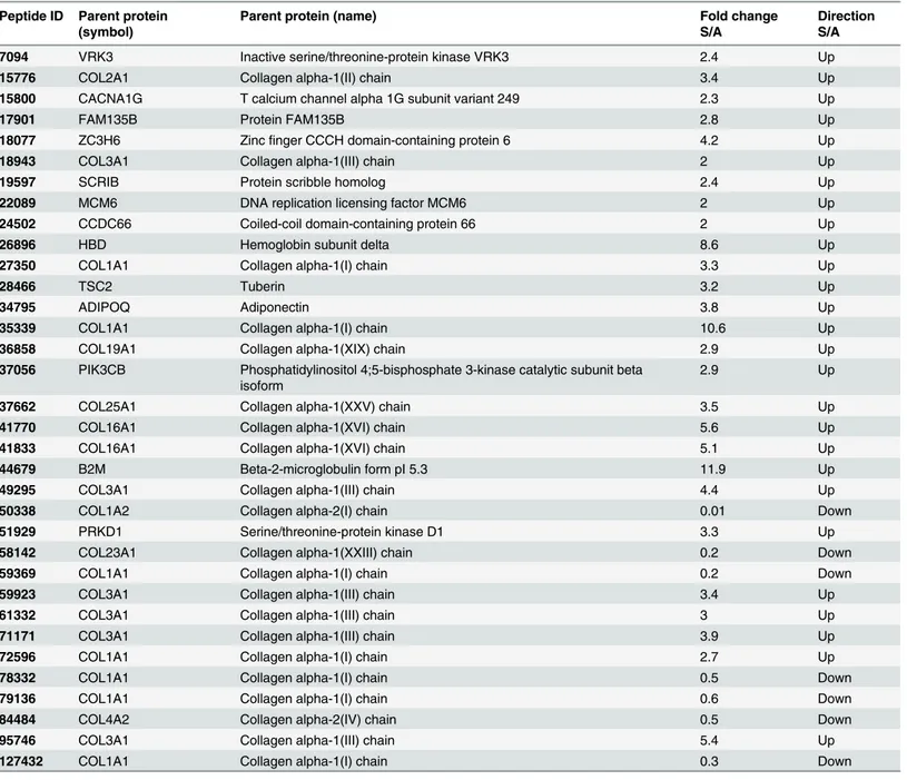
infected fetuses allowed the detection of a total of 4076 different peptides in all samples (Fig 1A). The samples were further divided in a discovery and validation cohorts (Table 1). The discovery cohort consisted of 26 pregnancies with primary CMV infection including 13 severely symptomatic cases (Table 1) and 13 asymptomatic cases. All amniotic fluid samples were collected between 17 and 29 weeks of gestation with a mean of 22 weeks of gestation in the asymptomatic group and of 23 weeks in the severely symptomatic group (p = 0.43, not sig-nificant) (Table 1andS1 Table). Follow-up of asymptomatic infants was obtained up to 12 to 53 (mean 24) months of age. In the severely symptomatic group, 12 fetuses underwent TOP for severe brain lesions and all but one were confirmed at necropsy (Table 2). One child was born alive but developed as severely handicapped (Table 2). Comparison of the amniotic fluid pep-tides content of the discovery cohort led to the identification of 76 peppep-tides that were consis-tently differentially excreted between severely symptomatic and asymptomatic cases between these two groups (Fig 1B,S1andS2Datasets). These 76 amniotic fluid peptides are identified by a unique tag composed of specific mass and migration time. In the next step, using a combi-nation of LC-MS/MS and CE-MS/MS, sequence information could be obtained in 37 of these 76 peptides. Most of these peptides were fragments of various collagens. However, a number of other non collagen-related peptides (13 out of 37) were also observed, including a fragment of hemoglobin subunit delta and of beta-2-microglobulin displaying a strong increase in abun-dance in amniotic fluid of symptomatic patients (>8-fold change)(Table 3). The 37 sequenced
peptides were reduced to a support vector machine (SVM) classifier with 34 peptides, called
candidates minus one. Peptides that did not influence the accuracy of the classifier in the total cross-validation of the training data were left out of the final classifier. Scoring the patients from the discovery cohort with the resulting CMV34 classifier clearly separated the majority of the symptomatic from asymptomatic patients (Fig 1C).
Fig 1. Detected peptides in amniotic fluid and differences between symptomatic and asymptomatic cases in the discovery cohort.(A) Representation of 4076 peptides, detected in all amniotic fluid samples (n = 47) by CE-MS. Each peptide was identified by a unique identifier based on the migration time (min) and specific mass (kDa), with a peak height representing the relative abundance. (B) In the discovery cohort, 76 amniotic fluid peptides were identified as differentially secreted between symptomatic and asymptomatic patients. (C) Cross-validation score of a SVM peptide classifier called CMV34 consisting of 34 of the 37 sequenced peptides obtained from the analysis of the discovery cohort.***p<0.0001, Mann-Whitney test for independent samples. Asympt., asymptomatic; Sympt., symptomatic.
doi:10.1371/journal.ppat.1005395.g001
Table 1. Patient characteristics.
Discovery cohort Validation cohort
Asympt. Sympt. Asympt. Sympt. Hearing loss and vestibular dysfunction
Number 13 13 12 9 9
Gestational age at sampling (weeks of gestation) 22.4±0.9 23.0±0.5 24.7±1.4 23.4±1.0 22.6±0.9
CMV DNA levels in amnioticfluid (log10 copies/mL) 5.4±0.9 7.4±0.3** 5.8±0.3 7.2±0.5* 7.3±0.4
CMV DNA levels in fetal blood (log10 copies/mL) 4.2±0.3 5.2±0.2* 4.3±0.3 5.6±0.2* 4.9±0.2
*p<0.05 and**p<0.01 compared to asymptomatic group, Mann-Whitney test for independent samples. Data are presented as mean±SEM. Asymt.: asymtomatic; Sympt.: symptomatic.
Validation of the multidimensional amniotic fluid peptide classifier in a
blinded validation cohort
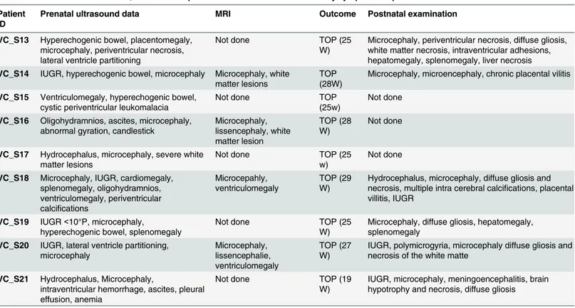
In the next step the CMV34 classifier was validated in a separate blinded cohort using 21 amni-otic fluid samples of primary CMV infection pregnancies. Amniocentesis was performed between 18 and 30 weeks of gestation with means of 25 and 23 weeks in the asymptomatic and in the severely symptomatic groups respectively (p = 0.49, not significant)(Table 1andS1 Table). The cohort consisted of 9 severely symptomatic and 12 asymptomatic fetuses that were not used in the discovery phase (Table 1). In the severely symptomatic group, all cases under-went TOP for severe brain lesions (Table 4). Six out of the 9 symptomatic fetuses had an autopsy and all confirmed the severity of the lesions. In the other 3 cases, although autopsy was not performed, prenatal imaging described indisputably severe cerebral lesions on ultrasound
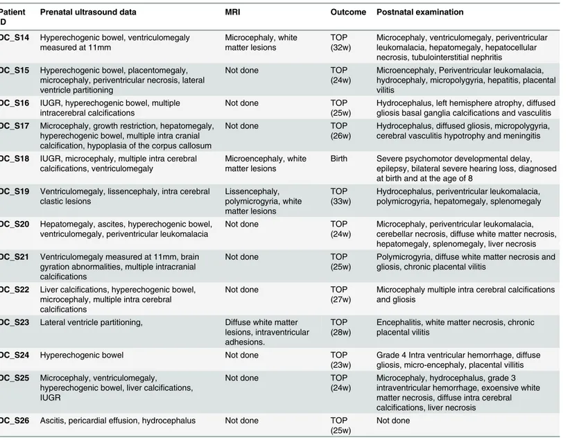
Table 2. Prenatal ultrasound data, outcome and postnatal examination of the severely symptomatic patients from the discovery cohort.
Patient ID
Prenatal ultrasound data MRI Outcome Postnatal examination
DC_S14 Hyperechogenic bowel, ventriculomegaly measured at 11mm
Microcephaly, white matter lesions
TOP (32w)
Microcephaly, ventriculomegaly, periventricular leukomalacia, hepatomegaly, hepatocellular necrosis, tubulointerstitial nephritis DC_S15 Hyperechogenic bowel, placentomegaly,
microcephaly, periventricular necrosis, lateral ventricle partitioning
Not done TOP
(24w)
Microencephaly, Periventricular leukomalacia, hydrocephaly, micropolygyria, hepatitis, placental vilitis
DC_S16 IUGR, hyperechogenic bowel, multiple intracerebral calcifications
Not done TOP
(25w)
Hydrocephalus, left hemisphere atrophy, diffused gliosis basal ganglia calcifications and vasculitis DC_S17 Microcephaly, growth restriction, hepatomegaly,
hyperechogenic bowel, multiple intra cranial calcification, hypoplasia of the corpus callosum
Not done TOP
(26w)
Hydrocephalus, diffused gliosis, micropolygyria, cerebral vasculitis hypotrophy and meningitis
DC_S18 IUGR, microcephaly, multiple intra cerebral calcifications, ventriculomegaly
Microencephaly, white matter lesions
Birth Severe psychomotor developmental delay, epilepsy, bilateral severe hearing loss, diagnosed at birth and at the age of 8
DC_S19 Ventriculomegaly, lissencephaly, intra cerebral clastic lesions Lissencephaly, polymicrogyria, white matter lesions TOP (33w)
Hydrocephalus, periventricular leukomalacia, polymicrogyria, hepatomegaly, splenomegaly
DC_S20 Hepatomegaly, ascites, hyperechogenic bowel, ventriculomegaly, periventricular leukomalacia
Not done TOP
(24w)
Microcephaly, periventricular leukomalacia, cerebellar necrosis, diffuse white matter necrosis, hepatomegaly, splenomegaly, liver necrosis DC_S21 Ventriculomegaly measured at 11mm, brain
gyration abnormalities, multiple intracranial calcifications
Not done TOP
(25w)
Polymicrogyria, diffuse white matter necrosis and gliosis, chronic placental vilitis
DC_S22 Liver calcifications, hyperechogenic bowel, microcephaly, multiple intra cerebral calcifications
Not done TOP
(27w)
Microcephaly multiple intra cerebral calcifications and gliosis
DC_S23 Lateral ventricle partitioning, Diffuse white matter lesions, intraventricular adhesions.
TOP (28w)
Encephalitis, white matter necrosis, chronic placental vilitis
DC_S24 Hyperechogenic bowel Not done TOP
(23w)
Grade 4 Intra ventricular hemorrhage, diffuse gliosis, micro-encephaly, placental villitis DC_S25 Microcephaly, ventriculomegaly,
hyperechogenic bowel, liver calcifications, IUGR
Not done TOP
(24w)
Microcephaly, hydrocephalus, grade 3 intraventricular hemorrhage, exoensive white matter necrosis, diffuse intra cerebral calcifications, liver necrosis
DC_S26 Ascitis, pericardial effusion, hydrocephalus Not done TOP (25w)
Not done
TOP, termination of pregnancy; IUGR, intra uterine growth restriction; w, weeks of pregnancy
that correlate well with all other cases terminated on the same basis and confirmed by postmor-tem examination. We have therefore no reason to believe that such a rich cerebral semeiology could be a false positive interpretation. The mean postnatal follow-up of the asymptomatic group was 8 months.
Amniotic fluid samples were analyzed by CE-MS (S3andS4Datasets), scored using the CMV34 classifier and then compared with the clinical data. We first verified that the CMV34 classifier score was independent of gestational age (Spearman r = -0.3992, not significant) (Fig 2A). The CMV34 classifier predicted a symptomatic/asymptomatic outcome with 89%
Table 3. Peptides used in the CMV34 classifier.
Peptide ID Parent protein (symbol)
Parent protein (name) Fold change
S/A
Direction S/A
7094 VRK3 Inactive serine/threonine-protein kinase VRK3 2.4 Up
15776 COL2A1 Collagen alpha-1(II) chain 3.4 Up
15800 CACNA1G T calcium channel alpha 1G subunit variant 249 2.3 Up
17901 FAM135B Protein FAM135B 2.8 Up
18077 ZC3H6 Zincfinger CCCH domain-containing protein 6 4.2 Up
18943 COL3A1 Collagen alpha-1(III) chain 2 Up
19597 SCRIB Protein scribble homolog 2.4 Up
22089 MCM6 DNA replication licensing factor MCM6 2 Up
24502 CCDC66 Coiled-coil domain-containing protein 66 2 Up
26896 HBD Hemoglobin subunit delta 8.6 Up
27350 COL1A1 Collagen alpha-1(I) chain 3.3 Up
28466 TSC2 Tuberin 3.2 Up
34795 ADIPOQ Adiponectin 3.8 Up
35339 COL1A1 Collagen alpha-1(I) chain 10.6 Up
36858 COL19A1 Collagen alpha-1(XIX) chain 2.9 Up
37056 PIK3CB Phosphatidylinositol 4;5-bisphosphate 3-kinase catalytic subunit beta isoform
2.9 Up
37662 COL25A1 Collagen alpha-1(XXV) chain 3.5 Up
41770 COL16A1 Collagen alpha-1(XVI) chain 5.6 Up
41833 COL16A1 Collagen alpha-1(XVI) chain 5.1 Up
44679 B2M Beta-2-microglobulin form pI 5.3 11.9 Up
49295 COL3A1 Collagen alpha-1(III) chain 4.4 Up
50338 COL1A2 Collagen alpha-2(I) chain 0.01 Down
51929 PRKD1 Serine/threonine-protein kinase D1 3.3 Up
58142 COL23A1 Collagen alpha-1(XXIII) chain 0.2 Down
59369 COL1A1 Collagen alpha-1(I) chain 0.2 Down
59923 COL3A1 Collagen alpha-1(III) chain 3.4 Up
61332 COL3A1 Collagen alpha-1(III) chain 3 Up
71171 COL3A1 Collagen alpha-1(III) chain 3.9 Up
72596 COL1A1 Collagen alpha-1(I) chain 2.7 Up
78332 COL1A1 Collagen alpha-1(I) chain 0.5 Down
79136 COL1A1 Collagen alpha-1(I) chain 0.6 Down
84484 COL4A2 Collagen alpha-2(IV) chain 0.5 Down
95746 COL3A1 Collagen alpha-1(III) chain 5.4 Up
127432 COL1A1 Collagen alpha-1(I) chain 0.3 Down
S/A, symptomatic/asymptomatic.
sensitivity and 75% specificity with an area under the curve (AUC) of 0.90 [95% CI: 0.68 to 0.98] (Fig 2BandTable 5). The distribution of CMV34 scores for the validation cohort also showed highly significant separation of the 2 populations (p = 0.0025,Fig 2C). Among the 12 asymptomatic fetuses, 3 were wrongly classified as symptomatic by the CMV34 classifier when
Table 4. Prenatal ultrasound data, outcome and postnatal examination of the severely symptomatic patients from the validation cohort.
Patient ID
Prenatal ultrasound data MRI Outcome Postnatal examination
VC_S13 Hyperechogenic bowel, placentomegaly, microcephaly, periventricular necrosis, lateral ventricle partitioning
Not done TOP (25
W)
Microcephaly, periventricular necrosis, diffuse gliosis, white matter necrosis, intraventricular adhesions, hepatomegaly, splenomegaly, liver necrosis VC_S14 IUGR, hyperechogenic bowel, microcephaly Microcephaly, white
matter lesions
TOP (28W)
Microcephaly, microencephaly, chronic placental vilitis
VC_S15 Ventriculomegaly, hyperechogenic bowel, cystic periventricular leukomalacia
Not done TOP
(25w)
Not done
VC_S16 Oligohydramnios, ascites, microcephaly, abnormal gyration, candlestick
Microcephaly, lissencephaly, white matter lesion TOP (28 W) Not done
VC_S17 Hydrocephalus, microcephaly, severe white matter lesions
Not done TOP (25
w)
Not done
VC_S18 Microcephaly, IUGR, cardiomegaly, splenomegaly, oligohydramnios, ventriculomegaly, periventricular calcifications
Microcepahly, ventriculomegaly
TOP (29 W)
Hydrocephalus, microcephaly, diffuse gliosis and necrosis, multiple intra cerebral calcifications, placental villitis, IUGR
VC_S19 IUGR<10°P, microcephaly,
hyperechogenic bowel, splenomegaly
Not done TOP (25
W)
Microcephaly, diffuse gliosis, hepatomegaly, splenomegaly
VC_S20 IUGR, lateral ventricle partitioning, microcephaly Microcephaly, lissencephalie, ventriculomegaly TOP (27 W)
IUGR, polymicrogyria, microcephaly diffuse gliosis and necrosis of the white matte
VC_S21 Hydrocephalus, Microcephaly,
intraventricular hemorrhage, ascites, pleural effusion, anemia
Not done TOP (19
W)
IUGR, microcephaly, meningoencephalitis, brain hypotrophy and necrosis, diffuse gliosis
TOP, termination of pregnancy; IUGR, intra uterine growth restriction; w, weeks of pregnancy
doi:10.1371/journal.ppat.1005395.t004
Fig 2. Performance of the CMV34 classifier in the validation cohort.(A) Correlation analysis of the CMV34 classifier and gestational age. (B) ROC curve for the CMV34 classifier. (C) Box-whisker plot for classification of symptomatic and asymptomatic patients in the validation set according to the CMV34 score.**p<0.01, Mann-Whitney test for independent samples.
considering a score above/below 0 as threshold (VC_AS2, 1.523; VC_AS6, 0.122; VC_AS8, 0.004 (S1 Table). Imaging data showed that for the first misclassified asymptomatic fetus, the only ultrasound symptom was hyperechogenic bowel. MRI done at 32 weeks of gestation did not show any abnormality. The child was asymptomatic at 24 months of age. For the second misclassified fetus, no abnormalities were seen at antenatal ultrasound nor at MRI. The child was still asymptomatic at 8 months of age. The third fetus showed no abnormalities were seen at antenatal ultrasound nor at MRI. The child was asymptomatic at birth and then lost for fol-low-up. Among the 9 fetuses considered as severely symptomatic, 2 were classified as asymp-tomatic by the classifier with a score of -0.136 (VC_S16) and of -1.164 (VC_S20) (Table 4and
S1 Table).
Comparison of the multidimensional amniotic fluid peptide classifier with
other clinical parameters
We next compared the performance of the CMV34 classifier with other frequently used, but controversial [8,9] [10–13], parameters used for the assessment of the severity of the CMV infection including CMV DNA levels in amniotic fluid and fetal blood and the fetal platelet count. Since the number of missing values for these parameters was high, mostly in the symp-tomatic fetuses (S1 Table) we combined the discovery and validation cohorts. The classifier dis-played a higher AUC than the clinical parameters (Fig 3andTable 5). However, the sensitivity of CMV DNA levels in fetal blood was slightly higher, 92 versus 89% for CMV34 (Table 5). Based on a prevalence of 13% risk of being symptomatic at birth among infected fetuses [1], we also assessed the positive (PPV) and negative (NPV) predictive values. All four parameters dis-played high NPV, while the classifier and CMV DNA levels in amniotic fluid showed highest and comparable positive predictive value (Table 5).
Classification of primary CMV infection with moderately symptomatic
neonates
A small group of 9 cases with moderate postnatal symptoms (i.e. hearing loss and/or vestibular
dysfunction) was scored separately using the CMV34 classifier (Table 1andS2 Table). Amnio-centesis was carried out between 19 and 28 (mean 23) weeks. Postnatal follow-up was of 12 to 84 (mean 34) months. All 4 children suffering from vestibular dysfunction and 2 of 5 with
Table 5. Sensitivity, specificity, AUC, positive predictive value (PPV) and negative predictive value (NPV) of CMV34 and other clinical parameters associated to postnatal outcome.
Sensitivity (% [95% CI])
Specificity (% [95% CI])
AUC [95% CI] PPV*(% [95% CI])
NPV*(% [95% CI])
CMV34° 89 [51.8–99.7] 75 [42.8–94.5] 0.90 [0.68–
0.98]
0.35 [0.03–0.85] 0.98 [0.62–1.00]
CMV DNA levels in amnioticfluid°° 79 [54.4–93.9] 84 [63.9–95.5] 0.84 [0.70–
0.93]
0.42 [0.05–0.88] 0.96 [0.72–1.00]
CMV DNA levels in fetal blood°° 92 [64.0–99.8] 59 [36.4–79.3] 0.81 [0.65–
0.92]
0.25 [0.02–0.71] 0.98 [0.55–1.00]
Fetal platelet count°° 82 [48.2–97.7] 70 [45.7–88.1] 0.77 [0.58–
0.90]
0.29 [0.01–0.83] 0.96 [0.54–1.00]
° Data given for validation cohort only.
°° Data given for both discovery and validation cohort since missing values did not allow a separate analysis of the discovery and validation cohort. *Based on a prevalence of 13% [1] of symptomatic CMV infected individuals; confidence intervals were calculated using variable numbers of cases: 22 for CMV34; 19 for CMV DNA levels in amnioticfluid, 13 for CMV DNA levels in fetal blood and 11 for fetal platelet count.
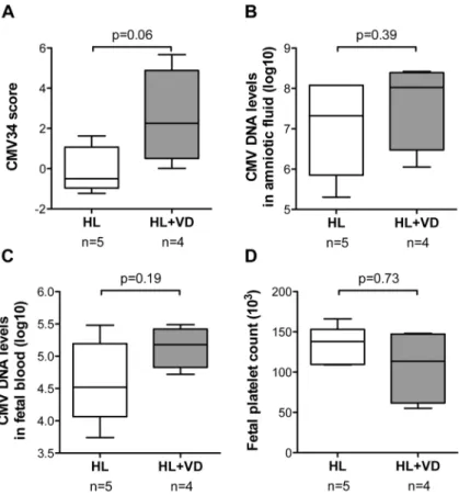
isolated hearing loss were classified as symptomatic prenatally by the CMV34 peptide classifier (S2 Table). The comparison of the combined CMV34 scores of these 2 sub-groups (hearing loss and vestibular dysfunction, HL+VDversushearing loss only, HL) showed a trend towards
higher scores for children with vestibular dysfunction (p = 0.06)(Fig 4A). Interestingly such trend could not be observed for CMV DNA levels in amniotic fluid and fetal blood and the fetal platelet count (Fig 4B–4DandS2 Table).
Pathway analysis
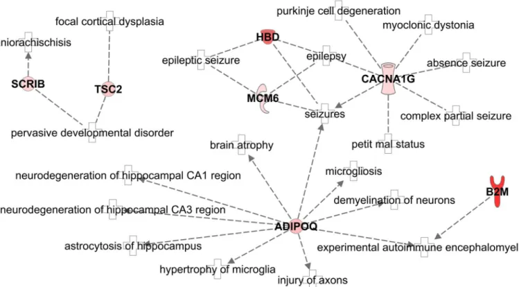
An advantage of non-targeted approaches leading to panels of peptides is that such data can be subjected to pathway analysis aiming at a deeper understanding of the underlying pathophysi-ology of the disease. The 13 non-collagen peptides (considering peptides as proteins) were sub-mitted to Ingenuity Pathway Analysis software. The second top canonical pathway (ignoring the first pathway related to HER-2 signaling in breast cancer) was“Virus Entry via Endocytic Pathways”confirming the relation of 3 (beta-2-microglobulin (B2M); Phosphatidylinositol 4;5-bisphosphate 3-kinase (PIK3CB) and protein kinase and Serine/threonine-protein kinase D1 (PRKD1)) out of the 13 peptide markers with the disease under study. When focusing on enrichment of pathways related to diseases,“Organismal Injury and Abnormalities”was among the top pathways suggesting a general connection to the potential lesions observed in symptomatic CMV. Another major enrichment of the peptide biomarkers was found to be in
“Neurological Disease”with 7 out of 13 peptides significantly enriched (Fig 5): beta-2-micro-globulin (B2M), hemoglobin delta subunit (HBD), adiponectin (ADIPOQ), protein scribble homolog (SCRIB), T calcium channel alpha-1G (CACNA1G), DNA replication licensing fac-tor (MCM6) and tuberin (TSC2). Neurological lesions are the main prognostic lesions among the manifestations observed in symptomatic CMV patients including epilepsy.
Discussion
The management of fetal CMV infection remains controversial. One difficulty is to establish the prognosis timely and accurately before birth. Prognostic factors are mainly derived from prenatal ultrasound or MRI imaging of the fetal brain and these can either appear late in the pregnancy and/or be objectified only after birth. Laboratory markers of the severity of infection including thrombocytopenia and high serum levels of beta-2-microglobulin in fetal blood have been suggested to precede the development of brain lesions. These can be obtained following
Fig 3. Performance of other frequently used parameters in the validation cohort.ROC curves for CMV DNA levels in amniotic fluid (A) and fetal blood (B) and the fetal platelet count (C) in the combined discovery and validation cohort. AF, amniotic fluid; FB, fetal blood; Plat., platelet.
cordocentesis, another invasive procedure performed under ultrasound guidance and separate from the amniocentesis performed for diagnosis [8,9,26]. We therefore aimed at identifying reliable protein-based prognostic factors of fetal CMV infection in the amniotic fluid at the time of prenatal diagnosis by amniocentesis.
Amniotic fluid of 26 fetuses infected with CMV contained 76 peptides showing a clearly dis-tinct abundance between those leading to either severely symptomatic or asymptomatic chil-dren by the age of 12 months. Thirty-four of the 37 sequenced peptides were combined in a prognostic classifier called CMV34. The classifier was independent of gestational age and reached 89% sensitivity, 75% specificity and an area under the curve of 0.90 to discriminate severely symptomatic from asymptomatic neonates in a validation cohort. The CMV34 classi-fier showed better AUC and NPV than the other biomarkers of severity of CMV fetal disease including fetal platelets, CMV DNA levels in fetal blood and in amniotic fluid. High NPV, lead-ing to unambiguous identification of asymptomatic neonates, is crucial for clinical manage-ment. PPV values were low and comparable for all four biomarkers, which could be expected due to the low prevalence of symptomatic neonates among CMV infected fetuses. Hence, a score suggesting a symptomatic fetus should always to be confirmed by targeted imaging. These data support that the CMV34 classifier may be of substantial value in the clinical context of intrauterine CMV infection.
Fig 4. Classification of primary CMV infection with moderately symptomatic neonates.(A) The amniotic fluid peptide content of CMV-infected fetuses with moderate neonatal symptoms (hearing loss (HL) or hearing loss and vestibular dysfunction (HL+VD)) was scored with the CMV34 classifier. The scores allowed a nearly significant difference after analysis by Mann-Whitney test for independent samples (p = 0.06). CMV DNA levels in amniotic fluid and fetal blood (BandC, respectively) and the fetal platelet count (D) were clearly not different between hearing lossversushearing loss and vestibular dysfunction (p values of 0.32, 0.19 and 0.73, respectively, Mann-Whitney test for independent samples).
The selected peptides included 21 collagen fragments suggestive of tissue remodeling differ-ences between symptomatic and asymptomatic patients. The other 13 peptides that were signif-icantly more abundant in amniotic fluid of severely symptomatic fetuses included fragments of beta-2-microglobulin, hemoglobin subunit delta and adiponectin. The beta-2-microglobulin and the hemoglobin subunit delta fragments were highly up-regulated (>8 fold) suggesting an
important role of these peptides in congenital CMV disease. Beta-2-microglobulin is the light chain of class I major histocompatibility complex and is present in the majority of T- and B-lymphocytes. Fetal blood levels of beta-2-microglobulin have previously been reported as a prognostic marker of fetal CMV infection [27] and beta-2-microglobulin has also been described as an important antibacterial protein in amniotic fluid [28]. Increased serum levels of beta-2-microglobulin is also a predictive factor of CMV infection in adult renal transplant recipients [29]. In the context of this study, high levels of circulating beta-2-microglobulin might be suggestive of lymphoid cell stimulation. One other hypothesis could be that increased beta-2-microglobulin reflects intense viral replication since it has been shown that CMV binds to beta-2-microglobulin [30]. However, in our study the abundance of the beta-2-microglobu-lin fragment (ID 44679) was neither correlated to CMV DNA levels in amniotic fluid nor to CMV DNA levels in fetal blood (S1 Fig).
In association with hemoglobin subunit alpha, hemoglobin subunit delta constitutes hemo-globin A2, which represents the adult hemohemo-globin. The hemohemo-globin subunit delta peptide found in amniotic fluid is therefore of maternal origin. The high specificity and the large differ-ences between symptomatic and asymptomatic groups (8.6-fold change) led us to hypothesize
Fig 5. Alteration of the neurological disease pathway.Ingenuity Pathway Analysis software showed a significant activation of neurological disease pathway in symptomatic fetuses compared with asymptomatic, with 7 out of 13 non-collagen peptide parent proteins being associated to the network. Red: increased abundance. B2M, beta-2-microglobulin; HBD, hemoglobin delta subunit; ADIPOQ, adiponectin; SCRIB, protein scribble homolog; CACNA1G, T calcium channel alpha-1G; MCM6, DNA replication licensing factor; TSC2, tuberin.
that those findings are not related to contamination during amniocentesis. Vaibuch et al. have demonstrated that total hemoglobin can be detected in the amniotic fluid of all pregnant women, increasing in concentration with gestational age. They also demonstrated in the same study [31] that the total hemoglobin level in amniotic fluid was significantly higher in women with intra-amniotic infection and/or inflammation. We suggest that this finding of higher hemoglobin subunit delta in amniotic fluid from symptomatic fetuses could be due to a more severe inflammation related to CMV infection.
There is emerging evidence indicating that the study of biological networks can provide insight into the pathobiology of disease and improve biomarker discovery. We used the Inge-nuity Pathway Analysis software to identify networks related to the peptides identified within this classifier. The second top canonical pathway was related to virus entry via endocytic path-way. Mechanisms of CMV entry into the cells are not completely understood. There is evidence that CMV enters into fibroblasts using a pathway involving membrane fusion, whereas it enters epithelial and endothelial cells via an endocytic pathway involving macropinocytosis [32]. Up-regulation of the canonical pathways“virus entry”in the more severe cases probably reflects intensive viral multiplication making the case for prenatal antiviral treatment. Moreover, one of the top disease pathways was identified as“neurological”and included 7 proteins all up-reg-ulated in the more severe cases: beta-2-microglobulin (B2M), hemoglobin delta subunit (HBD), adiponectin (ADIPOQ), protein scribble homolog (SCRIB), T calcium channel alpha-1G (CACNAalpha-1G), DNA replication licensing factor (MCM6) and tuberin (TSC2).
A previous publication reported elevated level of adiponectin in the amniotic fluid of women with intra-amniotic infection compared to women without infection [33]. Adiponectin appears to be intimately involved in several neurodegenerative disorders that are associated with CMV brain disease, such as epilepsy and ischaemic stroke [34] Therefore, adiponectin up-regulation in severe cases probably underlines the potential importance of inflammatory response on fetal brain lesions; this inflammatory response could be a target for future antena-tal therapeutic intervention. The protein scribble homolog (SCRIB) is involved in planar cell polarity and Scrib knockout animal models present with neural tube defects [35]. Up-regula-tion of SCRIB in fetuses with severely affected brains could be a counter-mechanism of the putative effect of CMV on the differentiation and the migration of neuronal stem cells. Tuberin is the product of gene TSC2 and is highly expressed in fetal neural tissue [36]. Mutation of TSC2 is responsible for the development of Tuberous Sclerosis whereas activation of TSC2 has been reported to increase autophagy of the neurons in a model of Parkinson‘s disease [37]. Finally, T calcium channel alpha-1G (CACNA1G) protein belongs to the neuronal T-type cal-cium channels that are critical contributors to membrane excitability in neuronal cells. These T-type channels have a role in ischemic neuronal cell death in brain undergoing oxygen-glu-cose deprivation [38] as could be the case in CMV infection. Up-regulation of T calcium chan-nel alpha-1G protein might thus predict an activation of seizures, which are frequent
consequences of severe CMV brain disease. Molecules that counteract the effect of T calcium channel alpha-1G could therefore also be an interesting field of development to help treating the symptoms of children with severe CMV brain disease.
In this study, we did not seek for diagnostic markers of fetal CMV infection but we were investigating prognostic markers in established CMV infected cases. However, it would be interesting to further study whether these markers are specific for CMV infection or could rep-resent, at least partially, global perturbations related toin uteroinfection. Therefore, the
In conclusion, the analysis of the peptidome in the amniotic fluids infected by CMV led to the identification of a prognostic classifier based on 34 peptides that yielded a better perfor-mance than the currently used fetal biomarkers to predict the asymptomatic or symptomatic status at birth. This classifier highlights the importance of the intensity of viral replication. A subset of those highly up-regulated proteins is also linked within a neurological disease net-work. This study serves as basis for future investigations to assess the prospective prognosis value of the CMV34 in larger cohorts. Moreover, these findings provide new insight on the pathobiology of CMV-induced fetal brain lesions and they open the perspective of new thera-peutic options.
Material and Methods
Patients
Forty-seven amniotic fluid samples collected in the second trimester of pregnancy with CMV infected fetuses were extracted from the Necker Virology laboratory database and analyzed ret-rospectively. Prenatal data were reviewed, including fetal ultrasound and MRI examination and follow-up. Outcome was assessed by targeted neonatal examination or postmortem exami-nation following termiexami-nation of pregnancy (TOP). All infected fetuses were followed-up in the Fetal Medicine Unit at Necker Hospital using the same management. Amniocentesis was per-formed either because of suggestive fetal ultrasound symptoms or in the context of docu-mented maternal primary infection. Fetal blood sample by cordocentesis under continuous ultrasound guidance is part of the management proposed in all infected cases. Fetal blood viral DNA-load and fetal platelet count are measured to be used as part of the prognostic assessment of all infected fetuses in the Fetal Medicine Unit at Necker Hospital. Cerebral MRI was per-formed after 30–32 weeks of pregnancy in all cases that were not terminated.
Amniotic fluid samples from women carrying a CMV infected fetus were divided into two cohorts. The discovery cohort was composed of 26 amniotic fluid samples obtained from infected fetuses that led to either termination of pregnancy (TOP) for severe brain lesions or symptomatic neonates (n = 13) or to asymptomatic neonates (n = 13). A second blinded cohort was used for validation and was composed of 21 amniotic fluid samples obtained from infected fetuses that led to TOP or to symptomatic neonates (n = 9) or from asymptomatic neonates (n = 12).
Ethics
All women undergoing amniocentesis in the Fetal Medicine Unit at Necker Hospital gave writ-ten informed consent for CMV prenatal diagnosis to be performed on amniotic fluid and for the amniotic fluid sample left-over to be used for research purposes. According to French laws, an ethics statement from an Institutional Review board was not required for this work. More-over, all women gave written informed consent for the use of clinical data as they were included in different clinical studies on congenital CMV in the Fetal Medicine Unit at Necker Hospital. These studies were approved by the Institutional Review Board of University Paris Ouest (IRB N°2011-001610-34 and 2013-A213-42).
Classification between symptomatic and asymptomatic infection
of TOP, severity was confirmed if postmortem examination showed microcephaly<4SD,
ven-triculomegaly, white matter necrosis, associated with diffuse lesions of vasculitis and of enceph-alitis. Indeed, it has been documented in the literature that 77% of infected newborns with severe abnormalities on neonatal CT scan (intracranial calcifications, ventricular dilatation, white-matter abnormalities, cortical atrophy and migration abnormalities) develop at least one psychomotor sequela [39]. In another study 100% of newborns with microcephaly had severe mental retardation [40]. Therefore, severely abnormal brain imaging both on ultrasound and on MRI in utero is likely to be found mainly in cases destined to become neurologically symp-tomatic neonates.
Symptomatic or asymptomatic status at birth was evaluated by clinical examination, biolog-ical assessment (platelet count, liver enzymes, and bilirubin serum levels) and audiometric assessment by automated auditory brainstem response (AABR). Neonates with normal clinical examination, normal biological assessment and normal hearing were considered asymptom-atic. Neonates with abnormal clinical examination and/or abnormal biological assessment, including petechiae, microcephaly, seizures, lethargy/hypotonia, poor suck, hepatosplenome-galy, thrombocytopenia and hearing loss were considered as symptomatic. Infected children are routinely followed-up with serial clinical examination and audiometric evaluation (AABR) performed at 4, 12, 18, 24 and 36 months of age.
For the purpose of the study, cases were classified into 3 groups. Cases with severe brain anomalies identified either at prenatal ultrasound or MRI, at necropsy or at birth were classi-fied as severely symptomatic. Cases with isolated unilateral hearing loss>40 decibels and/or
vestibular syndrome identified at birth or at last follow-up visit were classified as moderately symptomatic. Asymptomatic cases at birth or at last available follow-up visit were classified as asymptomatic.
CMV DNA quantification in fetal blood and in amniotic fluid
CMV DNA was extracted from 200μl of amniotic fluid with the MagNaPure LC using the
Total Nucleic Acid extraction kit (Roche Diagnostic, Meylan, France) and from 200μl of fetal
whole blood using the QiaAmp DNA mini blood kit (Qiagen, Courtaboeuf, France). CMV DNA was amplified by CMV-R Gene (Argene BioMerieux, France) a real time commercial quantitative CMV PCR assay.
Sample preparation, CE-MS and data processing
We have recently developed a sample preparation protocol for the analysis of the fetal urinary peptidome by CE-MS starting from 150μl of fetal urine [41]. This protocol was also used for
amniotic fluid. Briefly, immediately before preparation, amniotic fluid aliquots kept at–80°C were thawed and 150μl aliquots were diluted with the same volume of 2 M urea, 10 mM
NH4OH containing 0.2% SDS. Subsequently, samples were passed over a Centristat 20-kDa cut-off centrifugal filter device (Sartorius) in order to eliminate high molecular weight com-pounds. The filtrate was desalted using a NAP-5 gel filtration column (GE Healthcare) to remove urea and electrolytes. Lyophilisation of the sample was performed using a Savant speedvac SVC100H connected to a Virtis 3L Sentry freeze dryer (Fisher Scientific) and stored at 4°C until use. Shortly before CE-MS analysis, the samples were re-suspended in 10μL of
HPLC grade H2O.
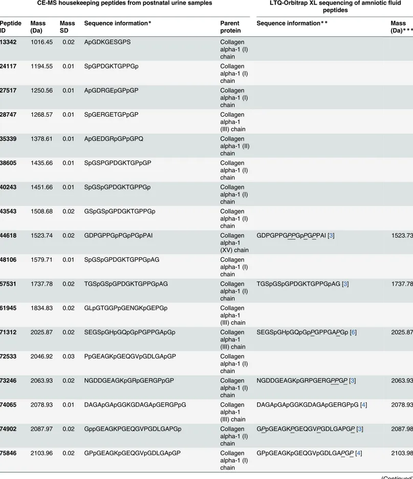
MS acquisition methods were automatically controlled by the CE via contact-close-relays. Spectra were accumulated every 3 s, over a range of m/z 350 to 3000. Mass spectral ion peaks representing identical molecules at different charge states were deconvoluted into single masses using MosaiquesVisu software [44]. The software automatically examined all mass spectra from a CE-MS analysis for signals with a signal-to-noise ratio of at least 4 present in 3 consecu-tive spectra. Furthermore, the isotopic distribution was assessed, and charge was assigned based on the isotopic distribution, as well as conjugated masses, using a probabilistic clustering algorithm. This operation resulted in a list wherein all signals that could be interpreted are defined by mass/charge, charge, migration time, and signal intensity (ion counts). Time-of-flight mass spectrometry (TOF-MS) data were calibrated utilizing Fourier transform ion cyclo-tron resonance mass spectrometry (FT-ICR-MS) data as reference masses applying linear regression. Both CE-migration time and ion signal intensity (amplitude) showed high variabil-ity, mostly due to different amounts of salt and peptides in the sample. Normalization of the amplitude of the amniotic fluid peptides was based on sequenced endogenous“housekeeping”
peptides (Table 6) that varied little among the samples. Based on the sequences most of the housekeeping peptides found in amniotic fluid were similar to those observed in urine, there-fore allowing the application of a CE-MS procedure for sample analysis and data processing similar to the procedure used for urine [42,45].
Statistical analysis and biomarker identification
Normalized levels of amniotic fluid peptides were compared between symptomatic and asymp-tomatic patients’groups for the identification of amniotic fluid proteome biomarkers. Only peptides with a P-value<0.05 corrected for multiple testing (Benjamini- Hochberg) were
con-sidered significant [46]. The number of peptides with differential abundance was reduced to a support vector machine (SVM) classifier with 34 peptides (CMV34) by a take-one-out proce-dure that had similar performance for the classification of the patients in the discovery CMV patient cohort. Sensitivity and specificity of the previously defined biomarker classifiers, and 95% confidence intervals (95% CI) were calculated using receiver operating characteristic (ROC) plots (MedCalc version 14.8.1, MedCalc Software, Belgium).
Sequencing of peptide biomarkers
Table 6. Amniotic fluid peptides used for normalization and their correspondence with postnatal urinary peptides used for normalization.
CE-MS housekeeping peptides from postnatal urine samples LTQ-Orbitrap XL sequencing of amnioticfluid peptides
Peptide ID
Mass (Da)
Mass SD
Sequence information* Parent protein
Sequence information** Mass (Da)***
13342 1016.45 0.02 ApGDKGESGPS Collagen alpha-1 (I) chain 24117 1194.55 0.01 SpGPDGKTGPPGp Collagen
alpha-1 (I) chain 27517 1250.56 0.01 ApGDRGEpGPpGP Collagen
alpha-1 (I) chain 28747 1268.57 0.01 SpGERGETGPpGP Collagen
alpha-1 (III) chain 35339 1378.61 0.01 ApGEDGRpGPpGPQ Collagen alpha-1 (II) chain 38605 1435.66 0.01 SpGSPGPDGKTGPpGP Collagen
alpha-1 (I) chain 40243 1451.66 0.01 SpGSpGPDGKTGPPGp Collagen
alpha-1 (I) chain 43543 1508.68 0.02 GSpGSpGPDGKTGPPGp Collagen
alpha-1 (I) chain 44618 1523.74 0.02 GDPGPPGpPGpPGpPAI Collagen
alpha-1 (XV) chain
GDPGPPGPPGpPGPPAI [3] 1523.73
48106 1579.71 0.01 SpGSpGPDGKTGPPGpAG Collagen alpha-1 (I) chain 57531 1737.78 0.02 TGSpGSpGPDGKTGPPGpAG Collagen
alpha-1 (I) chain
TGSpGSpGPDGKTGPPGpAG [3] 1737.78
61945 1834.83 0.02 GLpGTGGPpGENGKpGEPGp Collagen alpha-1 (III) chain 71312 2025.87 0.02 SEGSpGHpGQpGpPGPPGApGp Collagen alpha-1 (III) chain
SEGSpGHpGQpGpPGPPGAPGp [6] 2025.87
72533 2046.92 0.03 PpGEAGKpGEQGVpGDLGApGP Collagen alpha-1 (I) chain 73246 2063.93 0.02 NGDDGEAGKpGRpGERGPpGP Collagen
alpha-1 (I) chain
NGDDGEAGKpGRPGERGPPGP[3] 2063.93
74065 2078.93 0.01 DAGApGApGGKGDAGApGERGPpG Collagen alpha-1 (III) chain
DAGApGApGGKGDAGApGERGPpG [4] 2078.93
74902 2087.97 0.02 GppGEAGKPGEQGVPGDLGAPGp Collagen alpha-1 (I) chain
GPpGEAGKPGEQGVPGDLGAPGP[3] 2087.98
75846 2103.96 0.02 GPpGEAGKpGEQGVpGDLGApGP Collagen alpha-1 (I) chain
GPpGEAGKpGEQGVpGDLGAPGP[4] 2103.98
For further validation of peptide identification, the strict correlation between peptide charge at pH 2 and CE-migration time was utilized to minimize false-positive identification rates [48]. Calculated CE-migration time of the sequence candidate based on its peptide sequence (num-ber of basic amino acids) was compared to the experimental migration time. Peptides were accepted only if they had a mass deviation below ±50 ppm and a CE-migration time deviations below ±2 min.
Ingenuity pathway analysis
Ingenuity Pathway Analysis software (version 24390178, last release 18/06/2015,www. ingenuity.com) was performed using the parent proteins peptides with differential abundance
Table 6. (Continued)
CE-MS housekeeping peptides from postnatal urine samples LTQ-Orbitrap XL sequencing of amnioticfluid peptides
Peptide ID
Mass (Da)
Mass SD
Sequence information* Parent protein
Sequence information** Mass (Da)***
78332 2159.00 0.03 AGPpGEAGKpGEQGVpGDLGAPGP Collagen alpha-1 (I) chain
AGPpGEAGKPGEQGVPGDLGAPGP[3] 2159.02
78843 2169.98 0.02 NSGEpGApGSKGDTGAKGEpGPVG Collagen alpha-1 (I) chain
NSGEpGApGSKGDTGAKGEpGPVG [3] 2169.98
79136 2175.01 0.02 AGPpGEAGKpGEQGVpGDLGApGP Collagen alpha-1 (I) chain
AGPpGEAGKpGEQGVpGDLGApGP [4] 2175.01
81758 2220.99 0.02 ADGQpGAKGEpGDAGAKGDAGPpGP Collagen alpha-1 (I) chain
ADGQpGAKGEpGDAGAKGDAGPpGP [3] 2220.99
82026 2226.99 0.01 GNSGEpGApGSKGDTGAKGEpGPVG Collagen alpha-1 (I) chain 85761 2292.02 0.02 ADGQpGAKGEpGDAGAKGDAGPpGPA Collagen
alpha-1 (I) chain
ADGQpGAKGEpGDAGAKGDAGPpGPA [3] 2292.03
98660 2564.15 0.03 GApGQNGEpGGKGERGApGEKGEGGPpG Collagen alpha-1 (III) chain
GApGQNGEpGGKGERGApGEKGEGGPpG [4]
2564.15
104786 2679.20 0.03 NRGERGSEGSPGHpGQpGppGpPGAPGP Collagen alpha-1 (III) chain 105352 2695.20 0.02 NRGERGSEGSpGHpGQpGppGPPGAPGp Collagen alpha-1 (III) chain
NRGERGSEGSPGHpGQpGppGPPGAPGP[6] 2695.20
107460 2742.25 0.03 KNGETGPQGPpGPTGPGGDKGDTGPpGPQG Collagen alpha-1 (III) chain 111001 2825.27 0.03 ERGEAGIpGVpGAKGEDGKDGSpGEpGANG Collagen alpha-1 (III) chain
ERGEAGIpGVpGAKGEDGKDGSpGEpGANG [4]
2825.27
*Lower case p indicated hydroxyproline.
**Lower case p indicated unambiguous hydroxylated proline sites and at the same proline residues than sequence obtained from postnatal urine. Hydroxylated proline sites underlined and in italic indicated different possible site. The total number of hydroxyproline are indicated in square brackets. ***Experimental mass (Da) are obtained from the mean of all MS/MS data for this species on a LTQ-Orbitrap XL with a mass accuracy<4 ppm.
in the symptomatic CMV infections. As urine, amniotic fluid is very rich in different collagen fragments. Therefore, collagens were omitted from the Ingenuity pathway analysis to avoid a bias in the analysis towards fibrosis and conjunctive tissue remodeling. All Ingenuity output related to cancer was omitted.
Supporting Information
S1 Table. Patient data of the severely symptomatic and asymptomatic fetuses from the dis-covery and validation cohorts.
(XLS)
S2 Table. Patient data of the moderately symptomatic fetuses from the additional cohort.
(XLS)
S1 Dataset. Peptidome analysis of the asymptomatic fetuses from the discovery cohort.
(XLS)
S2 Dataset. Peptidome analysis of the symptomatic fetuses from the discovery cohort.
(XLS)
S3 Dataset. Peptidome analysis of the asymptomatic fetuses from the validation cohort.
(XLS)
S4 Dataset. Peptidome analysis of the symptomatic fetuses from the validation cohort.
(XLS)
S1 Fig. Correlation analysis of the beta-2-microglobulin fragment (ID 44679) and CMV DNA levels in amniotic fluid (A) and fetal blood (B).
(TIF)
Acknowledgments
We thank O. Schiltz (IPBS, CNRS) for technological assistance.
Author Contributions
Conceived and designed the experiments: MLV JPS YV. Performed the experiments: MLV CL BB CF. Analyzed the data: CD JK ART JLB YV. Contributed reagents/materials/analysis tools: MLV. Wrote the paper: CD JK MLV JLB JPS YV.
References
1. Dollard SC, Grosse SD, Ross DS. New estimates of the prevalence of neurological and sensory sequelae and mortality associated with congenital cytomegalovirus infection. Rev Med Virol. 2007; 17: 355–63. doi:10.1002/rmv.544PMID:17542052
2. Kenneson A, Cannon MJ. Review and meta-analysis of the epidemiology of congenital cytomegalovi-rus (CMV) infection y. Rev Med Virol. 2007; 253–276. PMID:17579921
3. Benoist G, Leruez-Ville M, Magny JF, Jacquemard F, Salomon LJ, Ville Y. Management of pregnancies with confirmed cytomegalovirus fetal infection. Fetal Diagn Ther. 2013; 33: 203–214. doi:10.1159/ 000342752PMID:23571413
4. Noyola DE, Demmler GJ, Nelson CT, Griesser C, Williamson WD, Atkins JT, et al. Early predictors of neurodevelopmental outcome in symptomatic congenital cytomegalovirus infection. J Pediatr. 2001; 138: 325–31. doi:10.1067/mpd.2001.112061PMID:11241037
6. Lipitz S, Yinon Y, Malinger G, Yagel S, Levit L, Hoffman C, et al. Risk of cytomegalovirus-associated sequelae in relation to time of infection and findings on prenatal imaging. Ultrasound Obstet Gynecol Off J Int Soc Ultrasound Obstet Gynecol. 2013; 41: 508–514. doi:10.1002/uog.12377
7. Farkas N, Hoffmann C, Ben-Sira L, Lev D, Schweiger A, Kidron D, et al. Does normal fetal brain ultra-sound predict normal neurodevelopmental outcome in congenital cytomegalovirus infection? Prenat Diagn. 2011; 31: 360–366. doi:10.1002/pd.2694PMID:21413035
8. Benoist G, Salomon LJ, Jacquemard F, Daffos F, Ville Y. The prognostic value of ultrasound abnormali-ties and biological parameters in blood of fetuses infected with cytomegalovirus. BJOG Int J Obstet Gynaecol. 2008; 115: 823–829. doi:10.1111/j.1471-0528.2008.01714.x
9. Fabbri E, Revello MG, Furione M, Zavattoni M, Lilleri D, Tassis B, et al. Prognostic markers of symptom-atic congenital human cytomegalovirus infection in fetal blood. BJOG Int J Obstet Gynaecol. 2011; 118: 448–456. doi:10.1111/j.1471-0528.2010.02822.x
10. Goegebuer T, Van Meensel B, Beuselinck K, Cossey V, Van Ranst M, Hanssens M, et al. Clinical pre-dictive value of real-time PCR quantification of human cytomegalovirus DNA in amniotic fluid samples. J Clin Microbiol. 2009; 47: 660–665. doi:10.1128/JCM.01576-08PMID:19109474
11. Guerra B, Lazzarotto T, Quarta S, Lanari M, Bovicelli L, Nicolosi A, et al. Prenatal diagnosis of symp-tomatic congenital cytomegalovirus infection. Am J Obstet Gynecol. 2000; 183: 476–482. doi:10.1067/ mob.2000.106347PMID:10942490
12. Lazzarotto T, Varani S, Guerra B, Nicolosi A, Lanari M, Landini MP. Prenatal indicators of congenital cytomegalovirus infection. J Pediatr. 2000; 137: 90–95. doi:10.1067/mpd.2000.107110PMID:
10891828
13. Revello MG, Zavattoni M, Furione M, Baldanti F, Gerna G. Quantification of human cytomegalovirus DNA in amniotic fluid of mothers of congenitally infected fetuses. J Clin Microbiol. 1999; 37: 3350–
3352. PMID:10488204
14. Ville Y, Leruez-Ville M. Managing infections in pregnancy. Curr Opin Infect Dis. 2014; 27: 251–257. PMID:24781057
15. Walker SP, Palma-Dias R, Wood EM, Shekleton P, Giles ML. Cytomegalovirus in pregnancy: to screen or not to screen. BMC Pregnancy Childbirth. 2013; 13: 96. doi:10.1186/1471-2393-13-96PMID:
23594714
16. Klein J, Buffin-Meyer B, Mullen W, Carty DM, Delles C, Vlahou A, et al. Clinical proteomics in obstetrics and neonatology. Expert Rev Proteomics. 2014; 11: 75–89. doi:10.1586/14789450.2014.872564
PMID:24404900
17. Cho C-KJ, Smith CR, Diamandis EP. Amniotic fluid proteome analysis from Down syndrome pregnan-cies for biomarker discovery. J Proteome Res. 2010; 9: 3574–3582. doi:10.1021/pr100088kPMID:
20459121
18. Cho C-KJ, Drabovich AP, Karagiannis GS, Martínez-Morillo E, Dason S, Dimitromanolakis A, et al. Quantitative proteomic analysis of amniocytes reveals potentially dysregulated molecular networks in Down syndrome. Clin Proteomics. 2013; 10: 2. doi:10.1186/1559-0275-10-2PMID:23394617
19. Buhimschi CS, Bhandari V, Hamar BD, Bahtiyar M-O, Zhao G, Sfakianaki AK, et al. Proteomic profiling of the amniotic fluid to detect inflammation, infection, and neonatal sepsis. PLoS Med. 2007; 4: e18. doi:10.1371/journal.pmed.0040018PMID:17227133
20. Buhimschi CS, Bhandari V, Han YW, Dulay AT, Baumbusch MA, Madri JA, et al. Using proteomics in perinatal and neonatal sepsis: hopes and challenges for the future. Curr Opin Infect Dis. 2009; 22: 235–
243. PMID:19395960
21. Tambor V, Kacerovsky M, Andrys C, Musilova I, Hornychova H, Pliskova L, et al. Amniotic fluid catheli-cidin in PPROM pregnancies: from proteomic discovery to assessing its potential in inflammatory com-plications diagnosis. PloS One. 2012; 7: e41164. doi:10.1371/journal.pone.0041164PMID:22815956
22. Cecconi D, Lonardoni F, Favretto D, Cosmi E, Tucci M, Visentin S, et al. Changes in amniotic fluid and umbilical cord serum proteomic profiles of foetuses with intrauterine growth retardation. Electrophore-sis. 2011; 32: 3630–3637. doi:10.1002/elps.201100256PMID:22180211
23. Decramer S, Gonzalez de Peredo A, Breuil B, Mischak H, Monsarrat B, Bascands J-L, et al. Urine in clinical proteomics. Mol Cell Proteomics MCP. 2008; 7: 1850–1862. doi: 10.1074/mcp.R800001-MCP200PMID:18667409
24. Mischak H, Schanstra JP. CE-MS in biomarker discovery, validation, and clinical application. Proteo-mics Clin Appl. 2011; 5: 9–23. doi:10.1002/prca.201000058PMID:21280234
25. Schanstra JP, Mischak H. Proteomic urinary biomarker approach in renal disease: from discovery to implementation. Pediatr Nephrol Berl Ger. 2014; doi:10.1007/s00467-014-2790-y
27. Dreux S, Rousseau T, Gerber S, Col J-Y, Dommergues M, Muller F. Fetal serum beta2-microglobulin as a marker for fetal infectious diseases. Prenat Diagn. 2006; 26: 471–474. doi:10.1002/pd.1441
PMID:16652403
28. Kim J-Y, Park S-C, Lee J-K, Choi SJ, Hahm K-S, Park Y. Novel antibacterial activity of β(2)-microglobu-lin in human amniotic fluid. PloS One. 2012; 7: e47642. doi:10.1371/journal.pone.0047642PMID:
23144825
29. Matos ACC, Durão MS, Pacheco-Silva A. Serial beta-2 microglobulin measurement as an auxilliary method in the early diagnosis of cytomegalovirus infection in renal transplant patients. Transplant Proc. 2004; 36: 894–895. doi:10.1016/j.transproceed.2004.03.110PMID:15194307
30. Grundy JE, McKeating JA, Griffiths PD. Cytomegalovirus strain AD169 binds beta 2 microglobulin in vitro after release from cells. J Gen Virol. 1987; 68 (Pt 3): 777–784. doi:10.1099/0022-1317-68-3-777
PMID:3029304
31. Vaisbuch E, Romero R, Erez O, Kusanovic JP, Gotsch F, Than NG, et al. Total hemoglobin concentra-tion in amniotic fluid is increased in intraamniotic infecconcentra-tion/inflammaconcentra-tion. Am J Obstet Gynecol. 2008; 199: 426.e1–426.e7. doi:10.1016/j.ajog.2008.06.075
32. Vanarsdall AL, Johnson DC. Human cytomegalovirus entry into cells. Curr Opin Virol. 2012; 2: 37–42. doi:10.1016/j.coviro.2012.01.001PMID:22440964
33. Chaiworapongsa T, Romero R, Kusanovic JP, Savasan ZA, Kim SK, Mazaki-Tovi S, et al. Unexplained fetal death is associated with increased concentrations of anti-angiogenic factors in amniotic fluid. J Matern-Fetal Neonatal Med Off J Eur Assoc Perinat Med Fed Asia Ocean Perinat Soc Int Soc Perinat Obstet. 2010; 23: 794–805. doi:10.3109/14767050903443467
34. Thundyil J, Pavlovski D, Sobey CG, Arumugam TV. Adiponectin receptor signalling in the brain. Br J Pharmacol. 2012; 165: 313–327. doi:10.1111/j.1476-5381.2011.01560.xPMID:21718299
35. Murdoch JN, Damrau C, Paudyal A, Bogani D, Wells S, Greene NDE, et al. Interactions between planar cell polarity genes cause diverse neural tube defects. Dis Model Mech. 2014; doi:10.1242/dmm. 016758
36. Kerfoot C, Wienecke R, Menchine M, Emelin J, Maize JC, Welsh CT, et al. Localization of tuberous sclerosis 2 mRNA and its protein product tuberin in normal human brain and in cerebral lesions of patients with tuberous sclerosis. Brain Pathol Zurich Switz. 1996; 6: 367–375.
37. Ha J-Y, Kim J-S, Kang Y-H, Bok E, Kim Y-S, Son JH. Tnfaip8 l1/Oxi-βbinds to FBXW5, increasing autophagy through activation of TSC2 in a Parkinson’s disease model. J Neurochem. 2014; 129: 527–
538. doi:10.1111/jnc.12643PMID:24444419
38. Cataldi M. The changing landscape of voltage-gated calcium channels in neurovascular disorders and in neurodegenerative diseases. Curr Neuropharmacol. 2013; 11: 276–297. doi:10.2174/
1570159X11311030004PMID:24179464
39. Ross S a, Novak Z, Kumbla R a, Zhang K, Fowler KB, Boppana S. GJB2 and GJB6 mutations in chil-dren with congenital cytomegalovirus infection. Pediatr Res. 2007; 61: 687–91. doi:10.1203/pdr. 0b013e3180536609PMID:17426645
40. Noyola DE, Demmler GJ, Nelson CT, Griesser C, Williamson WD, Atkins JT, et al. Early predictors of neurodevelopmental outcome in symptomatic congenital cytomegalovirus infection. J Pediatr. 2001; 138: 325–331. doi:10.1067/mpd.2001.112061PMID:11241037
41. Klein J, Lacroix C, Caubet C, Siwy J, Zürbig P, Dakna M, et al. Fetal urinary peptides to predict postna-tal outcome of renal disease in fetuses with posterior urethral valves (PUV). Sci Transl Med. 2013; 5: 198ra106. doi:10.1126/scitranslmed.3005807PMID:23946195
42. Theodorescu D, Wittke S, Ross MM, Walden M, Conaway M, Just I, et al. Discovery and validation of new protein biomarkers for urothelial cancer: a prospective analysis. Lancet Oncol. 2006; 7: 230–240. doi:10.1016/S1470-2045(06)70584-8PMID:16510332
43. Kolch W, Neusüss C, Pelzing M, Mischak H. Capillary electrophoresis-mass spectrometry as a power-ful tool in clinical diagnosis and biomarker discovery. Mass Spectrom Rev. 2005; 24: 959–977. doi:10. 1002/mas.20051PMID:15747373
44. Neuhoff N v, Kaiser T, Wittke S, Krebs R, Pitt A, Burchard A, et al. Mass spectrometry for the detection of differentially expressed proteins: a comparison of surface-enhanced laser desorption/ionization and capillary electrophoresis/mass spectrometry. Rapid Commun Mass Spectrom RCM. 2004; 18: 149–
156. doi:10.1002/rcm.1294PMID:14745763
46. Dakna M, Harris K, Kalousis A, Carpentier S, Kolch W, Schanstra JP, et al. Addressing the challenge of defining valid proteomic biomarkers and classifiers. BMC Bioinformatics. 2010; 11: 594. doi:10.1186/ 1471-2105-11-594PMID:21208396
47. Klein J, Papadopoulos T, Mischak H, Mullen W. Comparison of CE-MS/MS and LC-MS/MS sequencing demonstrates significant complementarity in natural peptide identification in human urine. Electropho-resis. 2014; 35: 1060–1064. doi:10.1002/elps.201300327PMID:24254231

