online | memorias.ioc.fiocruz.br Sputum smear microscopy is the most cost-effective
method of screening for pulmonary tuberculosis (TB) and is used in most developing countries where cul-turing is usually impossible (Collins & Grange 1983, Daborn & Grange 1993, Cosivi et al. 1995, 1998, Roxo 1997, Wedlock et al. 2002). When primary isolation of mycobacteria is performed, Löwenstein-Jensen medium containing glycerol is the most widely available and is used for tuberculosis culture that generally does not sup-port growth of field strains of Mycobacterium bovis.
Although most cases of human TB are caused by My-cobacterium tuberculosis, concerns over M. bovis have been expressed and are based on several observations. Firstly, occurrence of outbreaks of multidrug-resistant (MDR) M. bovis strains among hospitalized human im-human im-munodeficiency virus (HIV)-infected patients have been
observed (Blázquez et al. 1997, Samper et al. 1997, Rivero et al. 2001). This fact highlights the high spread risk of MDR M. bovis, especially in parts of Africa where M. bovis animal diseases and HIV human infection co-exist. Secondly, transmission from patients with pulmonary M. bovis disease to immune-competent contacts also appears to occur (Long et al. 1999, Evans et al. 2007). Thirdly, the reemergence of human M. bovis carried by immigrants from regions where bovine tuberculosis is still prevalent have been documented in Europe and also on the United States-Mexico border (Wilkins et al. 1986, Dankner et al. 1993, Dankner & Davis 2000, LoBue et al. 2003). Finally, some 7,000 new cases of TB due to M. bovis may arise each year in Latin America (PAHO 1991).
Identification of mycobacteria by conventional bio-chemical identification methods is laborious and time-consuming and is not often performed by diagnostic laboratories. Therefore, other methods to differentiate M. bovis from other members of M. tuberculosis com-plex (MTC) that are faster and at least as equally or more sensitive than the classical methods are urgently needed. Such methods would aid in the identification and treat-ment of patients due to the intrinsic resistance of M. bo-vis to pyrazinamide (Konno et al. 1967) and for purposes of conducting epidemiological investigations (Espinosa de los Monteros et al. 1998).
Financial support: CNPq (410595/2006-3)
+ Corresponding author: [email protected] Received 25 April 2010
Accepted 12 November 2010
Identification of
Mycobacterium tuberculosis
complex based
on amplification and sequencing of the
oxyR
pseudogene from stored
Ziehl-Neelsen-stained sputum smears in Brazil
1*Marcio Roberto Silva1,2/+, Mark Drew Crosland Guimarães2,
Vania Maria de Oliveira1,Aline dos Santos Moreira3, Ronaldo Rodrigues da Costa4,5, Kelly Cristina Ferreira Abi-Zaid5, Adalgiza da Silva Rocha6, Philip Noel Suffys6
1EMBRAPA Gado de Leite, Ministério da Agricultura, Pecuária e Abastecimento, Juiz de Fora, MG, Brasil 2Programa de Pós-Graduação em Saúde Pública, Departamento de Medicina Preventiva e Social, Faculdade de Medicina, Universidade Federal de Minas Gerais,
Belo Horizonte, MG, Brasil 3Laboratório de Genômica Funcional e Bioinformática 6Laboratório de Biologia Molecular Aplicada a Micobactérias, Instituto Oswaldo Cruz-Fiocruz, Rio de Janeiro, RJ, Brasil 4Hospital Universitário, Universidade Federal de Juiz de Fora,
Juiz de Fora, MG, Brasil 5Hospital João Penido, Fundação Hospitalar do Estado de Minas Gerais, Juiz de Fora, MG, Brasil
A cross-sectional analysis of stored Ziehl-Neelsen (ZN)-stained sputum smear slides (SSS) obtained from two public tuberculosis referral laboratories located in Juiz de Fora, Minas Gerais, was carried out to distinguish My-cobacterium bovis from other members of the Mycobacterium tuberculosis complex (MTC). A two-step approach was used to distinguish M. bovis from other members of MTC: (i) oxyR pseudogene amplification to detect MTC and, subsequently, (ii) allele-specific sequencing based on the polymorphism at position 285 of this gene. The oxyR pseudogene was successfully amplified in 100 of 177 (56.5%) SSS available from 99 individuals. No molecular profile of M. bovis was found. Multivariate analysis indicated that acid-fast bacilli (AFB) results and the source laboratory were associated (p < 0.05) with oxyR pseudogene amplification. SSS that were AFB++ SSSshowed more oxyR pseudogene amplification than those with AFB0, possibly due to the amount of DNA. One of the two source laboratories presented a greater chance of oxyR pseudogene amplification, suggesting that differences in sputum conservation between laboratories could have influenced the preservation of DNA. This study provides evidence that stored ZN-SSS can be used for the molecular detection of MTC.
Key words: oxyR pseudogene - polymerase chain reaction - sputum smear - Mycobacterium tuberculosis -
In Escherichia coli and Salmonella typhimurium, the oxyR gene functions as a sensor and transcriptional regu-lator of proteins involved in the oxidative stress response (Farr & Kogoma 1991). This protective response of oxyR was evident in the saprophytic Mycobacterium smegmati but absent in the MTC and the Mycobacterium avium com-plex (MAC). However, homologues of the oxyR gene have been isolated from MTC and MAC (Sherman et al. 1995). While the MAC oxyR appears intact, the oxyR homologue of MTC contains numerous deletions and frameshifts and probably does not encode a functional protein; it is re-ferred to as a pseudogene (Sherman et al. 1995). Inasmuch as pseudogenes accumulate mutations at an increased rate compared with functional genes, Sreevatsan et al. (1996, 1997) described a polymorphic nucleotide located at po-sition 285 of the oxyR pseudogene that differentiates M. bovis from other complex members. All M. bovis strains had an adenine residue at nucleotide 285 while all M. tu-berculosis strains had guanine in the same position.
Espinosa de los Monteros et al. (1998) described the use of an allele-specific polymerase chain reaction (PCR) method for detecting polymorphisms in oxyR to quickly and easily differentiate M. bovis from MTC. They found that the system based on oxyR could differ-entiate all of the M. bovis strains tested (including those isolated from goats).
Genetic analyses for the identification of mycobac-teria species have been more common from isolates (Espinosa de los Monteros et al. 1998), but were rarely used in primary biological specimens or stored materials [sputum smear slides (SSS) or paraffin blocks]. The sys-tems based on amplification and sequencing of the oxyR pseudogene from stored Ziehl-Neelsen (ZN)-stained SSS for identifying strains and distinguishing M. bovis from other members of the MTC have been implemented by research laboratories in Brazil (Da Silva Rocha 2009). Public health programs and epidemiological studies may benefit from such methods. This paper presents explora-tory data describing the characterization of mycobacte-ria by molecular methods using DNA extraction, ampli-fication and sequencing of the oxyR pseudogene using stored ZN-stained SSS obtained from patients with TB diagnosed at two public referral laboratories from Juiz de Fora, state of Minas Gerais (MG).
The choice of MG for the present study was due to the fact that a tuberculin survey (1999) was performed on 1,600 herds and 23,000 cattle, including Juiz de Fora. The estimated prevalence of tuberculin reactors was 0.85%. Meanwhile, M. bovis infection in cattle affects 5% of all herds and 15% of herds that presented greater ability to produce milk (MAPA 2006). Although no zoonotic TB cases due to M. bovis have been reported by Municipal Health Department of Juiz de Fora, the authors suspect-ed that pulmonary cases could be overlooksuspect-ed because sputum acid-fast bacilli (AFB) microscopy has been a major criterion for defining TB diagnosis.
PATIENTS, MATERIALS AND METHODS
This is a cross-sectional analysis of stored ZN-stained SSS obtained from patients with TB diagnosed at two public referral laboratories from Juiz de Fora, in 2007. Laboratory 1 is located in a TB regional hospital
and performs SSS for hospitalized patients only, while laboratory 2 is a local referral centre and performs SSS in response to outpatient demands. The SSS used in this study are comprised of leftover slides sent by the two laboratories to the State Public HealthReference Labo-ratory of Minas Gerais, Ezequiel Dias Foundation, for quality control after proper authorization. Confidential-ity was assured and the study was approved by the Ethi-cal Research Review Board of the Federal University of Juiz de Fora (protocol 819.125.2006). Sociodemographic data were obtained from notification records at the De-partment of Epidemiology/Municipal Health Depart-ment. No personal information was disclosed and only codes were used for analysis.
Preparation and packing of SSS by laboratories - The preparation of the SSS (smear preparation, fixation and staining of smears) by two involved laboratories was in accordance with the Tuberculosis Guidelines, Ministry of Health (MS 2005). Both laboratories used blank glass slides for preparing the SSS. Following a microscopic exam, these slides were separated with tissue paper and stored at room temperature.
DNA extraction - The available SSS were sent to the Laboratory of Molecular Biology Appliedto Myco-bacteriaatOswaldo Cruz Foundation (Fiocruz, Rio de Janeiro, Brazil) for DNA extraction, amplification and sequencing of the oxyR pseudogene.
The oil present on the SSS was removed with xylene. The ZN-stained material was scraped off from the mi-croscopic slides after the addition of 25 µL of distilled sterile water. Seventy-five microliters of Chelex suspen-sion was added and after thorough mixing of the sam-ples, the samples were incubated for 30 min at 97ºC. The samples were centrifuged at 14,000 g for 10 min. The supernatant was transferred to another tube and stored at -20ºC until use in the PCR.
OxyR pseudogene amplification - The primers used were CGCGCTGTCAGAGCTGACTTT and TCTGCG-GAATCAGTGTCACC. Forty-five cycles of amplifica-tion were performed. The stages were 94ºC for 10 s, 62ºC for 30 s and 72ºC for 15 s and a final elongation cycle was performed at 72ºC for 2 min, according to Taylor et al. (1999). A fragment of 150 bp was amplified. The bands generated were analyzed by 2% agarose gel electrophore-sis and visualized by ethidium bromide fluorescence.
Sequencing Ready Reaction (Applied Biosystems) and data were generated with an automated instrument ABI PRISM® 3730 Genetic Analyzer, Applied Biosystems.
The sequence data were assembled and edited electroni-cally with the SeqScape 2.6V program and were compared with a published sequence of the oxyR pseudogene of M. tuberculosis H37Rv strain (accession U16243).
Epidemiological data collection - Patients’ data were obtained from notification records at the Department of Epidemiology/Municipal Health Department of Juiz de Fora. Information such as age, sex, city of residence, AFB results (0, +, ++, +++), types of co-morbidities, type of case (if new case, reentry after dropout or recurrence) and information on whether they were institutionalized or referred to Directly Observed Therapy-Short Course (DOTS) were also collected.
The oxyR pseudogene amplification was consid-ered as the main event. Additionally, AFB results were considered the main explanatory variable that could be directly associated with the amount of existing bacilli and, consequently, DNA in the SSS. The interpretation of AFB results by each of the two laboratories that pro-vided the SSS was in accordance with the Tuberculosis Guidelines, Ministry of Health (MS 2005). It is recom-mended that the reading should cover at least 100 micro-scopic fields (observation time of approximately 5 min) and the results were reported according to the following scale: absence of AFB per field in 100 fields observed (0), presence of less than 1 AFB per field in 100 fields observed (+), presence of 1-10 AFB per field in 50 fields observed (++) and presence of more than 10 AFB per field in 20 fields observed (+++).
Other possible explanatory variables for oxyR pseu-dogene amplification were taken into consideration in the analysis, including the laboratory that produced the SSS, age, sex and other medical characteristics (co-mor-bidities, type of case, DOTS and whether patients were referred from other institutions such as a psychiatric hospital, penitentiary or shelter).
Statistical analysis - An overall description of all available SSS was initially carried out to verify the per-centage of oxyR pseudogene amplification between ei-ther AFB results (positive or negative) or laboratories.
For the statistical comparisons, we chose only one of the available SSS for each patient to assure the independ-ence of observations based on the AFB results (0, +, ++ and +++). The AFB results in either duplicate, triplicate or greater were ordered according to numerical codes, and only one of the observations was randomly selected. Only observations with positive AFB smear microscopy or negative AFB smear microscopy with confirmed TB diagnosis by complementary exams were included in the statistical comparisons.
Finally, the percentage of oxyR pseudogene amplifica-tion was estimated among the populaamplifica-tion, while factors associated with amplification were assessed using logis-tic regression models for both univariate and multivariate analysis. The difference in proportions was assessed us-ing the Chi-square test and the magnitude of the associa-tions was estimated by the odds ratio (OR) with 95% con-fidence interval (CI). The level of significance was 0.05.
Multivariate analysis attempted to identify the in-dependent association between selected explanatory variables and the presence of oxyR pseudogene ampli-fication. The variables that were associated with oxyR pseudogene amplification in the univariate analysis with significance level < 0.20 and those with biological or epidemiological relevance were considered for the multivariate logistic regression using manual backward modeling. Only variables with 0.05 significance level remained in the final model. Epi Info was used for data entry (Dean et al. 1994) and R was used for analysis (R Development Core Team 2005).
RESULTS
Overall description of all available SSS - An over-view of all SSS available (177) will be presented. They were obtained from two public tuberculosis referral lab-oratories located in Juiz de Fora (57.1% and 42.9% from laboratory 1 and laboratory 2, respectively). Among all SSS available, 156 (88.1%) were positive and 21 (11.8%) were negative for AFB smear microscopy. The oxyR pseudogene was detected in 91 (58.3%) and nine (42.8%) in each group, respectively. The proportions of AFB-positive SSS were also similar among laboratories 1 and 2 (88.1%). However, the frequency of oxyR pseudogene detection was higher among SSS from laboratory 1 (76.2%) compared to laboratory 2 (30.2%).
Descriptive characteristics of the population - Figure shows a flowchart conducting from the initial 177 speci-mens to the final selection of 93 slides from 93 valid source patients and the distribution of their frequencies of oxyR pseudogene amplification stratified by laborato-ries and positive/negative AFB smear microscopy results. The 177 AFB slide results originated from 99 individu-als. The AFB results (n = 6) from six individuals were excluded from the statistical analysis because they were negative for AFB smear microscopy without any other confirmatory diagnosis of TB. The remaining 171 AFB results were from 93 patients either with single (54.8%), duplicate (21.5%) or triplicate or greater (23.8%) results. Only one of the SSS was randomly selected among AFB results in duplicate, triplicate or more. Among all of the 93 AFB results selected, 83 (89.3%) were positive and 10 (10.7%) were negative for AFB smear microscopy. The oxyR pseudogene was detected in 48 (57.8%) and six (60%) in each group, respectively. The frequency of oxyR pseudogene detection was higher among AFB-positive slides from laboratory 1 (84.6%) compared to laboratory 2 (34%). Among AFB0 slides, only the laboratory 1 had some amplification of the pseudogene oxyR (75%).
The AFB results indicated that 10.8%, 45.2%, 32.3% and 11.8% were AFB0, AFB+, AFB++ and AFB+++, respec-tively. Approximately half (50.5%) of the patients were referred from laboratory 1. Finally, the oxyR pseudogene was detected in 58.1% patients.
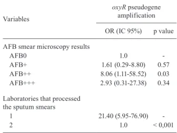
Univariate and multivariate analysis - Univari-ate analysis (n = 93) indicUnivari-ated that the presence of oxyR pseudogene amplification was statistically higher for pa-tients who had alcohol use (OR = 2.92; 95% CI = 1.02-8.3), for those who were referred for DOTS (OR = 4.37; 95% CI = 1.70-11.22) and for patients who had SSS prepared in laboratory 1 (OR = 10.7; 95% CI = 3.78-26.82) (Table I). Finally, multivariate analysis indicated two variables that were statistically associated with presence of the oxyR amplification: AFB++ results by smear microscopy (OR = 8.06; 95% IC = 1.11-58.52) and SSS prepared in labora-tory 1 (OR = 21.40; 95% IC = 5.95-76.90) (Table II).
DISCUSSION
We have demonstrated the capability of amplifying and sequencing the oxyR pseudogene from stored SSS. However, we could not identify any molecular profile consistent with M. bovis.
The observed relationship between alcohol use and DOTS with oxyR pseudogene amplification was only verified in the univariate analysis and can potentially be attributed to a confounding effect of the source labora-tory. Patients who reported alcohol use and those who were referred for DOTS were preferentially diagnosed in laboratory 1 (p value < 0.05). As shown, the association between these explanatory variables and the outcome of interest did not remain significant when adjusting for laboratory in the stratified and multivariate analyses.
The expected trend of the higher chance of oxyR pseudo-gene amplification in AFB-positive patients was not evi-denced in the univariate analysis, perhaps due to a lack of statistical power. However, the independent association be-tween AFB++ and the oxyR pseudogene amplification was evidenced in the multivariate analysis, a model that also took into account the laboratory that processed the SSS. Zink and Nerlich (2004) were able to distinguish the molecular profile of M. bovis from the rest of the MTC using formalin-fixed and paraffin wax-embedded biopsy tissue samples and concluded that different amounts and varying preservation states of the DNA samples could more easily affect the am-plification of single copy genes, such as oxyR, compared to multi copy genes. The lack of association between AFB+++ and amplification of the pseudogene oxyR in the multivari-ate analysis was most likely due to the small sample size.
The greater chance of the oxyR pseudogene amplifica-tion found in patients who had SSS prepared in laboratory 1 compared to laboratory 2 deserves attention. A potential explanation for such a difference may lie in the packing, transportation and/or processing of specimens, which may have affected the DNA integrity of the specimens. In labo-ratory 1, the preparation of the SSS was done no later than 24 h after sputum collection. However, in laboratory 2, the sputum samples were sent from the primary health unit to a central unit where they were kept for a week before being forwarded to the laboratory where the SSS were prepared for microscopy. This excessive delay between sputum col-This excessive delay between sputum col-lection and smear preparation is of public health importance and quality assurance should be a concern in such settings.
Zink and Nerlich (2004) argued that the chemical processing of stored materials is another factor that in-duces changes in DNA, potentially resulting in its par-tial or complete degradation. However, as the SSS of the two laboratories were apparently prepared using the same process of standardized staining (ZN), chemical processing should not be a factor influencing the differ-ent levels of oxyR pseudogene amplification.
It is feasible that the positive AFB smears that did not have the amplified oxyR pseudogene may be a result of atypical mycobacteria, which also could be differen-tially distributed between the two laboratories. Howev-er, another study conducted by the same team in Juiz de Fora, involving 156 patients with TB diagnosed from March 2008-February 2010 in the same laboratories, revealed that the levels of atypical mycobacteria were
177 AFB slides (99 individuals)
171 AFB slides (93 source patients)
93 AFB slides (93 patients)
83 AFB Pos 10 AFB 0
6 AFB slides (6 individuals)
excluded
51 patients in single AFB
results
20 patients in duplicate AFB
results
22 patients in tripletes or more
AFB results
33 (84,6%)
oxyR Pos 39 LAB 1
15 (34,0%)
oxyR Pos 44 LAB 2
6 (75,0%)
oxyR Pos 8 LAB 1
0 (0,0%)
oxyR Pos 2 LAB 2
low and evenly distributed between laboratories. The classical biochemical methods showed 151 (96.79%) M. tuberculosis, three (1.92%) M. avium intracellulare, one (0.64%) M. tuberculosis-M. avium intracellulare co-infection and one (0.64%) M. tuberculosis-M. bovis co-infection (unpublished observations). This percent-age of atypical mycobacteria (6%) was slightly lower than that found by Froes et al. (2003).
Also of public health and medical interest is the am-plification of the oxyR pseudogene in a considerable
por-tion (75%) of patients from laboratory 1 who had both negative results for AFB smear microscopy and a posi-tive culture for mycobacteria. These results suggest a potential ability of the oxyR pseudogene amplification in detecting the MTC even in SSS from paucibacillary patients. This identification of mycobacteria by molecu-lar methods in paucibacilmolecu-lary sputum from either fresh or stored specimens could be an important strategy to be used among patients who cannot wait for the results of traditional diagnosis such as a mycobacteria culture.
TABLE I
Univariate analysis of the oxyR pseudogene amplification, Juiz de Fora, Minas Gerais, 2007 (n = 93)
Variables
oxyR pseudogene amplification
p value Total Posa (%) OR (CI 95%)
Sex
Female 17 7 (41.2) 1.00
-Male 76 47 (61.8) 2.31 (0.79-6.75) 0.12
Age
> 42 42 23 (54.8) 1.00
-≤ 42 42 26 (61.9) 1.34 (0.51-3.52) 0.50
History of HIV infectionb
No 74 42 (56.8) 0 (0.0-5.73) 0.22
Yes 2 2 (100) 1.00
-Alcohol use (ever)b
No 47 22 (46.8) 1.00
-Yes 25 18 (72) 2.92 (1.02-8.3) < 0.05
Referred from other institutionb
No 63 33 (52.4) 1.00
-Yes 18 14 (77.8) 3.18 (0.94-10.73) 0.06
DOTSb
No 33 13 (39.4) 1.00
-Yes 50 37 (74) 4.37 (1.70-11.22) < 0.01
Type of caseb
Recurrence/reentry after dropout 19 9 (47.4) 1.00 -New case 66 41 (62.1) 1.82 (0.65-5.09) 0.25
AFB results
AFB0 10 6 (60) 1.00
-AFB+ 42 23 (54.8) 0.80 (0.19-3.28) 0.76 AFB++ 30 19 (63.3) 1.15 (0.26-4.99) 0.85 AFB+++ 11 6 (54.5) 0.80 (0.14-4.53) 0.80
Laboratories
1 47 39 (83) 10.07 (3.78-26.82) < 0.001
2 46 15 (32.6) 1.00
Some limitations of this study should be discussed. This is an exploratory study in which the samples were not randomly drawn but were remnants of SSS sent to the Public HealthCenter Laboratory for quality control. Most of the slides were AFB-positive because in the absence of culture, the result of AFB was a major criterion for defin-ing diagnosis of TB. It should be noted that only pulmo-nary cases were included in which M. bovis is less fre-quent. The study also only involved laboratories located in urban areas, which may reduce the inclusion of patients from rural areas where M. bovis may have a higher preva-lence. In addition, the small sample size from only one municipality and lack of representativeness may jeopard-ise generalization of our results.
Although M. bovis was not found in the examined sam-ples, the high percentage of pseudogene oxyR amplification (MTC) in our samples may indicate that the analysis of nu-cleotide polymorphisms at position 285 of oxyR in stored SSS can be used to obtain estimates of the prevalence or incidence rates of human infections caused by M. bovis. In addition to Brazil, this strategy could be expanded for epi-demiological purposes in areas such as Africa, Southeast Asia, Central America, South America and other regions where transmission of M. bovis from animals to humans and vice-versa can occur or is a suspected problem.
With patients who fail in the first regimen for tuber-culosis treatment or develop AFB-negative paucibacillary tuberculosis, it may be difficult to obtain new positive cultures due to the effect of therapy on mycobacteria and difficulties in collecting new samples, even by bronchial lavage. In these situations, the stored SSS can also be an alternative for the identification of involved mycobacte-rium species by molecular methods; this will be a particu-larly useful strategy for most developing countries where the isolation and characterization of mycobacteria are not routinely practiced at the start of tuberculosis treatment.
Sreevatsan et al. (1996) analyzed the polymorphism at nucleotide 285 in the oxyR pseudogenes of MTC organ-isms from widespread geographic sources and host spe-cies and indicated that it was 100% sensitive and specific for distinguishing M. bovis isolates from other members of the MTC. Either direct sequencingof amplifiedDNA or PCR-restriction fragment length polymorphism strategies were used to identify the polymorphism at nucleotide 285 in the oxyR pseudogene. While those strategies have been extensively applied to DNA extracted from MTC isolates (Sreevatsan et al. 1996, Espinosa de los Monteros et al. 1998), they are rarely used in primary clinical specimens and archived fixed tissues (Zink & Nerlich 2004).
Finally, despite the limitations of this study and the need for further work to assess the validity and reliabil-ity of these findings, the amplification and sequencing of the oxyR pseudogene from stored ZN-stained SSS has a potential public health application as a complementary method of diagnosis for tuberculosis, especially in areas or populations where M. bovis is more prevalent.
REFERENCES
Blázquez J, Espinosa de Los Monteros LE, Samper S, Martín C, Guerre-ro A, Cobo J, Van Embden J, BaqueGuerre-ro F, Gómez-Mampaso E 1997. Genetic characterization of multidrug-resistant Mycobacterium bovis strains from a hospital outbreak involving human immunode-ficiency virus-positive patients. J Clin Microbiol35: 1390-1393.
Collins CH, Grange JM 1983. The bovine tubercle bacillus. J Appl Bacteriol55: 13-29.
Cosivi O, Grange JM, Daborn CJ, Raviglione MC, Fujikura T, Cousins D, Robinson RA, Huchzermeyer HF, de Kantor I, Meslin FX 1998. Zoonotic tuberculosis due to Mycobacterium bovis in developing countries. Emerg Infect Dis4: 59-70.
Cosivi O, Meslin FX, Daborn CJ, Grange JM 1995. Epidemiology of Mycobacterium bovis infection in animals and humans, with particular reference to Africa. Rev Sci Tech14: 733-746.
Da Silva Rocha A 2009. Doenças micobacterianas de relevância máxima: I - Tuberculose humana e Mycobacterium bovis. II - Hanseníase, recidiva, re-infecção e resistência bacteriana a drogas dos esquemas terapêuticos propostos pela Organização Mundial da Saúde, PhD Thesis, Universidade Federal do Rio de Janeiro, Rio de Janeiro, 308 pp.
Daborn CJ, Grange JM 1993. HIV/AIDS and its implications for the control of animal tuberculosis. Br Vet J149: 405-417.
Dankner WM, Davis CE 2000. Mycobacterium bovis as a significant cause of tuberculosis in children residing along the United States-Mexico border in the Baja California region. Pediatrics105: E79.
Dankner WM, Waecker NJ, Essey MA, Moser K, Thompson M, Da-vis CE 1993. Mycobacterium bovis infections in San Diego: a clinicoepidemiologic study of 73 patients and a historical review of a forgotten pathogen. Medicine(Baltimore) 72: 11-37.
Dean AG, Dean JA, Coulombier D, Brendel KA, Smith DC, Burton AH, Dicker RC, Sullivan K, Fagan RF, Arner TG 1994. Epi Info Version 6, Centers for Diseases Control and Prevention, Atlanta, GA.
Espinosa de los Monteros LE, Galán JC, Gutiérrez M, Samper S, García Marín JF, Martín C, Domínguez L, de Rafael L, Baquero F, Gómez-Mampaso E, Blázquez J 1998. Allele-specific PCR method based on pncA and oxyR sequences for distinguishing Mycobacterium bovis from Mycobacterium tuberculosis: in-traspecific M. bovispncA sequence polymorphism. J Clin Mi-crobiol 36: 239-242.
TABLE II
Multivariate logistic regression model for the amplification of the oxyR pseudogene, Juiz de Fora, Minas Gerais, 2007 (n = 93)
Variables
oxyR pseudogene amplification
OR (IC 95%) p value
AFB smear microscopy results
AFB0 1.0
-AFB+ 1.61 (0.29-8.80) 0.57 AFB++ 8.06 (1.11-58.52) 0.03 AFB+++ 2.93 (0.31-27.38) 0.34
Laboratories that processed the sputum smears
1 21.40 (5.95-76.90)
-2 1.0 < 0,001
Evans JT, Smith EG, Banerjee A, Smith RM, Dale J, Innes JA, Hunt D, Tweddell A, Wood A, Anderson C, Hewinson RG, Smith NH, Hawkey PM, Sonnenberg P 2007. Cluster of human tuberculosis caused by Mycobacterium bovis: evidence for person-to-person transmission in the UK. Lancet369: 1270-1276.
Farr SB, Kogoma T1991. Oxidative stress responses in Escherichia coli and Salmonella typhimurium. Microbiol Rev55: 561-585.
Froes GC, Coutinho RL, Nardy de Ávila M, Cançado LR, Spíndola de Miranda S 2003. Perfil e seguimento dos pacientes portado-res de Mycobacterium sp.do Hospital das Clínicas da Universi-dade Federal de Minas Gerais. J Bras Pneumol29: 365-370.
Konno K, Feldmann FM, McDermott W 1967. Pyrazinamide sus-ceptibility and amidase activity of tubercle bacilli. Am Rev Re-spir Dis95: 461-469.
LoBue PA, Betacourt W, Peter C, Moser KS 2003. Epidemiology of Mycobacterium bovis disease in San Diego County, 1994-2000. Int J Tuberc Lung Dis7: 180-185.
Long R, Nobert E, Chomyc S, van Embden J, McNamee C, Duran RR, Talbot J, Fanning A 1999. Transcontinental spread of mul-tidrug-resistant Mycobacterium bovis. Am J Respir Crit Care Med 159: 2014-2017.
MAPA - Ministério da Agricultura, Pecuária e Abastecimento, Bra-sil 2006. Programa Nacional de Controle e Erradicação da Brucelose e da Tuberculose Animal (PNCEBT), MAPA/SDA/ DSA, Brasília, 188 pp.
MS - Ministério da Saúde, Brasil 2005. Manual de bacteriologia da tuberculose, 3rd ed., Centro de Referência Professor Hélio Fra-ga, Secretaria de Vigilância em Saúde, Rio de Janeiro, 240 pp.
PAHO - Pan American Health Organization 1991. Health conditions in the Americas, 1990, vol. I, scientific publication 524, Pan American Health Organization, Washington DC, 404 pp.
R Development Core Team 2005. R: a language and environment for statistical computing. R Foundation for Statistical Comput-ing, Vienna, Austria. Available from: http://www.R-project.org. [accessed 2005].
Rivero A, Márquez M, Santos J, Pinedo A, Sánchez MA, Esteve A, Samper S, Martín C 2001. High rate of tuberculosis reinfection dur-ing a nosocomial outbreak of multidrug-resistant tuberculosis caused by Mycobacterium bovis strain B. Clin Infect Dis32: 159-161.
Roxo E 1997. Mycobacterium bovis como causa de zoonose. Rev Cienc Farm18: 101-108.
Samper S, Martín C, Pinedo A, Rivero A, Blázquez J, Baquero F, van Soolingen D, van Embden J 1997. Transmission between HIV-infected patients of multidrug-resistant tuberculosis caused by Mycobacterium bovis. AIDS11: 1237-1242.
Sherman DR, Sabo PJ, Hickey MJ, Arain TM, Mahairas GG, Yuan Y, Barry CE 3rd, Stover CK 1995. Disparate responses to oxida-tive stress in saprophytic and pathogenic mycobacteria. Proc Natl Acad SciUSA 92: 6625-6629.
Sreevatsan S, Escalante P, Pan X, Gillies DA 2nd, Siddiqui S, Khalaf CN, Kreiswirth BN, Bifani P, Adams LG, Ficht T, Perumaalla VS, Cave MD, van Embden JD, Musser JM 1996. Identification of a polymorphic nucleotide in oxyR specific for Mycobacterium bovis. J Clin Microbiol34: 2007-2010.
Sreevatsan S, Pan X, Zhang Y, Deretic V, Musser JM 1997.Analysis of the oxyR-ahpC region in isoniazid-resistant and susceptible Mycobacterium tuberculosis complex organisms recovered from diseased humans and animals in diverse localities. Antimicrob Agents Chemother41: 600-606.
Taylor GM, Goyal M, Legge AJ, Shaw RJ, Young D 1999. Genotypic analysis of Mycobacterium tuberculosis from medieval human remains. Microbiology145: 899-904.
Wedlock DN, Skinner MA, de Lisle GW, Buddle BM 2002. Control of Mycobacterium bovis infections and the risk to human popula-tions. Microbes Infect4: 471-480.
Wilkins EG, Griffiths RJ, Roberts C 1986. Bovine variants of Myco-bacterium tuberculosis isolated in Liverpool during the period 1969 to 1983: an epidemiological survey. Q J Med59: 627-635.