Published online December 16th, 2010 © http://www.ijav.org
Case Report
International Journal of Anatomical Variations (2010) 3: 200–202
Introduction
According to the classical description in the textbooks of anatomy, the abducent nerve emerges from the brainstem at the pontomedullary junction as a single trunk, traverses the cranial cavity and enters the orbit through the superior orbital fissure to supply the lateral rectus muscle [1]. Multiple roots of the nerve have been reported at the exit or the nerve may split into branches after its exit somewhere along its course [2–5]. Duplication of the nerve at its exit has been reported by many workers [2–6]. Nathan et al. reported a case in which the abducent nerve had split into three branches in the petro-clival region [4]. In the present study, four roots of the abducent nerve were observed at its exit from the brainstem. There is no such report available in literature and this prompted us to report this case.
Recent advances in imaging techniques, cranial base surgery, endovascular interventions etc., have allowed neurosurgeons to attempt intervention in lesions involving the cavernous sinus. The knowledge of variations in the intracranial course of the abducent nerve is, therefore, important for both anatomical knowledge and for neurosurgical procedures.
Most of the descriptions of the abducent nerve in literature are based on microanatomic dissection. The nerve has also been examined by endonasal endoscopic approach [6].
Aaijaz Ahmed KHAN [1]
Pal Hillol KANTI [2]
Muzammil ULLAH [1]
Asma Haji HASSAN [1]
Departments of Anatomy [1] and Neurosciences [2], School of Medical Sciences, Universiti Sains Malaysia, Kelantan, MALAYSIA.
Aaijaz Ahmed Khan Senior Lecturer Department of Anatomy School of Medicine Universiti Sains Malaysia 16150 Kubang Kerian, Kelantan, Malaysia.
+60 9 7676080 [email protected]
Received July 14th, 2010; accepted September 3rd, 2010
ABSTRACT
The abducent nerve occupies a strategic position at the middle third of the clivus. With the recent advances in the field of imaging techniques and endoscopic skull base surgery it is important to understand the neurovascular relationships and variations in its course. Very limited literature is available on the cisternal course of abducent nerve as studied by an endoscope. A few studies have described the course of abducent nerve endoscopically through an endonasal approach. In this study we attempted to explore its cisternal course endoscopically through a retrosigmoid approach to the cerebello-pontine angle with emphasis on its neurovascular relationship. Duplication and triplication of the abducent nerve have been reported by some authors, but four roots of abducent nerve have not been reported. In the present study, in one of the cadavers it was found that on the left side the abducent nerve emerged as four roots from the pontomedullary sulcus. © IJAV. 2010; 3: 200–202.
Key words [abducent nerve] [cerebellopontine angle] [duplication] [roots] [cisternal]
eISSN 1308-4038
Four roots of abducent nerve at its exit from the pontomedullary sulcus: endoscopic case study
In the present study, its cisternal course and relations were examined by the endoscopy of the cebello-pontine (CP) angle through the retrosigmoid approach. Through this approach it is possible to define its cisternal course and its relations with anterior inferior cerebellar artery (AICA).
Case Report
Five formalin-fixed cadaveric heads were subjected to endoscopic exploration of intracranial course of the abducent nerve in the CP angle through a retrosigmoid approach at the Neuroanatomy Laboratory of the Department of Anatomy, School of Medical Sciences, Universiti Sains Malaysia. The study was performed using Richard Wolff 2.8 and 5 mm diameter rigid endoscopes at viewing angles of 0° and 30°. The video was recorded on a Pioneer HDD DVD 530H Recorder and still images were captured using Pinnacle software Version 9. The retrosigmoid approach:
201 Four roots of abducent nerve
The retrosigmoid endoscopic approach provided a good view of the entire cisternal course and relations of the abducent nerve.
In one of the four cadavers, four roots (R1, R2, R3 and R4) of the abducent nerve were observed on the left side, and two roots were observed on the right side at its exit from the pontomedullary sulcus (Figures 1, 2, 3). They formed two divisions (VIa and VIb) of the left abducent nerve. The VIa was larger than VIb. The R1 was smaller than other roots and continued as division VIb, whereas, R2, R3 and R4 joined to form the division VIa of the nerve. The R2 and R3 joined the VIa shortly after their exit whereas R4 joined it after travelling a short distance. The VIa was found to pass dorsal to the AICA and
entered the Dorello’s canal at the petro-clival region. The VIb was found to make a loop around the ventral aspect of the AICA and entered the Dorello’s canal separately a little (approximately 2 mm) below the entrance of VIa. Discussion
Most of the textbooks of anatomy state that the abducent nerve exits from the brainstem as a single trunk, and
during its course it traverses the cisterna pontis, Dorello’s canal, cavernous sinus, superior orbital fissure before reaching the orbit to supply the lateral rectus muscle. Iaconetta et al. described five segments of the abducent nerve: 3 intracranial (cisternal, gulfar and cavernous) and two orbital (fissural and intraconal) [6]. The cisternal segment extends from the pontomedullary sulcus to the dural foramen (Dorello’s canal).
The incidences of duplication of abducent nerve have been reported by many authors to range between 8 and 18 percent [4–6].
Iaconetta et al. studied 50 human formalin fixed cadavers (100 specimens) [3]. They found duplicated abducent nerve in eight specimens (8%). In two (25%) of these eight specimens, the nerve originated at the pontomedullary sulcus as two independent trunks, in one case they found that the superior trunk was thicker than the inferior and in the other it was thinner. In six cases (75%) the nerve originated as a single trunk, splitting into two trunks in its cisternal course.
Ozveren et al. examined 100 specimens in 50 autopsies [2]. They found that the duplication of the abducent nerve was bilateral in four of 50 cases (8 specimens), and was unilateral in 7 cases. In 9 specimens they found that the nerve emerged from the brainstem as a single trunk, entered the subarachnoid space and splits into two branches. In 6 specimens they found that the nerve emerged from the brainstem as two separate roots that joined in the cavernous sinus.
Iaconetta et al. examined 55 cadaveric human heads [6]. They found duplication of the nerve in 8 cases (5 on the right side and 3 on the left side). They also found that in 2 cases the nerve originated at the pontomedullary sulcus as two independent trunks and in other six cases, the nerve originated as a single trunk and then had split into two trunks in the cisternal segment.
Jain reported 5 (1.67%) of his 18 cases to have bilateral duplication of the abducent nerve [5].
Nathan et al. studied the abducent nerve in 62 cadavers [4]. They did not find the bilateral duplication of the
Figure 1. A panoramic endoscopic view of the left abducent nerve showing its roots and divisions. (AICA: anterior inferior cerebellar artery; R1–R4: roots; VIa:–large division; VIb: small division; VA:
vertebral artery)
AICA
R4
R4
R3
R3
VIa
VIa
VA
VA
R2
R2
R1
R1
VIb
VIb
PONS
PONS
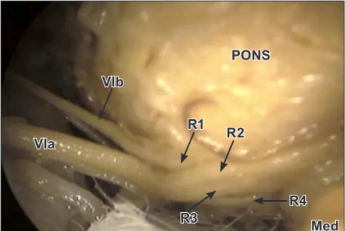
Figure 2. Endoscopic view showing the roots of the left abducent nerve at the pontomedullary sulcus. (R1–R4: roots of abducent nerve; VIa: large division; VIb: small division; Med: medulla)
R4 R3
VIa
R2 R1 VIb
PONS
Med
Figure 3. A schematic diagram of the multiple roots of the left abducent nerve. (DC: Dorello’s canal; VA: vertebral artery; VIa:
large division; VIb: small division; R1–R4: roots of the abducent nerve; MED: medulla; AICA: anterior inferior cerebellar artery; CB:
central branches)
AICA
MED DC
202 Khan et al.
abducent nerve in any case. They classified the abducent nerve into six types as follows:
Type I: The nerve emerged from the brainstem as a single trunk and there was no duplication of the nerve during its course. They found it in 86.5% cases.
Type II: The nerve emerged from the brainstem as a single trunk but had split into two branches before piercing the dura mater and then rejoined in the cavernous sinus. They found this variant in 6% of cases.
Type III: The nerve emerged from the brainstem as two separate roots that joined in the cavernous sinus. They found this variant in 7.5% of cases.
Type IV: The nerve emerged from the brainstem as two separate roots that continued upto lateral rectus muscle without joining during their course. They did not find this variant but Jain reported one such case [5].
Type V: Tillack et al. reported a case in which the abducent nerve emerged from the pontomedullary sulcus and joined the oculomotor nerve [7]. Fibers of the abducent nerve travelled along with the fibers of the oculomotor nerve and after passing through the superior
References
[1] Williams PL, Warwick R. Gray’s Anatomy. 36th Ed., Edinburgh, Churchill Livingstone. 1980; 1068–1070.
[2] Ozveren MF, Sam B, Akdemir I, Alkan A, Tekdemir I, Deda H. Duplication of the abducens nerve at the petroclival region: an anatomic study. Neurosurgery. 2003; 52: 645–652.
[3] Iaconetta G, Tessitore E, Samii M. Duplicated abducent nerve and its course: microanatomical study and surgery-related considerations. J Neurosurg. 2001; 95: 853–858.
[4] Nathan H, Ouaknine G, Kosary IZ. The abducens nerve: Anatomical variations in its course. J Neurosurg. 1974; 41: 561–566.
[5] Jain KK. Aberrant roots of the abducens nerve. J Neurosurg. 1964; 21: 349–351.
[6] Iaconetta G, Fusco M, Cavallo LM, Cappabianca P, Samii M, Tschabitscher M. The abducens nerve: microanatomic and endoscopic study. Neurosurgery. 2007; 61: ONS7–ONS14.
[7] Tillack TW, Winer JA. Anomaly of the abducens nerve. Yale J Biol Med. 1962; 34: 620–624.
orbital fissure, they innervated the lateral rectus muscle. Nathan et al. did not find such a variant [4].
Type VI: The nerve had split into three branches in the petro-clival region to reunite again in the cavernous sinus. Nathan et al. found one such case [4].
In the present study the variation was bilateral as reported by many authors referred above, but on the left side four roots of the abducent nerve were observed instead of two or three roots reported in literature cited above. In terms of the classification of the abducent nerve described by Nathan et al. cited above [4], the present variant may be treated as type VII.
Conclusion