Composed of
Staphylococcus aureus
Poly-N-Acetyl-Glucosamine and Clumping Factor A
Toma´s Maira-Litra´n*., Leticia V. Bentancor., Cagla Bozkurt-Guzel, Jennifer M. O’Malley, Colette
Cywes-Bentley, Gerald B. Pier
Channing Laboratory, Department of Medicine, Brigham and Women’s Hospital, Harvard Medical School, Boston, Massachusetts, United States of America
Abstract
The increasing frequency, severity and antimicrobial resistance of Staphylococcus aureus infections has made the development of immunotherapies against this pathogen more urgent than ever. Previous immunization attempts using monovalent antigens resulted in at best partial levels of protection againstS. aureusinfection. We therefore reasoned that synthesizing a bivalent conjugate vaccine composed of two widely expressed antigens ofS. aureuswould result in additive/ synergetic activities by antibodies to each vaccine component and/or in increased strain coverage. For this we used reductive amination, to covalently link theS. aureusantigens clumping factor A (ClfA) and deacetylated poly-N-b -(1–6)-acetyl-glucosamine (dPNAG). Mice immunized with 1, 5 or 10mg of the dPNAG-ClfA conjugate responded in a dose-dependent manner with IgG to dPNAG and ClfA, whereas mice immunized with a mixture of ClfA and dPNAG developed significantly lower antibody titers to ClfA and no antibodies to PNAG. The dPNAG-ClfA vaccine was also highly immunogenic in rabbits, rhesus monkeys and a goat. Moreover, affinity-purified, antibodies to ClfA from dPNAG-ClfA immune serum blocked the binding of threeS. aureusstrains to immobilized fibrinogen. In an opsonophagocytic assay (OPKA) goat antibodies to dPNAG-ClfA vaccine, in the presence of complement and polymorphonuclear cells, killed S. aureus Newman and, to a lower extent, S. aureus Newman DclfA. A PNAG-negative isogenic mutant was not killed. Moreover, PNAG antigen fully inhibited the killing ofS. aureusNewman by antisera to dPNAG-ClfA vaccine. Finally, mice passively vaccinated with goat antisera to dPNAG-ClfA or dPNAG-diphtheria toxoid conjugate had comparable levels of reductions of bacteria in the blood 2 h after infection with three differentS. aureusstrains as compared to mice given normal goat serum. In conclusion, ClfA is an immunogenic carrier protein that elicited anti-adhesive antibodies that fail to augment the OPK and protective activities of antibodies to the PNAG cell surface polysaccharide.
Citation:Maira-Litra´n T, Bentancor LV, Bozkurt-Guzel C, O’Malley JM, Cywes-Bentley C, et al. (2012) Synthesis and Evaluation of a Conjugate Vaccine Composed of
Staphylococcus aureusPoly-N-Acetyl-Glucosamine and Clumping Factor A. PLoS ONE 7(9): e43813. doi:10.1371/journal.pone.0043813 Editor:Riccardo Manganelli, University of Padova Medical School, Italy
ReceivedJune 16, 2012;AcceptedJuly 26, 2012;PublishedSeptember 6, 2012
Copyright:ß2012 Maira-Litra´n et al. This is an open-access article distributed under the terms of the Creative Commons Attribution License, which permits
unrestricted use, distribution, and reproduction in any medium, provided the original author and source are credited.
Funding:This work was supported by grants from the National Institutes of Health, National Institute of Allergy and Infectious Diseases numbers AI46706 (GBP) and AI057159, a component of Award Number U54 AI057159. The content is solely the responsibility of the authors and does not necessarily represent the official views of the National Institute of Allergy and Infectious Diseases or the National Institutes of Health. The funders had no role in study design, data collection and analysis, decision to publish, or preparation of the manuscript.
Competing Interests:Toma´s Maira-Litra´n (TML) and Gerald B. Pier (GBP) are inventors of Intellectual Property (IP) (PNAG Vaccine and human monoclonal antibody to PNAG) that is licensed by Brigham and Women’s Hospital (BWH) to Alopexx Vaccines LLC, and Alopexx Pharmaceuticals LLC, companies in which GBP also owns equity. As inventors of the IP, they also have the right to receive a share of licensing related income (royalties, fees) through BWH from Alopexx Pharmaceuticals and Alopexx Vaccines. Their interests were reviewed and are managed by the BWH and Partners Healthcare in accordance with their conflict of interest policies. This does not alter the authors’ adherence to all the PLoS ONE policies on sharing data and materials.
* E-mail: [email protected]
.These authors contributed equally to this work.
Introduction
Staphylococcus aureus is a leading etiology of hospital-acquired infections worldwide. This versatile organism, including strains with a troublesome pattern of antibiotic resistance as accentuated by methicillin-resistantS. aureus(MRSA), cause a wide spectrum of diseases that range from mild skin and soft tissue infections to more severe invasive ones such as endocarditis, blood and lower respiratory tract infections, septic arthritis, osteomyelitis or deep-seated abscesses among others [1,2]. With the emergence of these many difficult-to-treat strains the need to develop new antimicro-bials and/or immunotherapeutic approaches to combat infections is more urgent than ever.
binding protein (FnBP), collagen binding protein (CnBP), clump-ing factor A (ClfA), and the iron surface determinant B protein (IsdB) [3]. Some vaccine antigens including CP5 and CP8 conjugate vaccines, the IsdB antigen, human polyclonal antibodies to ClfA and a humanized monoclonal antibody to LTA have reached phase III human trials. Despite promising results obtained in pre-clinical studies, all have failed to meet their defined endpoints in preventingS. aureusinfection [3]. As a result of these disappointing outcomes with monovalent vaccine components, a shift towards use of polyvalent vaccines has garnered significant interest.
To asses if additive or synergistic protection could be engendered in a multivalent vaccine, we evaluated a conjugate vaccine composed of two highly conserved S. aureus surface antigens, poly-N-b-(1–6)-acetyl-glucosamine (PNAG) and ClfA by using the protection-inducing deacetylated glycoform of PNAG (dPNAG) [4] conjugated to ClfA as a vaccine candidate againstS. aureusinfections. Previous work carried out in our laboratory has already demonstrated the ability of a vaccine composed of dPNAG conjugated to the carrier protein diphtheria toxoid (dPNAG-DT) to induce high titers of opsonic and protective antibodies [4]. In addition to the potential additive/synergistic activity between antibodies to dPNAG and ClfA the synthesis of a bivalent dPNAG-CflA vaccine is also aimed to potentially expand the coverage of a single-component vaccine to includeS. aureusstrains expressing only one of the vaccine components.
Specifically this study investigated the value of ClfA as a carrier protein for dPNAG, the functional activity and specificity of antibodies to the dPNAG-ClfA vaccine using anti-adhesive and opsonic killing assays, and protective efficacy in a mouse model of S. aureusbloodstream infection.
Materials and Methods
Ethics Statement
This study was carried out in strict accordance with the recommendations in the Guide for the Care and Use of Laboratory Animals of the National Institutes of Health. All animal protocols were reviewed and approved by the Harvard Medical Area Standing Committee on Animals IACUC which has Animal Welfare Assurance of Compliance number A3431-01 on file with the Office of Laboratory Animal Welfare of the U.S. Public Health Service. During all animal experimentation procedures all efforts were made to minimize suffering.
Studies involving human subjects were approved by the Partners Health Care System Institutional Review Board (IRB). All subjects donating blood provided written informed consent to participate in the studies.
Bacterial strains and growth conditions
The strains used in this work (Table 1) were routinely grown to stationary phase in tryptic soy broth (TSB) supplemented with 1% glucose. When necessary, TSB as well as tryptic soy agar (TSA) were supplemented with ampicillin (100mg/ml), tetracycline (10mg/ml) or erythromycin (10mg/ml).
PCR detection of icaABCDandclfAgenes inS. aureus strains
Primers pairs icaF (59 -CCAGAGAAATTAGATATTCATT-GAACAAGAAGC-39)/icaR (59 -CATGCCGACACCTATACA-TAATCCTAAAATGAA-39) and ClfA-F (59 -CGGAAAAAATC-GATTGGCGTGGCTT-39)/ClfA-R (59 -GTAATGAACCTAT-TGATGCTAATAATCCCCA-39) were used to amplify the entire icaABCDlocus or theclfAgene, respectively, from genomic DNA
extracted from the strainsS. aureusNewman, 476 and MN8. The genomic DNA was isolated by using the Wizard genomic DNA purification kit (Promega) according to the manufacturer’s instructions.
Purification of clumping factor A
Recombinant clumping factor A (ClfA) protein, residues (221– 559), containing an N-terminal extension of six histidine residues was expressed from the plasmid pCF41carried byE. coliXL-1 Blue and kindly provided by Dr Timothy Foster.E. coliwas grown in LB supplemented with ampicillin in a 6-liter fermentor at 37uC until the optical density at 650 nm (OD650nm) reached 1. The
temperature was maintained at 37uC, isopropylb -D-1-thiogalac-topyranoside (IPTG) was added to a final concentration of 0.5 mM, and growth was continued for 3 h. Cells were pelleted (6,000g610 min at 4uC), resuspended in 30 ml of phosphate-buffered saline (PBS) with complete EDTA-free protease inhibitor cocktail (Roche Applied Science) and sonicated using 3 cycles of 20 s at 4 W (Sonic Dismembrator) followed by 10 s of rest in an ice-water bath. After cell debris was removed by centrifugation (6,000g610 min at 4uC), the supernatant was filtered and ClfA purified by affinity chromatography on a Ni2-chelate column
(Novagen) following the manufacture’s instructions. The protein was further purified by size exclusion chromatography (SEC) on Sephacryl S-200 (Amersham Biosciences) and endotoxin removed using Detoxi-Gel (Pierce, Rockford, IL).
Coupling of dPNAG to ClfA
ClfA was coupled to purified dPNAG using the reductive amination reaction. Aldehyde groups were introduced onto the surface of ClfA by treating the protein with an excess of glutaraldehyde and the activated ClfA was subsequently reacted with dPNAG using the abundant free amino groups present on this polysaccharide in the presence of sodium cyanoborohydride (NaBH3CN) (Matreya, Pleasant Gap, PA).
Activation of ClfA with glutaraldehyde. ClfA was dissolved in 0.1 M carbonate buffer pH 10 at a concentration of 10 mg/ml and glutaraldehyde (Sigma) added to a final concentration of 1.25% v/v. This reaction was allowed to proceed for 2 h at room temperature and the gluraldehyde-activated protein dialyzed against PBS pH 7.4.
Coupling of glutaraldehyde-activated ClfA to dPNAG. dPNAG was first dissolved in 5M HCl, neutralized with an equal volume of 5 M NaOH and diluted in PBS pH 7.5 to a final concentration of approximately 5 mg/ml. This dPNAG solution was then mixed with an equal amount of gluraldehyde-activated ClfA in PBS and the pH adjusted to 7.5. Purified NaBH3CN was added to the mixture in a 20-fold excess over the
amount of each of the components and the reaction allowed to proceed in the dark for 14 h at 37uC with mixing. After the conjugation reaction was completed the high molecular weight (MW) conjugate was purified from uncoupled dPNAG and ClfA by gel filtration chromatography on a Superose 6-prep-grade column (Amersham Biosciences). Fractions containing dPNAG-ClfA conjugate vaccine, which were identified on the basis of the earlier elution of both polysaccharide and protein compared with the elution of the non-conjugated components, were pooled, dialyzed against 20 mM HEPES buffer plus 50 mmM NaCl pH 8 and stored frozen at220uC.
and for protein by the Bradford method [6] with bovine serum albumin (BSA) as the standard.
Antiserum production
Antisera to purified dPNAG-ClfA were raised in various animal hosts including two rabbits, two rhesus monkeys and a goat. Antibodies to dPNAG-ClfA were raised in New Zealand White rabbits by subcutaneous (SC) immunization with two 10mg doses of conjugated polysaccharide emulsified for the first dose in complete Freund’s adjuvant and for the second dose in incomplete Freund’s adjuvant, followed 1 week later by three intravenous (IV) injections of antigen in saline spaced 3 days apart. Rabbits were bled every 2 weeks and sera tested by the enzyme-linked immunosorbent assay (ELISA). Immune sera to dPNAG-ClfA was also raised in two rhesus monkeys by SC vaccination with a single dose of 100mg of dPNAG-ClfA vaccine in aluminum hydroxide gel adjuvant (Alhydrogel 1.3%, Brenntag Biosector). Finally, a goat was immunized with the dPNAG-ClfA conjugate vaccine following the protocol described earlier for rabbits but with doses of 50mg per injection in Freund’s incomplete adjuvant. Serum samples used to compare the immunogenicity of the dPNAG-ClfA vaccine in rabbits, monkeys and a goat were collected four weeks after the last immunization.
Immunogenicity of dPNAG-ClfA vaccine in mice
Groups of 10 mice (Swiss-Webster; female, 5–7 weeks of age) were immunized SC 3 times, 1 week apart, with 1, 5 or 10mg of conjugated polysaccharide suspended in PBS. Blood was with-drawn weekly for a month and antibody titers to both dPNAG and ClfA determined by ELISA. Control groups received a mixture of unconjugated polysaccharide and protein in PBS in the same ratio as in the conjugate vaccine.
ELISA
dPNAG and ClfA-specific antibodies were measured in sera obtained from mice, rabbits, monkeys and a goat by ELISA as described previously [7]. Purified dPNAG and ClfA were used to sensitize the ELISA plates at a concentration of 0.3 or 10mg/ml respectively in phosphate buffer, pH 7.4.
Phagocyte-dependent killing assays
Polymorphonuclear cells (PMNs) were prepared from fresh human blood collected from healthy adult volunteers as described earlier [4] under a protocol approved by the Institutional Review Board of Partner’s Healthcare System and the concentration
adjusted to 2.56107/ml in modified minimal essential medium (MEM)+1% BSA.
The complement source (1 ml of rabbit serum, Sigma) was adsorbed three times withS. aureusNewman at 4uC for 30 min in order to remove all non-specific antibodies present. After adsorption, the complement solution was centrifuged and filter sterilized.
The test sera for the OPK, goat antibodies to dPNAG-ClfA conjugate vaccine and normal goat sera (NGS), were first heated at 56uC for 30 min to inactivate endogenous complement activity and then absorbed three times at 4uC for 30 min with the PNAG-negative strain Newman Dica or the DclfA mutant DU5852 to remove antibodies not directed to the PNAG or ClfA antigens, respectively. The bacteria were then removed by centrifugation and the test sera filter sterilized.
Overnight cultures of the bacterial strains to be evaluated in OPKA were diluted 1:100 in fresh TSB+1% glucose, grown to an OD650nm of 0.4 (,36108colony forming units (CFU)/ml), and the final concentration adjusted to 46106CFU/ml in MEM-1% BSA for use in the killing assay.
The actual phagocytic killing assay was performed by mixing 100ml (each) of the PMNs suspension, target bacteria, dilutions of test sera, and the complement source. The reaction mixture was incubated on a rotor rack at 37uC for 90 min; samples were taken at time zero and after 90 min. A 10-fold dilution was made in TSB with 0.05% Tween to prevent bacterial aggregation, and samples were plated onto TSA plates. Tubes lacking any serum and tubes with NGS were used as controls, as were tubes containing serum and complement but lacking PMNs to control for potential aggregation of bacteria by the antibody, which would reduce the apparent CFU counts at the end of the assay. At the concentra-tions of antisera used in the opsonic killing assay, there was no reduction in CFU of .10% in samples lacking phagocytes but containing antibody and complement, indicating little agglutinat-ing activity of the antibodies raised to dPNAG-ClfA vaccine. The percentage of killing was calculated by determining the ratio of the number of CFU surviving in the tubes with bacteria, leukocytes, complement, and goat anti-dPNAG-ClfA sera to the number of CFU surviving in tubes with the same components but containing NGA.
For inhibition studies, goat antiserum raised to dPNAG-ClfA was diluted 1:10 and incubated for 90 min at 4uC with an equal volume of a solution containing 25 to 200mg/ml of either purified dPNAG or ClfA. Subsequently, the antiserum was centrifuged, Table 1.Bacterial strains used in this study.
S. aureusstrains Description Source
Newman Serotype 5 strain [19]
NewmanDica NewmanDica::tet; TetR [20]
DU5852 NewmanclfA::Tn917; EryR [21]
MN8 Serotype 8 strain [22]
476 Non-typable strain [23]
E. colistrains
E. coliXL1-Blue pCF41-(221–559) Derivative of expression plasmid pQE30 (Qiagen Inc.) containing codons for ClfA amino acids 221–559. Used for the purification of rClfA221–559protein contains an N-terminal extension of six histidine residues; AmpR
[24]
and the supernatant was used in the opsonophagocytic assay as described above.
Murine bacteremia model
Groups of eight mice (FVB; female; 3–5 weeks of age) were immunized intraperitoneally (IP) with 0.4 ml of heat-inactivated (56uC for 30 min) immune goat sera raised to dPNAG-ClfA, dPNAG-DT (dPNAG conjugated to diphtheria toxoid protein) vaccines, or NGS 48 and 24 h before infection. Mice were challenged IV with theS. aureusstrains Newman (4.36107CFUs), MN8 (8.66106CFUs), 476 (6.76105CFUs), Newman Dica (7.56107CFUs) or DU5852 (3.56107CFUs), at the doses indicated, in 0.2 ml of PBS. Two hours later mice were sacrificed and samples of 0.5 ml blood obtained from the heart, mixed with 20ml of heparin (Sigma), and plated onto TSA plates. Bacteremia was quantified by colony counts after overnight growth and values expressed as CFU/ml of blood.
Preparation of affinity purified antibodies to ClfA Three grams of CNBr-activated Sepharose 4B gel (Pharmacia Biotech, Uppsala, Sweden) was suspended in 250 ml of 1 mM HCl for 30 min at 4uC, and then the gel was washed with alternating amounts of 750 ml of 1 mM HCl, 100 ml of distilled water, and 300 ml of reaction buffer containing 0.1 M NaHCO3
and 0.5 M NaCl (pH 8.3). The gel was suspended in 10 ml of the reaction buffer (pH 8.3), incubated with 5 mg of ClfA overnight at 4uC, and then the coupled gel was packed into a low-pressure column and washed with 250 ml of 1 M glycine in reaction buffer (pH 8.3) to block unused activation sites. Subsequently, the coupled gel was washed three times with alternating 200 ml amounts of borate buffer containing 0.1 M boric acid and 1 M NaCl (pH 8.5), 100 ml of distilled water, 200 ml of acetate buffer containing 0.1 M sodium acetate and 1 M NaCl (pH 4.0), and 100 ml of distilled water and then was suspended in PBS.
Ten ml of rabbit antiserum raised to dPNAG-ClfA was passed through a CNBr-activated Sepharose 4B-packed affinity column that was immobilized with ClfA. After intensive washing with PBS, the specific anti-ClfA immunoglobulins, which bound to the beads were eluted with 0.1 M glycine-HCI, pH 2.7 buffer and collected into fractions that were immediately neutralized with 1 M Tris base buffer ammonium bicarbonate, pH 9 and monitored for their absorbance at OD280nm. Fractions containing affinity-purified
rabbit antibodies specific to ClfA were then pooled and quantified for protein content with the Bradford assay [6] using rabbit IgG as standart.
Adherence ofS. aureusto immobilized fibrinogen Adherence ofS. aureusto fibrinogen (Fn) (Sigma) was assessed in 96-well polystyrene plates coated for with 400 ng/well of Fn for 1 h at 37uC after which the remaining sites were blocked by with 200ml of PBS 1% BSA for 1 h at 37uC.
The S. aureus strains Newman, MN8, 476 and the Newman DclfAderivative DU5852 were grown overnight on TSA plates, harvested by centrifugation, washed with PBS pH 7.2 and resuspended in PBS to an OD650nm of 1.0. S. aureus cultures
were then further concentrated 5 fold and pre-incubated with either rabbit affinity-purified antibodies to ClfA or control non-immune polyclonal rabbit IgG at concentrations ranging from 0.8–500mg/ml in PBS for 1 h at room temperature. After Fn-coated plates were incubated with theS. aureusstrains for 1 h at room temperature, wells were washed three times with PBS and bound cells fixed with 200ml of glutaraldehyde (2% v/v in PBS) for 1 h and stained with crystal violet (Sigma) (0.5% v/v) for 5 minutes. Plates were rinsed with water, air-dried and the
absorbance at 595 nm (OD595nm) determined using an ELISA
plate reader (BioTek Instruments, Winoski, Ill).
Confocal laser scanning microscopy
Confocal laser scanning microscopy (CLSM) was used to evaluate the production of PNAG and ClfA in three S. aureus strains, Newman, MN8 and 476.S. aureusstrains were grown overnight at 37uC in TSB supplemented with 1% glucose, washed twice with PBS and blocked overnight with PBS 1% BSA and 10% normal rabbit sera (NRS). Cells were washed twice with PBS and a 10ml aliquot of the cell suspension was air-dried onto a glass slide. Bacteria were fixed to the slide with methanol for 1 min at room temperature. At this pointS. aureussamples were incubated for 2 h at room temperature with antibodies to PNAG (human monoclonal antibody (mAb) F598 conjugated to Alexa Fluor 488) and ClfA (goat dPNAG-CflA sera that had been previously absorbed four times at 4uC with the ClfA-negativeS. aureusstrain DU5852) in PBS with 0.5% BSA and 10% NRS. As controls we includedS. aureussamples labeled with a human alginate-specific mAb F429 conjugated to Alexa Fluor 488 and NGS that had been previously absorbed with S. aureusDU5852. Samples were washed three times with PBS and further incubated for 1 h at room temperature with a secondary donkey anti-goat IgG conjugated to Alexa Fluor 568 (Invitrogen) diluted 1:250 in PBS 0.5% BSA plus the nucleic acid stain Syto 62, (Invitrogen) at a final concentration of 5mM. Samples were washed twice with PBS, and mounted with Mowiol mounting media and a glass coverslip and observed with a Zeiss LSM 5 Pascal confocal inverted microscope equipped with an Argon 488 nm laser, a HeNe1 543 nm laser, and a HeNe2 633 nm laser. Samples were viewed with a Plan Apochromat 636/1.4 oil objective and data analyzed with Zeiss LSM Imaging software.
Statistical analysis
All statistical analyses were performed with Prism 4.0 (GraphPad Software, http://www.graphpad.com/). An unpaired t test was used to compare the IgG antibody titers generated against ClfA between mice immunized with the conjugate dPNAG-ClfA vaccine and the mixture of unconjugated dPNAG+ClfA. One-way analysis of variance (ANOVA) was applied for comparisons of the opsonophagocytic killing among the isogenic S. aureus Newman strains and the protective efficacy of dPNAG-DT and dPNAG-ClfA antibodies versus NGS in bacteremia studies followed by Tukey’s post-hoc test. AP-value of,0.05 was considered significant.
Results
Synthesis of dPNAG-ClfA conjugate vaccine
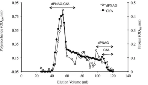
ClfA and dPNAG were covalently linked via reductive amination in the presence of NaBH3CN. Results presented in
Figure 1 show that both ClfA (OD595nm) and dPNAG
(OD650nm) co-eluted in the void volume of the column (void
Pooled fractions containing the purified dPNAG-ClfA conjugate were dialyzed overnight against HEPES buffer pH 8 and analyzed for polysaccharide and protein content using the Gilkerson-Smith and Bradford assays, respectively. The dPNAG-ClfA conjugate vaccine contained 31% dPNAG and 69% ClfA.
Immunogenicity of dPNAG-ClfA conjugate vaccine in various animal species
Mice (n = 10) were immunized three times with 1, 5 or 10mg of either the dPNAG-ClfA conjugate or a mix of comparable amounts of unconjugated dPNAG plus ClfA (dPNAG+ClfA) as a
control. As seen in Fig. 2A mice vaccinated with the dPNAG-ClfA conjugate responded with IgG antibodies to dPNAG in a dose-dependent manner. No antibodies to dPNAG were detected in the mice receiving the unconjugated dPNAG and ClfA mixture (Titer below level of detection of 25) (Fig. 2A). Similarly, mice vaccinated with the dPNAG-ClfA conjugate developed dose-dependent antibody titers to ClfA while the animals given the dPNAG+ClfA mix responded with significantly lower antibodies to ClfA (Antibody titers to ClfA in dPNAG-ClfA conjugate versus dPNAG+ClfA mixture ***P,0.0001 unpaired t test in all three doses and time points post-immunization except for mice
Figure 1. Size exclusion chromatography profile of dPNAG conjugated to ClfA through a Superose 6 gel column.Fractions were assayed for polysaccharide by the hexosamine assay (OD650nm) and for protein with the Bradford assay (OD595nm). Double-headed arrows indicate
the fractions containing conjugated dPNAG and ClfA, as well as where the unconjugated polysaccharide (dPNAG) and protein (ClfA) eluted. doi:10.1371/journal.pone.0043813.g001
Figure 2. Immunogenicity of dPNAG-ClfA vaccine in mice.Mean titers of IgG antibodies to PNAG and ClfA in sera from mice (n = 10) immunized three times at weekly intervals with 1, 5 or 10mg of dPNAG2ClfA conjugate vaccine or dPNAG mixed with ClfA (dPNAG+ClfA). Sera were
collected weekly for 4 weeks starting 1 week after the last immunization and were tested by ELISA for antibodies to dPNAG and ClfA. A) IgG antibody levels to dPNAG in sera of mice vaccinated with dPNAG2ClfA conjugate vaccine or a mixture of unconjugated dPNAG+ClfA. Horizontal line depicts
the limit of detection (titer = 25). For clarity, titers from mice receiving the mixture dPNAG+ClfA, which were below the limit of detection, were assigned a value of the limit of detection. B) IgG antibody titers to ClfA in sera of mice given the dPNAG2ClfA conjugate vaccine or control dPNAG+ClfA. Bars represent means and error bars indicate standard deviations.Pas determined by unpairedttest. *P,0.05, **P,0.005 and *** P,0.0001. All pre-immune titers were,25.
immunized with 1mg at week 2 and 3 post immunization that were *P,0.05 and **P,0.005, respectively) (Fig. 2B).
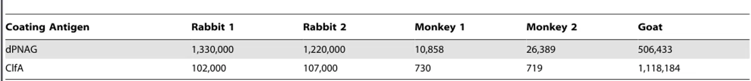
The immunogenicity of dPNAG-ClfA was also evaluated in two rabbits, two rhesus monkeys and a goat. As shown in Table 2 the dPNAG-ClfA vaccine was highly immunogenic in all three of these animal species, eliciting high antibody titers to both dPNAG and ClfA antigens. Interestingly there were differences in the levels and the relative response to each vaccine component among the three animal hosts. These differences might be explained in terms of doses, immunization regimes and/or host-specific differences among the various animal hosts.
Characterization of PNAG and ClfA production inS.
aureusNewman, MN8 and 476 strains
PCR amplification of theicaABCD locus and cflAgenes from genomic DNA extracted from three representativeS. aureusstrains Newman (CP5), MN8 (CP8) and 476 (non-typable) resulted in amplification bands with sizes consistent of those of theicaABCD (3,415 bp) andclfA(2,802 bp) genes ofS. aureus(data not shown). In addition to PCR we conducted confocal laser scanning microscopy (CLSM) studies to investigate the production of PNAG and ClfA by these three S. aureus strains. For these experiments PNAG was labeled with the human IgG1 mAb F598 specific to PNAG directly conjugated to Alexa fluor 488 (F598-AF488) and ClfA was labeled with goat antibodies raised to the dPNAG-ClfA antibodies that had been previously absorbed with S. aureus Newman DU5852 (DclfA) to remove all antibodies raised to dPNAG followed by a secondary donkey anti-goat IgG AF568. As controls, bacterial samples were stained with a human IgG1 mAb to Pseudomonas aeruginosa alginate, mAb F429, also directly conjugated to AF488 and with non-immune goat serum. As presented in Figure 3A (first and second columns) there were high
levels of ClfA (red channel) and PNAG (green channel) antigens detected by specific antibody binding in all threeS. aureusstrains. Conversely,S. aureusstaining with control mAb F429-AF488 and NGS resulted in almost undetectable levels of red or green fluorescent labels, (Fig. 3B first and second columns, respectively). Labeling with the nucleic acid-specific Syto 62 fluorescent dye was also included in the experiments and results shown in the third columns of both panels.
Inhibition ofS. aureusbinding to fibrinogen by antibodies to ClfA
Affinity purified antibodies specific to ClfA or control rabbit IgG were incubated at concentrations ranging from 0.8 to 500mg/ ml with S. aureusbacteria which were then added to Fn-coated microtiter plates. Bacterial binding was detected by crystal violet staining and quantification at OD595nm. As shown in Figure 4
affinity-purified antibodies to ClfA were highly effective at blocking the binding of the three S. aureus strains Newman, MN8 and 476 to immobilized Fn in a dose-dependent manner. In addition, there was no detectable binding of the ClfA-negative strainS. aureusDU5852 to immobilized Fn (data not shown).
Phagocyte-dependent killing by goat antibodies to the dPNAG-ClfA conjugate vaccine
As shown in Figure 5A, antisera to dPNAG-ClfA at a 1:10 dilution promoted the killing of wild typeS. aureusNewman (54% killing) but there was no killing of the PNAG-negative strain NewmanDica (5.4% killing) (Killing ofS. aureus Newman versus Dica;P,0.001, one-way ANOVA). This antiserum also killed the ClfA-negative mutant DU5852 but to a lower extent (35% killing) (Killing of S. aureus Newman versus DU5852; P,0.01, one-way ANOVA). This reduction in susceptibility of the ClfA-negative Table 2.IgG titers to dPNAG and ClfA in various animal host species including two rabbits, two rhesus monkeys and a goat vaccinated with dPNAG2ClfA.
Coating Antigen Rabbit 1 Rabbit 2 Monkey 1 Monkey 2 Goat
dPNAG 1,330,000 1,220,000 10,858 26,389 506,433
ClfA 102,000 107,000 730 719 1,118,184
Serum samples from all animal hosts, collected four weeks after the last immunization, were measured by ELISA for antibody titers specific to dPNAG and ClfA. Titers determined by end-point linear regression.
doi:10.1371/journal.pone.0043813.t002
Figure 3. CLSM imaging of ClfA and PNAG expression byS. aureusNewman, MN8 or 476 strains.A)S. aureuslabeled with mAb F598 to PNAG directly conjugated to Alexa Fluor 488 (F598-AF488), goat antibody to dPNAG2ClfA conjugate vaccine that had been previously absorbed with S. aureusNewman DU 5852 to remove antibody to dPNAG and a secondary donkey antibody to goat IgG conjugated to AF568 and the nucleic acid-specific fluorescent dye Syto 62. B)S. aureusstained with mAb F429 toP. aeruginosaalginate directly conjugated to AF488, NGS and a secondary donkey antibody to goat IgG conjugated to AF568 and Syto 62. For both panels A and B, the first, second and third columns representS. aureus samples viewed in the red (ClfA), green (PNAG) and blue (bacterial DNA) channels, respectively. Bar = 10mm.
strain DU5852 in comparison to wild typeS. aureus Newman is most likely attributable to the variation in PNAG retention on the bacterial cell surface when other factors are changed, as retention is often due to post-synthetic effects such as that of the extracellular IcaB deacetylase [8,9]. Based on our previous experience in testing mAbs and polyclonal sera to dPNAG in the OPKA we found that opsonic killing levels.30% identify antisera within vivoprotective activity [4,10].
To ascertain the specificity of the OPKA, purified dPNAG or ClfA were added to diluted antisera then tested for killing activity. Purified dPNAG inhibited the OPKA of goat antisera to dPNAG-ClfA conjugate, with inhibition titering out in a dose-dependent manner (Fig. 5B). ClfA failed to inhibit the opsonic killing ofS. aureus Newman by antibodies elicited by the dPNAG-ClfA
conjugate at all concentrations tested, indicating that the OPKA measured here was all due to antibody to PNAG.
Protective efficacy of goat antiserum to the dPNAG-ClfA conjugate vaccine in a murine bacteremia model
We evaluated the ability of goat antisera to the dPNAG-ClfA conjugate vaccine along with goat antisera raised to a conjugate of dPNAG and diphtheria toxoid (dPNAG-DT) that had comparable antibody titers to PNAG, to reduce bacterial levels in the blood of mice 2 h after IV injection. As shown in Figure 6, mice injected with antibodies to the dPNAG-ClfA conjugate vaccine had a significant reduction in blood CFU levels ofS. aureusNewman 2 h post infection in comparison to animals given control NGS. A comparable reduction in the blood levels ofS. aureusNewman was achieved with the antiserum to the dPNAG-DT conjugate vaccine, suggesting there was an immeasurable contribution of the antibodies to ClfA to the protective efficacy of the antibodies to the dPNAG-ClfA vaccine. Comparable results were also seen with theS. aureusstrains MN8 and 476 (Fig. 6).
When similar protection studies were carried out withS. aureus DU5852 which does not elaborate ClfA, mice immunized with dPNAG-DT or dPNAG-ClfA had significantly and comparable lower bacterial blood levels than animals given non-immune NGS (dPNAG-DT and dPNAG-ClfA vs NGSP,0.01). On the other hand mice that had been passively immunized with antisera raised to the dPNAG-DT vaccine then infected with the PNAG-deficient strain NewmanDicahad no significant changes in blood CFU levels in comparison to the control NGS (dPNAG-DT vs NGS; ns). However, mice infected with the Dica strain after passive immunization with antibodies raised to the dPNAG-ClfA vaccine exhibited significant bacterial blood clearance in comparison to the NGS-immunized group (dPNAG-ClfA vs NGS, P,0.01). These findings indicate that in the presence of the PNAG antigen, antibodies to ClfA are not effective at mediating protection from bacteremia. However in the absence of PNAG, the ClfA antigen could be either significantly more expressed or exposed on the surface of bacteria, allowing the antibody to ClfA to be protective in the bacteremia model of infection. Presumably this is by a non-OPK mechanism as antibody to ClfA does not appear to have significant opsonic activity against wild type orDica S. aureusstrains.
Discussion
In this work we report the synthesis of a conjugate vaccine covalently linking twoS. aureussurface components, ClfA and the
Figure 4. Antibody to ClfA inhibitsS. aureusbinding to immobilized fibrinogen.Affinity-purified rabbit anti-ClfA IgG’s (%) and control non-immune rabbit IgG’s (&) were tested at various concentrations (0.8 to 500mg/ml) for the their ability to inhibit the binding ofS. aureusNewman, MN8 or 476 strains to fibrinogen. Fibrinogen-coated plates were incubated withS. aureusfor 1 h, fixed with glutaraldehyde for 1 h and stained with crystal violet (0.5% v/v) for 5 minutes. After plates were air-dried the absorbance at 595 nm (OD595nm) was determined using an ELISA plate reader.
Data points represent the average of three independent experiments6SEM. doi:10.1371/journal.pone.0043813.g004
Figure 5. Opsonophagocytic killing activity and specificity of goat sera raised to dPNAG2ClfA conjugate vaccine.A) Opsonic killing ofS. aureusNewman,Dicaand DU 5852 (DclfA), by a 1:10 dilution of goat antiserum raised to dPNAG2ClfA in the presence of polymorphonuclear cells and complement. Bars represent mean percentages of killing6 SEM.P as determined by one-way ANOVA with Tukey’s post-hoc analysis. B) Inhibitory capacity of dPNAG and ClfA antigens in the opsonophagocytic assay. Inhibition of opsonic killing of S. aureusNewman by the goat serum raised to dPNAG2ClfA conjugate vaccine with purified dPNAG (solid bars) or ClfA (open bars) antigens at the indicated concentration. Bars represent means of three indepen-dent experiments6SEM.
deacetylated derivative of PNAG, and showed that immunization of mice with this conjugate vaccine dramatically enhanced the immunogenicity of both components of the vaccine.
The increased immunogenicity of ClfA in dPNAG-ClfA-vaccinated mice in comparison to animals receiving dPNAG plus ClfA may be explained in part by the likely enhenced immuno-genicity of the high molecular weight, highly cross-linked, three-dimensional structure of the dPNAG-ClfA conjugate in compar-ison to the mixture of unconjugated ClfA and dPNAG.
On the other hand, the lack of immunogenicity of unconjugated dPNAG in mice immunized with 1, 5 or 10mg doses of a mixture of dPNAG-ClfA was not entirely surprising. Previous studies carried out in our laboratory have demonstrated that mice immunized with 100mg doses of the fully acetylated PNAG, which was used to prepare the dPNAG for the present study, failed to elicit a detectable IgG response [7].
The dPNAG-ClfA vaccine was also highly immunogenic in multiple animal species including rabbits, rhesus monkeys and a
goat wherein vaccination elicited high antibody titers to both dPNAG and ClfA. Although the total number of animals immunized was limited to two rabbits, two monkeys and a goat we found relative differences in antibody responses to dPNAG and ClfA among the different animal hosts. These could relate to differences in vaccine dosage (10, 50 and 100mg doses for rabbits, goat and monkeys respectively), immunization protocol (2 SC injections followed by 3 IV doses in rabbits and goat versus one single SC dose for the monkeys), type of adjuvant (Freund’s incomplete for rabbits and the goat or alum for monkeys), used and/or to host-specific responses.
The goat antiserum was found to mediate opsonic killing ofS. aureus that was specific to the dPNAG antigen. In addition, we found that affinity-purified antibodies to ClfA inhibited the adherence of three S. aureus strains to immobilized Fn in vitro. However, antibody to ClfA had negligible opsonic activity against S. aureusNewman and did not augment the protective efficacy of
Figure 6. Protective efficacy of dPNAG2ClfA vaccine in a murine bloodstream infection model.Comparative protective efficacy elicited by goat immune sera raised to dPNAG2ClfA and dPNAG-DT conjugate versus NGS againstS. aureusstrains Newman, MN8, 476, NewmanDica, or DU5852 in a murine bacteremia model. Groups of eight FVB mice were immunized IP with 0.4 ml of heat-inactivated antisera raised to dPNAG-DT, dPNAG2ClfA vaccines or with control NGS, 48 and 24 h before IV challenge with theS. aureusstrains Newman (4.36107CFUs), MN8 (8.66106CFUs),
476 (6.76105CFUs), NewmanDica(7.56107CFUs), or DU5852 (3.56107CFUs), sacrificed 2 h post infection and the number of CFU/ml of blood
estimated by serial dilutions and plating. Bars indicate the mean CFU per ml of blood, error bars the SEM.Pvalues as determined by one-way ANOVA with Tukey’s post-hoc analysis. (ns: not significant).
dPNAG-specific sera in a murine model of bacteremia against three PNAG- and ClfA-positive strains ofS. aureus
Previous reports have demonstrated that the use of single vaccine antigens such as CP or ClfA resulted in protection of animals againstS. aureusinfections [11–14] but single component vaccines have failed in all human trials to date [3]. We therefore reasoned that the combination of PNAG and ClfA in the form of a bivalent dPNAG-ClfA conjugate vaccine might be required to confer full protection againstS. aureusinfection and/or to broaden the vaccine coverage to include S. aureus strains which do not fabricate either PNAG or ClfA. Moreover sinceS. aureuscapsular polysaccharide have been shown to mask clumping factor A-mediated adherence ofS. aureus to fibrinogen and platelets [15] then the addition of PNAG to ClfA-based vaccines might be advantageous.
The evidence presented in this work is in full agreement with previous reports showing that either polyclonal or monoclonal antibodies against ClfA inhibit the binding of S. aureus to immobilized fibrinogen in vitro [11,16,17], thought to be one correlate of protective efficacy. However, the sera raised to the dPNAG-ClfA conjugate was no better than antibodies raised to dPNAG conjugated to DT at reducing blood levels of S. aureus following IV challenge with three strains. These findings are consistent with a previous report showing that passive immuniza-tion of mice with affinity-purified ClfA specific antibodies could not prolong the survival in a sepsis model after receiving an intravenous lethal challenge ofS. aureus N315 orS. aureus MW2 [18].
In conclusion the conjugation of dPNAG to ClfA, enhanced the immunogenicity of each component of the vaccine and elicited functional anti-adhesive and opsonic/protective antibodies specific
to ClfA and dPNAG, respectively. However the lack of opsonic activity of antibodies to ClfA against wild typeS. aureusNewman combined with their negligible protective efficacy against wild type S. aureus strains expressing PNAG suggests no major role for immunity to ClfA in S. aureus bacteremia. While levels of the PNAG-defective mutant ofS. aureusNewman were reduced in the blood of mice passively administered the antibody to dPNAG-ClfA, this situation is not representative of human infections which occur almost exclusively with PNAG-producingS. aureus. Antibody to ClfA might be useful in other settings ofS. aureusinfections but incorporation of this bacterial component into a vaccine is made difficult by the poor availability of the antigen to antibodies when surface capsules, including both CP5 or CP8 and PNAG are made [15]. Coupled with the failure of the clinical trial using ClfA-enriched human immune globulins for prevention of S. aureus infections in neonates it appears that validating use of either passive antibody to this antigen or inclusion of ClfA in a vaccine will require better definition of where, when and how immunity to ClfA could augment human resistance toS. aureusinfection.
Acknowledgments
We would like to acknowledge Dr. Timothy J. Foster for the strainE. coli XL1-Blue pCF41-(221–559) which was used for the purification of recombinant ClfA.
Author Contributions
Conceived and designed the experiments: TML LVB GBP. Performed the experiments: TML LVB CBG JMO CCB. Analyzed the data: TML LVB GBP. Wrote the paper: TML GBP.
References
1. Lowy FD (1998)Staphylococcus aureusinfections. N Engl J Med 339: 520–532. 2. Lowy F (2005) Staphylococcal infections. Harrisons Principles of Internal
Medicine.
3. Proctor RA (2012) Challenges for a universalStaphylococcus aureusvaccine. Clin Infect Dis 54: 1179–1186.
4. Maira-Litran T, Kropec A, Goldmann DA, Pier GB (2005) Comparative opsonic and protective activities of Staphylococcus aureus conjugate vaccines containing native or deacetylated Staphylococcal Poly-N-acetyl-b -(1–6)-glucos-amine. Infect Immun 73: 6752–6762.
5. Smith RL, Gilkerson E (1979) Quantitation of glycosaminoglycan hexosamine using 3-methyl-2-benzothiazolone hydrazone hydrochloride. Anal Biochem 98: 478–480.
6. Bradford MM (1976) A rapid and sensitive method for the quantitation of microgram quantities of protein utilizing the principle of protein-dye binding. Anal Biochem 72: 248–254.
7. Maira-Litran T, Kropec A, Abeygunawardana C, Joyce J, Mark G, et al. (2002) Immunochemical properties of the staphylococcal poly-N-acetylglucosamine surface polysaccharide. Infect Immun 70: 4433–4440.
8. Cerca N, Jefferson KK, Maira-Litran T, Pier DB, Kelly-Quintos C, et al. (2007) Molecular basis for preferential protective efficacy of antibodies directed to the poorly acetylated form of staphylococcal poly-N-acetyl-b-(1–6)-glucosamine. Infect Immun 75: 3406–3413.
9. Vuong C, Kocianova S, Voyich JM, Yao Y, Fischer ER, et al. (2004) A crucial role for exopolysaccharide modification in bacterial biofilm formation, immune evasion, and virulence. J Biol Chem 279: 54881–54886.
10. Kelly-Quintos C, Cavacini LA, Posner MR, Goldmann D, Pier GB (2006) Characterization of the opsonic and protective activity againstStaphylococcus aureusof fully human monoclonal antibodies specific for the bacterial surface polysaccharide poly-N-acetylglucosamine. Infect Immun 74: 2742–2750. 11. Hall AE, Domanski PJ, Patel PR, Vernachio JH, Syribeys PJ, et al. (2003)
Characterization of a protective monoclonal antibody recognizingStaphylococcus aureusMSCRAMM protein clumping factor A.Infect Immun 71: 6864–6870. 12. Lee J-C, Park JS, Shepherd SE, Carey V, Fattom A (1997) Protective efficacy of
antibodies to theStaphylococcus aureustype 5 capsular polysaccharide in a modified model of endocarditis in rats. Infect Immun 65: 4146–4151.
13. Fattom AI, Sarwar J, Ortiz A, Naso R (1996) AStaphylococcus aureuscapsular polysaccharide (CP) vaccine and CP-specific antibodies protect mice against bacterial challenge. Infect Immun 64: 1659–1665.
14. Josefsson E, Hartford O, O’Brien L, Patti J, Foster T (2001) Protection against experimentalStaphylococcus aureusarthritis by vaccination with clumping factor A, a novel virulence determinant. J Infect Dis 184: 1572–1580.
15. Risley AL, Loughman A, Cywes-Bentley C, Foster TJ, Lee JC (2007) Capsular polysaccharide masks clumping factor A-mediated adherence ofStaphylococcus aureusto fibrinogen and platelets. J Infect Dis 196: 919–927.
16. Domanski PJ, Patel PR, Bayer AS, Zhang L, Hall AE, et al. (2005) Characterization of a humanized monoclonal antibody recognizing clumping factor A expressed byStaphylococcus aureus. Infect Immun 73: 5229–5232. 17. Vernachio JH, Bayer AS, Ames B, Bryant D, Prater BD, et al. (2006) Human
immunoglobulin G recognizing fibrinogen-binding surface proteins is protective against bothStaphylococcus aureusand Staphylococcus epidermidisinfections in vivo. Antimicrob Agents Ch 50: 511–518.
18. McAdow M, Kim HK, Dedent AC, Hendrickx APA, Schneewind O, et al. (2011) Preventing Staphylococcus aureus sepsis through the inhibition of its agglutination in blood. PLoS Pathog
19. Duthie ES, Lorenz LL (1952) Staphylococcal coagulase; mode of action and antigenicity. J Gen Microbiol 6: 95–107.
20. Kropec A, Maira-Litran T, Jefferson KK, Grout M, Cramton SE, et al. (2005) Poly-N-acetylglucosamine production in Staphylococcus aureus is essential for virulence in murine models of systemic infection. Infect Immun 73: 6868–6876. 21. McDevitt D, Francois P, Vaudaux P, Foster TJ (1994) Molecular characteriza-tion of the clumping factor (fibrinogen receptor) ofStaphylococcus aureus. Mol Microbiol 11: 237–248.
22. Kreiswirth BN, Lo¨fdahl S, Betley MJ, O’Reilly M, Schlievert PM, et al. (1983) The toxic shock syndrome exotoxin structural gene is not detectably transmitted by a prophage. Nature 305: 709–712.
23. Holden MTG, Feil EJ, Lindsay JA, Peacock SJ, Day NPJ, et al. (2004) Complete genomes of two clinical Staphylococcus aureus strains: evidence for the rapid evolution of virulence and drug resistance. Proc Natl Acad Sci USA 101: 9786– 9791.