Generate Ag/Ag
2
O Nanoparticles
G. Yamal1,2, P. Sharmila1, K. S. Rao2, P. Pardha-Saradhi1*
1Department of Environmental Studies, University of Delhi, Delhi, India,2Department of Botany, University of Delhi, Delhi, India
Abstract
We discovered that Yeast Extract Mannitol (YEM) medium possessed immense potential to generate silver nanoparticles from AgNO3upon autoclaving, which was evident from (i) alteration in color of the medium; (ii) peak at,410 nm in UV-Vis spectrum
due to surface plasmon resonance specific to silver nanoparticles; and (iii) TEM investigations. TEM coupled with EDX confirmed that distinct nanoparticles were composed of silver. Yeast extract and mannitol were key components of YEM medium responsible for the formation of nanoparticles. PXRD analysis indicated crystalline geometry and Ag/Ag2O phases in nanoparticles generated with YEM medium, yeast extract and mannitol. Our investigations also revealed that both mannitol and yeast extract possessed potential to convert,80% of silver ions in 0.5 mM AgNO3to nanoparticles, on autoclaving for 30 min at
121uC under a pressure of 1.06 kg/cm2. Addition of filter sterilized AgNO
3under ambient conditions to pre-autoclaved YEM medium and yeast extract brought about color change due to the formation of silver nanoparticles, but required prolonged duration. In general, even after 72 h intensity of color was significantly less than that recorded following autoclaving. Silver nanoparticles formed at room temperature were more heterogeneous compared to that obtained upon autoclaving. In summary, our findings demonstrated that (i) YEM medium and its constituents promote synthesis of silver nanoparticles; and (ii) autoclaving enhances rapid synthesis of silver nanoparticles by YEM medium, yeast extract and mannitol.
Citation:Yamal G, Sharmila P, Rao KS, Pardha-Saradhi P (2013) Inbuilt Potential of YEM Medium and Its Constituents to Generate Ag/Ag2O Nanoparticles. PLoS ONE 8(4): e61750. doi:10.1371/journal.pone.0061750
Editor:Vishal Shah, Dowling College, United States of America
ReceivedFebruary 8, 2013;AcceptedMarch 13, 2013;PublishedApril 23, 2013
Copyright:ß2013 Yamal et al. This is an open-access article distributed under the terms of the Creative Commons Attribution License, which permits unrestricted use, distribution, and reproduction in any medium, provided the original author and source are credited.
Funding:The authors acknowledge the support rendered by the Director and staff of University Science Instrumentation Facility, University of Delhi. The authors are thankful to Mr. Rahul Bhardwaj for providing special assistance during TEM analysis. The authors also thank Mr. Harsh for PXRD studies. GY is thankful to Council for Scientific and Industrial Research (Government of India), New Delhi, for providing research fellowship. The authors acknowledge the University of Delhi for providing financial support under Research & Development and Doctoral Research Programme. The funders had no role in study design, data collection and analysis, decision to publish, or preparation of the manuscript.
Competing Interests:The authors have declared that no competing interests exist.
* E-mail: [email protected]
Introduction
Nanotechnology is one of the most advancing areas of research in modern material science. Nanoparticles exhibit completely new or improved physical (in particular optical, electronic, magnetic) and chemical properties. Silver (Ag) nanoparticles possess strong antimicrobial effects, making them suitable for use in medical devices such as catheters, wound dressings, skin donation, prostheses, contraceptives, in daily commodities such as shampoos, soaps, toys, paints, textile, shoe insoles, washing machines etc. [1– 3]. Nanoparticles of silver also show the property of surface plasmon resonance, rendering them to be used in diagnostics and sensing applications [3–6].
Various physical, chemical and physico-chemical approaches such as the use of laser ablation, mechanical milling, inert gas condensation, thermal irradiation, laser irradiation, chemical reduction, photochemical reduction and electrochemical tech-niques have been demonstrated to generate metal nanoparticles [7–11]. But, the majority of these methods are expensive and/or the byproducts are potentially dangerous to the environment [11]. Sodium borohydrate and sodium citrate methods originally discovered by Brust et al. [12] and Turkevich et al. [13], respectively remain the most popularly used reductants for generation of metal nanoparticles. Owing to the wide applicability and increased commercial demand, the desire to generate
nanoparticles in most economically viable and environmentally friendly ways has gained impetus.
During an attempt to assess the potential of a symbiotic bacteriumRhizobium sp. to synthesize silver nanoparticles [using silver nitrate (AgNO3)], we noted the change in color of yeast extract mannitol (YEM) medium supplemented with AgNO3from pale yellow to brown up on autoclaving. Such a change in color of medium supplemented with AgNO3 is often associated with the formation of silver nanoparticles. This prompted us to carry out investigations for identifying the component(s) of YEM medium responsible for generation of silver nanoparticles.
In this communication, we report for the first time that the YEM medium and two of its components namely mannitol and yeast extract have potential to synthesize silver nanoparticles from Ag+under aqueous conditions. Our investigations revealed that autoclaving promotes rapid synthesis of stable, well dispersed silver nanoparticles in aqueous phase and the same has been compared with the production of silver nanoparticles under ambient conditions.
Materials and Methods
broth composed of 53 mM Mannitol, 2.8 mM K2HPO4, 0.81 mM MgSO4.7H2O, 1.7 mM NaCl and 1 g/L yeast extract [14]. For evaluating the interaction of Ag+
with YEM medium, the later was supplemented with 0, 0.1, 0.25 and 0.5 mM AgNO3. In order to establish the component of YEM medium that resulted in the synthesis of silver nanoparticles upon autoclaving, individual components namely, Mannitol, Yeast Extract, K2HPO4, MgSO4.7H2O and NaCl were used (as per the concentrations in YEM medium) with 0.25 mM AgNO3. The pH in all cases was adjusted to 6.8. The media were dispensed into test tubes and placed in ,75uC pre-heated vertical autoclave (Yorco, Delhi,
India) and autoclaved for 30 min at 121uC under a pressure of 1.06 kg/cm2(,10 min was required for attaining a temperature
of 121uC). About 20–30 min was taken for bringing down the autoclave to atmospheric pressure and temperature. After initial screening, the potential of mannitol and yeast extract to generate silver nanoparticles was evaluated with 0, 0.1, 0.25 and 0.5 mM AgNO3.
In order to test, if silver nanoparticles can be generated at room temperature, YEM medium, yeast extract and mannitol were autoclaved and after cooling different levels of filter sterilized AgNO3 were added under sterile conditions and incubated for different time intervals under ambient conditions.
Characterization of Silver Nanoparticles
The presence of silver nanoparticles in the resultant colloidal solution was established through UV-Vis spectroscopy, transmis-sion electron microscopy (TEM) and powder X-ray diffraction (PXRD) analysis.
The production of silver nanoparticles was initially confirmed by the UV-Vis spectrum of the resultant colored colloidal solutions. The spectra of the media were recorded using Analytikjena Specord 200 at a resolution of 10 nm and a scan rate of 20 nm/sec, to ascertain the presence of peaks arising due to surface plasmon resonance specific to silver nanoparticles.
The colored colloidal solutions obtained on autoclaving AgNO3 in combination with YEM broth, yeast extract or mannitol were centrifuged at 15,0006g for 30 min at 25uC. The resultant pellet
formed was suspended in desired amount of water and used for TEM and PXRD studies.
For TEM investigations the suspended pellet was subjected to sonication for 30 min. 10ml of the sonicated solution was drop
coated onto a 200 mesh copper TEM grid with an ultrathin continuous carbon film and allowed to dry in a desiccator. The grids were viewed in the transmission electron microscope (Technai G2 T30 U-TWIN) at a voltage of 300 kV. The hardware associated with the machine also allowed (i) energy dispersive X-ray (EDX) to measure the elemental composition and (ii) selected area electron diffraction (SAED) pattern to reveal the crystalline nature of nanoparticles.
For PXRD studies the pellet suspended in water, was drop coated on the silica surface, and dried in the desiccator. The PXRD pattern was collected using RIGAKU ROTAFLEX RAD-B using Cu target CuK(a) 1 radiation with tube voltage 40 kV and
60 mA in the 2h(degree) range of 20–80u.
Yield
For estimating the product yield, resultant colloidal solutions were centrifuged at 574386gin Sigma 3K30 centrifuge to obtain
supernatant and the pellet. Silver left in the supernatant was determined with GBC Scientific Equipment SensAA atomic absorption spectrophotometer (AAS) at 328.1 nm.
Results
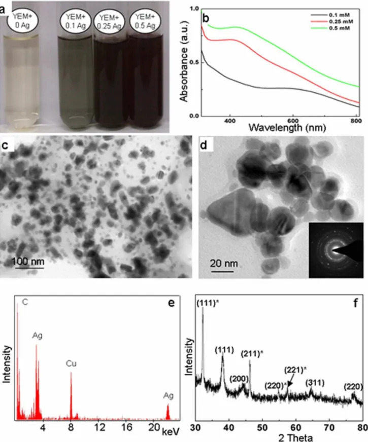
The color of the YEM broth in combination with AgNO3 changed from pale yellow to brown on autoclaving (Figure 1a). However, YEM broth and AgNO3solutions retained their original color when autoclaved individually at 121uC under a pressure of 1.06 kg/cm2for 30 min. It is well known, that colorless AgNO3 solutions turn brown due to the formation of silver nanoparticles. Accordingly, distinct peaks were seen in UV-Vis spectra at
,410 nm (Figure 1b), which are well documented to be due to the
surface plasmon resonance in silver nanoparticles [15]. As is evident from figure 1b, the intensity of peaks increased with increase in concentration of AgNO3.
YEM is a complex medium with mannitol, yeast extract, K2HPO4, MgSO4.7H2O and NaCl as its constituents [14], therefore it was necessary to identify the key component(s) of YEM medium responsible for the formation of silver nanoparti-cles. Alteration in color of different components of YEM medium when used independently in combination with AgNO3is depicted in figure 2a. As is evident from figure 2a, color of 0.25 mM AgNO3turned brown in the presence of mannitol or yeast extract, but remained unaltered in combination with other components. Accordingly, the UV-Vis spectra of colored colloidal solution formed with AgNO3 in combination with yeast extract and mannitol showed absorption peak at,410 nm, while no peaks
were seen with K2HPO4, NaCl and MgSO4.7H2O (Figure 2b). In order to evaluate if autoclaving is essential for formation of silver nanoparticles by YEM medium and its constituents, different levels of filter-sterilized AgNO3 were added to pre-autoclaved, cooled YEM medium, yeast extract and mannitol and incubated under ambient conditions. Incubation of pre-autoclaved YEM medium and yeast extract with filter sterilized AgNO3 under ambient conditions also lead to the color change and the synthesis of silver nanoparticles. However, the color changed only after a prolonged duration of incubation. As evident from figure (Figure 3a) pre-autoclaved YEM medium as well as yeast extract although initiated color change in a duration of ,16 h, the
intensity of color of these media/solutions remained lower than that noted through autoclaving even after 72 h of incubation (Figures 3b–4a). Pre-autoclaved mannitol incubated with filter-sterilized AgNO3 under ambient conditions showed only minor color change. It is important to appreciate that the total duration required for generation of silver nanoparticles from beginning till the end of autoclaving was ,1 h. In general, the silver
nanoparticle specific absorption peak was more broader with the solutions containing silver nanoparticles formed under ambient conditions compared to those formed following autoclaving. The TEM investigations revealed the presence of nanoparticles in the YEM medium (Figure 3d) and yeast extract (Figure 4c) incubated with AgNO3for 72 h under ambient conditions. It is also evident from the TEM images that the silver nanoparticles formed under ambient conditions were highly heterogeneous and agglomerated. In light of these negative aspects of formation of silver nanoparticles by YEM medium and yeast extract under ambient conditions, further investigations for generation and detailed characterization were restricted to the nanoparticles generated via autoclaving.
Figure 5a shows yeast extract and mannitol autoclaved with different levels of AgNO3. A distinct increase in the intensity of color was recorded with increasing concentration of AgNO3 (Figure 5a). Accordingly, intensity of absorption peak due to surface plasmon resonance of silver nanoparticles was intensified with increasing concentration of AgNO3, both in case of mannitol
Figure 1. Evaluation of silver nanoparticles formed by autoclaving AgNO3with Yeast Extract Mannitol (YEM) medium. a: Alteration in
color of YEM medium autoclaved in presence of 0, 0.1, 0.25 and 0.5 mM AgNO3. b: UV-Vis absorption spectra of 0.1, 0.25 and 0.5 mM AgNO3
autoclaved with YEM medium showing the characteristic peaks for silver nanoparticles.c & d: TEM images of the silver nanoparticles.Inset in d: SAED pattern of nanoparticles.e: The EDX spectrum of nanoparticles showing characteristic peaks of Ag, indicating that the nanoparticles to be composed of silver. Other prominent peaks of C and Cu, noted in the figure arose from the carbon coated copper grids.f: The PXRD pattern of nanoparticles.
concentration of AgNO3, the intensity of the color of the solution and absorption peak due to silver nanoparticles was significantly higher with yeast extract compared to mannitol. The absorption peak obtained due to surface plasmon resonance in silver nanoparticles was broad in case of yeast extract and sharp in case of mannitol.
TEM analysis further established the presence of silver nanoparticles in YEM medium autoclaved with AgNO3. Figures 1c–d are the representative TEM images of silver nanoparticles formed in YEM medium supplemented with AgNO3. As is evident from these figures, the nanoparticles varied in size from 10–50 nm. The EDX pattern (Figure 1e) collected from these nanoparticles showed distinct peaks at 3.40 keV and 22 keV corresponding to Ag, while the peaks situated at binding energies of 8.06 and 1 keV correspond to Cu and C, respectively. The peaks of C and Cu arose due to their presence as an integral component of carbon coated copper grids.
The SAED pattern revealed the crystalline nature of silver nanoparticles (inset Figure 1d), which was further corroborated by the PXRD studies (Figure 1f). The PXRD pattern showed the Bragg reflections that can be assigned to (111), (200), (220) and (311) and matched with the standard diffraction pattern of Joint Committee on Powder Diffraction Standards (JCPDS) No. 04– 0783, confirming the face-centered cubic (fcc) structure. The
position of 32.31, 46.28, 54.83, 57.97, 67.95 which can be due to Ag2O cubic structure with Bragg reflections (111), (211), (220), (221) and (222).
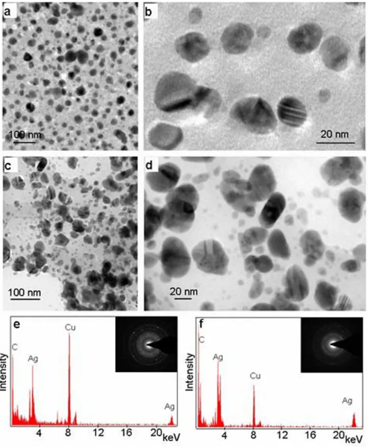
TEM images of silver nanoparticles formed on autoclaving AgNO3in combination with 53 mM (,0.9%) mannitol or 0.1%
yeast extract are shown in figures 6a–b and figures 6c–d, respectively. Silver nanoparticles formed in presence of mannitol were nearly spherical and varied in size range from 10–20 nm, while those formed with yeast extract varied in shape and were in the size range of 10–50 nm. The SAED pattern (insets in figures 6e–f) indicated the crystalline nature of these silver nanoparticles. The EDX spectra (Figures 6e–f) collected from these nanoparticles showed peaks for Ag, confirming that the nanoparticles were composed of silver.
The PXRD pattern showed Bragg reflections that can be assigned to (111), (200), (220), (311) planes, and matched with JCPDS No. 04–0783. These results confirmed that silver nano-particles synthesized with mannitol as well as yeast extract were crystalline with fcc structure. In a manner similar to YEM medium, the PXRD patterns of silver nanoparticles formed with mannitol and yeast extract (Figure 7a) also showed additional peaks, indicating biphasic nature of nanoparticles.
The AAS studies reveal that yeast extract as well as mannitol have equal potential to generate silver nanoparticles. Lower Figure 2. Evaluation of contribution of YEM medium components for silver nanoparticles generation upon autoclaving with AgNO3. a: Alteration in color of 0.25 mM AgNO3upon autoclaving with different components of YEM medium namely, yeast extract (YE), mannitol,
MgSO4, NaCl and K2HPO4 medium individually.b: UV-Vis absorption spectra of 0.25 mM AgNO3after autoclaving with YEM medium, yeast extract,
mannitol, MgSO4, NaCl and K2HPO4. doi:10.1371/journal.pone.0061750.g002
independently with yeast extract and mannitol did not show the presence of any silver ions in the supernatant, as they might have been converted into nanoparticles that pelleted down following
centrifugation at 574386g. However, 0.5 mM AgNO3autoclaved
independently with yeast extract and mannitol retained about 20% silver in ionic state.
Figure 3. Evaluation of silver nanoparticles formed by incubating filter-sterilized AgNO3with pre-autoclaved YEM medium under ambient conditions. a–b: Alteration in color of YEM medium incubated in presence of 0, 0.1, 0.25 and 0.5 mM AgNO3after,8 h (a) and 72 h (b).c: UV-Vis absorption spectra of 0, 0.1, 0.25 and 0.5 mM AgNO3incubated with YEM medium for 72 h.d: TEM image of silver nanoparticles formed under
ambient conditions.
doi:10.1371/journal.pone.0061750.g003
Figure 4. Evaluation of silver nanoparticles formed by incubating filter-sterilized AgNO3with pre-autoclaved yeast extract (YE) under ambient conditions. a: Alteration in color of YE with 0, 0.1, 0.25 and 0.5 mM AgNO3after 72 h.b: UV-Vis absorption spectra of YE incubated
with 0, 0.1, 0.25 and 0.5 mM AgNO3for 72 h.c: TEM images of silver nanoparticles formed under ambient conditions.
The silver nanoparticles formed with mannitol as well as yeast extract were well dispersed and stable, and showed no signs of agglomeration even on storing for a period of over a month under ambient conditions (Figure 7b).
Discussion
During present investigations, we observed alteration in color of YEM medium supplemented with AgNO3 from pale yellow to brown just upon autoclaving even without the inoculation of any microorganism. It has been established that colorless AgNO3 solution turns yellow to brown due to the formation of silver nanoparticles [16]. TEM investigations coupled with EDX studies confirmed the presence of silver nanoparticles in the YEM medium autoclaved with AgNO3 and accordingly this medium showed absorption peak at,410 nm, which is known to arise due
to surface plasmon resonance in the silver nanoparticles [15-17]. In addition to the role of bacteria in the synthesis of silver nanoparticles demonstrated in earlier investigations, the present study also demonstrates the important ability of media
compo-nents (YEM medium and two of its compocompo-nents viz. yeast extract and mannitol) to synthesize silver nanoparticles.
YEM is a complex medium constituted of yeast extract, mannitol, MgSO4, K2HPO4and NaCl [14]. AgNO3autoclaved independently with different components of YEM medium clearly indicated that two of its constituents namely, mannitol and yeast extract possessed potential to generate silver nanoparticles. As expected, silver nanoparticles generated with mannitol and yeast extract also showed absorption peaks at ,410 nm due to their
specific surface plasmon resonance. However, the absorption peak obtained with yeast extract was broader when compared to mannitol, indicating that the nanoparticles formed with the former were heterogeneous. The heterogeneity of silver nanoparticles formed by yeast extract was also evident from TEM images which not only showed variation in shape but also exhibited significant variation in size. Such a heterogeneous population of nanoparti-cles might have resulted due to the complex composition of yeast extract [18], [19].
Both yeast extract and mannitol possessed potential to convert cent percent of silver ions into nanoparticles when autoclaved with Figure 5. Evaluation of silver nanoparticles formed by autoclaving AgNO3with yeast extract (YE) and mannitol. a: Alteration in color
of 0.1, 0.25 and 0.5 mM AgNO3autoclaved with 53 mM mannitol and 0.1% yeast extract.b–c: UV-Vis absorption spectra of 0.1, 0.25 and 0.5 mM
AgNO3after autoclaving with 53 mM mannitol and 0.1% yeast extract, respectively.
doi:10.1371/journal.pone.0061750.g005
Figure 6. Characterization of silver nanoparticles formed by autoclaving AgNO3with mannitol and yeast extract (YE). a–b: TEM
images of silver nanoparticles synthesized with 53 mM Mannitol.c–d: TEM images of silver nanoparticles synthesized with 0.1% YE.e–f: EDX spectra of nanoparticles formed with mannitol and YE, respectively, showing the peaks for Ag, indicating nanoparticles to be composed of silver. Other prominent peaks of C and Cu, noted in the figure are due to the carbon coated copper grids.Inset e–f: SAED pattern of the silver nanoparticles formed with mannitol and YE, respectively, showing the crystalline nature of the nanoparticles.
Figure 7. PXRD pattern and stability of silver nanoparticles formed upon autoclaving AgNO3with mannitol and yeast extract (YE). a: PXRD pattern of the silver nanoparticles formed with (i) YE and (ii) mannitol showing bragg reflections characteristic of crystalline face-centred cubic structure of Ag () and cubic structure of Ag2O ()*.b: Ag nanoparticles formed from 0.1, 0.25 and 0.5 mM AgNO3autoclaved independently with
53 mM mannitol and 0.1% YE after 1 month with no signs of agglomeration. doi:10.1371/journal.pone.0061750.g007
of silver ions were converted into nanoparticles with 0.5 mM AgNO3within a duration of 1 h on autoclaving. The retention of 20% of silver ions in 0.5 mM AgNO3indicated that the level of yeast extract and mannitol used was not sufficient for reducing 100% silver into nanoparticles. We believe that it is important to increase the level of mannitol or yeast extract depending on the concentration of AgNO3used for obtaining optimal yield of silver nanoparticles, i.e. for converting cent percent of silver ions into nanoparticles.
During the course of present studies, experiments were also carried to assess, if silver nanoparticles can be generated under ambient conditions by adding and incubating filter sterilized AgNO3 to cooled, pre-autoclaved YEM medium, yeast extract and mannitol. This revealed that the former two possessed potential to alter the color and generate silver nanoparticles, but this alteration in color and the formation of silver nanoparticles under ambient conditions required prolonged duration. Infact, the intensity of the color of YEM medium as well as yeast extract incubated with AgNO3under ambient conditions for a duration of 72 h was significantly less than that noted in a duration of 1 h through autoclaving. Further, TEM investigations revealed that silver nanoparticles formed under ambient conditions were more heterogenous when compared to those formed through autoclav-ing.
Advantages of Autoclaving for Generation of Silver Nanoparticles
Addition of AgNO3to pre-autoclaved cooled medium required prolonged duration for the generation of silver nanoparticles under ambient conditions and reasonable quantity of silver nanoparticles could be generated after,72 h. In contrast, autoclaving AgNO3
independently with YEM medium, yeast extract and mannitol promoted generation of large quantities of silver nanoparticles within a duration of 1 h. Further, the nanoparticles generated under ambient conditions were more heterogeneous when compared to those obtained through autoclaving.
The stability of nanoparticles depends on the medium, inter-particle forces and chemical reactivity, which affect aggregation, size and shape of the nanoparticles [20]. The production and stability of silver nanoparticles in aqueous phase remained a major challenge and it is important to develop novel strategies for their wide application. The silver nanoparticles produced by autoclav-ing durautoclav-ing present investigations were well dispersed and retained stability even after a duration of more than a month (without showing any signs of agglomeration) in aqueous phase at room temperature.
For apt application in medicine and pharmaceutical industry it is important to generate silver nanoparticles under sterile conditions. We believe that autoclaving is a suitable approach for generation of metal nanoparticles under sterile conditions, as autoclaving is widely used for general sterilization purposes.
Mechanism of Formation of Silver Nanoparticles by Yeast Extract
Yeast extract being autolysate of yeast cells is a cocktail of proteins/polypetides, amino acids, carbohydrates, vitamins etc. [18], [19] and few of these biomolecules must have played a vital role in the generation of silver nanoparticles. It is well established that various biomolecules such as peptides, amino acids, carbo-hydrates, vitamins etc. possess capacity to synthesize silver nanoparticles and the mechanism of synthesis of metal nanopar-ticles (from their ions) by different biomolecules vary significantly from each other as has been extensively elaborated by Duran et al.
[21]. This accounts for the heterogeneity in the shape and size of nanoparticles obtained with yeast extract during present investi-gations. Some of the key groups of biomolecules in the cocktail of yeast extract namely (i) carboxyl group of glutamate and aspartate, and hydroxyl group of tyrosine, serine and threonine present individually or associated with polypeptides; and (ii) aldehyde, keto and hydroxyl groups associated with the carbohydrates, must have contributed significantly towards the reduction of Ag+
and formation and stabilization of silver nanoparticles. Similarly, vitamins such as riboflavin present in yeast extract could have also played an important role in generation of silver nanoparticles as has been reported earlier [22].
Mechanism of Formation of Silver Nanoparticles by Mannitol
The nanoparticles formed with mannitol were relatively uniform in shape and size. In all likelihood mannitol might have generated silver nanoparticles through reduction of Ag+
to Ag0 following Fetizon oxidation mechanism which involves oxidation of a primary alcohol to an aldehyde group followed by further oxidation to form lactone/ketone [23], [24]. Ag0 can further nucleate to generate silver nanoparticles.
For effective commercial endeavors, it is important to generate nanoparticles of uniform size and shape. It is equally important to ensure that the molecules/agent used for generating nanoparticles are completely safe and compatible with living systems and must be eco-friendly. Unlike yeast extract, which is a complex mixture of several biomolecules, mannitol has been well established to be a compatible solute, which has potential to protect living systems against various stresses [25–29].
The generation of stable nanoparticles with good dispersion potential under aqueous conditions is one of the key concerns associated with practical applications of nanoparticles in medicine and industry. Researchers across the globe have been looking for appropriate green protocols for improving the dispersion stability of nanoparticles for wider application and performance [30]. During present investigations, we noted that the silver nanopar-ticles formed with mannitol as well as yeast extract were well dispersed and stable, and showed no signs of agglomeration even on storing for a period of over a month at room temperature (Figure 4b). Some of the biomolecules including polyols are known to act as stabilizing agents [31–33].
Conclusions
In summary, our results demonstrated for the first time that (i) inclusion of Ag+
in YEM medium prior to autoclaving lead to generation of silver nanoparticles and therefore, it is not appropriate to include silver salt in the culture medium prior to autoclaving for evaluating the impact of Ag+
on microorganisms; (ii) YEM medium and two of its constituents namely, yeast extract and mannitol promote rapid generation of nanoparticles by autoclaving (as autoclaving promotes generation of silver nano-particles under strict sterile conditions, the same can be used directly in medicine and pharmaceutical industry); and (iii) the silver nanoparticles generated on autoclaving AgNO3 with yeast extract and mannitol remain stable in aqueous phase under ambient conditions for long durations.
Acknowledgments
Author Contributions
Conceived and designed the experiments: GY PS PPS. Performed the experiments: GY PS PPS. Analyzed the data: GY PS PPS. Contributed
reagents/materials/analysis tools: PPS KSR. Wrote the paper: GY PS PPS.
References
1. Singh N, Manshian B, Jenkins GJS, Griffiths SM, Williams PM, et al. (2009) NanoGenotoxicology: The DNA damaging potential of engineered nanomater-ials. Biomaterials 30: 3891–3914.
2. Song JY, Kwon E-Y, Kim BS (2010) Biological synthesis of platinum nanoparticles using Diopyros kaki leaf extract. Bioprocess Biosyst Eng 33: 159–164.
3. Tolaymat TM, Badawy AME, Genaidy A, Scheckel KG, Luxton TP, et al. (2010) An evidence-based environmental perspective of manufactured silver nanoparticle in synthesis and applications: A systematic review and critical appraisal of peer-reviewed scientific papers. Sci Total Environ 408: 999–1006. 4. Alvarez-Puebla RA, Aroca RA (2009) Synthesis of silver nanoparticles with controllable surface charge and their application to surface-enhanced raman scattering. Anal Chem 81: 2280–2285.
5. Basavaraja S, Balaji SD, Lagashetty A, Rajasab AH, Venkataraman A (2008) Extracellular biosynthesis of silver nanoparticles using the fungus Fusarium semitectum. Mater Res Bull 43: 1164–70.
6. Jiang X, Zeng Q, Yu A (2007) Thiol-frozen shape evolution of triangular silver nanoplates. Langmuir 23: 2218–23.
7. Daniel MC, Astruc D (2004) Gold nanoparticles: assembly, supramolecular chemistry, quantum-size-related properties, and applications toward biology, catalysis, and nanotechnology. Chem Rev 104: 293–346.
8. Masala O, Seshadri R (2004) Synthesis routes for large volumes of nanoparticles. Annu Rev Mater Res 34: 41–81.
9. Wani IA, Khatoon S, Ganguly A, Ahmed J, Ganguli AK, et al. (2010) Silver nanoparticles: Large scale solvothermal synthesis and optical properties. Mater Res Bull 45: 1033–1038.
10. Tavakoli A, Sohrabi M, Kargari A (2007) A review of methods for synthesis of nanostructured metals with emphasis on iron compounds. Chem Pap 61: 151– 170.
11. Iravani S (2011) Green synthesis of metal nanoparticles using plants. Green Chem 13: 2638–2650.
12. Brust M, Walker M, Bethell D, Schiffrin DJ, Whyman R (1994) Synthesis of Thiol-derivatised gold nanoparticles in a two-phase liquid-liquid system. J Chem Soc Chem Commun 7: 801–802.
13. Turkevitch J, Stevenson PC, Hillier J (1951) Nucleation and growth process in the synthesis of colloidal gold. Discuss. Faraday Soc. 11: 55–75.
14. Bagyaraj DJ (2011) Microbial Biotechnology for Sustainable Agriculture, Horticulture and Forestry. New India Publishing Agenecy (NIPA). New Delhi. 15. Sastry M, Mayya KS, Bandyopadhyay K (1997) pH dependent changes in the optical properties of carboxylic acid derivatized silver colloidal particles. Colloid Surface A 127: 221–228.
16. Shahverdi AR, Minaeian S, Shahverdi HR, Jamalifar H, Nohi AA (2007) Rapid synthesis of silver nanoparticles using culture supernatants ofEnterobacteria: A novel biological approach. Process Biochem 42: 919–923.
17. Nanda A, Saravanan M (2009) Biosynthesis of silver nanoparticles from Staphylococcus aureusand its antimicrobial activity against MRSA and MRSE. Nanomed: Nanotechnol Biol Med 5: 452–456.
18. Gellert G, Stommel A, Trujillano AB (1999) Development of an optimal bacterial medium based on the growth inhibition assay with Vibrio fischeri. Chemosphere 39: 467–476.
19. Lindan O, Work E (1951) The amino-acid composition of two yeasts used to produce massive dietetic liver necrosis in rats. Biochem 48: 337–341. 20. Stevenson APZ, Bea DB, Civit S, Contera SA, Cerveto AI, et al. (2012) Three
strategies to stabilise nearly monodispersed silver nanoparticles in aqueous solution. Nanoscale Res Lett 7: 151.
21. Duran N, Marcato PD, Duran M, Yadav A, Gade A, et al. (2011) Mechanistic aspects in the biogenic synthesis of extracellular metal nanoparticles by peptides, bacteria, fungi, and plants. Appl Microbiol Biotechnol 90: 1609–1624. 22. Chatterjee S, Nandi AK (2011) Tuning of the morphology of a
riboflavin-melamine equimolar supramolecular assembly by in situ silver nanoparticle formation. Chem Commun 47: 11510–11512.
23. Donati I, Travan A, Pelillo C, Scarp T, Coslovi A, et al. (2009) Polyol synthesis of silver nanoparticles: mechanism of reduction by alditol bearing polysaccha-rides. Biomacromolecules 10: 210–213.
24. Park Y, Hong YN, Weyers A, Kim YS, Linhardt RJ (2011) Polysaccharides and phytochemicals: a natural reservoir for the green synthesis of gold and silver nanoparticles. IET Nanobiotechnol 5: 69–78.
25. Maheswari M, Varalaxmi Y, Vijayalakshmi A, Yadav SK, Sharmila P, et al. (2010). Metabolic engineering using mtlD gene enhances tolerance to water deficit and salinity in sorghum. Biol Plant 54: 647–652.
26. Pardha Saradhi P, Sharmila P (2003) Improvement of crop plants for abiotic stress tolerance through introduction of glycine betaine pathway. In: Singh RP, Jaiwal PK (eds.) Plant Genetic Engineering. Sci Tech Publishing Lic., U.S.A., 147–177.
27. Sharmila P, Anwar F, Sharma KR, Pardha Saradhi P (2008). Management of abiotic stresses in grain legumes through manipulation of genes for compatible solutes. In Kirti PB (Ed.) Handbook of New Technologies for Genetic Improvement of Legumes. CRC Press, Taylor and Francis Group, USA, 577– 603.
28. Sivakumar P, Sharmila P, Jain V, Paradha Saradhi P (2002) Compatible sugars and sugar alcohols curtail oxygenase activity of Rubisco as a strategy for stress adaptation. Biochem Biophys Res Commun 298: 247–250.
29. Stoop JMH, Williamson JD, Pharr DM (1996) Mannitol metabolism in plants: a method for coping with stress. Trends Plant Sci 1: 139–144.
30. Piao L, Lee KH, Kwon WJ, Kim S-H, Yoon S (2009) The simple and facile methods to improve dispersion stability of nanoparticles: Different chain length alkylcarboxylate mixtures. J Colloid Interf Sci 334: 208–211.
31. Badwaik VD, Bartonojo JJ, Evans JW, Sahi SV, Willis CB, et al. (2011) Single-step biofriendly synthesis of surface modifiable, near-spherical gold nanoparticles for applications in biological detection and catalysis. Langmuir 27: 5549–5554. 32. Sun Y, Xia Y (2002) Shape-controlled synthesis of gold and silver nanoparticles.
Science 298: 2176–2179.
33. Zhaoa T, Suna R, Yua S, Zhang Z, Zhouc L, et al. (2010) Size-controlled preparation of silver nanoparticles by a modified polyol method. Colloid Surface A 366: 197–202.