Volume 2013, Article ID 174398,9pages http://dx.doi.org/10.1155/2013/174398
Research Article
Antifungal Applications of Ag-Decorated
Hydroxyapatite Nanoparticles
C. A. Zamperini,
1,2R. S. André,
3V. M. Longo,
1E. G. Mima,
2C. E. Vergani,
2A. L. Machado,
2J. A. Varela,
1and E. Longo
11LIEC, UNESP-Univ Estadual Paulista, P.O. Box 355, 14801-907 Araraquara, SP, Brazil
2Department of Dental Materials and Prosthodontics, Araraquara Dental School, UNESP-Univ Estadual Paulista,
14801-903 Araraquara, SP, Brazil
3LIEC, CMDMC, Department of Chemistry, Universidade Federal de S˜ao Carlos (UFSCar), 13565-905, S˜ao Carlos, SP, Brazil
Correspondence should be addressed to C. A. Zamperini; cazamperini@yahoo.com.br
Received 1 February 2013; Revised 23 May 2013; Accepted 26 May 2013
Academic Editor: Mohamed Bououdina
Copyright © 2013 C. A. Zamperini et al. This is an open access article distributed under the Creative Commons Attribution License, which permits unrestricted use, distribution, and reproduction in any medium, provided the original work is properly cited.
Pure hydroxyapatite (HA) and hydroxyapatite decorated with silver (HA@Ag) nanoparticles were synthesized and characterized.
The antifungal effect of HA@Ag nanoparticles in a distilled water solution was evaluated againstCandida albicans. The origin of
the antifungal activity of the HA@Ag is also discussed. The results obtained showed that the HA nanorod morphology remained the same with Ag ions decorations on the HA structure which were deposited in the form of nanospheres. Interaction where occurred between the structure and its defect density variation in the interfacial HA@Ag and intrafacial HA region with the fungal medium resulted in antifungal activity. The reaction mechanisms involved oxygen and water adsorption which formed an active complex cluster. The decomposition and desorption of the final products as well as the electron/hole recombination process have an important role in fungicidal effects.
1. Introduction
HA, Ca10(PO4)6(OH)2, is the major mineral component of human bone and other calcified tissues [1]. Thus, due to the similarity with bone composition and its ability to bond strongly with human hard tissue, the synthesis of HA (a bioce-ramic material) is of great interest for several clinical applica-tions in the biomedical field [2,3]. Synthetic HA is a versatile inorganic material that also exhibits desirable properties such as biocompatibility, bioactivity, and osteoconductivity. In medicine and dentistry, due to its bioactive properties, HA has been successfully used for bone reconstructions and as coatings for dental implants; also, the structure is similar to the human bone mineral component. However, various sources of infections exist in these biomedical interventions, including environment surfaces and surgical equipment as well as microorganisms present in body tissues. Hence, the risk of infectious complications is high and may lead to failures which entail the need for subsequent surgery
and medical costs in addition to the pain and suffering of patients. Candida albicans is among the most frequently isolated microorganisms from human infections, mainly in immunocompromised patients [4–6]. AlthoughC. albicansis an eukaryotic commensal microorganism of human mucosal surfaces, when there is impairment of the host immunity, it can cause serious superficial and deep infections [4]. In general, the in vivo colonization of tissues and surfaces by microorganisms is related to biofilm formation. Biofilm-associatedC. albicanscells are more resistant to antifungals commonly used compared to cells grown in planktonic form [5]. Therefore, the synthesis of HA with antifungal properties against planktonic and biofilm cells would be an important strategy to inhibit fungal colonization and, consequently, the incidence of surgical infections.
ments, the microwave-assisted hydrothermal (HTMW) [14,
15] method has been used in recent years and is one of the most promising methods to prepare nanomaterials with controlled size and shape [16]. Nevertheless, the challenge remains to prepare multicomponent or hybrid structures where two or more nanocrystal domains of different materials with individually tailored properties are integrated into one nanostructure.
Recently, the synthesis of Ag-doped nanocrystalline HA nanoparticles by a coprecipitation method has been reported and the antibacterial and antifungal activities were evaluated [17–19]. In these investigations, an antimicrobial effect was observed against Gram-negative and Gram-positive bacteria [17, 18] and Candida krusei, a fungal species [19]. In the present study, we report the synthesis of HA nanoparticles decorated with silver through a co-precipitation method with an HTMW treatment using calcium nitrate and ammonium phosphate as precursors. The silver was incorporated into the HA matrix after the synthesis. The HA@Ag powder was structurally characterized by high-resolution transmission electron microscopy (HR-TEM), X-ray diffraction (XRD), FT-Raman spectroscopy, and UV-visible spectroscopy (UV-vis). The potential fungistatic and fungicidal effects of the HA@Ag in solution against C. albicans were evaluated by determining the minimum inhibitory concentration (MIC) and minimum fungicidal concentration (MFC). In addition, the effects of the HA@Ag solution against the biofilm for-mation of C. albicans were also evaluated. To the authors’ knowledge, such effects of HA@Ag onC. albicanshave not been widely evaluated [20].
2. Experimental Section
2.1. Materials and Synthesis. For the synthesis of HA, two aqueous solutions were prepared: a calcium nitrate tetrahy-drate solution (Mallinckrodt, 99.9%) and an ammonium phosphate solution (Mallinckrodit 99.4%). The ammonium phosphate solution was then dripped into the calcium solu-tion with constant agitasolu-tion. The system was kept under flowing N2 throughout the mixing, and the pH solution was maintained at approximately 11 with the addition of ammonium hydroxide. The suspension containing the white solid precipitate was placed in a 100 mL Teflon autoclave which was sealed and loaded into the HTMW system using 2.45 GHz microwave radiation with 800 W of power. The system was heated to 140∘C and maintained at this tem-perature for 1 minute at a heating rate of 140∘C/min under
characterizations were performed by transmission electron microscopy (TEM, Tecnai G2TF20, FEI). To confirm the presence of Ag, an energy dispersive X-ray (EDX) mea-surement was taken in the same apparatus. All measure-ments were taken at room temperature. HA@Ag samples were characterized by XRD using a Rigaku DMax 2500PC diffractometer at 40 kV and 150 mA with Cu K𝛼radiation, a graphite monochromator, and a rotary anode. Raman spectra were recorded on a RFS/100/S Bruker Fourier transform Raman (FT-Raman) spectrometer with a 1064 nm excitation wavelength using a Nd:YAG laser in a spectral resolution of 4 cm−1. UV-vis absorption was recorded using a Cary 5G spectrometer in total reflectance mode by the integration cell.
2.3. Antifungal Activity. C. albicans (ATCC 90028) was grown in Sabouraud dextrose agar (SDA, Acumedia Man-ufacturers Inc., Baltimore, MD, USA) containing 5𝜇g/mL gentamicin for 48 h at 37∘C, followed by 21 h at 37∘C in an RPMI-1640 culture medium. Cells of the resultant culture were harvested by centrifugation at 4000 g for 5 min, washed twice with a phosphate-buffered saline solution (PBS; pH 7.2), and resuspended in the RPMI-1640 culture medium. The estimation of minimum inhibitory concentration (MIC) and minimum fungicidal concentration (MFC), using a broth microdilution assay [21], was used to evaluate the antifungal activity of the HA@Ag solution. The determination of MIC and MFC was used because it is a standard methodology for antimicrobial tests.C. albicanswas incubated on a 96-well microtiter plate for 48 hours at 35∘C and exposed to a serial two-fold dilution in the RPMI-1640 culture medium of the HA@Ag solution (from 1000𝜇g/mL to 3.90𝜇g/mL). The MIC value was the lowest concentration of the HA@Ag solution which resulted in no visible growth (by visual inspection). Thereafter, aliquots from each well were diluted (10−1, 10−2, 10−3, and 10−4) in PBS and inoculated on SDA (in duplicate) in order to establish the MFC. After 48 h at 37∘C, the colony-forming units per milliliter (CFU/mL) were calculated, and a log10transformation was plotted. The MFC value was defined as the lowest concentration of the HA@Ag solution resulting in 99% reduction of fungal growth (UFC/mL). The assays were performed in triplicate on three separate occasions.
50nm
(a)
50nm
(b)
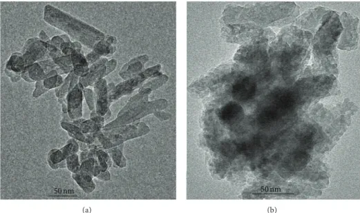
Figure 1: (a) TEM image of pure HA and (b) TEM image of HA@Ag.
microtiter plates and maintained for 90 min at 37∘C (adhesion phase) in an orbital shaker [5]. Then, the loosely adhered cells were removed by washing twice with 200𝜇L of PBS. Each one of the wells was filled with 200𝜇L of fresh RPMI-1640 culture medium containing different concentrations of the HA@Ag solution (from 1000𝜇g/mL to 3.90𝜇g/mL), and the plates were incubated at 37∘C in an orbital shaker at 75 rpm for 48 h. After biofilm formation, the wells were carefully washed twice with 200𝜇L of PBS to remove nonadherent cells.
The number of viableC. albicans and the total biomass of the biofilms exposed to different concentrations of the HA@Ag solution were evaluated by counting the colonies which formed units (CFU/mL) and measuring the absorbance of the crystal violet staining, respectively. To enumerate the CFU/mL, biofilms were scraped out of the wells of the 96-well microtiter plate and suspended by vigorous vortex mixing (1 min) in PBS. Thereafter, 10−1, 10−2, 10−3, and 10−4 dilutions were performed, and aliquots of 25𝜇L were inoculated on SDA plates (in duplicate). For the determination of the total biomass, the biofilms were fixed with methanol and stained with 1% violet crystal solution for 5 min. The stain bonded to biofilm was dissolved in acetic acid at 33% and the optical density was measured (570 nm) using a spectrophotometer (Thermo Plate—TP Reader). Data obtained from CFU/mL quantification and total biomass were statistically analyzed by ANOVA and Tukey’spost hoc
tests at 5% of significance.
The morphological characteristics of the biofilms formed in the presence of the HA@Ag solution also were evaluated using a field emission gun-scanning electron microscopy (FEG-SEM). For this procedure, C. albicans biofilms were cultured and exposed to different concentrations of HA@Ag solution on the bottom of a 24-well plate, as described previ-ously. Following biofilm formation, these products were fixed with 2.5% glutaraldehyde in PBS for 24 hours, dehydrated in
increasing concentrations of ethanol (70%, 85%, and 90%) for 5 min each and observed in FEG-SEM (Jeol, JSM 7500 F).
3. Results and Discussion
Figure 1 shows TEM morphologies of pure HA and the HA@Ag synthesized under the same conditions, respectively. Figure1shows that there was very little difference between the two samples in particle size and morphologies. The pure HA (see Figure1(a)) has a nanorod structure with a diameter varying from 12 to 27 nm. A closer look at a comparatively thin rod plate which provides sufficient transparency of electrons for high resolution is provided in Figure2; a layered structure is apparent.
However, silver is observed in the nanospherical mor-phology with an interplanar distance of 0.276 nm which cor-responds to the Ag (1 1 1) plane (the preferential growth plane) (see Figure2(c)) according to the JCPDS 65–2871. The EDX spectrum of HA@Ag confirms calcium (Ca), phosphor (P), oxygen (O), and silver (Ag) in the samples (see Figure2(a)). The presence of copper (Cu) and silicon (Si) was attributed to the sample grid support.
The characterization in a long-range order of the pure HA and HA@Ag samples was performed by XRD (see Figures
3(a) and3(b), resp.). The HA single phase formation has a hexagonal structure with a P 63/m space group even with Ag. No planes related to the Ag phase were located. Secondary phases such as calcium carbonate, CaCO3, were not found in XRD results.
(a)
Figure 2: (a) An EDX measurement of HA@Ag nanoparticles; (b) HRTEM image of Ag nanospheres; and (c) a zoom of a nanoparticle with (111) interplanar distance.
10 20 30 40 50 60 70
(431)
(512)
(413)
(510)
(214)
(331)
(501)
(313)
(322)
(203)
(113)
(311)
(301)
(111)
(200)
(101) (110)
(323)
(004)
(213)
(312)
(222)
(310)
(202)
(300)
(211)
(210)
(102)
(002)
(100)
(a) (b)
In
ten
si
ty (a.u
.)
I-Ca10(PO4)6(OH)2
2𝜃(∘)
Figure 3: XRD patterns for (a) pure HA and (b) HA@Ag sample.
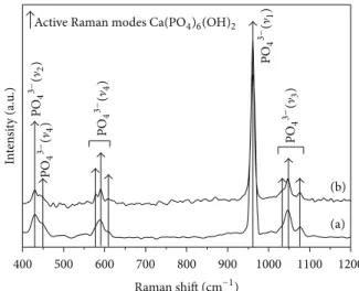
Raman modes are related to the HA phase. From Raman studies on carbonated apatite, two distinct wavenumbers of the ]1 carbonate mode have been suggested according
to OH− or PO43− site substitution at 1108 and 1070 cm−1, respectively. Variable numbers of bands in the V1PO43−
domain were also detected by the authors [22]. Since none of these characteristics were found in the Raman spectrum (see Figure4), we conclude that the HA@Ag obtained does not present the concurrent calcium carbonate phase as a second phase which agrees with XRD results that indicate the HA single phase formation.
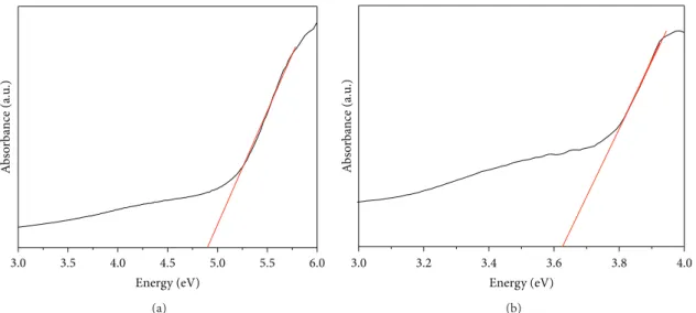
A UV-vis analysis was conducted (see Figures5(a)and
5(b)) to determine the electronic structure of HA@Ag. More-over, the GAP energy was calculated by using the Wood and Tauc method from the UV-spectra for pure HA and HA@Ag. The values obtained were 4.7 eV for pure HA and 3.6 eV for HA@Ag. There is a significant difference between band gap values for both samples. The exponential optical absorption edge and the optical band gap energy are controlled by the degree of structural disorder in the lattice. The decrease in the
400 500 600 700 800 900 1000 1100 1200
Raman shift (cm−1)
Figure 4: FT-Raman spectra for (a) pure HA and (b) HA@Ag.
band gap can be attributed to defects, local bond distortion, and intrinsic surface and/or interface states (HA and Ag) which yield localized electronic levels in the forbidden band gap.
We believe that this significant difference is attributed to Ag surface defects and (HA@Ag) interface defects [23] (see Figure6).
MIC and MFC values were 62.5𝜇g/mL and 250𝜇g/mL, respectively (Figure7). These results demonstrate that the HA@Ag solution has fungistatic and fungicidal effects against the microorganism tested and thus indicate that it may be a potential candidate for developing new antimicrobial agents with multiple applications in the biomedical field. Although these MIC and MFC values againstC. albicansare higher than those values obtained with commonly used antifungal agents [5] and different nanoparticles [24–26], other important aspects should be considered. First, when composites are used, the concentrations of individual nanoparticles can be decreased as well as the cytotoxicity to eukaryotic human cells [27]. Moreover, in this study, the amount of AgNO3used was 7.10−5mol with a Ag/HA ratio of 1. Hence, the combination of different nanoparticles could have a positive impact in avoiding the development of microbial resistance [27].
Bearing in mind the biofilm architecture and its role on resistance against antimicrobial agents, it was considered important to evaluate the possible effects of the HA@Ag solution against cells and extracellular polymeric matrix. The results demonstrated that, when theC. albicansbiofilms were exposed to different concentrations of the HA@Ag solution, a significant reduction in the number of CFU/mL was observed only at a concentration of 1000𝜇g/mL (see Figure 8). This result can be attributed to the higher resistance ofC. albicans
3.0 3.5 4.0 4.5 5.0 5.5 6.0
A
b
so
rba
n
ce (a.u
.)
Energy (eV)
(a)
3.0 3.2 3.4 3.6 3.8 4.0
A
b
so
rba
n
ce (a.u
.)
Energy (eV)
(b)
Figure 5: UV-vis absorbance spectra for (a) pure HA and (b) HA@Ag samples.
b a
c
Ag4Vx0/Ag
Ag4V∙∙0/Ag
Ca2+
P5+ H1+
Interface
Ag4V∙0/Ag
b a
c
O2−
Figure 6: Illustration of the system with the formation of a complex
cluster[Ag4]d.
of Ag-doped HA on the first step of bacterial adherence to the inert substratum and on a 24 h preformed biofilm. These authors also observed that the inhibitory effect against biofilm formation was dependent on the concentration of the nanoparticles evaluated. Other recent studies that evaluated the effect of silver nanoparticles againstC. albicansbiofilms also observed a high tolerance of this fungal specie grown in biofilms when compared to planktonic cells [24–26].
The results obtained after crystal violet staining showed significant decreases of the total biomass values when
C. albicans biofilms were exposed to HA@Ag solution at concentrations of 1000𝜇g/mL, 500𝜇g/mL, and 250𝜇g/mL (see Figure 9), showing clearly that the HA@Ag solution
1
2 3 4 5 6 7 8
0 3.91 7.82 15.63 31.25 62.5 125 250 500 1000
log
10
(CFU/mL)
HA@Ag concentration (𝜇g/mL)
∗ ∗
∗
Figure 7: Mean values of log10(CFU/mL) of planktonic cultures of
C. albicansexposed to HA@Ag solution. Errors bars: standard
devi-ation.∗: significant differences compared with control (0𝜇g/mL).
affected the extracellular matrix production. This result is important because biofilm formation is one of the major virulence factors ofC. albicans, and its reduction makes the microorganisms more susceptible to antifungal agents and more vulnerable to host defense mechanisms. Although the biofilms matrix was reduced by the HA@Ag solution, this effect was not observed in the terms of CFU/mL. This finding suggests that the cells exhibit recovery ability after the plating, and thus, the HA@Ag solution can have a temporary and reversible effect againstC. albicansbiofilms. These results are in agreement with those observed in a recent study in which Ag nanoparticles were effective in reducing biofilm biomass when applied to biofilms of C. albicans and C. glabrata. However, the effect of Ag nanoparticles in the number of viable biofilm cells ofC. albicanswas less evident [26].
Figure 8: Mean values of log10(CFU/mL) ofC. albicansbiofilm
exposed to HA@Ag solution. Errors bars: standard deviation. ∗:
significant difference compared with control (0𝜇g/mL).
3
2.5
2
1.5
1
0.5
0
A
b
so
rba
n
ce (
570
nm)
0 3.91 7.82 15.63 31.25 62.5 125 250 500 1000
HA@Ag concentration (𝜇g/mL)
∗ ∗ ∗ ∗
∗
Figure 9: Total biomass ofC. albicansbiofilms exposed to HA@Ag
solution. Errors bars: standard deviation.∗: significant differences
compared with control (0𝜇g/mL).
genes related to virulence factors, such as enzyme production [29] and biofilm formation [30].
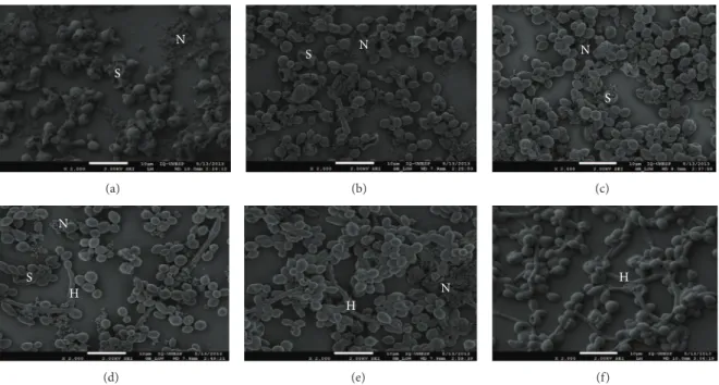
FEG-SEM images confirm the quantitative outcomes obtained with biofilms (see Figure 10) since alterations in cell morphology (“shriveled” cells appearance) were observed proportional to the concentration of HA@Ag nanoparticles. However, as can be seen in Figure10, some intact cells remain even in the presence of high concentrations of nanoparticles. Micrographs with none (control) or low HA@Ag content showed more cell aggregates and higher quantity of hyphal cells.C. albicansis able to change its morphology between yeast and filamentous (hyphae and pseudohyphae) forms in a process called polymorphism. Hyphal cells are responsible for infection in the host and are able to invade tissues. These findings show that the HA@Ag solution exhibited an antifungal property againstC. albicansbiofilms.
The performance of HA and silver is primarily dictated not only by physicochemical and mechanical properties, but also by biological activity [31]. HA is known as a biocompatible ceramic, and Ag has long been studied as
action. Membrane depolarization, the arrest of the fungal cell cycle by flow cytometry, the release of intracellular glucose and trehalose, a decrease in plasma membrane fluorescence with increasing concentrations of Ag nanoparticles, pits in the cell wall, and pores in the plasma membrane on the TEM images were observed. These findings demonstrate that Ag nanoparticles destruct the fungal membrane integrity and inhibit the normal budding process.
Disorders in surfaces and interfaces occur in HA@Ag synthesized by the HTMW method and create disordered sites [38]. This fact induces restructuring at the intermediate range which results in structural and electronic alterations to both the surface and interface. Disordered sites yield a local lattice distortion that is propagated along the overall material which pushes the surrounding clusters away from their ideal positions. Thus, complex clusters (disordered clusters) must move for these properties to occur which changes the electronic distribution along the network of these polar clusters [39]. This electronic structure may dictate the biological activity and plays a major role in determining the reactivity and stability of the cluster.
The mechanism of cluster complex [Ag4]∙activity with oxygen essentially depends on the complex cluster with the formation of a superoxide and/or a hydroxyl radical. [Ag4]∙d and[Ag4]d can create hydroxyl radicals (OH∗) and superoxide anions (O2H∗) by electron/hole reactions which can facilitate protein inactivation and eventual cell apoptosis where d = disorder and O = order.
Moreover, an effective charge separation requires an elec-tric field between the sample bulk and surface. Consequently, the effect of surface properties on electron/hole reaction performance should be considered in terms of the following:
[Ag4]𝑥0+ [Ag4]𝑥d→ [Ag4]d+ [Ag4]∙d (1)
The reactivity of molecular oxygen with a complex cluster [Ag4]∙don the surface of silver oxide results in a chemisorbed
species and subsequent oxygen incorporation into the lattice:
[Ag4]∙d+O2→ [Ag4]∙d⋅ ⋅ ⋅O2 (ads) (2)
[Ag4]∙d⋅ ⋅ ⋅O2 (ads)+ [Ag4]0→ [Ag4]∙d⋅ ⋅ ⋅O
2 (ads)+ [Ag4]
N
S
(a)
N S
(b)
N
S
(c)
N
S H
(d)
N H
(e)
H
(f)
Figure 10: FEG-SEM ofC. albicansbiofilms with different concentrations of the HA@Ag solution: (a) 1000, (b) 500, (c) 250, (d) 125, (e) 62.5,
and (f, control) 0𝜇g/mL. N: HA@Ag nanoparticles, S: “shriveled” cells appearance, H: Hyphae.
The clusters formed by the complex silver oxide also interact with water and split into hydroxyl radicals and hydrogen ions according to the following reactions:
[Ag4]∙+H2O→ [Ag4]∙⋅ ⋅ ⋅ H2O(ads) (4)
[Ag4]∙⋅ ⋅ ⋅ H2O(ads)→ [Ag4]
∙⋅ ⋅ ⋅
OH∗(ads)+H∙ (5)
Products of the partial oxidation reaction between water and a complex cluster[Ag4]∙dare hydroxyl radicals, OH∗, and hydrogen ions. These radicals exhibit high oxidation power which produces a microorganism mineralization in water (anodic oxidation) (5).
Primary reaction (cathodic) is the formation of a superox-ide species[Ag4]∙d⋅ ⋅ ⋅O2(2) and (3). These species then react with H∙(5) and produce the formation of hydrogen peroxide radicals (HO2∗) according to the following reactions:
[Ag4]∙d⋅ ⋅ ⋅O2 ( ads)+H∙→ [Ag4]
∙
d⋅ ⋅ ⋅O2H
∗
(ads) (6)
The radicals OH∗ and O2H∗ may react with the fungal cells which ultimately results in their oxidation.
The nature of the superoxide or hydroxyl radicals can be described using a complex cluster model where the electron transfer from the surface to the adsorbed molecular oxygen occurs [40–42].
These reaction mechanisms involve oxygen and water adsorptions that form an active complex cluster. Its decom-position, desorption of the final products, and the electron hole recombination process may have an important role in fungicidal effects.
Therefore, HA@Ag was successfully obtained by main-taining the structure of HA nanorods overlapped by nanosphere Ag as confirmed by TEM images. The HA@Ag
obtained shows efficient antifungal action. The results of antifungal tests can be explained by the interaction between the structure and the defect density variation in the interfa-cial (HA@Ag) and intrafainterfa-cial (HA) region with the fungal medium which results in antifungal activity.
4. Conclusions
HA@Ag was successfully obtained by maintaining the HA nanorods structure overlapped by nanosphere Ag through a co-precipitation method with HTMW. The HA@Ag solution showed fungistatic and fungicidal effects againstC. albicans
planktonic cells with MIC and MFC values of 62.5𝜇g/mL and 250𝜇g/mL, respectively. Additionally, the HA@Ag solution at concentrations of 1000𝜇g/mL, 500𝜇g/mL, and 250𝜇g/mL also exhibited antibiofilm activity, affecting mainly the extra-cellular matrix production. The morphological characteris-tics of cells were altered, and the density of hyphal cells was less noticeable in presence of the HA@Ag solution at higher concentrations. A mechanism was proposed to justify the antifungal activity by the interaction between the structure and the defect density variation in the interfacial (HA@Ag) and intrafacial (HA) region with the fungal medium.
Acknowledgment
This research was supported by grants 2011/06786-0, and 2011/24004-0, S˜ao Paulo Research Foundation (FAPESP).
References
long-term antiretroviral therapy, and with absent-negligible
immunodeficiency,”Brazilian Journal of Infectious Diseases, vol.
11, no. 6, pp. 605–609, 2007.
[5] J. Chandra, P. K. Mukherjee, S. D. Leidich et al., “Antifungal resistance of candidal biofilms formed on denture acrylic in
vitro,”Journal of Dental Research, vol. 80, no. 3, pp. 903–908,
2001.
[6] P. Badiee and A. Alborzi, “Invasive fungal infections in renal
transplant recipients,”Experimental and Clinical
Transplanta-tion, vol. 9, no. 6, pp. 355–362, 2011.
[7] I. Sondi and B. Salopek-Sondi, “Silver nanoparticles as
antimi-crobial agent: a case study on E. colias a model for
Gram-negative bacteria,”Journal of Colloid and Interface Science, vol.
275, no. 1, pp. 177–182, 2004.
[8] J. S. Kim, E. Kuk, K. N. Yu et al., “Antimicrobial effects of silver
nanoparticles,”Nanomedicine, vol. 3, no. 1, pp. 95–101, 2007.
[9] J. R. Morones, J. L. Elechiguerra, A. Camacho et al., “The
bactericidal effect of silver nanoparticles,”Nanotechnology, vol.
16, no. 10, pp. 2346–2353, 2005.
[10] M. A. Afzal, S. Kalmodia, P. Kesarwani, B. Basu, and K. Balani, “Bactericidal effect of silver-reinforced carbon nanotube and
hydroxyapatite composites,” Journal of Biomaterials
Applica-tions, vol. 27, no. 8, pp. 967–978, 2012.
[11] Z. Lu, Y. Liu, B. Liu, and M. Liu, “Friction and wear behavior of hydroxyapatite based composite ceramics reinforced with
fibers,”Materials and Design, vol. 39, pp. 444–449, 2012.
[12] D. Gopi, J. Indira, L. Kavitha, M. Sekar, and U. K. Mudali, “Synthesis of hydroxyapatite nanoparticles by a novel ultrasonic
assisted with mixed hollow sphere template method,”
Spec-trochimica Acta A, vol. 93, pp. 131–134, 2012.
[13] F. Ren, Y. Ding, X. Ge, X. Lu, K. Wang, and Y. Leng, “Growth of one-dimensional single-crystalline hydroxyapatite nanorods,” Journal of Crystal Growth, vol. 349, no. 1, pp. 75–82, 2012. [14] H. E. Wang, L. J. Xi, R. G. Ma et al., “Microwave-assisted
hydrothermal synthesis of porous SnO2 nanotubes and their
lithium ion storage properties,”Journal of Solid State Chemistry,
vol. 190, pp. 104–110, 2012.
[15] V. D. Maksimov, P. E. Meskin, and B. R. Churagulov,
“Microwave-assisted hydrothermal synthesis of fine BaZrO3
and BaHfO3 powders,”Inorganic Materials, vol. 43, no. 9, pp.
988–993, 2007.
[16] V. D. Ara´ujo, W. Avansi, H. B. de Carvalho et al., “CeO2
nanoparticles synthesized by a microwave-assisted
hydrother-mal method: evolution from nanospheres to nanorods,”
Crys-tEngComm, vol. 14, no. 3, pp. 1150–1154, 2012.
[17] C. S. Ciobanu, F. Massuyeau, L. V. Constantin, and D. Predoi, “Structural and physical properties of antibacterial Ag-doped
nano-hydroxyapatite synthesized at 100∘C,”Nanoscale Research
Letters, vol. 6, no. 1, article 613, pp. 1–8, 2011.
[21] CLSI,Reference Method for Broth Dilution Antifungal
Suscep-tibility Testing of Yeasts; Approved Standard, CLSI Document M27-A3, Clinical and Laboratory Standards Institute, Wayne, Pa, USA, 3rd edition, 2008.
[22] G. Penel, G. Leroy, C. Rey, and E. Bres, “MicroRaman spectral
study of the PO4and CO3vibrational modes in synthetic and
biological apatites,”Calcified Tissue International, vol. 63, no. 6,
pp. 475–481, 1998.
[23] C. W. Raubach, Y. V. B. de Santana, M. M. Ferrer et al., “Strutural
and optical approach of CdS@ZnS core-shell system,”Chemical
Physics Letters, vol. 536, pp. 96–99, 2012.
[24] A. F. Wady, A. L. Machado, V. Zucolotto, C. A. Zamperini,
E. Berni, and C. E. Vergani, “Evaluation ofCandida albicans
adhesion and biofilm formation on a denture base acrylic resin
containing silver nanoparticles,”Journal of Applied
Microbiol-ogy, vol. 112, no. 6, pp. 1163–1172, 2012.
[25] D. R. Monteiro, S. Silva, M. Negri et al., “Silver nanoparticles: influence of stabilizing agent and diameter on antifungal
activ-ity againstCandida albicansandCandida glabrata biofilms,”
Letters in Applied Microbiology, vol. 54, no. 5, pp. 383–391, 2012. [26] D. R. Monteiro, L. F. Gorup, S. Silva et al., “Silver col-loidal nanoparticles: antifungal effect against adhered cells and
biofilms ofCandida albicansandCandida glabrata,”Biofouling,
vol. 27, no. 7, pp. 711–719, 2011.
[27] M. A. Vargas-Reus, K. Memarzadeh, J. Huang, G. G. Ren, and R. P. Allaker, “Antimicrobial activity of nanoparticulate
metal oxides against peri-implantitis pathogens,”International
Journal of Antimicrobial Agents, vol. 40, no. 2, pp. 135–139, 2012. [28] G. Ramage, E. Mowat, B. Jones, C. Williams, and J. Lopez-Ribot,
“Our current understanding of fungal biofilms,”Critical Reviews
in Microbiology, vol. 35, no. 4, pp. 340–355, 2009.
[29] T. Wu, K. Wright, S. F. Hurst, and C. J. Morrison, “Enhanced extracellular production of aspartyl proteinase, a virulence
factor, byCandida albicansisolates following growth in
subin-hibitory concentrations of fluconazole,”Antimicrobial Agents
and Chemotherapy, vol. 44, no. 5, pp. 1200–1208, 2000. [30] L. Dong, Z. Tong, D. Linghu et al., “Effects of sub-minimum
inhibitory concentrations of antimicrobial agents on
Strep-tococcus mutans biofilm formation,” International Journal of Antimicrobial Agents, vol. 39, no. 5, pp. 390–395, 2012. [31] M. Roy, G. A. Fielding, H. Beyenal, A. Bandyopadhyay, and S.
Bose, “Mechanical, in vitro antimicrobial, and biological prop-erties of plasma-sprayed silver-doped hydroxyapatite coating,” ACS Applied Materials and Interfaces, vol. 4, no. 3, pp. 1341–1349, 2012.
[32] A. Ewald, D. H¨osel, S. Patel, L. M. Grover, J. E. Barralet, and U. Gbureck, “Silver-doped calcium phosphate cements with
antimicrobial activity,” Acta Biomaterialia, vol. 7, no. 11, pp.
[33] N. Matsumoto, K. Sato, K. Yoshida, K. Hashimoto, and Y. Toda,
“Preparation and characterization of𝛽-tricalcium phosphate
co-doped with monovalent and divalent antibacterial metal
ions,”Acta Biomaterialia, vol. 5, no. 8, pp. 3157–3164, 2009.
[34] W.-H. Song, S. R. Hyun, and S.-H. Hong, “Antibacterial prop-erties of Ag (or Pt)-containing calcium phosphate coatings
formed by micro-arc oxidation,”Journal of Biomedical Materials
Research A, vol. 88, no. 1, pp. 246–254, 2009.
[35] K. Das, S. Bose, A. Bandyopadhyay, B. Karandikar, and B. L. Gibbins, “Surface coatings for improvement of bone cell
materials and antimicrobial activities of Ti implants,”Journal
of Biomedical Materials Research B, vol. 87, no. 2, pp. 455–460, 2008.
[36] X. Bai, K. More, C. M. Rouleau, and A. Rabiei, “Functionally graded hydroxyapatite coatings doped with antibacterial
com-ponents,”Acta Biomaterialia, vol. 6, no. 6, pp. 2264–2273, 2010.
[37] K.-J. Kim, W. S. Sung, B. K. Suh et al., “Antifungal activity and
mode of action of silver nano-particles onCandida albicans,”
BioMetals, vol. 22, no. 2, pp. 235–242, 2009.
[38] P. G. Mendes, M. L. Moreira, S. M. Tebcherani et al., “SnO2
nanocrystals synthesized by microwave-assisted hydrothermal method: towards a relationship between structural and optical
properties,” Journal of Nanoparticle Research, vol. 14, no. 3,
article 750, 2012.
[39] T. Badapanda, S. K. Rout, L. S. Cavalcante et al., “Optical
and dielectric relaxor behaviour of Ba(Zr0.25Ti0.75)O3 ceramic
explained by means of distorted clusters,”Journal of Physics D,
vol. 42, no. 17, Article ID 175414, pp. 1–9, 2009.
[40] T. Bak, J. Nowotny, N. J. Sucher, and E. Wachsman, “Effect of
crystal imperfections on reactivity and photoreactivity of TiO2
(Rutile) with oxygen, water, and bacteria,”Journal of Physical
Chemistry C, vol. 115, no. 32, pp. 15711–15738, 2011.
[41] A. Bielanski and W. C. D. Hare, “Investigation of some antimi-crobial procedures on the in vitro development of early murine embryos aimed toward developing methods for the disinfection
of mammalian embryos prior to transfer,”Journal of In Vitro
Fertilization and Embryo Transfer, vol. 8, no. 1, pp. 24–32, 1991. [42] J. P. Kehrer, “The Haber-Weiss reaction and mechanisms of
Submit your manuscripts at
http://www.hindawi.com
Scientifica
Hindawi Publishing Corporation http://www.hindawi.com Volume 2014 Hindawi Publishing Corporationhttp://www.hindawi.com Volume 2014
Hindawi Publishing Corporation
http://www.hindawi.com Volume 2014
Ceramics
Journal ofNanoparticles
Journal of Hindawi Publishing Corporationhttp://www.hindawi.com Volume 2014
International Journal of
Biomaterials
Hindawi Publishing Corporationhttp://www.hindawi.com Volume 2014
Hindawi Publishing Corporation
http://www.hindawi.com Volume 2014
NANOSCIENCE
Journal of
Textiles
Hindawi Publishing Corporation
http://www.hindawi.com Volume 2014
Journal of
Hindawi Publishing Corporation
http://www.hindawi.com Volume 2014
Crystallography
Journal ofThe Scientific
World Journal
Hindawi Publishing Corporationhttp://www.hindawi.com Volume 2014
Hindawi Publishing Corporation
http://www.hindawi.com Volume 2014
Coatings
Journal of Advances inMaterials Science and Engineering
Hindawi Publishing Corporation
http://www.hindawi.com Volume 2014
Hindawi Publishing Corporation
http://www.hindawi.com Volume 2014
Hindawi Publishing Corporation
http://www.hindawi.com Volume 2014
Materials
Journal ofN
a
no
ma
te
ria
ls
Hindawi Publishing Corporation
http://www.hindawi.com Volume 2014
Journal of