J of Evolution of Med and Dent Sci/ eISSN- 2278-4802, pISSN- 2278-4748/ Vol. 4/ Issue 70/ Aug 31, 2015 Page 12164
PATTERN OF AEROBIC BACTERIAL INFECTION OF DIABETIC FOOT
Parmeshwari Patil1, Basavaraj Patil2HOW TO CITE THIS ARTICLE:
Parmeshwari Patil, Basavaraj Patil. Pattern of Aerobic Bacterial Infection of Diabetic Foot. Journal of Evolution of Medical and Dental Sciences 2015; Vol. 4, Issue 70, August 31; Page: 12164-12168,
DOI: 10.14260/jemds/2015/1752
ABSTRACT: BACKGROUND: Diabetic foot ulcer is one of the commonest complications of longstanding diabetes. Diabetic foot is a common cause of hospital admission in diabetic patients in India. The trio of problems leading onto diabetic foot is neuropathy, vascular changes and infections, which constitute the diabetic foot syndrome. OBJECTIVES: To determine prevalence of aerobic pathogens in diabetic foot lesions. METHODS: Tissue samples were taken from the affected foot of 109 diabetic patients and processed by routine microbiological methods. RESULTS: A total of 244 aerobic organisms were isolated from 100 cases with an average of 2.5 organisms per case. Staphylococcus aureus was the predominant aerobe isolated (25.4%) followed by Proteus mirabilis (21.3%). The other aerobes isolated were Pseudomonas aeruginosa (10.65%), Klebsiella pneumonia (8.6%), Escherichia coli (6.9%), Enterococcus faecalis (5.32%), Coagulase Negative Staphylococcus (4.5%), Proteus vulgaris (4.09%), Klebsiella oxytoca (3.27%), Citrobacter freudii (2.86%), Corynebacterium species (2.04%), Group A streptococci and Acinetobacter species (1.63%), and Enterobacter species (0.82%). MRSA was 17.8%. CONCLUSION: Diabetic foot infections are polymicrobial in nature. Staphylococcus aureus was the most common among gram-positive organisms, while Proteus mirabilis was the most frequent isolate among the gram-negative pathogens. Hence early identification of the risk factors and timely institution of appropriate treatment is indispensable to avoid amputations.
KEYWORDS: Diabetic foot ulcer, Aerobes.
INTRODUCTION: Diabetic foot is most simply defined as any infra-malleolar infection in a person with diabetes mellitus.1 India homes 33 million diabetics, ranking highest in the world and has a prevalence of about 8% in urban India. 20% of all diabetic complications involve feet.2 Diabetic foot includes paronychia, cellulitis, myositis, abscesses, necrotising fascitis, septic arthritis, tendonitis and osteomyelitis. The most common and classical lesion is the infected diabetic mal perforans foot ulcer.3
Neuropathy plays the central role with disturbances of sensory, motor and autonomic functions leading to ulceration due to trauma or excessive pressure on a deformed foot that lacks protective sensations. Once the protective layer of skin is breached, underlying tissues are exposed to bacterial colonisation. This wound may progress to become actively infected and by contiguous extension, the infection can involve deeper tissues. This sequence of events can be rapid, especially in an ischaemic limb and in the setting of various poorly characterized immunological disturbances involving polymorphonuclear leucocytes.4
Aerobes are commonly incriminated bacteria in pathogenesis of Diabetic foot. The most common aerobic organisms encountered are the Gram positive cocci including Staphylococcus aureus, Coagulase negative Staphylococci and Streptococcus species.
J of Evolution of Med and Dent Sci/ eISSN- 2278-4802, pISSN- 2278-4748/ Vol. 4/ Issue 70/ Aug 31, 2015 Page 12165 MATERIALS AND METHODS: The present study included 109 cases of patients diagnosed with diabetes ulcer having Foot ulcers of Grade I or more with evidence of purulent exudates and or oedema Diabetic patients with a foot lesions of grade 0 and/or limb amputation were excluded Two debrided tissue samples were taken from each patient and were subjected for smear preparation and aerobic culture.
The first tissue sample was used to prepare smears by crushing it between two sterile glass slides and heat fixing it. The heat fixed smears were stained by Gram staining technique and under oil immersion objective of the light microscope, the size, shape, arrangement of bacteria and presence of spores was noted.
The second tissue sample was inoculated into Brain heart infusion broth immediately after collection and was incubated overnight at 37oC in an incubator. The broth was looked for turbidity after overnight incubation. A smear was prepared from the turbid broth before sub culturing it. The smear was stained by gram staining and observed under oil immersion of light microscope for the presence of organisms.
The broth was then subcultured onto 5% sheep blood agar, Mac Conkey agar and chocolate agar plates which were pre-incubated in the incubator overnight to rule out the possibility of contamination of the plates. The inoculated media were incubated at 37oC overnight. Blood agar and chocolate agar plates were incubated under 5-10% CO2.
The colonies were examined under magnifying lens and identified using the standard microbiological procedures like colony morphology, Gram staining and biochemical reactions as described in Practical Microbiology of Mackie McCartney 14th volume.6
OBSERVATIONS: Of the 109 patients, 2/3rd of patients were male and maximum patients belonged to 6th decade of life followed closely by 7th & 5th decade. (Table: 1)
Age (in yrs.) Male Female Total No. % No. % No. %
0-10 0 0 0 0 0 0
11-20 0 0 0 0 0 0
21-30 1 100 0 0 1 0.92 31-40 5 71.4 2 28.6 7 6.42 41-50 14 68.9 9 31.1 23 21.10 51-60 25 69.4 11 30.6 36 33.03 61-70 17 60.7 11 39.3 28 25.69 71-80 7 50.0 7 50.0 14 12.84 Total 76 70 33 30 109 100.00 Table 1: Distribution of patients based on Age & Sex
J of Evolution of Med and Dent Sci/ eISSN- 2278-4802, pISSN- 2278-4748/ Vol. 4/ Issue 70/ Aug 31, 2015 Page 12166 Blood Sugar level (in mg %) No. No. of Isolates Isolates/Case
<200 22 45 2.05/case
>200 89 231 2.6/case
Total 109 276 2.5/case
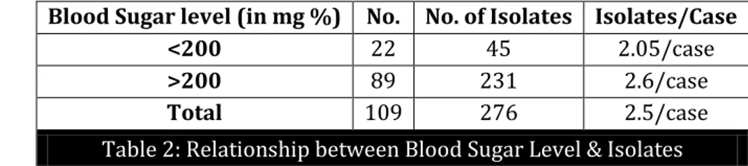
Table 2: Relationship between Blood Sugar Level & Isolates
Among Gram positive aerobes, Staphylococcus aureus was the predominant isolate (25.40%). Group A Streptococci was least isolated (1.63) Among Gram negative aerobes, Proteus mirabilis was the most common isolate (21.31%) and Enterobacter species and Bacillus species was the least common isolate (1.37%). (Table 3)
GRAM POSITIVE No % GRAM NEGATIVE
Staphylococcus aureus 62 25.40 Proteus mirabilis 52 21.31 Enterococcus faecalis 13 5.32 Pseudomonas aeruginosa 26 10.65 Coagulase negative Staphylococcus 11 4.50 Klebsiella pneumoniae 21 8.60
Group A Streptococci 4 1.63 Escherichia coli 17 6.96 Corynebacterium species 5 2.04 Proteus vulgaris 10 4.09 Klebshiella oxytoca 8 3.27 Citrobacter freundii 7 2.86 Acinobacter species 4 1.63 Enterobacter species 2 0.82 Bacillus species 2 0.82
Table 3: Number of Aerobic Bacilli Isolated
DISCUSSION: In the present study, the average age of patients was 51 with a range of 24-78 years. Age group of 51-60 years accounted for maximum number of patients that is 36 (33%). Ramani et al.7 reported the mean age of patients in their study as 58 years with a range of 28-87 years. Pathare et al.8 reported the mean age of patients in their study as 75.02 years. Dipali et al.9 reported the age range from 30 years to 86 years with an average of 58 years. Anandi et al.10 observed the mean age to be 43 years. Gadepalli et al.11 reported that mean age of patients to be 53.9 years. Bansal et al.12 reported mean age to be 57.04 years.
In the present study, we observed that 2.05 isolates per case was obtained from patients with BSL ≤200mg%, whereas 2.6 per case was obtained from patients with BSL> 200mg%. The difference between the mean isolates of BSL ≤200mg% and BSL >200mg% is significant (p< 0.05).
In the present study, Staphylococcus aureus was the predominant aerobic isolate (25.40%) which is in correlation with Wheat et al,13 Ramani et al,7 Dipali AC et al,9 Vijaya et al.14 and Raja et al.15 who isolated Staphylococcus aureus as their major aerobe.
J of Evolution of Med and Dent Sci/ eISSN- 2278-4802, pISSN- 2278-4748/ Vol. 4/ Issue 70/ Aug 31, 2015 Page 12167 CONCLUSION: Diabetic foot ulcers are one of the most common and dreaded complications of diabetes. However it is noted that good control of blood sugar level is desirable goal in the prevention of certain infections and to ensure maintenance of normal host defense mechanisms determining resistance and response to infection. Though it is known fact that diabetic foot is a polymicrobial nature of infection but majority of the isolates are aerobes.
Anaerobes are mostly isolated from higher grade ulcers. Staphylococcus aureus is the predominant gram positive organism and Proteus mirabilis is the predominant gram negative organism. It is utmost important to screen all elderly patients for diabetes and monitor blood sugar levels regularly and educate them about foot care. Early identification of the risk factors and timely institution of appropriate treatment is indispensable to avoid amputations.
REFERENCES:
1. Utpal De, Kamal K De, Nemai C Nath, Riju Dutta: Pathogenesis and Management of Diabetes Foot-A Review. JIMA 2005; 103(11):612,614,616.
2. Gaur DS, Verma A, Gupta P.: Diabetic foot in Uttaranchal. J K Science, Jan-Mar 2007; 9(1): 18-20. 3. Caputo G M, Cavangh P R, Ulbrecht J S, Gibbons G W, Karchmer A W: Assessment and
Management of foot disease in patients with diabtes. N Engl J Med 1994; 331: 854-60.
4. Schubert S, Heesemann J: Infections in diabetes mellitus (in German). Immun Infect 1995; 23: 200-4.
5. Lipsky BA. Medical treatment of diabetic foot infections. Clinical Infectious disease, 2004; 39: S104-14.
6. Collee JG, Duguid JP, Fraser AG, Marmion BP, Simmons A. (Eds). Mackie and Mc Cartney Practical Medical Microbiology, 14th Edn, Singapore: Churchill Livingstone 1989.
7. Ramani A, Ramani R, Shivananda PG, Kundaje GN.: Bacteriology of Diabetic Foot Ulcers. Indian J Pathol. Microbiol, 1991; 34(2): 81-87.
8. Pathare NA, Bal A, Tavalkar GV, Antani DU.: Diabetic foot infections: A study of microorganisms associated with the different Wagner grades. Indian J. Pathol. Microbial. 1998; 41(4): 437-441.
9. Dipali AC, Pal RB.: Study of fungal and bacterial infections of the diabetic foot. Indian J Pathol. Microbiol, 2002; 45(1): 15-22.
10.C Anandi, D Alaguraja, V Natarajan, M Ramanathan, C S Subramaniam, M Thulasiram, S Sumithra: Bacteriology of Diabetes Foot Lesions. IJMM 2004; 22(3): 175-178
11.Gadepalli R, Dhawan B, Sreenivas V, Kapil A, Ammini AC, Chaudhry R.: A Clinico-microbiological Study of Diabetic Foot Ulcers in an Indian Tertiary Care Hospital . Diabetes Care, 2006; 29(8): 1727-1731.
12.Bansal E, Garg A, Bhatia S, Attri AK, Chander J. Spectrum of microbial flora in diabetic foot ulcers. Indian J Pathol Microbiol. 2008 Apr-Jun; 51(2):204-8.
13.Wheat J, Allen SD, Henry M, Kernek CB: Diabetic foot infections - Bacteriological analysis. Arch Intern Med, 1986; 146: 1935-1940.
14.Vijaya D, Lakshmikanth, Sheshadri. Bacteriology of diabetic foot infection. Biomedine, 2000; 20(3): 176-179.
J of Evolution of Med and Dent Sci/ eISSN- 2278-4802, pISSN- 2278-4748/ Vol. 4/ Issue 70/ Aug 31, 2015 Page 12168
16.Gerding DN: Foot infections in diabetes patients: The role of anaerobes. Clin Infect Disease1995; 20: s 283-8.
AUTHORS:
1. Parmeshwari Patil 2. Basavaraj Patil
PARTICULARS OF CONTRIBUTORS:
1. Assistant Professor, Department of Department of Microbiology, M. R. Medical College, Gulbarga.
2. Professor and HOD, Department of Department of Microbiology, M. R. Medical College, Gulbarga.
FINANCIAL OR OTHER
COMPETING INTERESTS: None
NAME ADDRESS EMAIL ID OF THE CORRESPONDING AUTHOR:
Dr. Parmeshwari Patil, Assistant Professor,
Department of Microbiology, M. R. Medical College, Gulbarga-585103.
E-mail: [email protected]