Comparative study between computed tomography
and bronchoscopy in the diagnosis of lung cancer*
Estudo comparativo do diagnóstico de câncer pulmonar entre tomografia computadorizada e broncoscopia
Christopher Oliveira1, António Saraiva2
OBJECTIVE: To analyze the role of computed tomography and bronchoscopy in the diagnosis of lung can-cer, evaluating the effectiveness of these techniques in the presence of this disease. Parameters such as age, gender, smoking habits, histological types, staging and treatment were also analyzed. MATERIALS AND METHODS: The sample of the present study included 70 patients assisted at the Department of Pneumology of Hospital Distrital da Figueira da Foz, Coimbra, Portugal, who were submitted to both diagnostic methods, namely, computed tomography and bronchoscopy, to confirm the presence or the absence of lung cancer. RESULTS: Thirty-seven patients (23 men and 14 women) were diagnosed with lung cancer. Histologically 40.54% were adenocarcinoma, followed by squamous carcinoma (32.43% cases) and small-cell lung can-cer (18.92%). Staging showed 6.70% stage IB disease, 23.30% stage IIIA and 36.70% stage IIIB, and 33.30% stage IV. Chemotherapy alone was the first treatment of choice for 75.7% of patients. Bronchos-copy sensitivity was 83.8%, specificity 81.8%, and accuracy 82.8%. Computed tomography sensitivity was 81.1%, specificity 63.6%, and accuracy 72.8%. CONCLUSION: Bronchoscopy results corroborated the relevance of the method in the diagnosis of lung cancer, considering its dependence on the anatomopathological study of tissue or cells obtained through different biopsy techniques. Computed to-mography presented good sensitivity (81.1%), however the specificity of only 63.6% is related to the rate of false-positive results (36.4%).
Keywords: Lung cancer; Imaging diagnosis; Bronchoscopy.
OBJETIVO: Analisar a tomografia computadorizada e a broncoscopia no diagnóstico do câncer pulmonar e verificar a eficácia destas técnicas perante a presença desta doença. Os parâmetros idade, gênero, hábitos tabágicos, tipos histológicos, estadiamento e terapêutica foram, igualmente, analisados. MATERIAIS E MÉ-TODOS: Foram analisados 70 pacientes do Serviço de Pneumologia do Hospital Distrital da Figueira da Foz, Coimbra, Portugal, que realizaram ambas as técnicas em estudo, tendo-se confirmado ou não a presença de câncer pulmonar. RESULTADOS: Diagnosticaram-se 37 tumores pulmonares, 23 casos no gênero mascu-lino e 14 no feminino. Histologicamente, 40,54% eram adenocarcinomas, seguido do carcinoma escamoso (32,43% dos casos) e do carcinoma de pequenas células (18,92%). O estadiamento mostrou 6,70% no estádio IB, 23,30% no estádio IIIA comparativamente ao IIIB com 36,70%, encontrando-se 33,30% dos doentes no estádio IV. A quimioterapia isolada foi efetuada em 75,7% dos doentes. A sensibilidade da bron-coscopia foi de 83,8%, a especificidade, de 81,8%, e a precisão, de 82,8%. A sensibilidade da tomografia computadorizada foi de 81,1%, a especificidade, de 63,6%, e a precisão, de 72,8%. CONCLUSÃO: Os re-sultados da broncoscopia confirmaram a sua importância no diagnóstico do câncer pulmonar, pela depen-dência deste no exame anatomopatológico do tecido ou células, obtido por várias técnicas de biópsia. A tomografia computadorizada apresentou boa sensibilidade, de 81,1%, contudo, a sua especificidade, de apenas 63,6%, resulta do número de falso-positivos (36,4%).
Unitermos: Neoplasias pulmonares; Diagnóstico por imagem; Broncoscopia.
Abstract
Resumo
* Study developed at Hospital Distrital da Figueira da Foz (HDFF), Escola Superior de Tecnologia da Saúde de Coimbra (ESTeSC), Coimbra, Portugal.
1. Licentiate in Radiology, Radiologic Technologist, Escola Superior de Tecnologia da Saúde de Coimbra (ESTeSC), Coimbra, Portugal.
2. Master, Professor at Escola Superior de Tecnologia da Saúde de Coimbra (ESTeSC), Coimbra, Portugal.
Mailing address: António Saraiva. Escola Superior de Tecnologia da Saúde de Coimbra (ESTeSC). Rua 5 de Outubro. S. Martinho do Bispo, Apartado 7006. 3046-854 Coimbra, Portugal. E-mail: [email protected]
malignancy in both men and women. In the USA, lung cancer is the main cause of death by neoplasia (representing about 28% of deaths by neoplasia/year). In Por-tugal, the estimated incidence is 34 cases per 100,000 men, and 28 cases per 100,000 women. The highest incidence rate is ob-served in the age range from 55 to 65 years. Despite the recent therapeutic
develop-Oliveira C, Saraiva A. Comparative study between computed tomography and bronchoscopy in the diagnosis of lung can-cer. Radiol Bras. 2010;43(4):229–235.
INTRODUCTION
Lung cancer epidemiology
Lung cancer represents one of the most frequent neoplasias worldwide, accounting for the highest rate of deaths caused by
ments, the five-year survival rates among these patients remain as low as about 13%(1–4).
Lung cancer etiology
It was long ago that tobacco consump-tion was established as the main etiologi-cal factor in lung cancer. Smoking indi-viduals present a 22-time higher risk for death by this disease than non-smoking in-dividuals, and the strongest association with tobacco consumption has been ob-served in cases of small-cell lung cancer, squamous-cell carcinomas and large-cell lung carcinomas. Passive smoking is an increasingly relevant risk factor, represent-ing 3% to 5% of all cases of lung cancer(3,5,6).
It is important to note that tobacco con-sumption is not the sole cause of lung can-cer; other factors may be associated, affect-ing the disease incidence: exposure to ar-senic, chromium, nickel and asbestos, cica-tricial lesions of tuberculosis, bullous em-physema, and familial history of lung can-cer(2).
Clinical presentations
It should be highlighted that more than 90% of patients with lung cancer are as-ymptomatic at presentation, with clinical findings resulting from local, regional, metastatic or systemic effects of the tumor. Generally, the main symptom is persistent cough. The incidence of hemoptysis achieves 27–57%. Massive hemoptysis is less frequently found. Additionally, dysp-nea and paraneoplastic syndromes consti-tute the main symptoms in cases where the tumor is limited to the lung(3,5–9).
The presence of chest pain, hoarseness, brachial plexus neuropathies, Horner’s syndrome, dysphagia and pericardial tam-ponade is suggestive of mediastinal or chest wall invasion(3).
Main histological types of lung cancer
In the present study, the description of histological types will be based on the cri-teria of the World Health Organization (WHO) for histological classification of lung cancer(9).
The division of lung cancer into non-small cell lung carcinoma (NSCLC) and small cell lung carcinoma (SCLC) is cru-cial for the therapeutic planning(3,4).
The four main histological types in-clude: adenocarcinoma, squamous cell car-cinoma, SCLC and large-cell carcinoma. SCLC is considered as a histological sub-type with characteristics including an ag-gressive clinical behavior with fast cell proliferation, early metastatization and, nearly always, presentation at diagnosis as nonresectable disease(3,5).
Lung cancer diagnosis
The diagnostic evaluation of this dis-ease has two main objectives: the definition of the pathological type of tumor and stag-ing of the disease(7,10).
Chest computed tomography (CT) plays a relevant role in the determination of pres-ence and extent of lung cancer, demonstrat-ing the size and site of the tumor. However, this method presents some limitations such as high cost, utilization of ionizing radia-tion, contrast agent nephrotoxicity, besides the necessity of further procedures to con-firm the diagnosis(2,11).
The CT indispensability in the study of lung cancer is associated with the obliga-toriness of endoscopy of the respiratory tract with flexible endoscope. Endoscopic signs of cancer are quite variable, from a simple bright loss in a small region of the bronchial mucosa to a typical vegetative mass. Classically, three types of typical le-sions or direct signs of tumor are taken into consideration: mass, infiltration and ob-struction. The techniques associated with bronchofibroscopy include bronchial wash and brush cytology and bronchial bi-opsy(5,12–17).
Lung cancer staging
The staging analysis was based on the most recent revision of the TNM system dated of 1997. The staging is aimed at di-viding cancer patients into subgroups to define their prognosis and to determine the therapeutic options. It is important to note that imaging methods play an essential role in the staging and follow-up of patients with lung cancer(2,11,18).
The TNM system comprises several stages (IA, IB, IIA, IIB, IIIA, IIIB, IV) ac-cording to characteristics regarding: T – evaluation of the primary tumor; N – evalu-ation of lymph nodes involvement; M – evaluation of distant metastases(19).
In the clinical practice, the traditional TNM system is not useful in the staging of SCLC, limiting the staging to “limited dis-ease” and “extensive disdis-ease”(3,4).
Lung cancer therapeutic
An early diagnosis is the determining factor in the therapeutic selection. Surgery, chemotherapy and radiotherapy are the most frequently utilized types of treat-ment(5).
The most relevant therapeutic decision is related to the tumor resectability assess-ment, considering that surgery is the only potentially curative treatment. The type of surgical therapy is also dependent on the tumor type, site and extent, besides the patient’s age and general clinical condition. Many times a combination of two or even three treatment modalities is required(3–5,20).
MATERIALS AND METHODS
Study location and population
The present study was developed in the Unit of Pneumology of Hospital Distrital da Figueira da Foz, Coimbra, Portugal, in the period between January and July/2009, evaluating the clinical records of 70 tients. The study population included pa-tients referred to the Unit of Pneumology along a four-year period (January-2003 to January-2007) and submitted to CT and bronchoscopy for suspicion of lung cancer, confirmed or not by histopathological study. No other exclusion/inclusion crite-ria were considered.
Age, gender, smoking habits, histologi-cal types, staging and therapeutic options were also evaluated.
Search method and data analysis
A data record form was prepared for data collection and subsequent analysis. The data used in the present study were col-lected in the patients’ clinical records.
pre-pared the respective reports. On average, a two-day time interval was observed be-tween the performance of CT studies and bronchoscopy.
It is important to mention that chest, upper abdomen and skull CT, bronchos-copy, bone scintigraphy and mediastinos-copy were performed for the disease stag-ing.
Once the data collection was completed, the Statistical Package for the Social Sci-ences (SPSS) v17 was utilized for appro-priate data processing and analysis, allow-ing an accurate inference about the results of the present study.
Ethical considerations
The present investigation was fully compliant with the WMA Declaration of Helsinki – Ethical Principles for Medical Research Involving Human Subjects. Such declaration is worldwide recognized as the basic principles to be followed in medical research involving human subjects, includ-ing the respect for the human subject, be-sides the Principles of Beneficence and Justice. Thus patients were guaranteed their anonymity as well as the confidentiality of their clinical information.
RESULTS
In the present study, clinical records of 70 patients assisted in the Unit of Pneumol-ogy were analyzed. The patients (60% men and 40% women, in the age range between 35 and 84 years, mean = 66 years) had been referred with symptoms suggestive of lung cancer and were submitted to bronchos-copy and lung CT.
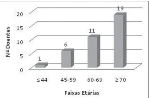
Among the 37 cases diagnosed with lung cancer, the mean age was 66.24 years (± 10.03), 23 were male and 14 female pa-tients. At the time of the diagnosis, only one patient was under 45 years of age and 19 patients were aged ≥ 70 years (Figure 1).
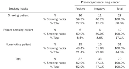
Table 1 demonstrates the sample char-acterization regarding smoking habits. Most of the patients smoked or had ever smoked, corresponding to 55.7% (n = 39), while the number of the non-smoking ones corresponded to 44.3% of the whole study sample. It is important to note that among the women participating in the present study, none smoked neither had ever
Table 1 Sample characterization according to smoking habits.
Gender
Male (n = 42) Female (n = 28)
Total (n = 70)
Smoking patient
27 (64.3%) 0 (0%)
27 (38.6%)
Former smoking patient
12 (28.6%) 0 (0%)
12 (17.1%)
Nonsmoking patient
3 (7.1%) 28 (100%)
31 (44.3%) Figure 1. Distribution of cancer patients by age range.
smoked (n = 28). Differently, only 7.1% of the male patients did not smoke.
Distribution of lung cancer types
Among the 70 patients submitted to bronchoscopy and CT, 37 presented lung cancer. Among these 37 cases, 30 (80.08%) were NSCLC and 7 were SCLC (18.92%).
Adenocarcinoma was the most frequent histological type observed (40.54%; n = 15), followed by squamous-cell carcinoma (32.43%; n = 12) and SCLC (18.92%; n = 7). As far as the rarest tumors are con-cerned, 5.41% were large-cell carcinomas and 2.70%, carcinoid tumors (Figure 2).
Figure 4. Lung cancer therapeutics.
Relation between smoking habits and presence/absence of lung cancer
In spite of the fact that the statistical analysis has indicated no statistical signifi-cance (p > 0.05) in the relation between smoking habits and presence/absence of lung cancer, it is essential to mention that the majority of the patients diagnosed with lung cancer, 59.46% (n = 22), smoked or had ever smoked, and 40.54% (n = 15) did not smoke (Table 2).
Staging
In the present study, it is important to evaluate the staging in the setting of NSCLC. Figure 3 demonstrates that the majority of the patients presented with ad-vanced stages of disease, i.e., stages IIIB (36.70%) and IV (33.30%) in a total of 70%. Eventually surgical stages correspond to 30%, with stage IB with 6.70% of cases, and stage IIIA with 23.30%.
Therapeutics
As regards therapeutics, it is important to note that surgery was performed as first therapeutic option (Figure 4) in 8.1% of the patients. Chemotherapy alone was per-formed in 75.7% of the cases. In 16.2% of the cases, the patients underwent combined therapy (combined surgery-chemotherapy, surgery-radiation therapy or association of chemotherapy and radiation therapy). Ra-diation therapy was not performed as a first therapeutic option, although this method has been utilized in combination with che-motherapy or surgery in certain cases. It is convenient to mention that the surgeries were performed in the Unit of Chest Sur-gery at Centro Hospitalar de Coimbra, the chemotherapy sessions at Hospital Distrital da Figueira da Foz, and the radiation therapy sessions at Instituto Português de Oncologia de Coimbra.
Relation between bronchoscopy results and presence/absence of lung cancer
The statistical analysis allowed the au-thors to observe that the relation between bronchoscopy results and the presence/ab-sence of lung cancer was statistically sig-nificant (p < 0.05). The kappa value 0.656 demonstrated a good rate of agreement between the obtained results.
Table 2 Relation between smoking habits and presence/absence of lung cancer (χ2 = 0.732; gl = 1; p = 0.694).
Presence/absence lung cancer
Smoking habits
Smoking patient
Former smoking patient
Nonsmoking patient
Total
N % Smoking habits
% Total
N % Smoking habits
% Total
N % Smoking habits
% Total
N % Smoking habits
% Total
Positive
16 59.3% 22.9%
6 50.0%
8.6%
15 48.4% 21.4%
37 52.9% 52.9%
Negative
11 40.7% 15.7%
6 50.0%
8.6%
16 51.6% 22.9%
33 47.1% 47.1%
Total
27 100.0%
38.6%
12 100.0%
17.1%
31 100.0%
44.3%
70 100.0% 100.0%
referred to the Unit of Pneumology of Hos-pital Distrital da Figueira da Foz for suspi-cion of lung cancer and submitted to both diagnostic methods. Additionally, the fol-lowing parameters were evaluated: pa-tients’ age, gender, staging and therapeutic options.
In the present study, the mean age ob-served in cases of lung cancer, 66.24 years, is similar to the one reported in other larger series, both at national and international levels(21,22). Men were most affected, with
23 cases of cancer, while 14 cases were ob-served among women. Studies in the litera-ture report that male individuals are most affected by lung cancer, despite the in-crease observed in the number of cases among women in the last decades(23,24).
As already mentioned in the introduc-tion of the present study, the smoking habit is the main factor involved in the etiology of lung cancer, but a small percentage of smoking individuals develop such disease, suggesting that the disease etiology is mul-tifactorial(25). Such data reported in the
lit-erature may corroborate the results ob-served in the evaluation of the relation be-tween smoking habits and presence/ab-sence of lung cancer which did not dem-onstrate statistical significance (p > 0.05). However, it is important to note that among the cases diagnosed as lung cancer, the majority of them (59.46%) corresponded to smoking and former smoking individuals. In the present series, adenocarcinoma was the prevalent histological type. The medical literature reports a remarkable in-crease in the number of cases of adenocar-cinomas and a corresponding decrease in the number of cases of squamous-cell car-cinomas(26,27).
As regards staging, the authors ob-served that most patients presented ad-vanced stages of disease, IIIB and IV, in agreement with data in the literature that as a matter of fact demonstrate their highest frequency in the clinical routine(26,28).
Re-cent studies have indicated that patients with stages III and IV present an extremely poor and limited five-year survival(28). The
present study does not approach the stag-ing of SCLC in cases of limited and exten-sive disease, since the authors could not collect the necessary data to analyze the subject.
Table 3 Relation between bronchoscopy results and presence/absence of lung cancer (χ2 = 30.125; gl = 1; p = 0; kappa = 0.656).
Presence/absence of lung cancer
Bronchoscopy Positive Negative Total N % Bronchoscopy % Total N % Bronchoscopy % Total N % Bronchoscopy % Total Positive 31 83.8% 44.3% 6 18.2% 8.6% 37 52.9% 52.9% Negative 6 16.2% 8.6% 27 81.8% 38.6% 33 47.1% 47.1% Total 37 100.0% 52.9% 33 100.0% 47.1% 70 100.0% 100.0%
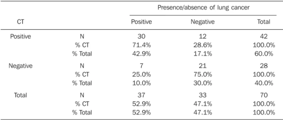
Tabela 4 Relation between CT results and the presence/absence of lung cancer (χ2 = 14.533; gl = 1; p = 0; kappa = 0.451).
Presence/absence of lung cancer
CT Positive Negative Total N % CT % Total N % CT % Total N % CT % Total Positive 30 71.4% 42.9% 7 25.0% 10.0% 37 52.9% 52.9% Negative 12 28.6% 17.1% 21 75.0% 30.0% 33 47.1% 47.1% Total 42 100.0% 60.0% 28 100.0% 40.0% 70 100.0% 100.0% The authors observed that, among the
37 positive bronchoscopic results, 31 (83.8%) corresponded to a positive diagno-sis of cancer, and 6 (16.2%) to a negative diagnosis of the disease. As regards the 33 negative bronchoscopic results, 27 (81.8%) really corresponded to a negative diagno-sis of lung cancer, and 6 (18.2%) to a posi-tive diagnosis of cancer (Table 3). In these 6 cases, the final diagnosis was achieved by means of transthoracic biopsy in 4 cases, and surgical biopsy in 2 cases. In the present study, bronchoscopy sensitivity was of 83.8%, specificity, 81.8%, and accuracy, 82.8%. False-positive results corresponded to 18.2%, and false-negative results, to 16.2%.
Relation between CT results and presence/absence of lung cancer
The statistical analysis demonstrated that the relation between CT results and presence/absence of lung cancer was sta-tistically significant (p < 0.05). Kappa value was of 0.451, corresponding to a weak agreement between results.
The authors observed that among the 42 CT studies interpreted as positive, 30 (71.4%) corresponded to a positive diagno-sis of lung cancer, and 12 (28.6%) corre-sponded to a negative diagnosis of the dis-ease. As regards the 28 tomographic stud-ies interpreted as negative, 21 (75%) really corresponded to absence of lung cancer, while 7 (25%) ended up demonstrating the presence of disease (Table 4). In these 7 cases, the final diagnosis was achieved by means of bronchial biopsy in 6 cases, and by means of transthoracic biopsy in 1 case. In the present study, the CT sensitivity was of 81.1%, specificity, 63.6%, and accuracy, 72.8%. False-positive results corresponded to 36.4% and false-negative results, to 18.9%.
DISCUSSION
In the present investigation, a small number of patients only 8.1% of cases -was submitted to surgery, while chemo-therapy - with 75.7% of cases - was the most utilized therapeutics. The medical lit-erature clearly describes the surgical therapy as critical in the treatment of lung cancer(29). However, studies report a small
number of patients submitted to surgery and demonstrate that resectability criteria are met in only 20% of cases at the time of diagnosis(23,24,30).
The situations observed regarding both the staging (predominance of most ad-vanced stages of disease) and therapeutic options (small number of patients submit-ted to surgery) demonstrates that the late diagnosis in most of cases still poses a chal-lenge, considering the difficulty in the achievement of an early diagnosis.
Bronchoscopy is thought to be funda-mental in the diagnosis of lung cancer and also in the staging and follow-up of the dis-ease(14,31,32). This rating found in studies in
the literature is corroborated by the results of the present study (sensitivity, 83.8%; specificity, 81.8%; accuracy, 82.8%).
Currently, new advances in techniques associated with bronchoscopy have al-lowed developments to improve the diag-nosis of lung cancer(31,33). The literature
indicates that in more than 70% of cases, carcinomas can be detected by bronchos-copy, and the performance of such method is enhanced, provided it is associated with bronchial biopsy, bronchial wash and brush cytology(13,29,31,34).
The false-negative results (16.2%) of bronchoscopy are due to the presence of peripheral lesions, particularly the smaller ones, because of the poor role played by bronchoscopy in these cases, corroborating data in the literature(15,35).
Imaging methods represent an essential diagnostic tool in the evaluation of lung cancer(36). Computed Tomography plays a
relevant role in the diagnosis of lung can-cer, as corroborated by studies in the litera-ture(36–38).
In the present study, CT presented sen-sitivity of 81.1% corroborating reports in the reference literature demonstrating that as lung cancer is detected by radiological methods, ¾ of the natural history of the disease were already completed(38,39).
The specificity is a result of the number of false-positive results (36.4%). This situ-ation demonstrated by the studies refer-enced herein is due to the fact that radio-logical findings suggestive of lung cancer (leading to the rating of a CT study as posi-tive) such as parenchymal mass with spicu-lated margins, microlobulations, thick-walled cavities, cavitary nodules and chest wall invasion may be observed in other dis-eases such as infections, pulmonary inflam-matory processes, infarction and lung ab-scesses(40,41).
CONCLUSION
Lung cancer diagnosis is essentially achieved by CT and bronchoscopic tech-niques. The association of bronchoscopy and CT is useful in the accurate diagnosis of lung cancer, since the occurrence of false-positive results of CT is minimized, improving the specificity of the method. On the other hand, the utilization of CT to detect the presence of peripheral lesions, which increase the incidence of false-nega-tive results of bronchoscopy, allows a bet-ter management of patients, increasing the diagnostic accuracy.
The association of these two methods, besides the discussion between pneumolo-gists and radiolopneumolo-gists constitute the best approach for lung cancer patients.
Acknowledgements
The authors would like to thank Hospi-tal DistriHospi-tal da Figueira da Foz, the Unit of Pneumology, particularly Dr. António An-tunes, Director for the mentioned Unit and Dr. Manuela Lopes, for the collaboration and willingness.
REFERENCES
1. Agarwal A, Ghotekar LH, Garbyal RS, et al. Evalu-ation of pulmonary malignancies in Kathmandu Valley and role of bronchoscopic techniques in di-agnosis of such cases. JIACM. 2003;4:127–33. 2. Figueiredo L, Bento MT. Neoplasia do pulmão.
In: Pisco JM, Sousa LA. Noções fundamentais de imagiologia. Lisboa: Lidel-Edições Técnicas; 1999. p. 195–209.
3. Alves AF, Silva MC. Cancro do pulmão no Ser-viço de Pneumologia do Hospital de Santarém: resultados de 4 anos (2003-2006). HDS InForma. 2008;(22).
4. Sotto-Mayor R. O lugar da quimioterapia na te-rapêutica do cancro do pulmão. Rev Port Pneu-mol. 2001;VII:558–94.
5. Barata F. O que o doente deve saber sobre o can-cro do pulmão. [acessado em 10 de outubro de 2008]. Disponível em: http://www. sppneumologia.pt/textos/?imc=51n81n 6. Uehara C, Jamnik S, Santoro IL. Câncer de
pul-mão. Medicina, Ribeirão Preto. 1998;31:266–76. 7. Beckles MA, Spiro SG, Colice GL, et al. Initial evaluation of the patient with lung cancer: symp-toms, signs, laboratory tests and paraneoplastic syndromes. Chest. 2003;123(1 Suppl);97S– 104S.
8. Kamath A, Chhajed PN. Role of bronchoscopy in the early diagnosis of lung cancer. Indian J Chest Dis Allied Sci. 2006;48:265–9.
9. Carvalho L. A nova classificação de tumores pul-monares – interesse meramente académico? Rev Port Pneumol. 2004;X(1 Supl 1):S9–13. 10. Westeel V. Diagnosis of lung cancer. Rev Prat.
2003;53:727–34.
11. Barcellos MG. Radiologia do câncer de pulmão. J Pneumol. 2002;28:94–9.
12. Cordeiro AJA Robalo. Pneumologia fundamen-tal. Lisboa: Fundação Calouste Gulbenkian; 1995. p. 349–73.
13. Herth FJ, Eberhardt R, Ernst A. The future of bronchoscopy in diagnosing, staging and treat-ment of lung cancer. Respiration. 2006;73:399– 409.
14. El-Bayoumi E, Silvestri GA. Bronchoscopy for the diagnosis and staging of lung cancer. Semin Respir Crit Care Med. 2008;29:261–70. 15. Rivera MP, Detterbeck F, Mehta AC; American
College of Chest Physicians. Diagnosis of lung cancer: the guidelines. Chest. 2003;123(1 Suppl 1):129S–36S.
16. Gaur DS, Thapliyal NC, Kishore S, et al. Efficacy of broncho-alveolar lavage and bronchial brush cytology in diagnosing lung cancers. J Cytol. 2007;24:73–7.
17. Cook RM, Miller YE. Flexible fiberoptic bron-choscopy in the diagnosis and staging of lung can-cer. In: Johnson EB, Johnson HD, editors. Lung cancer. New York, NY: Wiley-Lyss; 1995. p. 123– 44.
18. Wynants J, Stroobants S, Dooms C, et al. Stag-ing of lung cancer. Radiol Clin North Am. 2007;45:609–25.
19. Queiroga H. Factores de prognóstico “clássicos” no cancro do pulmão. Rev Port Pneumol. 2005;XI(3 Supl 1):S105–11.
20. Sá Vieira V. Cirurgia no doente com cancro do pulmão. [acessado em 10 de outubro de 2008]. Disponível em: http://www.sppneumologia.pt/ textos/?imc=51n81n
21. Provencio M, Camps C, Alberola V, et al. Lung cancer and treatment in elderly patients: the Achilles Study. Lung Cancer. 2009;66:103–6. 22. Barros JA, Valladares G, Faria AR, et al.
Diagnós-tico precoce do câncer de pulmão: o grande de-safio. Variáveis epidemiológicas e clínicas, esta-diamento e tratamento. J Bras Pneumol. 2006; 32:221–7.
23. Novaes FT, Cataneo DC, Ruiz Junior RL, et al. Câncer de pulmão: histologia, estádio, tratamento e sobrevida. J Bras Pneumol. 2008;34:595–60. 24. Westphal FL, Lima LC, Andrade EO, et al.
25. van Zandwijk N. Aetiology and prevention of lung cancer. Eur Respir Mon. 2001;17:13–33. 26. Stinchcombe TE, Socinski MA. Current
treat-ments for advanced stage non-small cell lung cancer. Proc Am Thorac Soc. 2009;6:233–41. 27. Kerr KM. Pulmonary adenocarcinomas:
classifi-cation and reporting. Histopathology. 2009;54: 12–27.
28. Wang T, Nelson RA, Bogardus A, et al. Five-year lung cancer survival: which advanced stage nonsmall cell lung cancer patients attain long-term survival? Cancer. 2010;116:1518–25. 29. Yasufuku K. Early diagnosis of lung cancer. Clin
Chest Med. 2010;31:39–47.
30. Thomas PA. Standards of surgery in lung cancer. Rev Prat. 2009;59:934–8.
31. Simon M, Simon I. Update in bronchoscopic tech-niques. Pneumologia. 2010;59:53–6.
32. Brandão DS, Haddad R, Marsico GA, et al. As-pectos clínico-patológicos do carcinoma bron-quioloalveolar e sobrevida em pacientes no es-tágio clínico I. J Bras Pneumol. 2010;36:167–74. 33. Herth FJ, Eberhardt R. Flexible bronchoscopy and its role in the staging of non-small cell lung can-cer. Clin Chest Med. 2010;31:87–100. 34. Fernandez A, Jatene FB, Zamboni M. Diagnóstico
e estadiamento do câncer de pulmão. J Pneumol. 2002;28:219–28.
35. Devbhandari MP, Quennell P, Krysiak P, et al. Implications of a negative bronchoscopy on wait-ing times to treatment for lung cancer patients: results of a prospective tracking study. Eur J Cardiothorac Surg. 2008;34:479–83.
36. Hansell DM, Boiselle PM, Goldin J, et al. Tho-racic imaging. Respirology. 2010;15:393–400. 37. Thiberville L, Salaun M, Lachkar S. New
diag-nostic tools in lung cancers. Rev Prat. 2009;59: 925–31.
38. Jiang F, Todd NW, Qiu Q, et al. Combined genetic analysis of sputum and computed tomography for noninvasive diagnosis of non-small-cell lung can-cer. Lung Cancan-cer. 2009;66:58–63.
39. Scagliotti G. Symptoms, signs and staging of lung cancer. Eur Respir Mon. 2001;17:86–119. 40. Madhusudhan KS, Gamanagatti S, Seith A, et al.
Pulmonary infections mimicking cancer: report of four cases. Singapore Med J. 2007;48:e327– 31.