Engineering More Stable, Selectable
Marker-Free Autoluminescent Mycobacteria by One
Step
Feng Yang1☯, Moses M. Njire1☯, Jia Liu1☯, Tian Wu1, Bangxing Wang1, Tianzhou Liu1,
Yuanyuan Cao1, Zhiyong Liu1, Junting Wan1, Zhengchao Tu1, Yaoju Tan2, Shouyong Tan2, Tianyu Zhang1*
1State Key Laboratory of Respiratory Disease, Guangzhou Institutes of Biomedicine and Health, Chinese Academy of Sciences, Guangzhou, Guangdong, China,2State Key Laboratory of Respiratory Disease, Department of Clinical Laboratory, The Guangzhou Chest Hospital, Guangzhou, Guangdong, China
☯These authors contributed equally to this work. *[email protected]
Abstract
In our previous study, we demonstrated that the use of the autoluminescentMycobacterium tuberculosisas a reporter strain had the potential to drastically reduce the time, effort, ani-mals and costs consumed in evaluation of the activities of drugs and vaccines in live mice. However, the strains were relatively unstable and lost reporter with time without selection. The kanamycin selection marker used wasn’t the best choice as it provides resistance to amino glycosides which are an important class of second line drugs used in tuberculosis treatment. In addition, the marker could limit utility of the strains for screening of new poten-tial drugs or evaluating drug combinations for tuberculosis treatment. Limited selection marker genes for mycobacterial genetic manipulation is a major drawback for such a mark-er-containing strain in many research fields. Therefore, selectable marker-free, more stable autoluminescent mycobacteria are highly needed. After trying several strategies, we creat-ed such mycobacterial strains successfully by using an integrative vector and removing both the resistance maker and integrase genes by Xer site-specific recombination in one step. The corresponding plasmid vectors developed in this study could be very convenient in constructing other selectable marker-free, more stable reporter mycobacteria with diverse applications.
Introduction
Many severe bacterial diseases, such as tuberculosis (TB), leprosy and Buruli ulcers are caused by mycobacteria. For example, TB, an infectious disease caused byMycobacterium tuberculosis
(MTB), is one of the greatest single infectious diseases causing morbidity and death in the world. The only TB vaccine in use for over 90 years,Mycobacterium bovisBCG (BCG), has very limited protection efficacy in older children and adults. The 9.0 million incident cases of
OPEN ACCESS
Citation:Yang F, Njire MM, Liu J, Wu T, Wang B, Liu T, et al. (2015) Engineering More Stable, Selectable Marker-Free Autoluminescent Mycobacteria by One Step. PLoS ONE 10(3): e0119341. doi:10.1371/ journal.pone.0119341
Academic Editor:Riccardo Manganelli, University of Padova, Medical School, ITALY
Received:October 29, 2014
Accepted:January 5, 2015
Published:March 11, 2015
Copyright:© 2015 Yang et al. This is an open access article distributed under the terms of the
Creative Commons Attribution License, which permits unrestricted use, distribution, and reproduction in any medium, provided the original author and source are credited.
Data Availability Statement:All relevant data are within the paper and its Supporting Information files
Funding:This work was supported by the Chinese Academy of Sciences 'One Hundred Talents Program' (Category A, to TZ), the National Great Research Program of China (2013ZX10003006) and the Key Program of the Chinese Academy of Sciences (KJZD-EW-L02). The funders had no role in study design, data collection and analysis, decision to publish, or preparation of the manuscript.
TB, 1.5 million deaths from TB patients in 2013 alone [1], and the appearance of multi drug-re-sistant (MDR) [2,3], extensively drug-resistant (XDR) [2,3] and even totally drug-resistant (TDR) TB [4] presents a striking reminder of the magnitude of destruction caused by TB. All these indicate that new, more effective drugs and vaccines are urgently needed.
Routine drug susceptibility testing for MTB depends on a positive culture for diagnosis after which a drug susceptibility test is performed which usually takes 3–6 weeks [5]. The slow diag-nosis and in some cases inaccurate or false negative phenotypic results [6], is a major contribu-tor to the current drug resistant epidemic and hindrance to mycobacterial research. The Buruli ulcers causing pathogen,Mycobacterium ulcerans, grows even much slower as 3 months are needed for counting the visible colonies after plating. The necessity to work under stringent biosafety level 3-containment also makes studies of MTB very expensive, especially for long-term use facilities. Therefore, the lack of an effective, rapid, reliable and inexpensive reporter strain in TB research, especially forin vivostudies, is a major drawback.
In our previous studies [7,8], we constructed autoluminescent MTB andMycobacterium ulceransas reporter strains which expressed theluxCDABEoperon fromPhotorhabdus lumi-nescens[9]. The operon encodes enzymes for both light production and for recycling reaction substrates. Therefore, use of the autoluminescent reporter strains for testing drugs does not need the addition of an exogenous substrate. The same samples can be monitored in real time, and the colony forming units (CFU) and the light intensity (relative light unit, RLUs) correlate very well. Use of this system demonstrates the potential to drastically reduce the time, effort, animals and costs consumed in evaluation of the activities of drugs and vaccines in live mice as it only takes 3 seconds to detect light in live mouse using an inexpensive device [7,8]. The auto-luminescent strains created have been proved to be essentially as virulent as their wild-type parent strains and the drug susceptibilities including for aminoglycosides such as streptomycin are not affected except for kanamycin (KAN) which was used as a selection marker. These properties make the reporter strains appealing for testing drug activity bothin vitroandin vivo
as only very small amount of samples, a few mice and short time are needed to infer the activity of a compound with very good reproducibility and no addition of exogenous substrate. Howev-er, the strains are relatively unstable probably as a result of excision of theluxCDABEoperon by the L5 mycobacteriophage integrase at a very low rate [10,7]. This assumption was recently approved in a similar study describing the construction of a recombinant MTB expressing fire-fly luciferase gene in an integrative plasmid with the integrase gene removed [11]. In addition, the selection marker used could have been inappropriate for molecular genetic manipulation [12], screening of potential drug combinations and testing therapeutic regimens containing KANin vivodue to possible cross drug resistance. The limited antibiotic resistance markers for mycobacterial genetic manipulation pose a serious challenge, and therefore, development of se-lectable marker-free, more stable, autoluminescent mycobacteria is highly needful.
The strategies for construction of selectable marker-free mycobacterial strains are summa-rized in our recently published report [12]. Herein, we tried 2 main strategies for constructing selectable marker-free mycobacteria. The antibiotic resistance cassette flanked by two short DNA sequences in direct orientation could possibly be recognized and removed either by the exogenous resolvase or the endogenous mycobacterial recombinases XerCD. The integrase gene also needed to be removed to make the autoluminescent mycobacteria more stable. We fi-nally succeeded using an integrative plasmid expressing the naturalluxCDABEoperon from
Photorhabdus luminescens[9] at the downstream ofHsp60promoter [13]. The L5 integrase gene (int) and hygromycin (HYG)-resistant gene in the same cassette were resolved by the en-dogenous XerC and XerD recombinases [14] using our recently published system [12]. The se-lectable marker-free strains were proved to be more stable than the previously reported ones
for patent (ZL 201210183007.2) and authorized. The patent title is "The plasmid pOPHI and construction of unmarked, more stable autoluminescent
and could be widely used in anti-mycobacterial drug screening and evaluation. Additionally, their derivative strains could possibly be used widely in many research fields of mycobacteria.
Materials and Methods
Bacterial strains (
Table 1
) and culture media
Escherichia colistrain DH5α[15] and the corresponding transformants were grown at 37°C in Luria-Bertani (LB) broth or on agar containing KAN (Invitrogen), ampicillin (Sigma-Aldrich, USA) or HYG (Roche Diagnostics, Switzerland) at final concentrations (μg/ml) of 40, 100 and
200, respectively. MTB H37Rv [16], MTB H37Ra [17] andMycobacterium bovisBCG Tice (BCG) [18,19] were grown at 37°C in Middlebrook 7H9 broth (Becton Dickinson, USA) sup-plemented with 10% oleic acid albumin dextrose catalase (OADC, Becton Dickinson, USA) and 0.05% Tween80 where indicated, or on 7H11 agar supplemented with OADC.M. smegma-tismc2155 (MSM) [20] was grown in LB broth or on LB agar or Middlebrook 7H11 agar (Difco) supplemented with albumin dextrose catalase at 37°C. KAN, HYG, carbenicillin and cycloheximide were added to agar when required to final concentrations (μg/ml) of 40, 50, 50
and 10 respectively for MTB and BCG, and the same concentrations for MSM except for HYG 150. The concentrations (μg/ml) in liquid broth were KAN 20 and HYG 100 for MSM and
HYG 10 for MTB and BCG.
Table 1. Bacterial strains in this study.
Strains Relevant characteristic(s) Source or
reference
E.coliDH5α General-purpose cloning strain; F-[φ80dlacZ
ΔM15]ΔD(lacZYA-argF)U169 deoR recA1 endA1 hsdR17 glnV44 thi-1 gyrA96 relA
[11]
M.smegmatis
mc2155 Highly transformable derivative of ATCC 607 [16]
MSM-OHP MSM cotransformed with pOHP and pInt This study
MSM-OHP MSM cotransformed with pOHP and pInt This study
AlMSMT1 MSM containing pOHIhd This study
AlMSMT2 MSM containing pOPHI This study
UAlMSM Selectable marker-free autoluminescent MSM This study
M.tuberculosis H37Rv
Widely used virulent laboratory MTB strain, ATCC 27294 [12]
AlRv Autoluminescent MTB H37Rv resistant to KAN [7]
AlRvT1 MTB H37Rv::pOHIhd, MTB H37Rv containing pOHIhd This study
AlRvT2 MTB H37Rv::pOPHI MTB, H37Rv containing pOPHI This study
UAlRv Selectable marker-free autoluminescent MTB H37Rv This study
M.tuberculosis H37Ra
Widely used avirulent laboratory MTB strain, ATCC25177 [13]
AlRaT2 MTB H37Ra::pOPHI MTB, H37Ra containing pOPHI This study
UAlRa Selectable marker-free autoluminescent MTB H37Ra This study
M.bovisBCG Tice The live attenuated TB vaccine [15]
AlBCGT2 BCG::pOPHI, BCG containing pOPHI This study
UABCG Selectable marker-free autoluminescent BCG This study
ATCC: The American Type Culture Collection.
General DNA techniques
For polymerase chain reaction (PCR) amplification reactions were performed withpfuDNA polymerase (Takara) and 5% DMSO was added due to the high G+C content of the mycobacte-rial genomes. The PCR products were analyzed by electrophoresis in agarose gels and purified using a DNA gel extraction kit (Bioflux). Plasmids were also extracted and purified using kits from the same company. Purified PCR products, plasmids or plasmids transformed intoE.coli
strains were sequenced at BGI, Shenzhen, China. MSM was transformed as previously de-scribed [20], while MTB and BCG were transformed as previously described [21] with some modifications. The competent MTB and BCG cells were first incubated at 37°C for 10 min be-fore electroporation, and transformation was performed at room temperature. The genomic mycobacterial DNA was extracted using the CTAB method as previously described [15].
Construction of marker-free autoluminescent mycobacteria using the
endogenous Xer recombinase system
This strategy was designed to deliver theHsp60-luxCDABEinto the mycobacterial genome by an integrative plasmid. TheHyggene could then either be removed by the endogenous Xer recombinase system to form selectable marker-free strains (plasmid pOHIhd,Table 2,Fig. 1), or alternatively, bothHyg+intgenes could be removed together by the same system to form se-lectable marker-free and more stable strains (plasmid pOPHI,Table 2,Fig. 1). In addition, we co-transformed the suicide plasmid pInt containing theintgene (Fig. 1,Table 2) with plasmid pOHP containing theattPsite, theluxCDABE(under the regulation of the strongHsp60 pro-moter,Hsp60-luxCDABE) [15], and thedif-OHYG-difcassette (Fig. 1,Table 2).
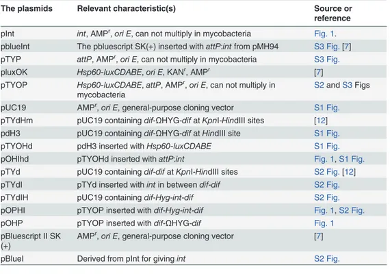
Table 2. Plasmids in this study.
The plasmids Relevant characteristic(s) Source or
reference
pInt int, AMPr,ori E, can not multiply in mycobacteria Fig. 1. pblueInt The pbluescript SK(+) inserted withattP:intfrom pMH94 S3 Fig.[7] pTYP attP, AMPr,ori E, can not multiply in mycobacteria S3 Fig.
pluxOK Hsp60-luxCDABE,ori E, KANr, AMPr [7]
pTYOP Hsp60-luxCDABE,attP, AMPr,ori E, can not multiply in mycobacteria
S2andS3Figs
pUC19 AMPr,ori E, general-purpose cloning vector S1 Fig. pTYdHm pUC19 containingdif-ΩHYG-difatKpnI-HindIII sites [12] pdH3 pUC19 containingdif-ΩHYG-difatHindIII site S1 Fig.
pTYOHd pdH3 inserted withHsp60-luxCDABE S1 Fig.
pOHIhd pTYOHd inserted withattP:int Fig. 1,S1 Fig.
pTYd pUC19 containingdif-difatKpnI-HindIII sites S2 Fig.[12] pTYdI pTYd inserted withintin betweendif-dif S2 Fig.
pTYdIH pUC19 containingdif-Hyg-int-dif S2 Fig.
pOPHI pTYOP inserted withdif-Hyg-int-dif Fig. 1,S2 Fig.
pOHP pTYOP inserted withdif-ΩHYG-dif Fig. 1
pBluescript II SK (+)
AMPr,ori E, general-purpose cloning vector [7]
pBlueI Derived from pInt for givingint S2 Fig.
AMP: ampicillin; KAN: kanamycin; HYG: hygromycin.
To construct plasmid pOHIhd, thedif-OHYG-difcassette was excised withHindIII from plasmid pTYdHm (Table 2) [12] and inserted into plasmid pUC19 (Table 2) digested with the same enzyme to form pdH3 (Table 2,S1 Fig.). TheKpnI-Hsp60-luxCDABE-PstI was then ex-cised from pluxOK (Table 2) [7] and inserted into pdH3 (Table 2) digested with the same en-zymes to form pTYOHd (S1 Fig.,Table 2). TheattP:Intexcised withKpnI andSmaI from pblueInt (S1 Fig.,Table 2) [7] was then inserted into pTYOHd digested withKpnI andScaI to form pOHIhd (S1 Fig.andFig. 1,Table 2). To construct the plasmid pOPHI (Table 2), theint
gene, amplified with primers Intf and Intr (Table 3) from the plasmid pblueInt (Table 2), was excised withXbaI andClaI, inserted into the common plasmid pBluescript II SK(+) to form pBlueI (Table 2), and then sequenced. Theintgene in pBlueI was then excised withXbaI and
ClaI and inserted into pTYd [12] to form pTYdI (S2 Fig.,Table 2). The optimizedHyggene [12] in pTYdHm (Table 2) was cut withXbaI and then inserted into theXbaI site of pTYdI to form pTYdIH (S2 Fig.,Table 2). The direction of theHyggene was verified by restriction map-ping analysis. Thedif-OHYG-int-difcassette was then excised from pTYdIH and inserted into pTYOP to form pOPHI (S2 Fig.andFig. 1,Table 2). To construct plasmid pTYOP, the plasmid pblueInt was digested withPstI and self-ligated to form pTYP (S3 Fig.,Table 2) withintgene removed. TheHsp60-luxCDABEexcised from pluxOK (Table 2) [7] withKpnI-XhoI was in-serted into pTYP to give pTYOP (S3 Fig.).Thedif-OHYG-difcassette was excised from the plasmid pTYd constructed in our previous work (Table 2) [12] withXhoI and inserted into the same site of pTYOP to give pOHP (Fig. 1,Table 2).
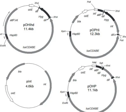
Fig 1. The plasmids constructed in this study for transforming into mycobacteria to create unmarked autoluminescent mycobacteria.oriE, origin region ofE.coli;Hsp60, the strong mycobacterial promoter; luxCDABE, the operon for producing autoluminescence;bla, ampicillin resistance gene;Kan, KAN resistance gene;res, the transposonγδresolvase action site;attP, mycobacteriophage L5 attachment site;int, integrase gene;int’, the remaining part of integrase gene;attB, attachment site from the mycobacterial genome corresponding toattP;oriM, origin region of mycobacteria;Hyg, HYG resistance gene;dif, the recombinases XerCD action site.
Construction and verification of the target autoluminescent strains
Mycobacteria transformed with pOHIhd or pOPHI or co-transformed with pInt and pOHP
(Table 2) were selected on HYG-containing plates. The autoluminescent mycobacterial
colo-nies with deleted HYG-resistant gene were selected by streaking them in the presence and ab-sence of HYG after several passages in plain 7H9 broth and further tested by PCR using appropriate primers (Table 3).The primer pair Hyg0702-f and Hyg0702-r was for testing the loss of theHyggene; Int0702-f and Int0702-r for testing the loss of theintgene; while noHI-f (corresponding to 170 bp from the end ofluxEgene) and noHI-r (corresponding to the end of
attPnearluxCDABEin plasmid pTYOP) was for testing the loss of bothHygandintgenes. A 579-bp fragment was expected from amplification of theHyggene open reading frame with Hyg0702-f and Hyg0702-r; a 586-bp fragment fromintwith Int0702-f and Int0702-r; and a 367-bp fragment from the genome of MTB::pOPHI with deleteddif-OHYG-int-difusing prim-ers noHI-f and noHI-r. Three randomly selected MTB H37Rv::pOHIhd colonies with lost HYG resistance gene were amplified with primers attB1210f and attB1210r (Table 3) and se-quenced to verify if the whole pOHIhd (Table 2) plasmid had been lost in these strains. Three MSM colonies co-transformed with pInt and pOHP (Table 2) were verified further by amplifi-cation with primers luxAB-f and luxAB-r (Table 3), and the expected PCR product was 750bp. The bioluminescence of the autoluminescent MSM/BCG/MTB H37Ra transformants was de-tected by GloMax 20/20 Luminometer (Promega) while for the autoluminescent MTB H37Rv transformants was detected by Orion II Microplate Luminometer (Titertek-Berthold).
Testing the stability of the selectable marker-free autoluminescent MSM,
BCG and MTB
Three single colonies of selectable marker-free autoluminescent MSM, BCG and MTB H37Rv were separately inoculated into 30 mL 7H9 medium and incubated at 37°C with shaking until the OD600reached over 0.7. An aliquot of 0.3 ml of the culture was then sub-cultured into 30 mL 7H9 medium under the same conditions. An appropriate dilution of the broth culture was obtained after several passages and then plated on plain 7H11 plates. The RLUs of approxi-mately 200 individual colonies picked up at each time point was detected using the above men-tioned luminometers. The proportion of autoluminescent colonies was then calculated as: the No. of positive colonies/the total number of colonies detected×100%. If>99% colonies were Table 3. DNA primers used in this study.
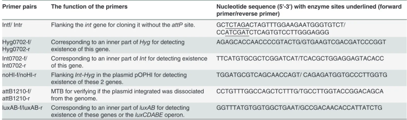
Primer pairs The function of the primers Nucleotide sequence (5'-3') with enzyme sites underlined (forward primer/reverse primer)
Intf/ Intr Flanking theintgene for cloning it without theattPsite. GCTCTAGACTAGTTTGGAAGAATGGGTGTCT/ CCATCGATCTCAGTGTCCTTGGGAGGG Hyg0702-f/
Hyg0702-r
Corresponding to an inner part ofHygfor detecting existence of this gene.
AGAGCACCAACCCCGTACTG/GTGAAGTCGACGATCCCGGT
Int0702-f/ Int0702-r
Corresponding to an inner part ofIntfor detecting existence of this gene.
TTCATGTGCGCTCGGATCAT/TCACGCTGGAGGAGTACACC
noHI-f/noHI-r FlankingInt-Hygin the plasmid pOPHI for detecting existence of these 2 genes.
TGGATGCGTCAGCAACCAGT/ CAGAGATGGTGCCCTTGGTG
attB1210-f/ attB1210-r
MTB for verifying if the plasmid integrated was dissociated from the genome.
CCTGTTTGGCCAGCTCTTTG/TGCCTTGGTACCGGACAGCA
luxAB-f/luxAB-r Corresponding to an inner part ofluxABfor detecting existence of these genes or theluxCDABEoperon.
GGTTTATGTGGTGGCTGAAT/GCCGACAACACCATTATCTG
still autoluminescent after 3 passages (~20 generations), this indicated that the strain was very stable.
Results
Construction of marker-free autoluminescent mycobacteria
We endeavored to create selectable marker-free mycobacteria by removing the resistance marker using the exogenous resolvase or the endogenous mycobacterial recombinases XerCD. In the first strategy, target strains were to be created by integrating theHsp60-luxCDABEand theres-OKAN-rescassette containing plasmids into the genomicattBsite withintgene in a separate plasmid. This was to be followed by the removal of the KAN resistance maker gene by the tnpR from resolvase of transposonγδsystem, and subsequent removal of the plasmid ex-pressing the resolvase [22]. Even though thistnp/ressystem had been proved successful in MSM [22], it was unsuccessful in this study using autoluminescent MSM and therefore we did not proceed with it using MTB.
On the other hand, we succeeded using the second strategy in which the target selectable marker-free autoluminescent strains were constructed by integrating theHsp60-luxCDABE
into the genome, followed by the removal of the resistance gene together with theintgene by the endogenous recombinases XerC and XerD [14].
Both MSM and MTB H37Rv were transformed with pOHIhd or pOPHI successfully (Fig. 1,
Table 2). Thereafter, BCG and MTB H37Ra were also transformed with pOPHI successfully.
All transformants colonies were verified further by detecting bioluminescence. We co-trans-formed pInt and pOHP (Fig. 1,Table 2) into MSM successfully and obtained MSM-OHP
(Table 1). However, none of them was bioluminescent. We therefore verified by PCR if the
lux-CDABEandHygfragments had been integrated into the MSM genome using primer pairs luxAB-f and luxAB-r (750-bp band), and Hyg0702-f and Hyg0702-r (579-bp band),
respective-ly (Table 3). All the 3 randomly selected MSM-OHP colonies gave right sized bands, which
meant that the plasmid pOHP (Table 2) had been integrated into the MSM genome.
Counter-selection of the selectable marker-free autoluminescent
mycobacteria
The selectable marker-free autoluminescent mycobacterial colonies with HYG-resistant gene rescued were screened by passing the corresponding parent strains several times in antibiotic-free broth culture, testing HYG susceptibility and the autoluminescence of each individual col-ony. For MSM transformants containing pOHIhd (Fig. 1,Table 2) and designated as
AlMSMT1 (Table 1), 90% colonies did not grow on HYG-containing plates anymore after just one passage, and had also lost their autoluminescence. No selectable marker-free autolumines-cent MSM was obtained through this technique route after multiple attempts. A similar phe-nomenon was observed in MTB H37Rv strain transformed with the same plasmid and designated as AlRvT1 (Table 1).
MSM transformed with pOPHI and designated as AlMSMT2 (Table 1) was passed twice in drug-free 7H9 broth and plated on plain agar. 56% colonies lost their HYG resistance and were still autoluminescent and one representative strain was designated as UAlMSM (Table 1). MTB H37Rv transformed with pOPHI was designated as AlRvT2 (Table 2), and all 200 AlRvT2 colonies had lost the HYG resistance and were also still autoluminescent after just one passage. One representative selectable marker-free autoluminescent MTB H37Rv strain was designated as UAlRv (Table 1). Similarly, we obtained AlRaT2 and AlBCGT2 by transforming MTB H37Ra and BCG respectively with pOPHI (Table 2) and the corresponding selectable marker-free autoluminescent UAlRa and UABCG (Fig. 1,Table 1). Two randomly selected UAlMSM, UABCG (Fig. 2), UAlRa and UAlRv colonies (Table 1) were verified further for the loss of theHygandintgenes by PCR using 3 primer pairs (Table 3): Hyg0702-f and Hyg0702-r for detecting loss ofHyg, Int0702-f and Int0702-r for detecting loss ofint, and noHI-f and noHI-r for detecting the loss of bothHygandintgenes. As expected, no right sized PCR prod-ucts were obtained using the first 2 primer pairs and a 367-bp fragment was obtained using the primers noHI-f and noHI-r (S5 Fig.). Sequence analysis showed that the randomly selected tar-get band from UAlRv1 (lane 2) was the same as deduced from AlRvT2 genome with the
dif-OHYG-int-difcassette lost (Table 1). At last, we obtained the selectable marker-free
autolumi-nescent MSM, BCG and MTB H37Rv (Table 1) withintgene lost using pOPHI. The selectable marker-free autoluminescent mycobacterial colonies were visible with naked eyes in a dark room and could be imaged using a normal camera.
Stability of the selectable marker-free autoluminescent mycobacteria
The stability of UAlMSM, UAlRa, UAlRv and UABCG was tested. For UAlMSM,>99% colo-nies were still autoluminescent after 6 passage (~40 generations) in about 4 weeks, which im-plied that the strain was very stable. About 100% (at least more than 99%) of the UAlRv and UAIRa colonies were still strongly autoluminescent after 2 (about 1 month, (~15 generations) and 5 (about 94 days,>35 generations) passages respectively. The UABCG was also very stable as it retained autoluminescence after several passages within 3 months.
Discussion
Studies in MTB and other mycobacteria, and especially the discovery of new anti-mycobacteri-al drugs and the mechanism of drug action are heavily hampered by their slow growth and the need of expensive biosafety laboratory at higher levels. Rapid, convenient, inexpensive and sen-sitive reporter strains would facilitate such studies in mycobacteria. We previously demonstrat-ed that a very sensitive autoluminescent MTB grew as fast and was as virulent as its parent strain, in which the RLUs produced by this strain accurately correlated with the CFU counts. The strain could not only be usedin vitrofor rapid evaluation but alsoin vivofor rapid drug and even vaccine testing noninvasively using the same batch of live mice in a larger scale [7]. Fig 2. Photograph of the more stable, selectable marker-free, autoluminescent BCG (UABCG).
However, there were 2 deficiencies in the autoluminescent strain. Firstly, the strain was not sta-ble, which would limit its applications, and secondly, it contained a KAN resistance marker gene, which further limited its utility in many fields, especially for MTB which has onlyHyg
and KAN resistance markers [12].
In this study, we demonstrate for the first time the successful construction of selectable marker-free autoluminescent mycobacteria including MTB, BCG and MSM (Table 1). More importantly, all the target strains were extremely stable. For example, 100% (at least>99%) of randomly selected individual colonies of the UAlRv were still autoluminescent after 5 passages with>35 generations in 94 days comprising both log phase and stationary phase culture. In contrast, only 95.7% colonies of the KAN-resistant autoluminescent AlRv (Table 1) were auto-luminescent after 37 days ofin vitrogrowth in broth without passage [7]. These results were ac-cordant with a previous study describing the construction of recombinant MTB expressing firefly luciferase gene in which the strains whoseintgene was removed were more stable than those whoseintgene was not removed [11]. One limitation of this study is that we did not test stability under diverse growth conditions, such as, low pH, macrophage infection model, non-replicating persistence as well as infection animal models. The macrophage infection model can not last for a very long time (usually within 14 days), and no loss of bioluminescence be-cause of instability was observed in our study thus indicating sufficient stability of our strain in the model. The integration of the transforming plasmid into the genomes of mycobacteria and subsequent removal of the integrase gene which excises the plasmid at a very low rate contrib-uted to the stability of the target strains in this study. However, further stability testing of such mycobacterial strains under the above diverse conditions would be needful.
Thedif-OHYG-int-difcassette could be widely used in constructing selectable marker-free and more stable recombinant MTB or BCG strains in just one transformation step, such as BCG-based vaccines and recombinant MTB reporter strains.
Previously, no autoluminescent mycobacteria were successfully constructed using extra-chromosomal plasmids as delivery vectors [7]. Whether this arose from the reaction of biolu-minescence triggering some unknown mechanism to eliminate the plasmids is unknown. Additionally, whether the extra-chromosomal plasmids are affected by the luminescence pro-duced in the autoluminescent mycobacteria challenging their stable existence is also unknown. Another possible cause of instability of the extrachromosomal plasmids expressingluxCDABE
in recombinant mycobacteria is the lack of enough energy and toxicity arising from the strong autoluminescence reaction [15]. An earlier study reported that the GFP is expressed at a much higher level when its gene is carried in an extrachromosomal plasmid than when carried in an integrative plasmid [23]. We also reported in our previous study that if a strong promoter is in front ofluxAB, such a plasmid could not be obtained even inE.colibecause of high toxicity [15]. In this study, we also transformed several types of extrachromosomal plasmids into the selectable marker free autoluminescent mycobacteria and found they could stably exist in them (data not shown). The findings of this study and the other two studies mentioned above sup-port the latter hypothesis about the instability of extrachromosomal plasmids expressing lux-CDABE. However, the real reason for this phenomenon still needs further verification as such extrachromosomal plasmids could be used in autoluminescent mycobacteria for studying mechanisms of drug action, such as over-expression and gene complementary experiments.
In a previous study, the authors reported the inability to recover the wild-typeattBsequence of MSM inE.coliacceptor cells due to consistent rearrangements [24]. They hypothesized that the wild-typeattBsequence of MSM is toxic toE.colidue to the presence of the mycobacterial tRNAgly within theattBsite. However, using our failed strategy, we obtained the wild-type
while we usedE.coliDH5α. The other difference is that the fragment containingattBsite in our study contained an intact tRNAgly, while in the reported study it only contained a partial tRNAgly [24], which could have been toxic toE.coli.
One interesting observation is that when the mycobacteria were transformed with pOHIhd
(Fig. 1,Table 2) in which theHygwas supposed to be removed by the XerCD, the
transfor-mants were autoluminescent. However, all the selectable marker-free derivatives could not give out light. When checked, the selectable marker-free colonies had lost the pOHIhd plasmid at theattBsite and were recovered as wild-type. Therefore, selectable marker-free autolumines-cent mycobacteria strains could not be obtained by this method. This phenomenon was howev-er not obshowev-erved with pOPHI (Fig. 1,Table 2) in which theintandHyggenes were lost together. The autoluminescence together withdifsequence (XerCD) could have affected the activity of the integrase in pOHIhd. Besides, we did not encounter a similar phenomenon in our previous study using eGFP contained in the integrative plasmid pTYGi9 instead of theluxCDABE[12]. The mycobacteria strains transformed with pTYGi9 can just lose thedif-OHYG-difcassette alone successfully instead of the whole plasmid. However, the exact reason for the above strange phenomenon is not fully established and still needs to be further investigated.
The selectable marker-free and more stable mycobacteria present several obvious advan-tages: Firstly, there is no need of regrowing the original autoluminescent MTB very often to avoid loss of bioluminescence during drug screening and evaluation. Secondly, the potential cross-resistance during drug screening arising from theKangene is eliminated. Additionally, the strains can be used to test regimens containing KAN or any drug combinations with KAN. Thirdly the selectable marker-free autoluminescent mycobacterial strains can be used for my-cobacterial recombineering [25] or for creating unmarked deletions in autoluminescent strains [26]. Mycobacterial recombineering is a very useful tool that was recently developed for knock-ing out mycobacterial gene(s) [25], and requires mycobacteria containing a plasmid expressing thegp60/61genes for increasing the recombination rate. The substrate for homologous ex-change usually contains another resistance marker, and as mentioned above, onlyKanand
Hygmarker genes are utilized in MTB which means that the parent strain should be selectable marker-free. Fourthly, the strains created here can be used for high efficient transposition ex-periments directly. The mycobacteriophage carrying the highly efficient transposon harbor a
Kanmarker gene, and when used to transpose MTB [27], the subsequent complementary ex-periments require the use of another drug resistant marker. Fifthly, some clinical isolates could already be resistant to KAN, and thus after transformation of such a KAN-resistant strain with aHyggene to make it autoluminescent; it would be very hard to do any further transformation. Sixthly, the strains have the potential to study the mechanism of drug action related genes more efficiently and quickly. For example, in the knocking out of a gene and complementing it with the corresponding mutant; or overexpressing a gene in the selectable marker-free stable autoluminescent mycobacteria; and then testing their susceptibilities to the corresponding drugs. According to our previous published data on the resistance gene marked autolumines-cent strains, it is very reasonable to infer that the new version of strains are more suitable for anti-mycobacterial drug research and for studing the functions or virulence of genes rapidly and more intuitively.
Supporting Information
S1 Fig. Construction of the plasmid pOHIhd.oriE, origin region ofE.coli;bla, ampicillin
re-sistance gene;Hyg, HYG resistance gene;dif, the recombinases XerCD action site;Hsp60, the strong mycobacterial promoter;luxCDABE, the operon for producing autoluminescence;attP, mycobacteriophage L5 attachment site;int, integrase gene. Commonly used restriction enzyme sites are indicated.
(TIF)
S2 Fig. Construction of the plasmid pOPHI.oriE, origin region ofE.coli;bla, ampicillin
resis-tance gene; lacZ, the beta-galactosidase gene; lacZ’and lacZ”, the remaining parts of beta-galac-tosidase gene;dif, the recombinases XerCD action site;int, integrase gene;int’, the remaining part of integrase gene;Hyg, HYG resistance gene;Hsp60, the strong mycobacterial promoter;
luxCDABE, the operon for producing autoluminescence. (TIF)
S3 Fig. Construction of the plasmid pTYOP.oriE, origin region ofE.coli;bla, ampicillin
re-sistance gene;attP, mycobacteriophage L5 attachment site;int, integrase gene;int’, the remain-ing part of integrase gene;Hsp60, the strong mycobacterial promoter;luxCDABE, the operon for producing autoluminescence was from plasmid pluxOK. Commonly used restriction en-zyme sites are indicated.
(TIF)
S4 Fig. Identification of AlRvT1.MTB H37Rv transformed the pOHIhd colonies that lost the
autoluminescnece by PCR with primers attB1210-f and attB1210-r. M, DNA marker; 1, wild type MTB H37Rv as a control; 2–4, three randomly selected AlRvT1 colonies from that lost the autoluminescnece.
(TIF)
S5 Fig. Identification of dif-OHYG-int-dif deletion in UAlRv, UABCG and UAlMSM using
primers noHI-f and noHI-r.Lane M, DNA marker (bp); Lane 1, PCR product from water as a
control (no template); Lane 2,3, PCR products from UAlRv colony 1 and colony2; Lane 4,5, PCR products from UABCG colony 1 and colony2; Lane 6,7, PCR products from UAlMSM colony 1 and colony2; Lane 8, product from wild-type BCG as a control. The right band from lane 2 was sequenced.
(TIF)
Acknowledgments
We are grateful to Professor Christophe Guilhot and Professor Brigitte Gicquel from Institut Pasteur, for providing us with thermosensitive plasmid pCG122 and pWM19 for use in the strategy using transposonγδsystem. We thank Professor Eric Nuermberger, William Bishai and Jacques Grosset from the Johns Hopkins University for providing plasmids such as pTYOK, pluxOK and pblueInt and the BCG Tice strain. We thank Professor Jiaoyu Deng at Wuhan Institute of Virology, Chinese Academy of Sciences for providing us with theM. smeg-matismc2155.
Author Contributions
JW ZT YT ST TZ. Contributed reagents/materials/analysis tools: ZL ZT YT ST TZ. Wrote the paper: FY MMN JL YT ST TZ.
References
1. WHO.Global tuberculosis report 2014. World Health Organization, Geneva, Switzerland. 2014. Avail-able:http://www.who.int/tb/publications/global_report/en/.
2. Hoffner S. Unexpected high levels of multidrug-resistant tuberculosis present new challenges for tuber-culosis control. Lancet. 2012; 380:1367–1369. doi:10.1016/S0140-6736(12)61069-1PMID:
22938756
3. WHO. Global tuberculosis report 2012. World Health Organization, Geneva, Switzerland. 2012.
4. Loewenberg S. India reports cases of totally drug-resistant tuberculosis. Lancet. 2012; 379: 205. PMID:22272391
5. Victor TC, van Helden PD, Warren R. Prediction of drug resistance inM.tuberculosis: molecular mech-anisms, tools, and applications. IUBMB Life. 2002; 53: 231–237. PMID:12121001
6. van Rie, Warren AR, Mshanga I, Jordaan AM, van der Spuy GD, Richardson M, et al. Analysis for a lim-ited number of gene codons can predict drug resistance ofMycobacterium tuberculosisin a high-inci-dence community. J Clin Microbiol. 2001; 39: 636–641. PMID:11158121
7. Zhang T, Li SY, Nuermberger EL. Autoluminescent Mycobacterium tuberculosis for rapid, real-time, non-invasive assessment of drug and vaccine efficacy. PLoS One. 2012; 7:e29774. doi:10.1371/ journal.pone.0029774PMID:22253776
8. Zhang T, Li SY, Converse PJ, Grosset JH, Nuermberger EL. Rapid, Serial, Non-invasive Assessment of Drug Efficacy in Mice with Autoluminescent Mycobacterium ulcerans Infection. PLoS Negl Trop Dis. 2013; 7(12): e2598. doi:10.1371/journal.pntd.0002598PMID:24367713
9. Winson MK, Swift S, Hill PJ, Sims CM, Griesmayr G, Bycroft BW, et al. Engineering theluxCDABE genes fromPhotorhabdus luminescensto provide a bioluminescent reporter for constitutive and pro-moter probe plasmids and mini-Tn5 constructs. FEMS Microbiol Lett. 1998; 163: 193–202. PMID: 9673022
10. Springer B, Sander P, Sedlacek L, Ellrott K, Bottger EC. Instability and site-specific excision of integra-tion-proficient mycobacteriophage L5 plasmids: Development of stably maintained integrative vectors. Int J Med Microbiol. 2001; 290: 669–675. PMID:11310445
11. Andreu N, Zelmer A, Sampson SL, Ikeh M, Bancroft GJ, Schaible UE, et al. Rapidin vivoassessment of drug efficacy againstMycobacterium tuberculosisusing an improved firefly luciferase. J Antimicrob Chemother. 2013; 68: 2118–2127. doi:10.1093/jac/dkt155PMID:23633686
12. Yang F, Tan Y, Liu J, Liu T, Wang B, Cao Y, et al. Efficient construction of unmarked recombinant my-cobacteria using an improved system. J Microbiol Methods. 2014; 103:29–36. doi:10.1016/j.mimet. 2014.05.007PMID:24873745
13. Stover CK, de la Cruz VF, Fuerst TR, Burlein JE, Benson LA, Bennett LT, et al. New use of BCG for re-combinant vaccines. Nature. 1991; 351: 456–460. PMID:1904554
14. Cascioferro A, Boldrin F, Serafini A, Provvedi R, Palu G, Manganelli R. Xer site-specific recombination, an efficient tool to introduce unmarked deletions into mycobacteria. Appl Environ Microb. 2010; 76: 5312–5316. doi:10.1128/AEM.00382-10PMID:20543044
15. Zhang T, Bishai WR, Grosset JH, Nuermberger EL. Rapid assessment of antibacterial activity against Mycobacterium ulceransby using recombinant luminescent strains. Antimicrob Agents Chemother. 2010; 54: 2806–2813. doi:10.1128/AAC.00400-10PMID:20421401
16. Tan Y, Hu Z, Zhang T, Cai X, Kuang H, Liu Y, et al. Role ofpncAandrpsAGene Sequencing in Detec-tion of Pyrazinamide Resistance inMycobacterium tuberculosisIsolates from Southern China. J Clin Microbiol. 2014; 52: 291–297. doi:10.1128/JCM.01903-13PMID:24131688
17. Zheng H, Lu L, Wang B, Pu S, Zhang X, Zhu G, et al. Genetic basis of virulence attenuation revealed by comparative genomic analysis ofMycobacterium tuberculosisstrain H37Ra versus H37Rv. PLoS One. 2008; 3: e2375. doi:10.1371/journal.pone.0002375PMID:18584054
18. Horwitz MA, Harth G. A new vaccine against tuberculosis affords greater survival after challenge than the current vaccine in the guinea pig model of pulmonary tuberculosis. Infect Immun. 2003; 71: 1672– 1679. PMID:12654780
20. Snapper SB, Melton RE, Mustafa S, Kieser T, Jacobs WR Jr. Isolation and characterization of efficient plasmid transformation mutants ofMycobacterium smegmatis. Mol Microbiol. 1990; 4: 1911–1919. PMID:2082148
21. Wards BJ, Collins DM. Electroporation at elevated temperatures substantially improves transformation efficiency of slow-growing mycobacteria. FEMS Microbiol. Lett. 1996; 145:101–105. PMID:8931333
22. Malaga W, Perez E, Guilhot C. Production of unmarked mutations in mycobacteria using site-specific recombination. FEMS Microbiol Lett. 2003; 219: 261–268. PMID:12620630
23. Huff J, Czyz A, Landick R, Niederweis M. Taking phage integration to the next level as a genetic tool for mycobacteria. Gene. 2010; 468:8–19. doi:10.1016/j.gene.2010.07.012PMID:20692326
24. Saviola B, Bishai WR. Method to integrate multiple plasmids into the mycobacterial chromosome. Nu-cleic Acids Res. 2004; 32: e11. doi:10.1093/nar/gnh005PMID:14718555
25. van Kessel JC, Hatfull GF. Recombineering inMycobacterium tuberculosis. Nat Methods. 2007; 4: 147–152. PMID:17179933
26. Jain P, Hsu T, Arai M, Biermann K, Thaler DS, Nguyen A, et al. Specialized transduction designed for precise high-throughput unmarked deletions inMycobacterium tuberculosis. MBio. 2014; 5:e01245– 14. doi:10.1128/mBio.01245-14PMID:24895308