Research Article
Jussara (Euterpe edulis
Mart.) Supplementation during
Pregnancy and Lactation Modulates the Gene and Protein
Expression of Inflammation Biomarkers Induced by
trans-Fatty Acids in the Colon of Offspring
Carina Almeida Morais,
1Lila Missae Oyama,
2Juliana Lopez de Oliveira,
2Márcia Carvalho Garcia,
3Veridiana Vera de Rosso,
3Laís Sousa Mendes Amigo,
3Claudia Maria Oller do Nascimento,
2and Luciana Pellegrini Pisani
31Programa de P´os-Graduac¸˜ao Interdisciplinar em Ciˆencias da Sa´ude, Universidade Federal de S˜ao Paulo, Santos, SP, Brazil 2Departamento de Fisiologia da Nutric¸˜ao, Escola Paulista de Medicina, Universidade Federal de S˜ao Paulo,
S˜ao Paulo, SP, Brazil
3Departamento de Biociˆencias, Instituto de Sa´ude e Sociedade, Universidade Federal de S˜ao Paulo, Rua Silva Jardim 136, Laborat´orio 311, Vila Mathias, 11015-020 Santos, SP, Brazil
Correspondence should be addressed to Luciana Pellegrini Pisani; [email protected]
Received 6 June 2014; Revised 10 July 2014; Accepted 24 July 2014; Published 7 September 2014
Academic Editor: F´abio Santos de Lira
Copyright © 2014 Carina Almeida Morais et al. his is an open access article distributed under the Creative Commons Attribution License, which permits unrestricted use, distribution, and reproduction in any medium, provided the original work is properly cited.
Maternal intake oftrans-fatty acids (TFAs) in the perinatal period triggers a proinlammatory state in ofspring. Anthocyanins
contained in fruit are promising modulators of inlammation. his study investigated the efect of Jussara supplementation in the maternal diet on the proinlammatory state of the colon in ofspring exposed to perinatal TFAs. On the irst day of pregnancy rats were divided into four groups: control diet (C), control diet with 0.5% Jussara supplementation (CJ), diet enriched with hydrogenated vegetable fat, rich in TFAs (T), or T diet supplemented with 0.5% Jussara (TJ) during pregnancy and lactation. We showed that Jussara supplementation in maternal diet (CJ and TJ groups) reduced carcass lipid/protein ratios, serum lipids, glucose,
IL-6, TNF-�, gene expression of IL-6R, TNF-�R (� < 0.05), TLR-4 (� < 0.01), and increaseLactobacillusspp. (� < 0.05) in the
colon of ofspring compared to the T group. he IL-10 (� = 0.035) and IL-10/TNF-�ratio (� < 0.01) was higher in the CJ group
than in the T group. he 0.5% Jussara supplementation reverses the adverse efects of perinatal TFAs, improving lipid proiles, glucose levels, body composition, and gut microbiota and reducing low-grade inlammation in the colon of 21-day-old ofspring, and could contribute to reducing chronic disease development.
1. Background
Variations in maternal nutrition during pregnancy and lactation may alter the physiological and morphological development of the fetus and the newborn by epigenetic modiication. his process, known as metabolic program-ming or metabolic imprinting, can alter gene expression and permanently afect the structure and function of organs and tissues, increasing an individual’s susceptibility to the
development of chronic diseases [1–3].
he composition of fatty acids in the maternal diet during pregnancy and/or lactation is thus a key factor in determining whether fetal and postnatal development proceeds normally.
We previously demonstrated that maternal intake of
trans-fatty acids (TFAs), obtained industrially by partial
hydro-genation of vegetable oils [4], can promote adverse efects in
ofspring as well as increasing the tumor necrosis factor-�
(TNF-�) mRNA expression, plasminogen activator
inhibitor-1 (PAI-inhibitor-1) mRNA expression, and TNF receptor-associated
factor-6 (TRAF-6) protein in the adipose tissue of
21-day-old ofspring [5,6]. Furthermore, in adult ofspring of dams
fed TFAs, increased PAI-1 mRNA expression in the adipose
tissue [6], increased serum endotoxin levels, NF-�Bp65,
TLR-4, and MyD88 protein expression, and induced hypothalamic
increases in IL-6, TNF-�, and IL-1�[7].
Other studies have demonstrated that nutritional fatty acid exposure at perinatal stages modulates the functionality and inlammatory status of a variety of tissues and organs, including white and brown adipose tissue, skeletal muscle,
and liver [5–9]. Additionally, diferences in maternal dietary
fat can change gut phospholipids, microbiota, intestinal permeability, and the colonic inlammatory response in
ofspring, particularly in animal disease models [10,11].
Furthermore, it has been established that dietary fats modulate the gut microlora, increase colonic permeability, and trigger low-grade colon inlammation in healthy adult
animals [12,13].
he gastrointestinal tract is the irst organ exposed to dietary components, and its functionality and integrity have systemic implications. In this sense, modiication of micro-biota, inlammation of the gut, and increases in intestinal per-meability could mediate or contribute to disease development
and metabolic disorders as has been proposed recently [14–
16]. his process evolves with damage to the integrity of the
intestinal barrier, causing an increase in bacterial transloca-tion, and therefore an increase in the serum concentration of the external cellular membranes of gram-negative intestinal bacteria, which include lipopolysaccharide (LPS) and result in Toll-like receptor-4- (TLR-4-) mediated inlammatory
responses [16–18].
LPS-induced TLR-4 provokes an inlammatory response
through activation of the NF-�B signaling pathway and
subsequent expression of proinlammatory cytokines such as
TNF-�and IL-6 [19,20]. Likewise, elevated serum levels of
free fatty acids or saturated fatty acids (SFAs) can stimulate
the TLR-4 and NF-�B signaling pathways and the
inlamma-tory response [20–22].
During the perinatal period, diets rich in lipids, particu-larly TFAs, increase TFA-free, long-chain SFAs and decrease
polyunsaturated fatty acids (PUFAs) in breast milk [23,24].
In addition, intake of TFAs changes the lipid proile and ele-vates LPS serum concentration, increasing proinlammatory
cytokines in ofspring [5–7].
In contrast,cis-unsaturated fatty acids in the diet reduce
the production of inlammatory cytokines and downregulate
inlammation by inhibiting the NF-�B signaling pathway [25,
26]. Dietary ibers, especially prebiotics, also have favorable
efects on the expression of inlammatory cytokines [27,
28] by decreasing colonic pH, stimulating the gut probiotic
bacterial colonization and reducing intestinal permeability
and consequently the migration of LPS to circulation [18,28–
30].
Evidence has highlighted the contribution of the maternal lora to gut growth and function in the newborn. Some strains of the mother’s bacterial lora are transferred through the maternal skin, fecal and vaginal contact, or breast milk
[31,32]. he transmission of maternal lora is an important
variable in ofspring development and health because the
mother’s microbiota and milk content can be afected by
dietary factors [10,32].
Foods rich in lavonoids have been identiied as
promis-ing modulators of inlammation and oxidative stress [33,34].
he fruit of the Jussara palm (Euterpe edulisMart.) is a species
native to the Atlantic Forest/Brazil. he fruits are rich incis
-unsaturated fatty acids, PUFAs, and dietary iber and are a source of anthocyanins, lavonoids that have been shown to have high antioxidant activity, inhibit cell proliferation, and play an important role in inlammation modulation in adult
animals [35–38].
Studies investigating the efect of supplementation of the maternal diet with fruits phenolics content during gestation and lactation on the inlammatory process of the ofspring and the inluence of early-life nutritional factors on the gut intestinal tract of healthy ofspring are rare. hus, the aim of this study was to investigate the efect of Jussara supplementation on the TFA-induced proinlammatory state in the intestinal tract of 21-day-old ofspring.
2. Materials and Methods
2.1. Animals and Treatments. All experimental procedures were approved by the Experimental Research Committee of the Federal University of Sao Paulo (Protocol number 859814).
Rats were kept under controlled conditions of light (12 : 12 h light-dark cycle with lights on at 07:00) and
temper-ature (24 ± 1∘C), withad libitumwater and food.
Twelve-week-old female Wistar rats of irst-order parity were let overnight to mate. Copulation was veriied the following morning by the presence of sperm in vaginal smears. On the irst day of gestation, rats were isolated in individual cages and randomly assigned to one of four groups receiving a control diet (C diet, C group), a control diet supplemented with Jussara 0.5% freeze-dried powder (CJ diet, CJ group), a diet enriched with hydrogenated vegetable fat (T diet, T group), or a T diet supplemented with 0.5% Jussara freeze-dried powder (TJ diet, TJ group).
he diets were prepared according to the recommen-dations of the American Institute of Nutrition (AIN-93G)
[39,40] and had similar caloric and lipid content. he source
of lipids for the C and CJ diets was soybean oil; the principal source for the T and TJ diets was partially hydrogenated vegetable fat rich in TFAs. he CJ and TJ diets were pre-pared by adding 5 g/kg of Jussara freeze-dried powder to
each diet. Jussara pulp (Euterpe edulisMart.) was obtained
from the agroecological Project Juc¸ara/IPEMA—Institute of Permaculture and Ecovillages of the Atlantic (Ubatuba, SP, Brazil) and then freeze-dried to powder using a lyophilizer.
Diets were then stored at−20∘C. he phenolic compounds
and anthocyanin contents of the Jussara pulp were previously
analyzed in our laboratory [36]. he centesimal composition
of the diets is presented in Table1. he fatty acid proile of C
and T diets was previously described by Pisani et al. [6].
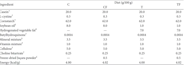
Table 1: Composition of the control diet (C), control diet supplemented with 0.5% freeze-dried Jussara powder (CJ), diet enriched with hydrogenated vegetable fat, TFAs (T), and diet enriched with TFAs supplemented with 0.5% freeze-dried Jussara powder (TJ) according to AIN-93.
Ingredient C Diet (g/100 g) TF
CF T
Casein∗ 20.0 20.0 20.0 20.0
L-cystine† 0.3 0.3 0.3 0.3
Cornstarch† 62.0 62.0 62.0 62.0
Soybean oil‡ 8.0 8.0 1.0 1.0
Hydrogenated vegetable fat$ — — 7.0 7.0
Butylhydroquinone† 0.0014 0.0014 0.0014 0.0014
Mineral mixture§ 3.5 3.5 3.5 3.5
Vitamin mixture# 1.0 1.0 1.0 1.0
Cellulose† 5.0 5.0 5.0 5.0
Choline bitartrate† 0.25 0.25 0.25 0.25
Freeze-dried Juc¸ara powder£ — 0.5 — 0.5
Energy (kcal/g) 4.00 4.02 4.00 4.02
∗Casein was obtained from Labsynth, S˜ao Paulo, Brazil.
†L-cystine, cornstarch, butylhydroquinone, cellulose and choline bitartrate were obtained from Viafarma, S˜ao Paulo, Brazil.
‡Oil was supplied from soybean (Lisa/Ind. Brazil).
$Hydrogenated vegetable fat was supplied from Unilever, S˜ao Paulo, Brazil.
§
Mineral mix (9 mg/kg diet): calcium, 5000; phosphorus, 1561; potassium, 3600; sodium, 1019; chloride, 1571; sulfur, 300; magnesium, 507; iron, 35; copper, 6.0; manganese, 10.0; zinc, 30.0; chromium, 1.0; iodine 0.2; selenium, 0.15; luoride, 1.00; boron, 0.50; molybdenum, 0.15; silicon, 5.0; nickel, 0.5; lithium, 0.1; vanadium, 0.1 (AIN-93G, mineral mix, Rhoster, Brazil).
#Vitamin mix (mg/kg diet): thiamin HCL, 6.0, ribolavin, 6.0; pyridoxine HCL, 7.0; niacin, 30.0; calcium pantothenate, 16.0; folic acid, 2.0; biotin, 0.2; vitamin
B12, 25.0; vitamin A palmitate 4000 IU; vitamin E acetate, 75; vitamin D3, 1000 IU; vitamin KI, 0.75 (AIN-93G, vitamin mix, Rhoster, Brazil).
£
Freeze-dried Juc¸ara powder: Juc¸ara pulp (Euterpe edulisMart.) was obtained from agroecological Project Juc¸ara/IPEMA—Institute of Permaculture and
Ecovillages of the Atlantic (Ubatuba, SP, Brazil)—and by freeze-drying to powder using a lyophilizer.
measured (nasoanal length) at birth and on postnatal days 7, 14, and 21. Ater 21 days the ofspring were decapitated. Trunk blood was collected and centrifuged. Serum was separated
and stored at−80∘C for later determination of triacylglycerol
(TAG), total cholesterol, HDL-cholesterol, and glucose levels. he colon was removed and the fecal content was isolated;
both were stored at−80∘C.
2.2. Biochemical Serum Analyses. Glucose, triacylglycerol, total cholesterol, and HDL-cholesterol serum concentrations were measured with an enzymatic colorimetric method using commercial kits (Labtest Brazil).
2.3. Carcass Lipid and Protein Content. he carcasses were eviscerated and the remnants were weighed and stored at
−20∘C. he lipid content was measured as described by
Stansbie et al. [41] and standardized using the method
described by Oller Do Nascimento and Williamson [42]. he
carcass was autoclaved at 120∘C for 90 min and homogenized
with water at a volume twice the carcass mass. Triplicate aliquots of approximately 3 g were digested in 3 mL of 30%
KOH and 3 mL of ethanol for≥2 h at 70∘C in capped tubes.
Ater cooling, 2 mL of 12 N H2SO4was added and the samples
were washed three times with petroleum ether to extract the lipids. he results are expressed as grams of lipid per 100 g of carcass. To measure the protein content, aliquots of the
same homogenate, approximately 1 g, were heated to 37∘C for
1 h in 0.6 N KOH with constant shaking. Ater clariication
by centrifugation, protein content was measured using the Bradford assay (Bio-Rad, Hercules, CA, USA) with bovine serum albumin as a reference.
2.4. RNA Extraction and Real-Time Polymerase Chain Reac-tion (RT-PCR). Total RNA was extracted from tissues with Tri-reagent (Sigma, St. Louis, MO, USA) and its concen-tration was determined from 260/280 nm absorbance ratios taken with a NanoDrop 2000/2000c (NanoDrop
Technolo-gies Inc., Wilmington, DE, USA). he TLR-4, TNF-�R, and
IL-6R mRNA expression from colons were quantiied by real-time polymerase chain reaction using a SYBR Green primer in StepOne Real-Time PCR Systems (Applied Biosystems, Foster City, CA, USA). Relative levels of the housekeeping gene hypoxanthine phosphoribosyl transferase (HPRT) were
measured. he PCR primers used are listed in Table 2.
Results were obtained using StepOne Sotware 2.1 (Applied Biosystems) and are expressed as a relative increase, using the
method of2−ΔΔCtdescribed by Livak and Schmittgen [43].
2.5. Genomic DNA Extraction from Fecal Samples and
RT-PCR. Genomic DNA was extracted from colon fecal samples
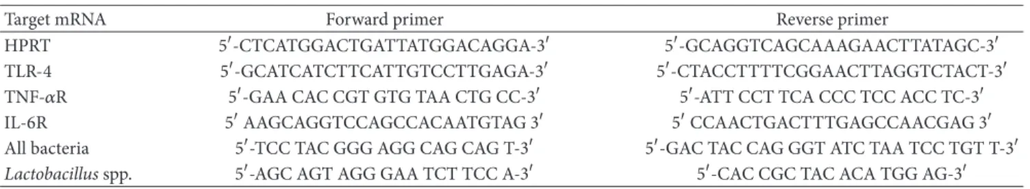
Table 2: Nucleotide sequence of the forward and reverse primers for the RT-PCR.
Target mRNA Forward primer Reverse primer
HPRT 5�-CTCATGGACTGATTATGGACAGGA-3� 5�-GCAGGTCAGCAAAGAACTTATAGC-3�
TLR-4 5�-GCATCATCTTCATTGTCCTTGAGA-3� 5�-CTACCTTTTCGGAACTTAGGTCTACT-3�
TNF-�R 5�-GAA CAC CGT GTG TAA CTG CC-3� 5�-ATT CCT TCA CCC TCC ACC TC-3�
IL-6R 5�AAGCAGGTCCAGCCACAATGTAG 3� 5�CCAACTGACTTTGAGCCAACGAG 3�
All bacteria 5�-TCC TAC GGG AGG CAG CAG T-3� 5�-GAC TAC CAG GGT ATC TAA TCC TGT T-3�
Lactobacillusspp. 5�-AGC AGT AGG GAA TCT TCC A-3� 5�-CAC CGC TAC ACA TGG AG-3�
Table 3: Serum glucose, total cholesterol, HDL-cholesterol, and triacylglycerols in 21-day-old ofspring.
C (15) CJ (20) T (19) TJ (19)
Glucose 110.19±3.44 103.99±1.73# 118.51±2.96 98.83±1.14∗#
Total cholesterol 120.05±4.25 114.43±3.57# 136.01±2.85∗ 108.79±2.23#
HDL-cholesterol 26.56±1.48 28.92±1.29 25.84±0.71 28.34±1.03
Triacylglycerols 180.20±11.37 131.11±4.06∗# 209.19±16.33 135.15±3.72∗#
C: ofspring of dams fed control diet; CJ: ofspring of dams fed control diet supplemented with 0.5% freeze-dried Jussara powder; T: ofspring of dams fed diet enriched with hydrogenated vegetable fat, TFAs; TJ: ofspring of dams fed diet enriched with TFAs supplemented with 0.5% freeze-dried Jussara powder. Data
are presented as mean±SEM. he number in parentheses refers to the sample value.
∗� < 0.05versus C.$� < 0.05versus CJ.#
� < 0.05versus T.&� < 0.05versus TJ.
230 nm. he purity was estimated by the 260/280 nm ratio, which must range between 1.8 and 2.0 for nucleic acids. All
samples were maintained at−80∘C.
2.6. Lactobacillus spp. Quantiied by RT-PCR. Relative levels ofLactobacillusspp. DNA were quantiied in real time, using a SYBR Green primer in an ABI Prism 7500 Sequence Detector (both from Applied Biosystems, Foster City, CA, USA). Relative levels of the housekeeping gene of all bac-teria were measured. he PCR primers used are listed in
Table2. he results were obtained using Sequence Detector
sotware (Applied Biosystems) and are expressed as a relative
increase, using the method of2−ΔΔCt, described by Livak and
Schmittgen [43].
2.7. Colon TNF-�, IL-6, and IL-10 Protein Levels by ELISA.
he colon was homogenized and centrifuged at 12,000 rpm
for 40 min at 4∘C; the supernatant was saved and the protein
concentration determined using the BCA assay (Bio-Rad, Hercules, CA, USA) with bovine serum albumin (BSA) as
a reference. Quantitative assessment of TNF-�, IL-6, and
IL-10 proteins was carried out by ELISA (DuoSet ELISA, R&D Systems, Minneapolis, MN, USA) following the rec-ommendations of the manufacturer. All samples were run as duplicates and the mean value was reported.
2.8. Statistical Analysis. Statistical analyses were performed using the Sigma Stat 3.5. he data were analyzed by ANOVA followed by a Bonferroni posthoc or Kruskal-Wallis test. All
results are presented as the mean±SEM and� ≤ 0.05was
considered statistically signiicant.
3. Results
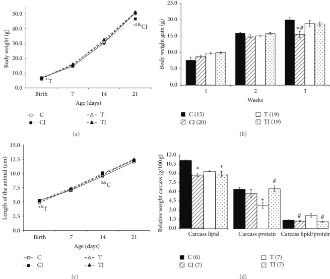
3.1. Body Weight, Body Weight Gain, Length of the Animal, and Carcass Lipid and Protein Content. At birth, ofspring of the
CJ group were longer than those of the T group (� < 0.05)
(Figure1(c)). he body weight (BW) and length were reduced
in the pups of the T group compared to those of the C group (� = 0.02and � < 0.05, resp.) (Figures1(a)and 1(c)). At postnatal day 21, the length of pups did not difer between the groups and the BW of the CJ group was lower than the C (� = 0.042), T (� = 0.006), and TJ (� = 0.006) groups
(Figure1(a)). Furthermore, the CJ and TJ groups displayed
longer body length than the C group (� < 0.001and� =
0.017) at postnatal day 14 (Figure1(c)). he CJ group also exhibited decreased BW gain compared with the T and C
groups (� < 0.001) three weeks ater birth (Figure1(b)).
he relative carcass lipid levels in the CJ and TJ groups
were signiicantly lower than in the C group (� < 0.05). he
TJ group exhibited higher relative carcass protein levels than
the T group (� = 0.003) and the T group contained less
relative carcass protein than the C group (� = 0.006). he
lipid to protein carcass ratio was lower in the CJ and TJ groups
than in the T group (� < 0.05) (Figure1(d)).
3.2. Biochemical Serum. he T group had increased serum concentrations of total cholesterol compared to the C group
at postnatal day 21 (� = 0.008) while the CJ and TJ groups
exhibited reduced serum levels of total cholesterol (� <
0.001) and glucose (� < 0.05) compared to the T group. Furthermore, in the TJ group the glucose concentration was
lower than in the C group (� < 0.05). Triacylglycerol levels
were also reduced in the CJ and TJ groups compared to the C
and T groups (� < 0.05). HDL-cholesterol levels in the serum
0.0 10.0 20.0 30.0 40.0 50.0
Birth 7 14 21
B
o
d
y w
eig
h
t (g)
Age (days)
C CJ
T TJ ∗
T
CJ
∗#&
(a)
0.0 5.0 10.0 15.0 20.0 25.0
1 2 3
B
o
d
y w
eig
h
t ga
in (g)
Weeks
C (15) CJ (20)
T (19) TJ (19)
# ∗
(b)
0.0 2.5 5.0 7.5 10.0 12.5 15.0
Birth 7 14 21
L
en
gt
h
o
f t
h
e a
n
ima
l (cm)
Age (days)
C CJ
T TJ T
∗$
C
$&
(c)
0.0 1.5 3.0 4.5 6.0 7.5 9.0 10.5 12.0
Rela
ti
ve
w
eig
h
t ca
rc
ass (g/100
g)
Carcass lipid Carcass protein Carcass lipid/protein
#
# #
∗
∗ ∗
C (6) CJ (7)
T (7) TJ (7)
(d)
Figure 1: Body weight (a), body weight evolution (b), length (c), and carcass lipid, protein content, and lipid/protein ratio (d). C: ofspring of dams fed control diet; CJ: ofspring of dams fed control diet supplemented with 0.5% freeze-dried Jussara powder; T: ofspring of dams fed diet enriched with hydrogenated vegetable fat, TFAs; TJ: ofspring of dams fed diet enriched with TFAs supplemented with 0.5% freeze-dried
Jussara powder. Data are means±SEMs. he number in parentheses refers to the sample value.∗� < 0.05versus C.$� < 0.05versus CJ.
#
� < 0.05versus T.&� < 0.05versus TJ.
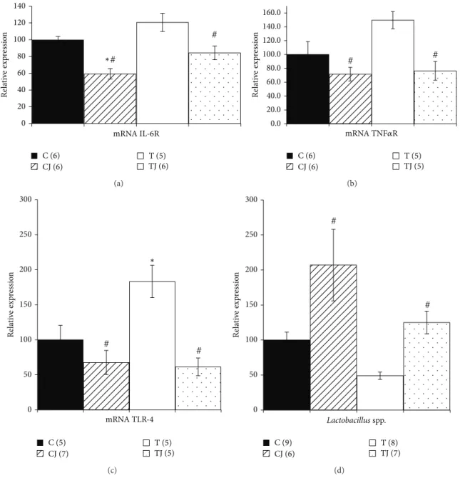
3.3. IL-6R, TNF-�R, and TLR-4 Gene Expression. he TLR-4 gene expression in the colon of 21-day-old ofspring was
higher in the T group (83.1%) than in the C group (� =
0.047). Levels of TNF-�R mRNA expression also increased in the T group (49.8%), but this diference was not signiicant. However, the CJ and TJ groups showed lower levels of
TNF-�R mRNA expression (CJ group 52.1%,� = 0.013versus T
group; TJ group 48.9%,� = 0.027versus T group) and
TLR-4 mRNA expression (CJ group 63.1%,� = 0.002versus T
group; TJ group 66.5%,� = 0.002versus T group) in ofspring
at postnatal day 21 (Figures2(b)and2(c)). In addition, the
IL-6R gene expression decreased in the CJ and TJ groups
compared to the T group (50.9%,� < 0.001; 30.2%,� = 0.02,
resp.) and also in the CJ group compared to C group (40.7%,
� = 0.005) (Figure2(a)).
3.4. Levels of Lactobacillus spp. in Colon. he levels of
Lactobacillusspp. genomic DNA in colon fecal content in the
CJ and TJ groups were 4.2-fold higher and 2.6-fold higher,
respectively, than in the T group (� < 0.05). he T group level
ofLactobacillusspp. genomic DNA was 2.1-fold lower than the C group level; however, this diference was not signiicant
(Figure2(d)).
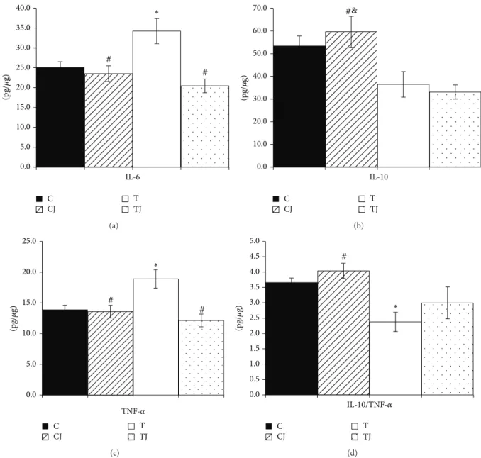
3.5. Cytokine Proile of the Colon. he protein levels of IL-6
(36.5%) and TNF-�(36.2%) were signiicantly higher (� =
0.048and � = 0.013, resp.) in the T group than in the C group in ofspring at postnatal day 21. However, the Jussara supplementation in the CJ and TJ groups reduced the levels of
IL-6 (CJ group 31.3%,� = 0.011; TJ group 40.2%,� < 0.001)
and TNF-�(CJ group 28.1%� = 0.013and TJ group 35.8%,
� < 0.001) compared to the T group (Figures3(a)and3(c)). Furthermore, in the CJ group, the expression of IL-10 protein
was higher than in both the T group (63.4%,� = 0.035)
and TJ group (80.2%,� = 0.011) (Figure3(b)). hus, the
0 20 40 60 80 100 120 140
Rela
ti
ve
exp
ressio
n
mRNA IL-6R
C (6) CJ (6)
T (5) TJ (6)
#
#
∗
(a)
0.0 20.0 40.0 60.0 80.0 100.0 120.0 140.0 160.0
Rela
ti
ve
exp
ressio
n
mRNA TNF�R
C (6) CJ (6)
T (5) TJ (5)
# #
(b)
0 50 100 150 200 250 300
Rela
ti
ve
exp
ressio
n
mRNA TLR-4
C (5) CJ (7)
T (5) TJ (5)
#
# ∗
(c)
0 50 100 150 200 250 300
Rela
ti
ve
exp
ressio
n
Lactobacillus spp.
C (9) CJ (6)
T (8) TJ (7)
#
#
(d)
Figure 2: Gene expression of IL-6 receptor (IL-6R) (a), tumor necrosis factor-�receptor (TNF-�R) (b), Toll-like receptor 4 (TLR4) (c), and
DNA levels ofLactobacillusspp. in 21-day-old ofspring colon (d). C: ofspring of dams fed control diet; CJ: ofspring of dams fed control
diet supplemented with 0.5% freeze-dried Jussara powder; T: ofspring of dams fed diet enriched with hydrogenated vegetable fat, TFAs; TJ:
ofspring of dams fed diet enriched with TFAs supplemented with 0.5% freeze-dried Jussara powder. Data are means±SEMs. he number
in parentheses refers to the sample value. Results are expressed in arbitrary units, stipulating 100 as the control value.∗� < 0.05versus C.
#
� < 0.05versus T.
group (70.2%,� = 0.003) and was reduced in the T group
compared to the C group (35.1%,� = 0.026) (Figure3(d)).
4. Discussion
In this study, supplementing the maternal diet with 0.5% Jussara attenuated the adverse efects of perinatal TFAs. We showed that the maternal intake of Jussara in perinatal period modulates the inlammatory state and improves the lipid proile, glucose levels, body composition, and intestinal microbiota of 21-day-old ofspring.
In our study, Jussara supplementation of the maternal diet did not afect the growth of pups at birth but in 21 day of life ofspring from Jussara-supplemented dams had lower BW gain and better body composition with lower lipid content
and higher carcass protein (Figure1).
Corroborating our data, a study with ac¸a´ı (Euterpe
Oler-acea Mart.) a fruit similar to Jussara, reported that
sup-plementation with hydroalcoholic extract (200 mg/kg/day) during the pregnancy does not change BW in ofspring at
birth [44]. However, Rahal et al. [45] found that
0.0 5.0 10.0 15.0 20.0 25.0 30.0 35.0 40.0
IL-6
C CJ
T TJ
#
# ∗
(pg/
𝜇
g)
(a)
0.0 10.0 20.0 30.0 40.0 50.0 60.0 70.0
IL-10
C CJ
T TJ
#&
(pg/
𝜇
g)
(b)
0.0 5.0 10.0 15.0 20.0 25.0
C CJ
T TJ
#
# ∗
TNF-�
(pg/
𝜇
g)
(c)
0.0 0.5 1.0 1.5 2.0 2.5 3.0 3.5 4.0 4.5 5.0
C CJ
T TJ
#
∗
IL-10/TNF-�
(pg/
𝜇
g)
(d)
Figure 3: IL-6 protein expression (a), IL-10 (b), TNF-�(c), and IL-10/TNF-�ratio (d) in 21-day-old ofspring colon. C: ofspring of dams
fed control diet; CJ: ofspring of dams fed control diet supplemented with 0.5% freeze-dried Jussara powder; T: ofspring of dams fed diet enriched with hydrogenated vegetable fat, TFAs; TJ: ofspring of dams fed diet enriched with TFAs supplemented with 0.5% freeze-dried
Jussara powder. Data are means±SEMs of 7–11 determinations per group.∗� < 0.05versus C.#� < 0.05versus T.&� < 0.05versus TJ.
the maternal diet during pregnancy and lactation in MMTV-Wnt1-transgenic mice does not afect BW of the ofspring at weaning. Furthermore, authors demonstrated that 2% Jussara supplementation in adult ApoE-deicient mice leads to no
change in BW during the entire experimental period [46].
Our indings also indicate that the addition of Jussara to the maternal diet restores total cholesterol to a normal range and reduces serum TAG and glucose in 21-day-old ofspring. Similar efects have been reported by De Souza
et al. [47] ater supplementation with 2% ac¸a´ı for 6 weeks,
with decreased total cholesterol observed in female Fischer rats. In fact, studies have indicated that the beneicial efect of similar fruit on the lipid proile is linked to diverse
components contained in the fruit, such ascis-unsaturated
fatty acids, polyunsaturated fatty acids (PUFAs), polyphenols, and dietary iber. hese components are associated with reduced intestinal absorption of fatty acids, greater balance in the synthesis and absorption of sterols, and increased expression of genes involved in cholesterol metabolism and
excretion in the adult animals [48–50].
Moreover, there is evidence that phenolic compounds, particularly the lavonoids, induces Glut 4 in the adipose tissue and skeletal muscle and can improve glucose home-ostasis and lipid metabolism via AMP-activated protein
kinase activation in adult animal models [51].
Our study found that the Jussara supplementation in maternal diet led to a reduction in proinlammatory
TLR-4 mRNA expression) induced from TFAs to normal levels, accompanied by an increase in anti-inlammatory
cytokines (IL-10, IL-10/TNF-� ratio) andLactobacillusspp.
genomic DNA levels in the colon of ofspring.
Indeed, TFAs are known for their ability to increase
expression of inlammatory markers such as IL-6 and TNF-�
[4,52] and there is evidence that exposure to a high-fat diet
increases inlammation in the colon [14,15,53] while
high-polyphenol diet reduces this process [54]. Previous studies
have shown that in adult ofspring of mothers fed TFAs dur-ing pregnancy and lactation, high levels of LPS activate
TLR-4 and mediate low-grade inlammation [7]. Additionally, as
described in other studies, changes in the composition of the microbiota and the subsequent alteration of membrane permeability damage intestinal barrier integrity. his damage can cause an increase in bacterial translocation and uptake of LPS, resulting in TLR4-mediated inlammatory responses
in the ofspring [16, 55]. hus, this suggests a potential
mechanism by which TFAs increase the inlammatory status of the ofspring colon.
In accordance with our results, we believe that the anti-inlammatory efect of Jussara also could be associated with
the high nutritional value,cis-unsaturated fatty acids, PUFAs,
bioactive compounds levels as phenolics (415 mg GAE/100 g
f.m.), particularly anthocyanins (239.16 ± 7.6mg C3R/100 g),
and dietary iber presence in the fruit of the Jussara palm [35,
36].
PUFAs can inluence synthesis of the proinlammatory
cytokines TNF-�and IL-6 in order to downregulate
inlam-matory transcription factors by actions upon intracellular
signaling through the inhibition of NF-�B pathway [56–58].
In this sense, the study performed by Fong et al. [59] in female
rats fed a diet containing DHA, polyunsaturated long chain fatty acids (DHA group), during pregnancy and lactation,
found decreased expression of TNF-�and IL-6 in 21-day-old
ofspring.
Likewise, polyphenols, especially anthocyanin, have been associated with the modulation of oxidative stress and inlam-mation in some studies from the use of similar fruits or
isolation form by inhibiting NF-�B activation [33,60,61].
Lee et al. [62] found that fruits containing diferent major
anthocyanins showed similar anti-inlammatory efects in
macrophages. Xie et al. [63] demonstrated that the diet
containing 5% freeze-dried ac¸a´ı (Euterpe oleraceaMart.) juice
powder reduced TNF-� and IL-6 in adult ApoE-deicient
mice model. he same authors reported that polyphenols isolated from the ac¸a´ı pulp reduces these LPS-induced
proin-lammatory cytokines by inhibiting NF-�B in macrophages
[64].
he intestinal microbiota modulation has been con-sidered as a possible mechanism by which polyphenols, particularly anthocyanins, may exert their beneic efect
[65]. In recent study, high-polyphenols apple, was associated
with reduction of inlammation markers and modulation in
intestinal microbiota in healthy adult mice [66]. Additionally,
Neyrinck et al. [67] demonstrated that the pomegranate
extract, rich in phenolic compounds, modulates the gut
microbiota in favor ofBiidobacteriumspp. and
downregu-lated IL-6 in the colon of adult mice. hese authors suggest
the inluence of the intestinal microbiota to reduce proin-lammatory cytokines by polyphenol in mice. Similarly, our indings suggest that the gut microbiota modulation have an important role in beneic efect of Jussara in ofspring.
Dietary iber has also been associated with beneic changes in intestinal microbiota, especially in the amount of biidobacteria and lactobacilli with a consequent
enhance-ment in colonic barrier functions [30,68]. Increases in
lac-tobacilli and reductions in colonic paracellular permeability have been linked to reductions in bacterial translocation and absorption of LPS, resulting in the downregulation of
TLR-4-mediated inlammatory responses [16]. Recently, Arora et
al. [69] demonstrated beneits in intestinal histology,
reduc-ing endotoxemia and inlammation in Female Wistar rats
exposed toLactobacillus plantarum. Pe˜na and Versalovic [70]
also reported an anti-inlammatory efect of Lactobacillus
rhamnosus GG in a macrophage model, with inhibition of
TNF-�production and a reduction in the TNF-�/IL-10 ratio.
hus, it is possible that the increase in Lactobacillus
spp. in Jussara-supplemented groups plays an important role in the downregulation of proinlammatory cytokines and the upregulation of anti-inlammatory interleukin markers in the colon of 21-day-old ofspring. his efect could be associated with the fortiication of the intestinal barrier integrity and intestinal mucosal permeability, which could result in reduced LPS translocation.
herefore, we demonstrated that Jussara supplementation during pregnancy and lactation was a natural alternative to reduce of inlammation biomarkers in colon in 21-day-old ofspring without altering the normality status.
5. Conclusion
In summary, we showed that supplementation of the maternal diet with the 0.5% Jussara during pregnancy and lactation reverses the adverse efects of perinatal TFAs. he maternal intake of Jussara in perinatal period improves lipid proiles, glucose levels, body composition, and gut microbiota and reduces low-grade inlammation in the colon of 21-day-old ofspring. hese efects are most likely a result of better fatty acid balance, the presence of ibers and phenolic compounds in Jussara favoring colonic bacterial population, and possibly the fortiication of the intestinal barrier integrity, which could result in reduced LPS translocation. hese indings support our hypothesis on the potential role of Jussara supplementation in modulating the adverse inlammatory efects of maternal TFA intake in ofspring. Our results could contribute to the control of inlammation and the prevention of chronic disease development until adulthood.
Conflict of Interests
Acknowledgments
his research was supported by FAPESP (Fundac¸˜ao de Amparo `a Pesquisa do Estado de S˜ao Paulo) and CAPES (Coordenac¸˜ao de Aperfeic¸oamento de Pessoal de N´ıvel Superior). he authors gratefully acknowledge the invaluable assistance of Valter Tadeu Boldarine.
References
[1] L. O’Sullivan, M. H. Little, A. N. Combes, and K. M. Moritz, “Epigenetics and developmental programming of adult onset
diseases,”Pediatric Nephrology, vol. 27, no. 12, pp. 2175–2182,
2012.
[2] K. M. Godfrey and D. J. P. Barker, “Fetal programming and adult
health,”Public Health Nutrition, vol. 4, no. 2B, pp. 611–624, 2001.
[3] D. J. P. Barker, “In utero programming of chronic disease,”
Clinical Science, vol. 95, no. 2, pp. 115–128, 1998.
[4] V. Remig, B. Franklin, S. Margolis, G. Kostas, T. Nece, and J. C. Street, “Trans fats in America: a review of their use,
consumption, health implications, and regulation,”Journal of
the American Dietetic Association, vol. 110, no. 4, pp. 585–592, 2010.
[5] J. L. De Oliveira, L. M. Oyama, A. C. L. Hachul et al., “Hydro-genated fat intake during pregnancy and lactation caused increase in TRAF-6 and reduced AdipoR1 in white adipose
tissue, but not in muscle of 21 days old ofspring rats,”Lipids
in Health and Disease, vol. 10, article 22, 2011.
[6] L. P. Pisani, C. M. O. do Nascimento, A. A. Bueno et al., “Hydrogenated fat diet intake during pregnancy and lactation modiies the PAI-1 gene expression in white adipose tissue of
ofspring in adult life,”Lipids in Health and Disease, vol. 7, no. 1,
article 13, 2008.
[7] G. D. Pimentel, F. S. Lira, J. C. Rosa et al., “Intake of trans fatty acids during gestation and lactation leads to hypothalamic
inlammation via TLR4/NF�Bp65 signaling in adult ofspring,”
Journal of Nutritional Biochemistry, vol. 23, no. 3, pp. 265–271, 2012.
[8] T. Priego, J. S´anchez, A. P. Garc´ıa, A. Palou, and C. Pic´o, “Maternal dietary fat afects milk fatty acid proile and impacts on weight gain and thermogenic capacity of suckling rats,”
Lipids, vol. 48, no. 5, pp. 481–495, 2013.
[9] E. M. Novak, B. O. Keller, and S. M. Innis, “Metabolic devel-opment in the liver and the implications of the n-3 fatty acid
supply,”he American Journal of Physiology—Gastrointestinal
and Liver Physiology, vol. 302, no. 2, pp. G250–G259, 2012. [10] S. Mozeˇs, D. Bujˇn´akov´a, Z. ˇSefˇc´ıkov´a, and V. Kmet,
“Develop-mental changes of gut microlora and enzyme activity in rat
pups exposed to fat-rich diet,”Obesity, vol. 16, no. 12, pp. 2610–
2615, 2008.
[11] K. Jacobson, H. Mundra, and S. M. Innis, “Intestinal responsive-ness to experimental colitis in young rats is altered by maternal
diet,” American Journal of Physiology—Gastrointestinal and
Liver Physiology, vol. 289, no. 1, pp. G13–G20, 2005.
[12] Y. Y. Lam, C. W. Y. Ha, C. R. Campbell et al., “Increased gut permeability and microbiota change associate with mesenteric fat inlammation and metabolic dysfunction in diet-induced
obese mice,”PLoS ONE, vol. 7, no. 3, Article ID e34233, 2012.
[13] K. Kim, W. Gu, I. Lee, E. Joh, and D. Kim, “High fat diet-induced gut microbiota exacerbates inlammation and obesity in mice
via the tlr4 signaling pathway,”PLoS ONE, vol. 7, no. 10, Article
ID e47713, 2012.
[14] Z. Liu, R. S. Brooks, E. D. Ciappio et al., “Diet-induced obesity
elevates colonic TNF-�in mice and is accompanied by an
activation of Wnt signaling: a mechanism for obesity-associated
colorectal cancer,”Journal of Nutritional Biochemistry, vol. 23,
no. 10, pp. 1207–1213, 2012.
[15] S. Ding, M. M. Chi, B. P. Scull et al., “High-fat diet: Bacteria interactions promote intestinal inlammation which precedes and correlates with obesity and insulin resistance in mouse,”
PLoS ONE, vol. 5, no. 8, Article ID e12191, 2010.
[16] J. Villena and H. Kitazawa, “Modulation of intestinal TLR4-inlammatory signaling pathways by probiotic microorganisms:
lessons learned fromLactobacillus jenseniiTL2937,”Frontiers in
Immunology, vol. 4, article 512, 2014.
[17] A. Nenci, C. Becker, A. Wullaert et al., “Epithelial NEMO links
innate immunity to chronic intestinal inlammation,”Nature,
vol. 446, no. 7135, pp. 557–561, 2007.
[18] Y. K. Nakamura and S. T. Omaye, “Metabolic diseases and
pro-and prebiotics: mechanistic insights,”Nutrition & Metabolism,
vol. 9, no. 1, article 60, 2012.
[19] O. Takeuchi and S. Akira, “Toll-like receptors; their
phys-iological role and signal transduction system,” International
Immunopharmacology, vol. 1, no. 4, pp. 625–635, 2001.
[20] T. Kawai and S. Akira, “Signaling to NF-�B by Toll-like
receptors,”Trends in Molecular Medicine, vol. 13, no. 11, pp. 460–
469, 2007.
[21] T. Kondo, T. Kawai, and S. Akira, “Dissecting negative
regula-tion of Toll-like receptor signaling,”Trends in Immunology, vol.
33, no. 9, pp. 449–458, 2012.
[22] G. Boden, P. She, M. Mozzoli et al., “Free fatty acids produce insulin resistance and activate the proinlammatory nuclear
factor-�b pathway in rat liver,”Diabetes, vol. 54, no. 12, pp. 3458–
3465, 2005.
[23] R. P. Assumpc¸˜ao, F. Duarte Dos Santos, P. D. M. M. Andrade, G. F. Barreto, and M. D. G. Tavares Do Carmo, “Efect of variation of trans-fatty acid in lactating rats’ diet on lipoprotein lipase
activity in mammary gland, liver, and adipose tissue,”Nutrition,
vol. 20, no. 9, pp. 806–811, 2004.
[24] K. Kavanagh, S. S. Soraya, A. K. A. J. Kurt et al., “Neonatal and fetal exposure to trans-fatty acids retards early growth and
adiposity while adversely afecting glucose in mice,”Nutrition
Research, vol. 30, no. 6, pp. 418–426, 2010.
[25] A. Hassan, A. Ibrahim, K. Mbodji et al., “An�-linolenic
acid-rich formula reduces oxidative stress and inlammation by
regulating NF-�B in rats with TNBS-induced colitis,”Journal of
Nutrition, vol. 140, no. 10, pp. 1714–1721, 2010.
[26] N. S. Kalupahana, K. J. Claycombe, and N. Moustaid-Moussa, “(n-3) Fatty acids alleviate adipose tissue inlammation and
insulin resistance: mechanistic insights.,”Advances in Nutrition,
vol. 2, no. 4, pp. 304–316, 2011.
[27] G. Jakobsdottir, J. Xu, G. Molin, S. Ahrne, and M. Nyman, “High-fat diet reduces the formation of butyrate, but increases succinate, inlammation, liver fat and cholesterol in rats, while
dietary ibre counteracts these efects,”PLoS ONE, vol. 8, no. 11,
Article ID e80476, 2013.
[28] P. D. Cani, S. Possemiers, T. van de Wiele et al., “Changes in gut microbiota control inlammation in obese mice through a mechanism involving GLP-2-driven improvement of gut
permeability,”Gut, vol. 58, no. 8, pp. 1091–1103, 2009.
[29] P. Dehghan, B. P. Gargari, and M. A. Jafar-Abadi, “Oligofructose-enriched inulin improves some inlammatory markers and metabolic endotoxemia in women with type 2 diabetes mellitus: a randomized controlled clinical trial,”
[30] J. M. Mariadason, A. Catto-Smith, and P. R. Gibson, “Modula-tion of distal colonic epithelial barrier func“Modula-tion by dietary ibre
in normal rats,”Gut, vol. 44, no. 3, pp. 394–399, 1999.
[31] F. F˚ak, S. Ahrn´e, G. Molin, B. Jeppsson, and B. Westr¨om, “Microbial manipulation of the rat dam changes bacterial colonization and alters properties of the gut in her ofspring,”
American Journal of Physiology: Gastrointestinal and Liver Physiology, vol. 294, no. 1, pp. G148–G154, 2007.
[32] C. L. J. Karlsson, G. Molin, F. F˚ak et al., “Efects on weight gain and gut microbiota in rats given bacterial supplements and a high-energy-dense diet from fetal life through to 6 months of
age,”British Journal of Nutrition, vol. 106, no. 6, pp. 887–895,
2011.
[33] J. F. D. C. Guerra, C. L. D. B. Magalh˜aes, D. C. Costa, M. E. Silva, and M. L. Pedrosa, “Dietary ac¸ai modulates ROS production by neutrophils and gene expression of liver antioxidant enzymes in
rats,”Journal of Clinical Biochemistry and Nutrition, vol. 49, no.
3, pp. 188–194, 2011.
[34] K. Vanhees, F. J. van Schooten, S. B. van Waalwijk Van Doorn-Khosrovani et al., “Intrauterine exposure to lavonoids modiies antioxidant status at adulthood and decreases oxidative
stress-induced DNA damage,”Free Radical Biology and Medicine, vol.
57, pp. 154–161, 2013.
[35] P. P. M. Silva, L. F. Carmo, G. M. Silva et al., “Composition of
ju�ara pulp,”Brazilian Journal of Food and Nutrition, vol. 24,
no. 1, pp. 7–13, 2013.
[36] N. A. Silva, E. Rodrigues, A. Z. Mercadante, and V. V. de Rosso, “Phenolic compounds and carotenoids from four fruits native
from the Brazilian Atlantic Forest,”Journal of Agricultural and
Food Chemistry, vol. 62, no. 20, pp. 4481–4832, 2014.
[37] J. He and M. Monica Giusti, “Anthocyanins: natural colorants
with health-promoting properties,” Annual Review of Food
Science and Technology, vol. 1, no. 1, pp. 163–187, 2010.
[38] S. M. Poulose, D. R. Fisher, J. Larson et al.,
“Anthocyanin-rich ac¸ai (Euterpe oleraceaMart.) fruit pulp fractions attenuate
inlammatory stress signaling in mouse brain BV-2 microglial
cells,”Journal of Agricultural and Food Chemistry, vol. 60, no. 4,
pp. 1084–1093, 2012.
[39] P. G. Reeves, F. H. Nielsen, and G. C. Fahey Jr., “AIN-93 puriied diets for laboratory rodents: inal report of the American Insti-tute of Nutrition ad hoc writing committee on the reformulation
of the AIN-76A rodent diet,”Journal of Nutrition, vol. 123, no.
11, pp. 1939–1951, 1993.
[40] P. G. Reeves, “Components of the AIN-93 diets as
improve-ments in the AIN-76A diet,”Journal of Nutrition, vol. 127, no.
5, pp. 838S–841S, 1997.
[41] D. Stansbie, R. M. Denton, B. J. Bridges, H. T. Pask, and P. J. Randle, “Regulation of pyruvate dehydrogenase and pyruvate dehydrogenase phosphate phosphatase activity in rat epididy-mal fat pads. Efects of starvation, alloxan diabetes and high fat
diet,”Biochemical Journal, vol. 154, no. 1, pp. 225–236, 1976.
[42] C. M. Oller Do Nascimento and D. H. Williamson, “Evidence for conservation of dietary lipid in the rat during lactation and the immediate period ater removal of the litter. Decreased
oxidation of oral [1-14C]triolein,”Biochemical Journal, vol. 239,
no. 1, pp. 233–236, 1986.
[43] K. J. Livak and T. D. Schmittgen, “Analysis of relative gene expression data using real-time quantitative PCR and the
2−����method,”Methods, vol. 25, no. 4, pp. 402–408, 2001.
[44] G. F. de Bem, C. A. da Costa, and P. R. B. de Oliveira, “Protective
efect ofEuterpe oleraceaMart (ac¸a´ı) extract on programmed
changes in the adult rat ofspring caused by maternal protein
restriction during pregnancy,”Journal of Pharmacy and
Phar-macology, 2014.
[45] O. M. Rahal, J. M. P.Pabona, T. Kelly et al., “Suppression of Wnt1-induced mammary tumor growth and lower serum insulin in ofspring exposed to maternal blueberry diet suggest early
dietary inluence on developmental programming,”
Carcino-genesis, vol. 34, no. 2, pp. 464–474, 2013.
[46] C. A. de Castro, A. J. Natali, L. M. Cardoso et al., “Aero-bic exercise and not a diet supplemented with Jussara aca´ı (Euterpe edulisMartius) alters hepatic oxidative and
inlam-matory biomarkers in ApoE-deicient mice,”British Journal of
Nutrition, vol. 110, no. 1, pp. 1–10, 2014.
[47] M. O. De Souza, L. Souza e Silva, C. L. de Brito Magalh˜aes
et al., “he hypocholesterolemic activity of ac¸a´ı (Euterpe
oler-acea Mart.) is mediated by the enhanced expression of the
ATP-binding cassette, subfamily G transporters 5 and 8 and
low-density lipoprotein receptor genes in the rat,” Nutrition
Research, vol. 32, no. 12, pp. 976–984, 2012.
[48] M. O. de Souza, M. Silva, M. E. Silva, R. de Paula Oliveira,
and M. L. Pedrosa, “Diet supplementation with ac¸ai (Euterpe
oleraceaMart.) pulp improves biomarkers of oxidative stress
and the serum lipid proile in rats,”Nutrition, vol. 26, no. 7-8,
pp. 804–810, 2010.
[49] C. A. Feio, M. C. Izar, S. S. Ihara et al., “Euterpe oleracea (ac¸ai) Modiies sterol metabolism and Attenuates
experimentally-induced atherosclerosis,”Journal of Atherosclerosis and
hrom-bosis, vol. 19, no. 3, pp. 237–245, 2012.
[50] D. Graf, S. Seifert, A. Jaudszus, A. Bub, and B. Watzl, “Anthocyanin-rich juice lowers serum cholesterol, leptin, and resistin and improves plasma fatty acid composition in ischer
rats,”PLoS ONE, vol. 8, no. 6, Article ID e66690, 2013.
[51] M. Takikawa, S. Inoue, F. Horio, and T. Tsuda, “Dietary anthocyanin-rich bilberry extract ameliorates hyperglycemia and insulin sensitivity via activation of amp-activated protein
kinase in diabetic mice,”Journal of Nutrition, vol. 140, no. 3, pp.
527–533, 2010.
[52] S. K. Gebauer, T. L. Psota, and P. M. Kris-Etherton, “he diversity of health efects of individual trans fatty acid isomers,”
Lipids, vol. 42, no. 9, pp. 787–799, 2007.
[53] U. Axling, C. Olsson, J. Xu et al., “Green tea powder and Lactobacillus plantarum afect gut microbiota, lipid metabolism
and inlammation in high-fat fed C57BL/6J mice,”Nutrition and
Metabolism, vol. 9, article 105, no. 1, 2012.
[54] M. Femia, C. Luceri, F. Bianchini et al., “Marie M´enard apples with high polyphenol content and a low-fat diet reduce 1,2-dimethylhydrazine-induced colon carcinogenesis in rats: efects
on inlammation and apoptosis,”Molecular Nutrition and Food
Research, vol. 56, no. 8, pp. 1353–1357, 2012.
[55] I. A. Myles, N. M. Fontecilla, B. M. Janelsins, P. J. Vithayathil, J. A. Segre, and S. K. Datta, “Parental dietary fat intake
alters ofspring microbiome and immunity,” he Journal of
Immunology, vol. 191, no. 6, pp. 3200–3209, 2013.
[56] J. Ren and S. H. Chung, “Anti-inlammatory efect of�-linolenic
acid and its mode of action through the inhibition of nitric oxide production and inducible nitric oxide synthase gene expression
via NF-�B and mitogen-activated protein kinase pathways,”
Journal of Agricultural and Food Chemistry, vol. 55, no. 13, pp. 5073–5080, 2007.
[57] W. Zhang, X. Hu, W. Yang, Y. Gao, and J. Chen, “Omega-3 polyunsaturated fatty acid supplementation confers long-term neuroprotection against neonatal hypoxic-ischemic brain
injury through anti-inlammatory actions,”Stroke, vol. 41, no.
[58] P. C. Calder, “N-3 Fatty acids, inlammation and immunity: new
mechanisms to explain old actions,”Proceedings of the Nutrition
Society, vol. 72, no. 3, pp. 326–336, 2013.
[59] L. Fong, B. S. Muhlhausler, R. A. Gibson, and C. J. Xian,
“Perinatal maternal dietary supplementation of�3-fatty acids
transiently afects bone marrow microenvironment, osteoblast and osteoclast formation, and bone mass in male ofspring,”
Endocrinology, vol. 153, no. 5, pp. 2455–2465, 2012.
[60] D. Esposito, A. Chen, M. H. Grace, and S. M. A. Lila, “Inhibitory efects of wild blueberry anthocyanins and other lavonoids on
biomarkers of acute and chronic inlammation in vitro,”Journal
of Agricultural and Food Chemistry, vol. 62, no. 29, pp. 7022– 7028, 2014.
[61] F. M. Dias, D. D. Lefa, F. Daumann et al., “Acerola (Malpighia
emarginata DC.) juice intake protects against alterations to proteins involved in inlammatory and lipolysis pathways in the
adipose tissue of obese mice fed a cafeteria diet,”Lipids in Health
and Disease, vol. 13, p. 24, 2014.
[62] S. G. Lee, B. Kim, Y. Yang et al., “Berry anthocyanins suppress the expression and secretion of proinlammatory mediators
in macrophages by inhibiting nuclear translocation of NF-�B
independent of NRF2-mediated mechanism,”Journ of Nutrition
Biochemistry, vol. 25, no. 4, pp. 404–411, 2014.
[63] C. Xie, J. Kang, R. Burris et al., “Ac¸a´ı juice attenuates atheroscle-rosis in ApoE deicient mice through antioxidant and
anti-inlammatory activities,”Atherosclerosis, vol. 216, no. 2, pp. 327–
333, 2011.
[64] C. Xie, J. Kang, Z. Li et al., “he ac¸a´ı lavonoid velutin is a potent
anti-inlammatory agent: Blockade of LPS-mediated TNF-�
and IL-6 production through inhibiting NF-�B activation and
MAPK pathway,”Journal of Nutritional Biochemistry, vol. 23, no.
9, pp. 1184–1191, 2012.
[65] G. Jakobsdottir, N. Blanco, J. Xu et al., “Formation of short-chain fatty acids, excretion of anthocyanins, and microbial diversity in rats fed blackcurrants, blackberries, and
raspber-ries,”Journal of Nutrition and Metabolism, vol. 2013, Article ID
202534, 12 pages, 2013.
[66] R. V. Espley, C. A. Butts, W. A. Laing et al., “Dietary lavonoids from modiied apple reduce inlammation markers and
modu-late gut microbiota in mice,”Journal of Nutrition, vol. 144, no. 2,
pp. 146–154, 2014.
[67] A. M. Neyrinck, V. F. Van H´ee, L. B. Bindels, F. De Backer, P. D. Cani, and N. M. Delzenne, “Polyphenol-rich extract of pomegranate peel alleviates tissue inlammation and hyperc-holesterolaemia in high-fat diet-induced obese mice: potential
implication of the gut microbiota,”British Journal of Nutrition,
vol. 109, no. 5, pp. 802–809, 2013.
[68] J. A. Parnell and R. A. Reimer, “Prebiotic iber modulation of the gut microbiota improves risk factors for obesity and the
metabolic syndrome.,”Gut microbes, vol. 3, no. 1, pp. 29–34,
2012.
[69] S. Arora, I. P. Kaur, K. Chopra, and P. Rishi, “Eiciency of double layered microencapsulated probiotic to modulate proinlammatory molecular markers for the management of
alcoholic liver disease,”Mediators of Inlammation, vol. 2014,
Article ID 715130, 11 pages, 2014.
[70] J. A. Pe˜na and J. Versalovic, “Lactobacillus rhamnosus GG
decreases TNF-� production in lipopolysaccharide-activated
murine macrophages by a contact-independent mechanism,”
Submit your manuscripts at
http://www.hindawi.com
Stem Cells
International
Hindawi Publishing Corporation
http://www.hindawi.com Volume 2014
Hindawi Publishing Corporation
http://www.hindawi.com Volume 2014 INFLAMMATION
Hindawi Publishing Corporation
http://www.hindawi.com Volume 2014
Behavioural
Neurology
Endocrinology
International Journal of Hindawi Publishing Corporationhttp://www.hindawi.com Volume 2014
Hindawi Publishing Corporation
http://www.hindawi.com Volume 2014
Disease Markers
Hindawi Publishing Corporation
http://www.hindawi.com Volume 2014
BioMed
Research International
Oncology
Journal ofHindawi Publishing Corporation
http://www.hindawi.com Volume 2014
Hindawi Publishing Corporation
http://www.hindawi.com Volume 2014
Oxidative Medicine and Cellular Longevity
Hindawi Publishing Corporation
http://www.hindawi.com Volume 2014
PPAR Research
The Scientiic
World Journal
Hindawi Publishing Corporation
http://www.hindawi.com Volume 2014
Immunology Research
Hindawi Publishing Corporation
http://www.hindawi.com Volume 2014
Journal of
Obesity
Journal ofHindawi Publishing Corporation
http://www.hindawi.com Volume 2014
Hindawi Publishing Corporation
http://www.hindawi.com Volume 2014 Computational and Mathematical Methods in Medicine
Ophthalmology
Journal ofHindawi Publishing Corporation
http://www.hindawi.com Volume 2014
Diabetes Research
Journal ofHindawi Publishing Corporation
http://www.hindawi.com Volume 2014
Hindawi Publishing Corporation
http://www.hindawi.com Volume 2014
Research and Treatment
AIDS
Hindawi Publishing Corporation
http://www.hindawi.com Volume 2014
Gastroenterology Research and Practice
Hindawi Publishing Corporation
http://www.hindawi.com Volume 2014
Parkinson’s
Disease
Evidence-Based Complementary and Alternative Medicine
Volume 2014