Prevalence and Genotype Allocation of
Pathogenic
Leptospira
Species in Small
Mammals from Various Habitat Types in
Germany
Anna Obiegala1,2☯*, Dietlinde Woll2☯, Carolin Karnath2, Cornelia Silaghi1¤, Susanne Schex3, Sandra Eßbauer3, Martin Pfeffer2
1Comparative Tropical Medicine and Parasitology, Ludwig-Maximilians-Universität München, Munich, Germany,2Institute of Animal Hygiene and Veterinary Public Health, University of Leipzig, Leipzig, Germany,3Department of Virology and Rickettsiology, Bundeswehr Institute of Microbiology, Munich, Germany
☯These authors contributed equally to this work.
¤ Current address: National Center of Vector Entomology, Institute of Parasitology, University of Zürich, Zurich, Switzerland
*Anna.Obiegala@vetmed.uni-leipzig.de
Abstract
Small mammals serve as most important reservoirs forLeptospiraspp., the causative
agents of Leptospirosis, which is one of the most neglected and widespread zoonotic dis-eases worldwide. The knowledge aboutLeptospiraspp. occurring in small mammals from
Germany is scarce. Thus, this study’s objectives were to investigate the occurrence of Lep-tospiraspp. and the inherent sequence types in small mammals from three different study
sites: a forest in southern Germany (site B1); a National Park in south-eastern Germany (site B2) and a renaturalised area, in eastern Germany (site S) where small mammals were captured. DNA was extracted from kidneys of small mammals and tested forLeptospira
spp. by real-time PCR. Positive samples were further analysed by duplex and conventional PCRs. For 14 positive samples, multi locus sequence typing (MLST) was performed. Alto-gether, 1213 small mammals were captured: 216 at site B1, 456 at site B2 and 541 at site S belonging to following species:Sorex(S.)araneus,S.coronatus,Apodemus (A.) flavicol-lis,Myodes glareolus,Microtus(Mi.)arvalis,Crocidura russula,Arvicola terrestris,A. agrar-ius,Mustela nivalis,Talpa europaea, andMi.agrestis. DNA ofLeptospiraspp. was detected
in 6% of all small mammals. At site B1, 25 small mammals (11.6%), at site B2, 15 small mammals (3.3%) and at site S, 33 small mammals (6.1%) were positive forLeptospiraspp.
Overall, 54 of the positive samples were further determined asL.kirschneri, nine asL. inter-rogansand four asL.borgpeterseniiwhile five real-time PCR-positive samples could not be
further determined by conventional PCR. MLST results revealed focal occurrence ofL. interrogansandL.kirschnerisequence type (ST) 117 whileL.kirschneriST 110 was
pres-ent in small mammals at all three sites. Further, this study provides evidence for a particular host association ofL.borgpeterseniito mice of the genusApodemus.
OPEN ACCESS
Citation:Obiegala A, Woll D, Karnath C, Silaghi C, Schex S, Eßbauer S, et al. (2016) Prevalence and Genotype Allocation of PathogenicLeptospira
Species in Small Mammals from Various Habitat Types in Germany. PLoS Negl Trop Dis 10(3): e0004501. doi:10.1371/journal.pntd.0004501
Editor:Mathieu Picardeau, Institut Pasteur, FRANCE
Received:October 20, 2015
Accepted:February 8, 2016
Published:March 25, 2016
Copyright:© 2016 Obiegala et al. This is an open access article distributed under the terms of the Creative Commons Attribution License, which permits unrestricted use, distribution, and reproduction in any medium, provided the original author and source are credited.
Data Availability Statement:All sequence files are available from the GenBank database (accession numbers KT804429-42).
Author Summary
Leptospirosis is one of the most widespread zoonotic diseases and is caused byLeptospira
spp. Small mammals often serve as maintenance hosts. We evaluated host-pathogen rela-tions forLeptospiraspecies and sequence types in different small mammal species cap-tured at three German study sites.Leptospiraspp. was detected in 5.9% of the captured small mammal species and at all investigated study sites. While a particular host associa-tion was observed forL.borgpeterseniitoApodemusspp., a broad host spectrum was detected forL.kirschneriwhich was the most common species detected in small mammals from this study. Besides this study is proving first evidence ofL.kirschnerisequence types (ST) 110 and 117 as well asL.interrogansST 24 in rodents from Germany. A focal occur-rence ofL.interrogansandL.kirschneriST 117 was detected. In contrast,L.kirschneriST 110 was present in most small mammal species at all three sites.
Introduction
Leptospiraspp. are helical-shaped bacteria and form a particular group of causative agents for the zoonotic disease Leptospirosis.Leptospiraspp. are transmitted through infected urine of small mammals or contaminated water via the direct contact to skin lesions or conjunctivae [1]. Small mammals are described as the most important maintenance reservoirs in nature and thus as an essential vector for several pathogenicLeptospiraspp. [2,3,4,5]. Leptospirosis is considered the most widespread zoonotic disease worldwide, which is of emerging concern [6]. In the past, Leptospirosis was described to be a disease of occupational risk for harvesters, min-ers, veterinarians and rodent control workers in Europe [2,7]. Nowadays, it is increasingly linked to recreational outdoor activities, such as water sports and adventure travels [5,8]. How-ever, partially due to the broad variety of clinical symptoms, which are nonspecific, the aware-ness for this disease is not yet present especially in temperate regions [5,9]. The estimated incidence of clinical cases per year is 0.2 / 100,000 in Germany [10]. Severe cases associated with rats have also been reported [8,11]. Recently, human cases, which were linked to contami-nated water or soil, occurred in Austria [12,13,14]. Furthermore leptospirosis outbreaks were reported among triathletes and strawberry harvesters in Germany [15,16]. The clinical severity ofLeptospiraspp. infection depends on the virulence of the infectingLeptospiraserovar as well as on the health status of the patient [3]. The taxonomy ofLeptospiraspp. is complex. To date, ten different pathogenicLeptospiraspecies with more than 300 serovars, grouped in 20 ser-ogroups are known [17]. The term serogroup is of taxonomic importance and defines groups with antigenetically related serovars. However identical serovars may belong to different Lep-tospiraspecies [2]. Duplex PCR [18] and detailed sequence typing are used for the characterisa-tion ofLeptospiraspp. strains and genotypes while the microscopic agglutination test (MAT) which is important for the categorisation of serovars, is still the gold standard in routine diag-nostics [19]. Most commonly, human clinical cases in Europe are caused byL.interrogansand/ orLeptospiraspp. serovar Grippotyphosa [12,13,16,20]. A recent study from Poland reported also antibody titres in humans against the serovars Australis, Autumnalis, Hebdomadis, Hardjo, Sejroe, Zanoni, Bataviae, Bratislava, Canicola and Grippotyphosa, belonging to 3 spe-cies,L.interrogans,L.borgpeterseniiandL.kirschneri[21]. Studies from Germany and France reported high prevalences forLeptospiraspp. in small mammals which are likely responsible for simultaneous human leptospirosis cases [16,20]. So far there are only a few studies report-ing moderate to high prevalences in small mammals, beavers (Castor fiber) and wild boars (Sus scrofa) from Germany [5,8,22]. Little is known about the prevalence and the geographic
distribution of pathogenicLeptospiraspp. in rodent maintenance hosts in Germany. Possible host-pathogen associations were not further determined thus far.
Therefore, this study’s objectives were:
1. Detection of prevalence rates for different pathogenicLeptospiraspp. in captured small mammals from three selected sites in Germany;
2. Comparison of the detectedLeptospiraspp. and their sequence types (ST) in relation to cap-tured small mammal species and the various study sites.
Materials and Methods
Study sites
Bavarian Site 1“Angelberger Forst”, Bavaria (B1). The site B1, located near Tussenhau-sen, Bavaria, is a large mixed forest (641 ha). The anthropogenic influence is low thus interac-tion between wild and domestic animals and humans is limited [23]. Details of this study area have been described before [24,25].
Bavarian Site 2“Bavarian Forest National Park”(B2). The second study site B2 was divided in three transects in the“Bavarian Forest National Park”(242,000 ha) from 629 to 1420 m a. s. l. It is also situated in Bavaria and is part of the Bohemian Massif. This National Park and the adjacent Czech Sumava National Park form one of the most homogenous and extensively forested landscapes in Central Europe. The study site is located in a forested mon-tane area and is often frequented by visitors for recreational activities. Further characteristics, GPS coordinates of trapping sites along the transects as well as ecologic properties have been described in detail before [26].
Renaturalised area in the city centre of Leipzig, Saxony (S). The third site (S) is divided in five areas located around the city centre of Leipzig formerly consecutively named from E to I [27]. The study site belongs to a renaturalised location (www.neuseenland.de), which was cre-ated out of a former brown coal mining area. Today, the surroundings of this site are the largest recreational area near Leipzig thus many visitors frequent this site. Detailed descriptions about the areas around Leipzig have been published elsewhere [27].
Sampling of small mammals
Small mammals were trapped with Sherman live animal traps (H. B. Sherman Traps, Inc., Tal-lahassee, Fla., U.S.A.) in 2012 at site B1, in 2010 at site B2 and from 2010 to 2012 at site S. Traps, baited with apple slices, were placed for at least two consecutive nights per month and site and were checked twice a day. For site B1, 50 traps were set up between July and October in 2012. At site B2 in plot sizes of 18 x 18 m, 16 traps were laid out in a grid. The traps were checked twice on two successive days once a month, from May to October 2010. At site S, small mammals were captured with at least 20 traps per subdivided area per month in August and October in 2010 and from March to June in 2011 (site E, H, I) and further in November 2011 and from March to October in 2012 (site E, F, G, H, I). Collected animals were euthanized in accordance with the German Animal Protection Act and stored at−80°C. Detailed trapping
(6.7%) including all bycaught small mammal species, which were not rodents, in order to verify morphological identification and to verify successful DNA extraction.
DNA extraction
Depending on the initial kidney size of the small mammals, kidney samples weighed 0.01– 0.05g and were homogenized either by cutting the samples into small pieces each with a sterile scalpel (site B1) or by the use of the Precellys 24 Tissue Homogenizer (Bertin Technologies, Montigny-le-Bretonneux, France) for which 600–800μl phosphate buffered saline (PBS, pH = 7.2) and 0.6 g of sterile ceramic beads, (PeqLab Biotechnologie GmbH, Erlangen, Ger-many) sized 1.4 mm, were added to the samples in advance (sites S and B2). DNA was extracted either with the Maxwell 16 LEV Blood DNA Kit (Promega GmbH, Mannheim, Ger-many) and the corresponding Maxwell 16 System (site B1) or manually with the QIAamp DNA Mini Kit (Qiagen, Hilden, Germany) as recommended by the manufacturers (sites S and B2) after addition of 300μl lysis buffer and 30μl proteinase K to each sample and incubation overnight at 56°C in a thermomixer (Eppendorf, Hamburg, Germany). For all samples, quan-tity and quality of the extracted DNA samples were determined with a spectrophotometer (NanoDrop 2000c respectively NanoDrop ND-1000, Peqlab Biotechnologie GmbH).
PCR methods
Real-Time PCR. For the initial screening of all samples, a real-time PCR targeting a partial sequence (242 bp) of the 32-kDa leptospiral major outer membrane lipoproteinlipl 32gene was performed as described [22,31] by the use of the Mx3000P QPCR System (Agilent Tech-nologies, Santa Clara, C.A., U.S.A.). Samples with a CT-value below 40 were regarded as positive.
Duplex PCR. In order to distinguishL.kirschnerifrom other pathogenicLeptospiraspp., samples tested positive by real-time PCR were further determined by a duplex PCR [18]. The duplex PCR was targeting a flagellin-encodingflaBgene fragment (563 bp) which exclusively amplifies inL.kirschnerias well as a preprotein translocase-encodingsecYgene fragment (285 bp), which also amplifies in several other pathogenicLeptospiraspp. such asL.interrogans,L.
weilii,L.noguchii,L.borgpetersenii,L.santarosaiandL.meyeri[5,18].
Conventional PCR. Samples which were positive only forsecYwere further determined by conventional PCR targeting a gene fragment (504 bp) which is encoding for the DNA gyrase subunit B (gyr B) [32]. Eight pathogenicLeptospiraspecies have been described to be detected by this PCR method:L.interrogans,L.borgpetersenii,L.weilii,L.santarosai,L.alexanderi,L. genomospecies 1, now known asL.alstonii,L.noguchiiandL.kirschneri[32,33]. PCR prod-ucts were purified with the NucleoSpin and PCR clean-up kit (MACHEREY-NAGEL, Düren, Germany) according to the manufacturer’s recommendations and subsequently commercially sequenced (Interdisziplinäres Zentrum für Klinische Forschung, Leipzig, Germany) with for-ward and reverse primers used for PCR amplification. Consensus sequences without primer sequences were analysed and aligned with the MegAlign Pro Software (DNASTAR, Inc., Madison, W.I., U.S.A.), compared to available sequences in the GenBank with BLASTn (National Center for Biotechnology Information, Bethesda MD, USA) and deposited in Gen-Bank under following Acc. No.: KT804429-42.
Multi Locus Sequence Typing (MLST). A set of seven primer pairs was used which amplify different housekeeping gene loci:glmU,pntA,sucA,fadD,tpiA,pfkBandmreA[17,
sequence type, sequences were trimmed accordingly, and further analysed and compared to sequences athttp://leptospira.mlst.net/.
Statistical analysis. Confidence intervals (95%CI) for prevalences in small mammals were determined by the Clopper and Pearson method using the Graph Pad Software Prism (Graph Pad Software Inc., San Diego, Ca., USA). Pearson’s chi-squared test was used with a type I errorαof 0.05 to test the independence of compared prevalences. Fisher’s exact test was used
for small sample sizes tested (n<30). The Bonferroni correction was used to control Pearson’s
chi-squared tests computed with multiple values.
Ethics statement. The permission of small mammal trapping was granted by the“ Landes-direktion Sachsen”at site S (permission number: AZ 36.11–36.45.12/4/12-001) and by the “Regierung von Schwaben”at site B1 (permission number: 55.1-8646-2/30). Permission for small mammal trapping was not needed at site B2 as this study site was investigated for federal concerns by the Bundeswehr. Collected animals were euthanized in accordance with the Ger-man Animal Protection Act.
Results
Trapping of small mammals
Altogether, 1213 small mammals of eleven different species (737M.glareolus, 12Microtus agrestis, 431 eitherA.flavicollis or A.sylvaticus, oneSorex coronatus, sevenSorex araneus, four
Crocidura russula, threeArvicola terrestris, twoTalpa europaea, twoMustela nivalis, seven
Apodemus agrarius, sevenMicrotus arvalis) were captured (Table 1). Altogether, 216 small mammals were captured at site B1 (three species), 456 at site B2 (four species) and 541 at site S (ten species). DNA was extracted from one kidney of all 1213 animals.
PCR analysis for
Leptospira
spp.
From altogether 1213 small mammals, 73 tested positive by real-time PCR (5.9%; 95%CI: 4.7– 7.4) (Table 2). Regarding the different sites, 11.6% (95%CI: 8–16.7) of the small mammals from site B1 (n: 25/216), 3.3% (95%CI: 1.9–5.44) from site B2 (n: 15/456) and 6.1% (95%CI: 4.32–8.49) from site S were positive (n: 32/542). Interestingly along the altitude gradient of B2
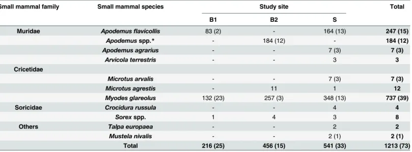
Table 1. Number of collected small mammal species from each study site with the number of small mammals positive forLeptospiraspp. as detected by real-time PCR in brackets.
Small mammal family Small mammal species Study site Total
B1 B2 S
Muridae Apodemusflavicollis 83 (2) - 164 (13) 247 (15)
Apodemusspp.* - 184 (12) - 184 (12)
Apodemus agrarius - - 7 (3) 7 (3)
Arvicola terrestris - - 3 3
Cricetidae
Microtus arvalis - - 7 (3) 7 (3)
Microtus agrestis - 11 1 12
Myodes glareolus 132 (23) 257 (3) 348 (13) 737 (39)
Soricidae Crocidura russula - - 4 4
Sorexspp. 1 4 3 8
Others Talpa europaea - - 2 2
Mustela nivalis - - 2 (1) 2 (1)
Total 216 (25) 456 (15) 541 (33) 1213 (73)
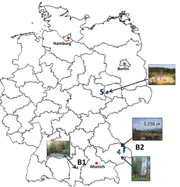
at two sampling sites at low altitude (both small beech forests at 379 m and 412 m a.s.l.,Fig 1) most small mammals of B2 were PCR positive (n: 12). However,Leptospiraspp. were found up to an altitude of 1.298 m a.s.l. (seeFig 1). From the altogether 73 PCR-positive samples, 67 could be further determined by duplex PCR. The other six samples did not yield any of the two amplicons most likely due to the high CT value achieved by real-time PCR (>39). Fifty-four
(80.3%; 95%CI: 69–88.3) of these 67 samples were identified asL.kirschneri. Four of the other 13 (19.7%; 95%CI: 11.8–31) samples were further determined asL.borgpetersenii, and 9 asL.
interrogansby conventional PCR of the partialgyrB gene and amplicon sequencing.
Leptospira borgpeterseniiwas exclusively andL.interroganswas mainly found inApodemus
spp. (n: 7/9, 77.8%, 95%CI: 44.3–94.7). Only twoM.glareoluswere positive forL.interrogans
(Table 2).Leptospira kirschneriwas mainly detected in positive small mammals (34 of 36 posi-tiveM.glareolus, 94.4%; 95%CI: 81–99.4; 14 of 25 positiveApodemusspp., 56%, 95%CI: 37.0– 73.35).Apodemus agrarius,Mustela nivalisandMi.arvalis, though captured in small numbers, showed high prevalences forL.kirschneri(42.86%; 95%CI: 15.75–75.02; 50%; 95%CI: 9.5–90.5 and 37.5%; 95%CI: 13.5–69.6 respectively (Table 1)).Leptospira kirschneriwas obtained in four of the five rodent species (A.agrarius,Mi.arvalis,M.glareolus,A.flavicollis). In contrast,L.
interrogansandL.borgpeterseniiwere detected only inA.flavicollisandM.glareolus. All other investigated small mammal species were negative.
Whereas mostL.kirschneri-positive rodents were found at site S (n: 27/54, 50.94%; 95%CI: 37.88–63.88), the majority of other pathogenicLeptospiraspp.-positive samples were found at site B2 (n: 10/13, 76.92%; 95%CI: 49.06–92.5) (χ2= 30.0823; p<0.00001), at a sampling site 379 m a.s.l.
Multi Locus Sequence Typing (MLST)
From 67 samples tested positive by duplex PCR, for 14 a complete MLST covering all seven housekeeping genes could be determined for rodents captured at all three study sites (tenM.
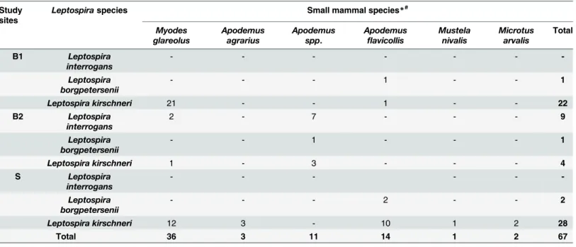
Table 2. Number of the Leptospira species detected in the small mammals per study site*#.
Study sites
Leptospiraspecies Small mammal species*#
Myodes glareolus
Apodemus agrarius
Apodemus spp.
Apodemus
flavicollis
Mustela nivalis
Microtus arvalis
Total
B1 Leptospira interrogans
- - -
-Leptospira borgpetersenii
- - - 1 - - 1
Leptospira kirschneri 21 - - 1 - - 22
B2 Leptospira interrogans
2 - 7 - - - 9
Leptospira borgpetersenii
- - 1 - - - 1
Leptospira kirschneri 1 - 3 - - - 4
S Leptospira
interrogans
- - -
-Leptospira borgpetersenii
- - - 2 - - 2
Leptospira kirschneri 12 3 - 10 1 2 28
Total 36 3 11 14 1 2 67
*numbers may differ from sum of positive samples detected by real-time PCR as some samples could not be further determined.
#from the following small mammal species none was positive forLeptospiraspp. detected by real-time PCR:Arvicola terrestris,Crocidura russula,Talpa
europaea,Sorexspp. andMicrotus agrestis.
glareolus, threeApodemusspp. and oneA.agrarius). Altogether sequence types for elevenL.
kirschneriand threeL.interroganspositive samples were detected. AllLeptospira interrogans
positive samples were found to be ST 24 (Table 2). ForL.kirschneritwo different sequence types were detected. Four samples were positive for ST 117 and seven for ST 110. While ST 110 could be detected at all three sites, ST 117 was only detected at site S. Here, ST 117 could be detected inA.flavicollis,A.agrariusandM.glareoluswhereas ST 110 could only be detected in
M.glareolus.Leptospira interrogansST 24 could be detected inA.flavicollisandM.glareolus
but exclusively at site B2 (Table 2).
Discussion
This study focussed on pathogenicLeptospiraspecies in small mammals from selected habitats in Germany. Studies on prevalences forLeptospiraspp. in mammals in Europe are rare and focussed mainly on larger rodent species such as rats (Rattus norvegicus) which are considered to be the major source ofLeptospirainfection for humans [34,35]. High prevalences (20–88%)
Fig 1. Map of Germany showing the trapping areas (blue dots) of the study sites B1, B2 and S.
inRattus norvegicushave been reported from different European countries such as Turkey, France and Denmark [36,37,38]. Studies from Germany, Switzerland, the Netherlands, Croa-tia and Austria showed the occurrence ofLeptospiraspp. in a wide range of different small mammal species includingM.glareolus,Apodemusspp.,Mi.arvalis,Mus musculus,Castor fiberandSorexspp. (2.9–71.4%)[5,22,39,40,41,42,43]. This study’s prevalences show a simi-lar wide range in prevalence regarding the investigated rodent species (5.3–42.9%). The highest prevalence in small mammals was detected at site B1, a forest in southern Bavaria in compari-son to the other two study sites. A recent German study showed high prevalences of leptospiral DNA inMi.arvalisandA.agrarius(12–14%) which are supposed to be the most common car-rier hosts forL.kirschneri[5]. In this study the highest prevalence was also found in both of these rodent species forL.kirschneri.Leptospira kirschneriwas detected in almost all investi-gated rodent species (M.glareolus,Mi.arvalis,A.flavicollis,A.agrarius) with the exception of
Mi.agrestissuggesting that thisLeptospiraspecies has a broad host range and is well adapted to a number of different small mammal species. Additionally,L.kirschneriwas found inMustela nivalisbut not inSorexspp. which therefore may play a subordinate role as maintenance host forL.kirschneri.
Human leptospirosis case reports caused byL.kirschneriare scarce. A recent study from Poland, however, reported antibody titres against ten serovars belonging toL.kirschneri,L.
borgpetersenii and L.interrogansin several healthy humans [21]. This argues for asymptomatic infections and a less pathogenic potential than in other pathogenicLeptospiraspecies. Leptos-pira kirschneriis known to cause unspecific clinical symptoms in dogs including diarrhoea, lethargy and dehydration [44]. Human cases caused byL.kirschnerimay likewise display such unspecific illness and Leptospirosis may well be overlooked or kept undiagnosed. Nevertheless, thisLeptospiraspecies should be taken into account as a possible cause of disease in mammals other than dogs and humans.
Leptospira interrogansis known to cause severe symptoms such as pneumonia, hepatitis and kidney failure in humans and dogs [45,46]. The hazardous impact ofL.interrogansto human health was recently described in France in human cases with symptoms such as lumbar myalgia and pneumonia [20]. Moreover unspecific clinical symptoms such as lethargy and fever in humans with previous outdoor activities (e.g. strawberry harvesters, triathletes) were reported in Germany and Austria [12,13,15,16]. High prevalences (33.3–100%) were detected inMi.arvalis,Mus musculusandRattus norvegicuswhich occurred sympatrically to human Leptospirosis outbreaks in France and Germany [16,20]. In former studiesMi.arvaliswas also pointed out to be one of the most important maintenance hosts forL.interrogansamong small mammal species [47,48]. In the current study, however, this highly pathogenicLeptospira spe-cies was mostly detected inApodemusspp. and exclusively at site B2, at four locations in a national park, which leads to the assumption thatL.interrogansin contrast toL.kirschneri(at least of ST 110, see below) rather occurs focally.
Leptospira borgpeterseniiand in particular serovar Hardjo type Hardjobovis is reported as the most causative leptospiral agent for infertility and abortion in cattle from North America [49].Leptospira borgpeterseniistrains were described to be associated withMus musculus[50]. In the present study thisLeptospiraspecies was detected at all three sites but, exclusively and significantly more often inApodemusspp. than in any other small mammal species which sug-gests that certainL.borgpetersenii strainshave probably a host preference for the genus Apode-musin the investigated habitats.
activities, such as strawberry harvesting [16].Leptospira kirschneriST 117 was formerly found inA.agrariusandA.flavicolliscollected in Croatia [42]. In our study this ST was also found in both of Apodemus species and additionally inM.glareolus. In SpainLeptospira interrogansST 24was detected in dogs and wild carnivores such asVulpes vulpesshowing clinical signs, thus several carnivore species were suggested to be not maintenance but dead end hosts for Leptos-pira interrogans[51]. Further ST 24 was detected inA.flavicollisfrom Croatia [42]. In the pres-ent study, this sequence type was found in two differpres-ent rodpres-ent species (M.glareolus,A.
flavicollis).
It should be taken into account that the comparison of our results between sites and species is limited due to different DNA extraction methods and as the animals examined were not caught in the same years.
In summary, in our study at three sites in Germany pathogenicLeptospiraspp. were detected in high prevalences in four of five investigated rodent species. Therefore humans could during leisure time activities get into contact with these pathogenicLeptospiraspp. if respective transmission conditions are optimal. Regarding theLeptospiraspp. prevalences this study’s results suggest a host preference forL.borgpeterseniiinApodemusspp. Moreover a broad host spectrum was detected forL.kirschneriwhich was the most common species detected in this study. Besides this study is proving first evidence ofL.kirschneriST 110 and 117 as well asL.interrogansST 24 in rodents from Germany.
Acknowledgments
The authors wish to thank Claudia Thiel, Pauline Bleichert, Daniela Eder, Stefan Frey, Mareike Pollaerts, Robin Reiter, Rahime Terzioglu, Florian Goldberg, Harald Weber, Tim Tiedemann, Jennifer Krieg, Daniela Sum, Franziska Eller and Claudia Kehler for their help in lab and field work.
Author Contributions
Conceived and designed the experiments: CS MP SE. Performed the experiments: MP SE DW SS CK AO. Analyzed the data: DW AO. Contributed reagents/materials/analysis tools: CS MP SE. Wrote the paper: AO SE CS MP.
References
1. Reis RB, Ribeiro GS, Felzemburgh RD, Santana FS, Mohr S, Melendez AX, et al. Impact of environ-ment and social gradient on Leptospira infection in urban slums. PLoS Negl Trop Dis. 2008; 2: e228. doi:10.1371/journal.pntd.0000228PMID:18431445
2. Levett PN. Leptospirosis. Clin Microbiol Rev. 2001; 14: 296–326. PMID:11292640
3. Faine S, Adler B, Bolin C, Perolat P, Leptospira and Leptospirosis. 2nd edition. Melbourne, Australia, MediSci., 1999; pp. 83–86.
4. Meerburg BG, Singleton GR, Kijlstra A. Rodent-borne diseases and their risks for public health. Crit Rev Microbiol. 2009; 35: 221–270. doi:10.1080/10408410902989837PMID:19548807
5. Mayer-Scholl A, Hammerl JA, Schmidt S, Ulrich RG, Pfeffer M, Woll D, et al. Leptospira spp. in Rodents and Shrews in Germany. Int J Environ Res Public Health. 2014; 11: 7562–7574. doi:10.3390/ ijerph110807562PMID:25062275
6. Vijayachari P, Sugunan AP, Shriram AN. Leptospirosis: an emerging global public health problem. J Biosci. 2008; 33: 557–569. PMID:19208981
7. Bharti AR, Nally JE, Ricaldi JN, Matthias MA, Diaz MM, Lovett MA, Levett PN, et al. Leptospirosis: A zoonotic disease of global importance. Lancet Infect Dis. 2003; 3: 757–771. PMID:14652202
8. Jansen A, Schöneberg I, Frank C, Alpers K, Schneider T, Stark K. Leptospirosis in Germany 1962– 2003. Emerg Infect Dis. 2005; 11: 1048–1054. PMID:16022779
10. Hoffmeister B, Peyerl-Hoffmann G, Pischke S, Zollner-Schwetz I, Krause R, Müller MC, Graf A, Kluge S, Burchard GD, Kern WV, Suttorp N, Cramer JP. Differences in clinical manifestations of imported ver-sus autochthonous leptospirosis in Austria and Germany. Am J Trop Med Hyg. 2010; 83: 326–35. doi: 10.4269/ajtmh.2010.10-0040PMID:20682876
11. Roczek A, Forster C, Raschel H, Hörmansdorfer S, Bogner KH, Hafner-Marx A, Lepper H, Dobler G, Büttner M, Sing A. Severe course of rat bite-associated Weil's disease in a patient diagnosed with a new Leptospira-specific real-time quantitative LUX-PCR. J Med Microbiol. 2008; 57:658–63. doi:10. 1099/jmm.0.47677-0PMID:18436602
12. Windpessl M, Prammer W, Nömeyer R, Dinkhauser P, Wimmer L, Müller P, et al. Leptospirosis and renal failure: a case series. Wien klin Wochenschr.2014; 126: 238–242. doi: 10.1007/s00508-014-0501-0PMID:24496714
13. Radl C, Müller M, Revilla-Fernandez S, Karner-Zuser S, de Martin A, Schauer U, et al. Outbreak of lep-tospirosis among triathlon participants in Langau, Austria, 2010. Wien klin Wochenschr. 2011; 123: 751–755. doi:10.1007/s00508-011-0100-2PMID:22105111
14. Hoenigl M, Wallner C, Allerberger F, Schmoll F, Seeber K, Wagner J, Valentin T, Zollner-Schwetz I, Flick H, Krause R. Autochthonous leptospirosis in South-East Austria, 2004–2012. PLoS One. 2014 Jan 20; 9(1): e85974. doi:10.1371/journal.pone.0085974PMID:24465820
15. Brockmann S, Piechotowski I, Bock-Hensley O, Winter C, Oehme R, Zimmermann S, et al. Outbreak of leptospirosis among triathlon participants in Germany, 2006. BMC Infect Dis. 2010; 10: 91. doi:10. 1186/1471-2334-10-91PMID:20380736
16. Desai S, van Treeck U, Lierz M, Espelage W, Zota L, Sarbu A, et al. Resurgence of field fever in a tem-perate country: an epidemic of leptospirosis among seasonal strawberry harvesters in Germany in 2007. Clin Infect Dis. 2009; 48: 691–697. doi:10.1086/597036PMID:19193108
17. Thaipadungpanit J, Wuthiekanun V, Chierakul W, Smythe LD, Petkanchanapong W, Limpaiboon R, et al. A dominant clone of Leptospira interrogans associated with an outbreak of human leptospirosis in Thailand. PLoS Negl Trop Dis. 2007; 1: e56. PMID:17989782
18. Gravekamp C, van de Kemp H, Franzen M, Carrington D, Schoone GJ, van Eys GJ, et al. Detection of seven species of pathogenic leptospires by PCR using two sets of primers. J Gen Microbiol. 1993; 139: 1691–1700. PMID:8409911
19. Bourhy P, Herrmann Storck C, Theodose R, Olive C, Nicolas M, Hochedez P, et al. Serovar diversity of pathogenic Leptospira circulating in the French West Indies. PLoS Negl Trop Dis. 2013; 7: e2114. doi: 10.1371/journal.pntd.0002114PMID:23516654
20. Dupouey J, Faucher B, Edouard S, Richet H, de Broucker C-A, Marié J-L, et al. Epidemiological investi-gation of a human leptospirosis case reported in a suburban area near Marseille. New Microbes New Infect. 2014; 2: 82–83. doi:10.1002/nmi2.45PMID:25356349
21. Wasiński B, Sroka J, Wójcik-Fatla A, Zając V, Cisak E, Knap JP, et al. Seroprevalence of leptospirosis
in rural populations inhabiting areas exposed and not exposed to floods in eastern Poland. Ann Agric Environ Med. 2012; 19.
22. Woll D, Karnath C, Pfeffer M, Allgöwer R. Genetic characterization of Leptospira spp. From beavers found dead in south-west Germany. Vet Microbiol. 2012; 158: 232–234. doi:10.1016/j.vetmic.2012.02. 022PMID:22410308
23. Forstdirektion Oberbayern-Schwaben: Managementplan zum FFH-Gebiet 7829–301“Angelberger Forst“, 2004.
24. Overzier E, Pfister K, Herb I, Mahling M, Böck G Jr, Silaghi C. Detection of tick-borne pathogens in roe deer (Capreolus capreolus), questing ticks (Ixodes ricinus) and ticks infesting roe deer in southern Ger-many. Ticks Tick Borne Dis. 2013a; 4: 320–328.
25. Overzier E, Pfister K, Thiel C, Herb I, Mahling M, Silaghi C. Diversity of Babesia and Rickettsia Species in Questing Ixodes ricinus: A Longitudinal Study in Urban, Pasture, and Natural Habitats. Vector Borne Zoonotic Dis. 2013b; 13: 559–564.
26. Thoma BR, Müller J, Bässler C, Georgi E, Osterberg A, Schex S, et al. Identification of factors influenc-ing the Puumala virus seroprevalence within its reservoir in a montane forest environment. Viruses. 2014; 6: 3944–3967. doi:10.3390/v6103944PMID:25341661
27. Silaghi C, Hamel D, Thiel C, Pfister K, Pfeffer M. Spotted fever group rickettsiae in ticks, Germany. Emerg Infect Dis. 2011; 17: 890–892. doi:10.3201/eid1705.101445PMID:21529404
29. Stresemann E., 1989, Exkursionsfauna von Deutschland, Wirbeltiere, Volume 3. Heidelberg: Spek-trum Akademischer Verlag, Gustav Fischer.
30. Parson W, Pegoraro K, Niederstätter H, Föger M, Steinlechner M. Species identification by means of the cytochrome b gene. Int J Legal Med. 2000; 114: 23–28. PMID:11197623
31. Stoddard RA, Gee JE, Wilkins PP, McCaustland K, Hoffmaster AR. Detection of pathogenic Leptospira spp. through TaqMan polymerase chain reaction targeting the LipL32 gene. Diagn Microbiol Infect Dis. 2009; 64:247–255. doi:10.1016/j.diagmicrobio.2009.03.014PMID:19395218
32. Slack AT, Symonds ML, Dohnt MF, Smythe LD. Identification of pathogenic Leptospira. species by con-ventional or real-time PCR and sequencing of the DNA gyrase subunit B encoding gene. BMC Micro-biol. 2006; 6: 95. PMID:17067399
33. Smythe L, Adler B, Hartskeerl RA, Galloway RL, Turenne CY, Levett PN, International Committee on Systematics of Prokaryotes Subcommittee on the Taxonomy of Leptospiraceae. Classification of tospira genomospecies 1, 3, 4 and 5 as Leptospira alstonii sp. nov., Leptospira vanthielii sp. nov., Lep-tospira terpstrae sp. nov. and LepLep-tospira yanagawae sp. nov., respectively. Int J Syst Evol Microbiol. 2013; 63: 1859–62.
34. Maiden MC, Bygraves JA, Feil E, Morelli G, Russell JE, Urwin R, et al. Multilocus sequence typing: a portable approach to the identification of clones within populations of pathogenic microorganisms. Proc Natl Acad Sci USA. 1998; 95: 3140–5. PMID:9501229
35. Tucunduva de Faria M, Athanazio DA, Gonçalves Ramos EA, Silva EF, Reis MG, Ko AI. Morphological alterations in the kidney of rats with natural and experimental Leptospira infection. J Comp Pathol. 2007; 137: 231–8. PMID:17996544
36. Sunbul M, Esen S, Leblebicioglu H, Hokelek M, Pekbay A, Eroglu C. Rattus norvegicus acting as reser-voir of leptospira interrogans in the Middle Black Sea region of Turkey, as evidenced by PCR and pres-ence of serum antibodies to Leptospira strain. Scand J Infect Dis. 2001; 33: 896–8. PMID:11868761 37. Aviat F, Blanchard B, Michel V, Blanchet B, Branger C, Hars J, et al. Leptospira exposure in the human
environment in France: a survey in feral rodents and in fresh water. Comp Immunol Microbiol Infect Dis. 2009; 32: 463–476. doi:10.1016/j.cimid.2008.05.004PMID:18639932
38. Krøjgaard LH, Villumsen S, Markussen MDK, Jensen JS, Leirs H, Heiberg A-C. High prevalence of Leptospira spp. in sewer rats (Rattus norvegicus). Epidemiol Infect. 2009; 137: 1586–1592. doi:10. 1017/S0950268809002647PMID:19393116
39. Kocianova E, Kozuch O, Bakoss P, Rehacek J, Kovacova E. The prevalence of small terrestrial mam-mals infected with tick-borne encephalitis virus and leptospirae in the foothills of the southern Bavarian forest. Germany Appl Parasitol. 1993; 34: 283–290. PMID:8298661
40. Adler H, Vonstein S, Deplazes P, Stieger C, Frei R. Prevalence of Leptospira spp. in various species of small mammals caught in an inner-city area in Switzerland. Epidemiol Infect. 2002; 128: 107–109. PMID:11895085
41. Hartskeerl PA, Terpstra WJ. Leptospirosis in wild animals. Vet Q. 1996; 18: 149–150. PMID:22077115
42. Turk N, Milas Z, Margaletic J, Staresina V, Slavica A, Riquelme-Sertour N, et al. Molecular characteri-zation of Leptospira spp. strains isolated from small rodents in Croatia. Epidemiol Infect. 2003; 130: 159–166. PMID:12613757
43. Schmidt S, Essbauer SS, Mayer-Scholl A, Poppert S, Schmidt-Chanasit J, Klempa B, et al. Multiple infections of rodents with zoonotic pathogens in Austria. Vector Borne Zoonotic Dis. 2014; 14: 467– 475. doi:10.1089/vbz.2013.1504PMID:24915446
44. Greenlee JJ, Bolin CA, Alt DP, Cheville NF, Andreasen CB. Clinical and pathologic comparison of acute leptospirosis in dogs caused by two strains of Leptospira kirschneri serovar grippotyphosa. Am J Vet Res. 2004; 65: 1100–1107. PMID:15334844
45. Schulze MH, Raschel H, Langen HJ, Stich A, Tappe D. Severe Leptospira interrogans serovar Ictero-haemorrhagiae infection with hepato‐renal‐pulmonary involvement treated with corticosteroids. Clin Case Rep. 2014; 2: 191–196. doi:10.1002/ccr3.91PMID:25614810
46. Geisen V, Stengel C, Brem S, Müller W, Greene C, Hartmann K. Canine leptospirosis infections– clini-cal signs and outcome with different suspected Leptospira serogroups (42 cases). J Small Anim Pract. 2007; 48: 324–328. PMID:17490440
47. Kuiken T, van Dijk JE, Terpstra WJ, Bokhout BA. The role of the common vole (Microtus arvalis) in the epidemiology of bovine infection with Leptospira interrogans serovar hardjo. Vet Microbiol. 1991; 28: 353–361. PMID:1949549
48. Treml F, Pejcoch M, Holesovska Z. Small mammals—natural reservoirs of pathogenic leptospiroses. Vet Med Czech. 2002; 47: 309–311.
50. Perez J, Brescia F, Becam J, Mauron C, Goarant C. Rodent abundance dynamics and leptospirosis carriage in an area of hyper-endemicity in New Caledonia. PLoS Negl Trop Dis. 2011; 5: e1361. doi: 10.1371/journal.pntd.0001361PMID:22039557