www.spsp.org.br
REVISTA PAULISTA
DE PEDIATRIA
1984-0462/$ - see front matter © 2014 Sociedade de Pediatria de São Paulo. Published by Elsevier Editora Ltda. All rights reserved. KEYWORDS
Asphyxia neonatorum; Apgar score;
Neonatal screening
Abstract
Objective: To evaluate the effects of perinatal asphyxia on the level of the response to transient otoacoustic emissions in infants.
Methods: Otoacoustic emissions in 154 neonates were performed: 54 infants who suffered asphyxia at birth, measured by Apgar score and medical diagnosis, and 100 infants without risk were compared. Scores less than 4 in the first minute and/or less than 6 in the fifth minute were considered as “low Apgar”. Statistical analysis of the data was performed using the Kruskal, Wilcoxon, and Mann-Whitney nonparametric tests.
Results: Lower levels of response were observed in transient otoacoustic emission in the group that suffered perinatal asphyxia, with significant values for the frequencies 2,000, 3,000, and 4,000 Hz in the right ear, and 2,000 and 4,000 Hz in the left ear.
Conclusions: The analysis of the intrinsic characteristics of the otoacoustic emissions evidenced low performance of outer hair cells in neonates who had perinatal asphyxia, which may affect the development of listening skills in this population.
© 2014 Sociedade de Pediatria de São Paulo. Published by Elsevier Editora Ltda. All rights reserved.
ORIgINAL ARTIcLE
Assessment of levels of otoacoustic emission response in neonates
with perinatal asphyxia
☆Georgea Espindola Ribeiro*, Daniela Polo Camargo da Silva, Jair Cortez Montovani
Universidade Estadual Paulista (UNESP), Botucatu, SP, Brazil
Received 8 January 2013; accepted 26 March 2014
DOI refers to: 10.1590/1984-0462201432307
☆Study conducted at: Departament of Oftalmology, Otorrinolaringology and Head and Neck Surgery, Faculdade de Medicina de Botucatu, Universidade Estadual Paulista, Botucatu, SP, Brazil.
*corresponding author.
E-mail: georgea_espindola@hotmail.com (g.E. Ribeiro). PALAVRAS-CHAVE
Asixia neonatal; Índice de Apgar; Triagem neonatal
Avaliação dos níveis de resposta das emissões otoacústicas em neonatos com asixia
perinatal
Resumo
Objetivo: Avaliar os efeitos da asfixia perinatal sobre os níveis de resposta das emissões otoacústicas por estímulo transiente em lactentes.
Apgar e diagnóstico médico ao nascimento, e 100 bebês sem risco. Escores abaixo de 4 no primeiro minuto e/ou menores que 6 no quinto minuto foram considerados “Apgar baixo”. A análise estatística do conjunto de dados foi efetuada utilizando-se os testes não paramétricos de Kruskal, Wilcoxon e Mann-Whitney.
Resultados: Foram observados menores níveis de resposta nas emissões otoacústicas transientes para o grupo que sofreu asfixia perinatal, com valores estatisticamente sig-nificantes para as frequências de 2000, 3000 e 4000Hz na orelha direita e 2000 e 4000Hz na orelha esquerda.
Conclusão: A análise das características intrínsecas do exame de emissões otoacústicas transientes mostrou baixo desempenho das células ciliadas externas em neonatos que tiveram asfixia perinatal, o que pode afetar o desenvolvimento das habilidades auditivas nessa população.
© 2014 Sociedade de Pediatria de São Paulo. Publicado por Elsevier Editora Ltda. Todos os direitos reservados.
Introduction
The Apgar score allows for the evaluation of newborn clini-cal status and identification of those in need of assistance, assessing the risks of perinatal asphyxia.1 It consists of five
criteria: heart rate, breathing effort, muscle tone, reflex irritability, and skin color. Each item is given values ranging from 0 to 2; the higher the score, the better the conditions at birth.2,3
This evaluation is performed by the neonatologist in the first, fifth, and tenth minute of life. However, perinatal asphyxia develops when there is significant tissue hypo-perfusion and decreased oxygen supply, resulting from sev-eral etiologies in the peripartum period, which can cause neurological lesions and damage cochlear hair cells.2 Apgar
scores lower than 4 in the first minute and/or less than 6 in the fifth minute are considered risk factors for hearing loss and deserve attention.4
The first hearing assessment should be performed in the hospital nursery, using the otoacoustic emissions test for assessing the integrity of outer hair cells.5,6 In most studies
on hearing screening with otoacoustic emissions, the cri-terion used to characterize a normal exam is based on the presence of response.7 However, for Aidan et al,8 one of the
criteria to assess the actual status of the inner ear is the analysis of the intrinsic characteristics of this examination, such as the response magnitude of these emissions.
According to Basseto et al,9 full-term newborns have
higher response amplitudes when compared to preterm newborns. The female gender and the right ear often have larger amplitudes.9,10 The use of ototoxic drugs can cause
lower amplitude response for otoacoustic emissions.11
From the perspective that analysis of the signal/noise ratio can provide additional information on the operation of outer hair cells, this study aimed to evaluate the levels of response to otoacoustic emissions evoked by transient stimuli in infants who had perinatal asphyxia.
Methods
This study was approved by the Research Ethics committee of Faculdade de Medicina de Botucatu (FMB) – Universidade
Estadual Paulista (UNESP), process No. 4156/2012. Data were collected at the Center for Rehabilitation of Hearing and Communication Disorders (Centro de Reabilitação dos Distúrbios da Audição e Comunicação – CERDAC), Hospital das Clínicas (HC) FMB-UNESP, from April 2012 to April 2013.
The study consisted of a non-concurrent cohort with a fixed population. Inclusion criteria were: a) having being born in the HC, b) presence of response in the otoacoustic emissions test, c) informed consent signed by the parents/ guardians of the neonate. The exclusion criteria were: a) middle ear disorders diagnosed by the otorhinolaryngolo-gist, b) presence of genetic syndromes, c) history of con-genital infections, and d) use of ototoxic drugs.
For exposed individuals, an Apgar score of less than 4 in the first minute and/or less than 6 in the fifth minute was considered, as they are at risk for hearing loss, and this score was deemed “low Apgar”. The medical diagnosis of perinatal asphyxia for this group was also taken into account. The diagnosis of perinatal asphyxia was consid-ered by the physician according to the clinical manifesta-tions of the newborn, and was classified as neurological, cardiovascular, respiratory, metabolic, renal, gastrointesti-nal, or hematological. For those not exposed, only neonates with Apgar scores >7 in the first minute were selected for comparison.
There was no criterion for sample pairing, but poten-tial confounders, gestational age, birth weight, and gender were tested (Tables 2, 3, and 4, respectively). As there was no evidence of significant probable associations with the outcome, the association between asphyxia and responses to otoacoustic emissions was analyzed without the need for correction.
Regarding the sample size calculation, since this was a non-concurrent cohort with a fixed population, no sam-pling dimension was required in the planning phase, but an estimate of test power, which ranged from 60% to 85%, depending on the frequency/ear. The power estimates con-sidered a simple random sample, normality of outcomes, type I error of 0.05, and equal standard deviation between individuals with and without low Apgar.
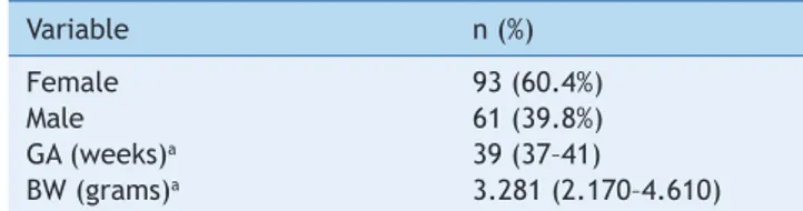
-Table 1 Sample characterization
Variable n (%)
Female 93 (60.4%)
Male 61 (39.8%)
gA (weeks)a 39 (37–41)
BW (grams)a 3.281 (2.170–4.610)
a Summary in median, minimum, and maximum;
GA, gestational age; BW, birth weight
ogy. Responses from both ears were recorded, with the infant in a state of post-prandial natural sleep, in their mothers’ arms, after 48 hours of life, in a silent room. If the infant awoke during the examination, the mother or guardian was advised to make the infant sleep again. The equipment used in all assessments was OtoRead/ Interacoustics (Interacoustics do Brasil, RJ, Brazil), which allows for the recording of responses by introduc-ing a probe, coupled with a microphone, in the ear canal. The parameter PASS/FAIL was used as a criterion of analysis, described in the equipment protocol, with click stimuli, at an intensity of 83 dB peSPL, and six bands of frequencies (from 1,500Hz to 4,000Hz) were evaluated. Values that were considered PASS were emissions present in a signal/noise ratio of 6 dB in at least three consecu -tive frequency bands, including the 4,000 Hz band. The examination lasted 64 seconds, at most.
The signal/noise ratios considered for the analysis were frequencies 2,000; 3,000 and 4,000 Hz in both ears.12
The values obtained at each frequency were compared between groups. Statistical analysis of the data set was performed using the nonparametric Kruskal-Wallis test, Spearman’s correlation, and the Mann-Whitney test. The nonparametric test was used because the probabilis-tic distribution of the outcome was not identified. The descriptive level was highlighted in all tests and a sig-nificance level of 0.05 (5%) was used to reject the null hypothesis.
Results
The study included 154 infants; sample characterization regarding gender, mean gestational age, and birth weight is shown in Table 1.
Before the analysis of perinatal asphyxia effect on the response level of otoacoustic emissions, the effect of ges-tational age and birth weight as potential confounders was investigated, but no statistical significance was observed (Tables 2 and 3).
However, when investigating gender, higher response amplitudes were observed for females at 2,000Hz, 3,000Hz, and 4,000Hz in the right ear, and 3,000Hz in the left ear (Table 4).
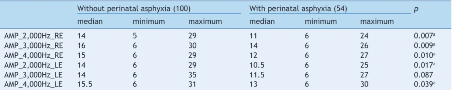
When comparing infants who suffered perinatal asphyxia with those who were healthy, lower response amplitudes were observed in individuals exposed to the risk indicator for hearing loss in all frequencies, except at 3,000Hz in the left ear (Table 5). In this case, the analysis was adjusted in relation to gender, as it was a potential confounding factor.
Discussion
One of the current methods to diagnose early hearing loss is hearing screening by otoacoustic emissions, which aims at identifying infants with possible hearing impairment. Its analysis is based on the categorization of responses as pres -ent or abs-ent,13 but only those considered absent are
can-didates for diagnostic hearing evaluation by other methods, except in cases with suspected auditory neuropathy.
In the study of the amplitude of otoacoustic emissions in relation to gender, significantly higher mean amplitudes were observed in newborn females, with a predominance of the right ear, as reported by other studies.8,14 According
to cassidy & Ditty,14 higher amplitudes in the examination
of transient otoacoustic emissions in females when com-pared to males can be attributed to increased sensitivity of the outer hair cells in females. The analysis of the sig-nal/noise ratio was also studied by other authors, such as Jiang et al,15 who observed significantly lower amplitudes
at frequencies of 1 kHz and 10 kHz in otoacoustic emis -sion testing by distortion products in neonates with low Apgar scores, suggesting cochlear impairment, even with the presence of response. The magnitude of the response was also shown to be influenced by other risk factors, such as hyperbilirubinemia, prematurity, and exposure to oto-toxic drugs.16-18
These findings demonstrate that it is necessary to bet-ter investigate the cribet-teria of normal cochlear function, especially the pass/fail criterion of emissions, as studies in adult individuals have shown loss of cochlear function in those exposed to noise and ototoxic medication19-21 long
before the decrease in psychoacoustic thresholds, a factor that is not possible to investigate in the neonatal stage.
Literature shows that perinatal asphyxia is a major cause of failure in the neonatal hearing screening exami-nation.22,23 However, when analyzing the amplitude of oto
-acoustic emissions, this study observed lower values than those found in newborns with no risk indicators for hearing loss at birth. This indicates the possibility of damage to cochlear cells caused by tissue hypoxia, an information not taken into account for the criteria of normal otoacoustic emissions. Therefore, these infants should undergo clini-cal follow-up, as proper development of auditory skills depends on the integrity of the peripheral auditory system, and thus, parents should be informed. It is believed that other tests, such as brainstem auditory evoked potentials (BAEPs), already used in the clinical routine in neonates with low Apgar scores and electrocochleography, could assist in interpreting these findings.
Acknowledgements
The authors would like to thank the children and their par-ents/guardians for participating in the study and Hospital das Clínicas da Faculdade de Medicina de Botucatu, for allowing the study to be performed.
Conlicts of interest
The authors declare to have no conflicts of interest.
References
1. Catlin EA, Carpenter MW, Brann BS, Mayield SR, Shaul PW, Goldstein M. The Apgar score revisited: inluence of gestacional age. J Pediatr 1986;109:865-8.
2. Corrêa RR, Salge AK, Ribeiro GA, Ferraz ML, Reis MA, Castro EC et al. Anatomic and pathological placenta alterations and Apgar score variations. Rev Bras Saude Mater Infant 2006;6:239-43. 3. Stoll BJ. Panorama da morbidade e mortalidade. In: Kligman
RM, Behrman RE, Jenson HB, Stanton BF, editors. Tratado de Pediatria. 18th ed. São Paulo: Elsevier; 2009. p. 679.
Table 2 Association between gestational age and amplitude per frequency
37 weeks 38 weeks 39 weeks 40 weeks 41 weeks p
med min max med min max med min max med min max med min max
AMP_2,000Hz_RE 13 6 28 12 5 24 11 6 29 14 6 29 11 6 24 0.764
AMP_3,000Hz_RE 15 6 23 15 6 26 15 6 30 16 6 29 17 6 21 0.933
AMP_4,000Hz_RE 14 6 26 14 7 26 15 6 27 15 6 29 14 6 27 0.986
AMP_2,000Hz_LE 11 6 28 10 6 24 15 6 28 14 6 28 11.5 6 29 0.337
AMP_3,000Hz_LE 16 7 20 14 6 27 15 6 26 12 6 35 13.5 6 30 0.847
AMP_4,000Hz_LE 13 6 29 13 7 26 17 6 31 15 6 27 14 7 30 0.289
(*) Kruskal Wallis; AMP, amplitude; RE, right ear; LE, left ear Summary in median (med), minimum (min), and maximum (max)
Table 3 Association between birth weight (BW) and amplitude of transient otoacoustic emissions
AMP 2,000Hz-RE
AMP 3,000Hz-OD
AMP 4,000Hz-OD
AMP 2,000Hz-OE
AMP 3,000Hz-OE
AMP 4,000Hz-OE
BW r 0.04 0.15 0.05 0.10 0.04 0.02
p 0.633 0.068 0.557 0.209 0.641 0.810
n 154 154 154 154 154 154
(*) Spearman’s correlation
BW, birth weight; AMP, amplitude; RE, right ear; LE, left ear; n, number of participants.
Table 4 Association between gender and amplitude
Female Male p
Median Minimum Maximum Median Minimum Maximum
AMP_2,000Hz_RE 14 6 29 11 5 28 0.012a
AMP_3,000Hz_RE 17 6 30 14 6 26 0.000a
AMP_4,000Hz_RE 17 7 29 14 6 26 0.000a
AMP_2,000Hz_LE 14 6 29 11 6 25 0.054
AMP_3,000Hz_LE 15 6 30 12 6 35 0.041a
AMP_4,000Hz_LE 15 6 30 14 6 31 0.167
a≤0.05 Mann-Whitney test
AMP, amplitude; RE, right ear; LE, left ear.
Table 5 comparison between newborns with and without perinatal asphyxia (in relation to amplitude of response)
Without perinatal asphyxia (100) With perinatal asphyxia (54) p
median minimum maximum median minimum maximum
AMP_2,000Hz_RE 14 5 29 11 6 24 0.007a
AMP_3,000Hz_RE 16 6 30 14 6 26 0.009a
AMP_4,000Hz_RE 15 6 29 12 6 27 0.010a
AMP_2,000Hz_LE 14 6 29 10.5 6 25 0.017a
AMP_3,000Hz_LE 14 6 35 11.5 6 27 0.087
AMP_4,000Hz_LE 15.5 6 31 13 6 30 0.039a
a≤0.05 Mann-Whitney test
4. Joint Committee on Infant Hearing. Year 2007 position statement: principles and guidelines for early hearing detection and intervention programs. Pediatrics 2007;120:898-921.
5. Garcia CF, Isaac ML, de Oliveira JA. Transient evoked otoacoustic emissions: tool for early detection of hearing alteration in full-term and preterm neonates. Rev Bras Otorrinolaringol 2002;68:344-52.
6. Costa JM, de Almeida VF, de Oliveira CA, Sampaio AL. Transient and distortion product evoked otoacoustic emissions in premature infants. Arq Int Otorrinolaringol 2009;13:309-16. 7. Simonek MC, de Azevedo MF. False-positive results in newborn
universal hearing screening: possible causes. Rev CEFAC 2011;13:292-8.
8. Aidan D, Lestang P, Avan P, Bonils P. Characteristics of transient-evoked otoacoustic emissions (TEOEs) in neonates. Acta Otolaryngol 1997;117:25-30.
9. Basseto MC, Chiari BM, Azevedo MF. Transient evoked otoacoustic emissions (TEOAE): response amplitude in term and pre-term neonates Rev Bras Otorrinolaringol 2003;69:84-92.
10. Melo AD, Alvarenga KF, Modolo DJ, Bevilacqua MC, Lopes AC, Agostinho-Pesse RS. Transient evoked otoacoustic emissions in full-term and preterm newborns. Rev CEFAC 2010;12:115-21. 11. Zorowka PG, Schmitt HJ, Gutjahr P. Evoked otoacustics
emissions and pure tone threshold audiometry in patients receiving cisplatinum therapy. Int J Pediatr Otorhinolaryngol 1993;25:73-80.
12. Côrtes- Andrade IF, Bento DV, Lewis. Emissions (TEOE): newborn hearing screening program protocols. Rev CEFAC 2013;15: 521-7.
13. Lima GM, Marba ST, Santos MF. Hearing screening in a neonatal intensive care unit. J Pediatr (Rio J) 2006;82:110-4.
14. cassidy JW, Ditty KM. gender differences among newborns on a transient evoked otoacoustic emissions test for hearing. J Music Ther (Spring) 2001;38:28-35.
15. Jiang ZD, Zang Z, Wilkinson AR. Distortion product otoacoustic emissions in term infants with a low Apgar score. Acta Otolaryngol 2006;126:1062-6.
16. Silva DP, Martins RH. Analysis of transient otoacoustic emissions and brainstem evoked auditory potentials in neonates with hyperbilirubinemia. Braz J Otorhinolaryngol 2009;75:381-6.
17. Santos AF, Durante AS, Almeida K, Taguchi CK, Greco MC. characteristics of otoacoustic emissions in infants exposed to ototoxic drugs. Rev Soc Bras Fonoaudiol 2009;14:521-7. 18. cavalcante JM, Isaac ML. Análise das emissões otoacústicas
transientes em recém-nascidos a termo e pré-termo. Braz J Otorhinolaryngol 2013;79:582-8.
19. Santos CF, Valete CM, Martins AG, Ferreira NG, Tomita S. Aspectos clínicos da ototoxicidade dos aminoglicosídios. Acta AWHO 2000;19:160-4.
20. Negrão MA, Soares E. Variation in amplitudes of evoked otoacoustic emissions and suceptibility to hearing loss induzed by nois-hlin. Rev CEFAC 2004;6:414-22.
21. Marques FP, da Costa EA. Exposure to occupational noise: otoacoustic emissions test alterations. Rev Bras Otorrinolaringol 2006;72:362-6.
22. Wroblewska-Seniuk K, Chojnacka K, Pucher B, Szczapa J, Gadzinowski J, Grzegorowski M. The results of newborn hearing screening by means of transient evoked otoacoustic emissions. Int J Pediatr Otorhinolaryngol 2005;69:1351-7.