The article was published by Academy of Chemistry of Globe Publications www.acgpubs.org/RNP © Published 10/12/2015 EISSN:1307-6167
Rec. Nat. Prod
. 10:3 (2016) 294-306
Bio-pesticidal and Antimicrobial Coumarins from
Angelica
dahurica
(Fisch. Ex Hoffm)
Qian
Xie
1, Shun-Xiang
Li
1, Duan-Fang Liao
1, 4, Wei Wang
1, 4*, Babu
Tekwani
2, Hui-Yong Huang
1, Abbas Ali
2, Junaid ur Rehman
2, Kevin K.
Schrader
3, Stephen O. Duke
3, Charles L. Cantrell
3and David E. Wedge
1, 3*1
TCM and Ethnomedicine Innovation & Development Laboratory, Sino-Luxemburg TCM Resaerch Center, School of Pharmacy , Hunan University of Chinese Medicine,
Changsha, Hunan, 410208, China 2
University of Mississippi, National Center for Natural Products Research, & Department of Pharmacology, University of Mississippi, MS, 38677. USA
3
United States Department of Agriculture, Agricultural Research Service, Natural Products Utilization Research Unit, National Center for Natural Products Research, University of Mississippi, MS, 38677.
USA 4
Division of Stem Cell Regulation and Application with Chinese Materia Medica, Hunan University of
Chinese Medicine,Changsha 410208,China
(Received January 19, 2014; Revised July 15, 2014; Accepted September 26, 2014)
Abstract: Angelica dahurica (Fisch. Ex Hoffm)is an important traditional Chinese herb which is widely used in curing acne, ulcers, carbuncles, rheumatism, headaches and toothaches. A systematic antifungal bioassay-guided fractionation of the ethyl acetate extract from A. dahurica led to the isolation of six coumarins, namely, suberosin, bergapten, alloimperatorin, xanthotoxol, 5-methoxy-8-hydroxypsoralen and pabulenol. These coumarins were subsequently subjected to evaluation for antifungal, antibacterial, antimalarial, antileishmanial and mosquito larvicidal activities. Bergapten and xanthotoxol showed good antifungal activity against
Phomopsis viticola,with 99.8% and 73.0% fungal growth inhibition at the concentration of 30 µM. Among all the compounds tested against Phomopsis obscurans, bergapten and suberosin showed the highest antifungal activity with 61.0% and 88.3% inhibition at a 30 µM concentration, respectively. 5-Methoxy-8-hydroxypsoralen displayed toxicity against Streptococcus iniae with an IC50 of 11.6 mg/L and MIC values of 2.32 mg/L.
Suberosin and alloimperatorin displayed moderate activity against Leishmania donovani promastigotes with IC50
of 4.43 µg/mL and 20.43 µg/mL. Herein, the antifungal activities of suberosin against P. obscurans and xanthotoxol against P. viticola, the antileishmanial activities of suberosin and alloimperatorin, and the antibacterial activity 5-methoxy-8-hydroxy-psoralen against the fresh water fish pathogen S. iniae were reported for the first time.
Keywords: Angelica dahurica; Apiaceae; biopesticide; antibacterial; coumarin; bio-guided isolation. © 2015 ACG Publications. All rights reserved.
1. Introduction
Increasing resistance to agrochemicals by insects, weeds, and plant pathogens and the loss of labeled use for biopesticides are factors that drive the need to search for new natural product-based pest management products. The necessity for a larger “tool box” of agrochemical interventions to
*
Corresponding author: E-Mail:[email protected]; Phone:1-662-915-1137 Fax:1-662-915-1035,
prevent plant pests and diseases is always needed, especially for insects that carry serious vector born diseases. Because pesticide registration is often far easier for natural products than synthetic chemicals, the agrochemical and pharmaceutical industry should be interested in agrochemicals and pharmaceutical compounds obtained from plants that have a long and safe history such as those used in Traditional Chinese Medicines (TCM). Bio-prospecting natural products allows for the discovery of new agrochemicals and drugs from plants used worldwide in traditional medicine.
The genus Angelica L. (Apiaceae) has been used in traditional medicines in Asia for centuries.
Angelica dahurica (Fisch. Ex Hoffm.) Benth. et Hook F. is native to northern China, Siberia, far eastern Russia, Korea and Japan. The species often grows along river banks and streams. Common plant names include Dahurian angelica (English), Bai Zhi (Chinese), and Gurisdae (Korean). The drug
is known as Xiang Bai Zhi (Chinese), Byakushi (Japansese), Paegchi (Korean) and Chinese Angelica
root (English) [1]. A. dahurica was primarily cultivated in Hubei, Liaoning, and Zhejiang provinces, but now due to ideal local climate and soil conditions, the major commercial production area in China is the Suining district of Sichuan Province. The root is usually harvested in the late summer or early autumn when the leaves turn yellow. The rootlets are cleaned and usually dried in the sun without processing. The best quality of A. dahurica root is identified as possessing a strong aromatic aroma along with a pungent and bitter taste [2]. Angelica root was used as a traditional medicine in ancient China as early as 400 BC for the treatment of illnesses such as toothache, headache, cough, asthma, pain relief, anti-inflammatory, sedative, an antifungal cream for the skin and alleviation of vaginal discharge [3-5]. The root of A. dahurica is also widely known to contain furanocoumarins[6-7].
We have experience in the study of Angelica species for agrochemical applications and several species were shown to be active as mosquito biting deterrents and antifungal in nature. Previously, we studied the chemical composition and antifungal activity of Angelica sinensis essential oil against
Colletotrichum spp [8]. Prior research evaluating the mosquito biting deterrent activity of A. sinensis
demonstrated (Z)-ligustilide to be a highly active molecule against Ae. Aegypti [9]. Therefore, we began another study of A. dahurica species with Hunan University of Chinese Medicine in 2012. Originally, the extracts from A. dahurica were tested in TLC-bioautography with Colletotrichum
species as the antifungal detection agents, and results demonstrated that the ethyl acetate (EtOAc) extract from A. dahurica showed strong antifungal activity (Figure 1). In order to isolate and identify the antifungal constituents in the root of A. dahurica, a systematic bioassay-guided fractionation of the EtOAc extract was performed. Our objective was to identify and characterize bio-pesticides, mosquito biting deterrent and repellent compounds, and to test for activity against leishmania and pathogens that cause opportunistic infections in humans.
The bioassay-guided fractionation and bio-pestidical activities of essential oils from several
Angelica species have been reported [8-9]. However, our results showed that antimicrobial activity seemed to follow the coumarin-containing fractions. Therefore, the focus of this project was to identify and characterize A. dahurica phytochemicals with potential bio-pesticide, antileishmania and antibacterial activity against common bacterial pathogens of fish.
2. Materials and Methods
2.1. Instrumentation
1
H- and 13C-NMR spectrum data were recorded on a 600 MHz Varian (Palo Alto, CA, USA) spectrometer. Column chromatography was performed using a Biotage, Inc. HorizonTM Pump (Charlottesville, Virginia, USA) equipped with a HorizonTM Flash Collector and a fixed wavelength (254and 365 nm) detector. Semi-preparative HPLC purifications and HPLC method development work was performed using an Agilent (Santa Clara, CA, USA) 1200 system equipped with a quaternary pump, auto-sampler, diode-array detector, and vacuum degasser. Biotage purifications were used with a SNAP cartridge (XP-Sil, 100g, 40−63 μm, 60Å, 40 x 150 mm, Biotage, LLC, 10430 Harris Oaks Blvd., Suite C, Charlotte, NC USA 28269). Fractions and purified compounds were
Figure 1. Direct TLC-bioautography using C. fragariae as the antifungal detection agent. (*Concentration: 20mg/mL; 1. Essential oil; 2. Residue; 3. n-BuOH extract; 4. EtOAc extract; 5. Petroleum ether extract; 6. 70% EtOH extract)
2.2. Plant Material
Roots of A. dahurica (Fisch. Ex Hoffm) (Lot number: 120401, April, 2012) were produced by the Hunan Province Songlingtang Traditional Chinese medicines Co. LTD (Changsha, Hunan Province, P.R.China) and was identified by professor Wei Wang, preserved in TCM and Ethnomedicine Innovation & Development Laboratory (No.201204628-1).
2.3. Plant Extraction
The air-dried roots of A. dahurica (3 kg) were ground followed by hydrodistillation in water (5h) providing 2.2 mL of essential oil (I), residual water and plant residue after filtration and centrifugation of the water. Residual water was concentrated (2000 mL) and to this was added 70% EtOH followed by filtration to remove the precipitate which provided 157.5 g of ethanol extract (II). Plant residue was air-dried and refluxed in excess 95% EtOH. The 95% ethanol extract (volume 100 mL) was suspended in 250 mL H2O and partitioned sequentially with petroleum ether (III), EtOAc (IV) and n-BuOH (VI).
2.4. Antifungal Bioautography-guided Fractionation
Initially, extracts I-VI were tested for antifungal activity using direct TLC-bioautography, the results showed antifungal activity in the EtOAc layer. The EtOAc extract was purified using a Biotage XP-Sil cartridge running at 40 mLmin-1 using a hexane: EtOAc gradient beginning with 100:0 to 70:30 over 1600 mL followed by 30:70 to 0:100 over 800 mL. Portions of 22 mL volume were collected in 16 x 150 mm text tubes. Test fractions were combined and concentrated on the basis of thin-layer chromatography (TLC) similarities, providing 10 fractions A - J, and fraction D yielded 10.7 mg of pure compound (1).
Fraction F was further purified using a Biotage XP-Sil, 100 g, SNAP cartridge running at 40 mLmin−1 using a hexane: EtOAc gradient beginning with 75:15 to 80:20 over 2200 mL and finishing with 80:20 to 80:83 over 50 mL. Three test tubes were collected and recombined on the basis of TLC similarities, providing 9.2 mg of pure compound (2). Four test tubes were collected and recombined on the basis of TLC, providing 36.9 mg of semi-pure compound (3). Semi-preparative HPLC was performed to purify compound (3) using a normal phase silica-gel HPLC column (Agilent, 9.4 x 250 mm, 5 μm) and running isocratic conditions (87/13, hex: EtOAc) while monitoring at 300 nm.
Fraction H was purified using a Biotage XP-Sil, 100g, SNAP cartridge running at 40 mL min−1 using a premixed solvent A: CHCl3/MeOH (95:5) and solvent B: CHCl3 gradient beginning with 100:
0 to 0:100 over 2000 mL, which yielded 87 mg fraction H1.
Fraction H1 was subsequently further purified using a Biotage XP-Sil SNAP cartridge running
at 40 mLmin−1 using a premixed solvent A: CHCl3/MeOH (95:5) and solvent B: CHCl3 (gradient
beginning with 25: 75 to 60: 40 over 2000 mL finishing with 60: 40 to 79:21 over 92 mL), which provided seven fractions H1A-H1G. Compounds 4 (7.4mg), 5 (14mg) and 6 (4.4mg) successfully
repurified from fraction H1A (35.2mg) using a normal phase silica-gel semi-preparative HPLC column
(Agilent, 9.4 x 250 mm, 5 μm), isocratic hex/EtOAc: 75/15, 300 nm using HPLC.
1 2 3
4ul
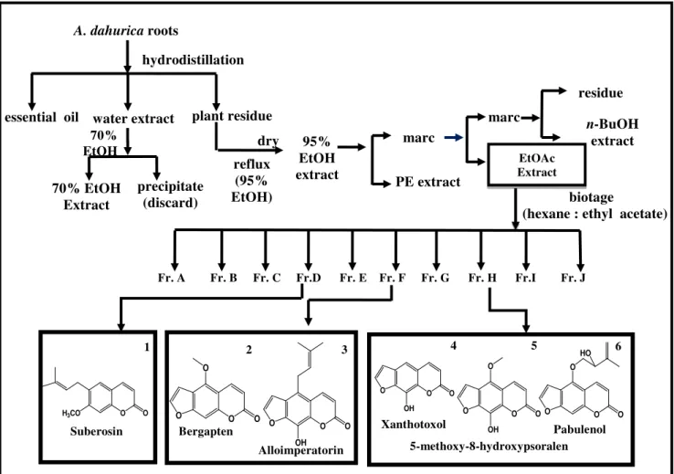
The structures of 6 compounds were identified as suberosin (1), bergapten (2), alloimperatorin (3), xanthotoxol (4), 5-methoxy-8-hydroxypsoralen (5) and pabulenol (6), by comparing 1H, 13C NMR and MS data with the literatures [10-12].
The bioassay-guided flow chart and the structures of active compounds found are in Figure2.
Figure 2. Flow chart for isolation of active compounds.
2.5. Antifungal bioassay
2.5.1. Fungal Pathogen production
We tested extracts and pure compounds on filamentous fungal plant pathogens that are serious problems in horticultural crops. Isolates of Colletotrichum acutatum Simmonds, Colletotrichum fragariae Brooks, and Colletotrichum gloeosporioides (Penz.) Penz. and Sacc. were obtained from B. J. Smith,USDA, ARS, Poplarville, MS. Cultures of P. viticola (Sacc.) Sacc. and P. obscurans (Ellis & Everh.) were obtained from Mike Ellis, The Ohio State University, OH, and Botrytis cinerea Pers.
and Fusarium oxysporum Schlechtend were isolated at USDA-ARS, NPURU Oxford, MS. The three
Colletotrichum species and P. obscurans were isolated from strawberry (Fragaria x ananassa
Duchesne), while P. viticola and B. cinerea were isolated from commercial grape (Vitis vinifera L.) and F. oxysporum from orchid (Cynoches sp.).
Fr. A Fr. B Fr. C Fr.D Fr. E Fr. F Fr. G Fr. H Fr.I Fr. J
biotage
(hexane : ethyl acetate) marc EtOAc Extract residue e n-BuOH extract reflux (95% EtOH) 95% EtOH extract PE extract marc essential oil water extract plant residue
70% EtOH Extract precipitate (discard) 70% EtOH
A. dahurica roots
hydrodistillation
1 2 3
dry
O O
H3CO O O O
O
O O O
OH
O O O
OH O O O
O
OH
O O O
O HO Suberosin Xanthotoxol 5-methoxy-8-hydroxypsoralen Pabulenol Bergapten Alloimperatorin
2.5.2. Standardization of Fungal Inoculum
Conidia of eachfungal species are harvested from 7-10 day-old cultures by flooding plates with 5 mL of sterile distilled water and dislodging conidia by softly brushing the colonies with an L-shaped glass rod. Conidial suspensions are then filtered through sterile Miracloth (Calbiochem-Novabiochem Corp., La Jolla CA) to remove mycelia. Conidia concentrations are determined photometrically from a standard curve based on the percent of transmittance (%T) at 625 nm and manual hemocytometer counts [13-14]. Conidial stock suspensions are then adjusted with sterile distilled water to a concentration of 1.0x10⁶ conidia/mL.
2.5.3. Direct Bioautography
A simple technique to visually follow antifungal components through the separation process was provided. A number of bioautography techniques are used as primary screening systems to detect antifungal compounds. Matrix, one-dimensional, and two-dimensional bioautography protocols on silica gel TLC plates and Colletotrichum spp. as the test organisms are used to identify the antifungal activity according to published bioautography methods [15-16]. Matrix bioautography is used to screen large numbers of crude extract at 20mg/mL. One-dimensional thin-layer chromatography (1-D TLC) was subsequently used to partially purify and identify the number of antifungal agents in an extract. Each plate was subsequently sprayed with a spore suspension (105 spores/mL) of the fungus of
interest and incubated in a moisture chamber for 4 days at 26°C with a 12 h photoperiod. Clear zones
of fungal growth inhibition on the TLC plate indicated the presence of antifungal constituents in each extract.
2.5.4. Micro-dilution Broth Assay
A standardized 96-well micro-dilution broth assay developed by Wedge and Kuhajek was used to evaluate antifungal activity towards B. cinerea, C. acutatum, C. fragariae, C. gloeosporioides, P. viticola, P. obscurans and F. oxysporum to the various antifungal agents in comparison with known fungicidal standards[14]. Captan, and azoxystrobin which represent two different modes of action, were used as standards in this experiment. Each fungus was challenged in a dose-response format using test compounds where the final treatment concentrations were 0.3, 3.0 and 30 uM. The commercial fungicide standard, captan was run at 0.3, 3.0 and 30 uM. After inoculation, microtiter plates (Nunc MicroWell, untreated; Roskilde, Denmark) were covered with a plastic lid and incubated in a growth chamber as described previously for fungal growth. Growth was then evaluated by measuring absorbance of each well at 620 nm using a microplate photometer (Packard Spectra Count, Packard Instrument Co., Downers Grove, IL). Microtiter plates were covered with a plastic lid and
incubated in a growth chamber at 24 ± 1 C and 12 h photoperiod under 60 ± 5 µmol light. Growth was
then evaluated by measuring absorbance of each well at 620 nm using a microplate photometer (Packard Spectra Count, Packard Instrument Co., Downers Grove, IL). Mean absorbance values and standard errors were used to evaluate fungal growth at 48 h and 72 h. Due to the slow germination and growth for
Phomospis species, P. obscurans and P. viticola growth was evaluated at 120 and 144 hrs. Analysis of variance of means for percent inhibition/stimulation of each fungus at each dose of test compound relative the untreated positive growth controls were used to evaluate fungal growth.
2.6. Antibacterial Bioassay
were used in this study. Final test concentrations of the crude extracts in the microplate wells were 0.1, 1.0, 10.0, and 100.0 mg/L. Three replications were used for each dilution of each crude extract and controls. Initially, dissolved test material or drug controls were micropippeted separately into
individual microplate wells (10 µL/well), and solvent was allowed to completely evaporate before 0.5 MacFarland bacterial culture [prepared as described previously by Schrader and Harries (2006)] was
added to the microplate wells (200 µL/well) [17]. Microplates were incubated at 29±1°C. A SpectraCount microplate photometer (Packard Instrument Company, Meriden, CT) was used to measure the absorbance (630 nm) of the microplate wells at time 0 and 24 h. The means and standard deviations of absorbance measurements were calculated, graphed, and compared to controls to help determine the 24-h IC50 and MIC for each crude extract
[17]
. The 24-h IC50 and MIC results for each
test extract were divided by the respective 24-h IC50 and MIC results obtained for the positive controls
florfenicol and oxytetracycline to determine the relative-to-drug-control florfenicol (RDCF) and relative-to-drug-control oxytetracycline (RDCO) values.
2.7. Antileishmanial Screen (LEM)
The anti-leishmanial screen (LEM) tests samples for their ability to inhibit L. donovani, a fly-borne protozoan that causes visceral leishmaniasis. Crude extracts are initially tested in a Primary Screen at 80µg/mLin duplicate and percent inhibitions (% inh.) are calculated relative to negative and positive controls. Extracts showing ≥50% inhibition proceed to the Secondary Assay. In the Secondary LEM Assay, all samples (2 and 20 mg/mL) are tested at 40, 8.0 and 1.6µg/mLand IC50s as well as
IC90s (test concentration that affords 90% inhibition of the protozoan relative to controls) are
reported. Samples that have an IC50of <1.6µg/mLin the Secondary LEM assay proceed to the Tertiary
Assay where the sample is tested at 40, 8, 1.6, 0.32, 0.064, 0.0128µg/mLand IC50s and IC90s are
reported. The in vitro antileismanial assay was done on a culture of L. donovani promastigotes by Alamar Blue assay as reported earlier [18]. In a 96 well microplate the samples with appropriate dilution were added to the leishmania promastigotes culture (2x10⁶ cell/mL). The compounds were tested at six concentrations ranging from 40 to 0.0128 µg/mL. The plates were incubated at 26°C for 72 hours (37°C for amastigote) and growth of leishmania promastigotes was determined. IC50 and IC90
values were computed from the dose response curves using the XLFit fit curve-fitting software. Pentamidine and amphotericine B were tested as positive antileishmanial drug controls.
2.8. Herbicide Bioassays
All bioassays were done in duplicate in sterile non-pyrogenic polystyrene 24-well cell culture plates (CoStar 3524, Corning Incorporated). One filter paper disk (Whatman Grade 1, 1.5 cm) was placed in each well to be used. The control wells contained 200 μL of Millipore water. The control + solvent well contained 180 μL of water and 20 μL of the solvent. All sample wells contained 180 μL of water and 20 μL of the appropriate dilution of the sample. Water was always pipetted into the well before the sample or solvent. All plate preparation was done in a sterile environment to lessen chances of any possible contamination. When prepping lettuce plates, five seeds were placed in each well. Lids were sealed with Parafilm. The plates were incubated in a Percival Scientific CU-36L5 incubator under continuous light conditions at 26C and 120.1 μmol s-1 m-2 average photosynthetically active radiation. Plates were incubated for at least seven days. Ranking of plant growth was subjective. Ranking was based on a scale of 0 to 5. A ranking of 0 indicated no apparent inhibition (sample well plants looked identical to the control + solvent well plants). A ranking of 5 indicated no growth or complete inhibition. A ranking of five was given only if no seeds germinated.
2.9. Mosquito biting bioassays and Larval Bioassays
in its absence. This is in contrast to a repellent, a chemical that causes insects to move away from a chemical or its source.
Larval Bioassays were conducted using system described by Pridgeon et al. [21] to determine the larvicidal activity of A. dahurica essential oil and extracts against Ae. aegypti. Proportion no biting data were analyzed using SAS Proc ANOVA, (SAS Institute 2007)and means were separated using the Ryan-Einot-Gabriel-Welsch Multiple Range Test. Control mortality was corrected by using
Abbott’s formula.
3. Results and Discussion
3.1. Antifungal activity
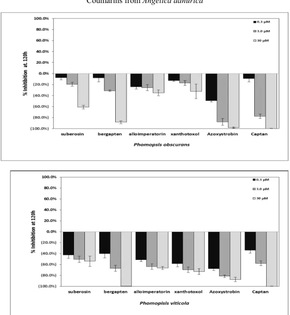
In bioautography assays, essential oil and EtOAc extracts showed strong antifungal activity against C. fragariae, fungal growth inhibition zones of the EtOAc extract applied to the TLC plate at 80 and 160 ug were 1.65 cm and 2.50 cm in diameter against C. fragariae (Figure 1), respectively. Essential oil was not available in sufficient amount for further separation. Biotage and semi-preparative HPLC were used for separating active fractions. Six pure compounds were obtained from the EtOAc extract using bioassay-guided fractionation. Only four of them showed valuable antifungal activity and the microtitter assay was performed to evaluate the antifungal activity of four compounds against seven fungal species. Compounds suberosin (1), bergapten (2), alloimperatorin (3) and xanthotoxol (4) displayed significant activity against P. viticola at 30 µM with values of inhibition 54.1%, 99.8%, 66.2%, 73.0%, respectively. Bergapten and xanthotoxol showed good antifungal activity against P. viticola with 99.8% and 73.0% fungal growth inhibition at the concentration of 30
µM, compared with inhibition 87.5 % and 100.0 % of the standard azoxystrobin and captan (Fig 3). Bergapten and xanthotoxol were the most active compounds against P. viticola, bergapten was even more active than the commercial fungicide azoxystrobin and captan. While compounds (1) - (4) appeared less active against P. obscurans at 30 µM with growth inhibition 61.0%, 88.3%, 34.9%, 32.5%, respectively. Bergapten and suberosin showed the most effective antifungal activity against P. obscurans with 61.0% and 88.3% inhibition respectively at 30µM concentration, compared with
inhibition 98.3% and 100.0% of the standard azoxystrobin and captan (Figure 3). Suberosin, bergapten, alloimperatorin and xanthotoxol showed antifungal activity against plant pathogenic fungi , these four coumarins are most likely to be the source antifungal activity of A. dahurica. In addition, weak antifungal activity was demonstrated against Cryptococcus neoformans by suberosin with IC50 values
of 13.43 µg/mL, as compared with IC50 0.239 µg/mL of the standard amphotericin, in addition,
compounds (1) - (4) did not show activity against other bacterial species(Staphylococcus aureus,
Methicillin-resistant Staphylococcus aureus (MRSa), Escherichia coli, Pseudomonas aeruginosa,
Mycobacterium intracellulare)and five fungi (Candida albicans, Candida glabrata, Candida krusei,
Aspergillus fumigatus, Cryptococcus neoformans) that are pathogenic in humans.
3.2. Antibacterial Results
Enteric septicemia of catfish (ESC) and columnaris disease in pond-raised channel catfish (Ictalurus punctatus) are caused by E. ictaluri and F. columnare, respectively, while S. iniae can cause streptococcosis in freshwater fish. Among the three test bacteria, the etheyl acetate extract had the most potent antibacterial activity against S. iniae, with a 24-h IC50 and MIC of 0.16 and 1.0 mg/L,
respectively (Table 1) and IC50 and MIC RDCO values of 2.3 and 12.5, respectively (Table 2).
Figure 3. Means and standard errors of fungal growth inhibition in response to four pure compounds from A. dahurica active against P. obscurans and P. viticola. Technical grade commercial fungicides azoxystrobin and
captan were used as standards.
Table 1. Results of the bioassay evaluation of crude extracts for toxicities against fish pathogenic bacteria. Numbers in parentheses are the standard error of the mean.
a
24-h IC50 = 50% inhibition concentration in mg/L. bMIC = Minimum inhibition concentration in mg/L. c
NA = Not applicable. d
PE =Petroleum ether.
Test bacteria
Test compound E. ictaluri F. columnare S. iniae
24-h IC50 a
MICb 24-h IC50 MIC 24-h IC50 MIC
Oxytetracycline HCl NAc
NA NA NA 0.07 (0.02) 0.08 (0.03)
Florfenicol 0.19 (0.01) 0.1 (0) 0.36 (0.1) 0.55 (0.45) NA NA
PEd Extract >100.0 100.0 (0) 24.0 (0) 10.0 (0) 48.0 (0) 10.0 (0)
Table 2. Relative-to-drug control values for the bioassay evaluation of crude extracts for toxicities against fish pathogenic bacteria.
Test bacteria
aRDCF = Relative-to-drug control florfenicol; values above and closer to “1.0” indicate stronger antibacterial activity. bRDCO = Relative-to-drug control oxytetracycline; values above and closer to “1.0” indicate stronger antibacterial activity. c24-h IC
50 = Mean 50% inhibition concentration (mg/L) of growth after 24 h of incubation. d
MIC = Minimum inhibition concentration (mg/L) of growth after 24 h of incubation.
ePE =Petroleum ether.
3.3. Antileishmanial assay Results (LEM).
Compounds suberosin and alloimperatorin displayed significant antileishmanial activity with half-maximal inhibitory concentration (IC50) values of 4.43 µg/mLand 20.43 µg/mL, respectively,
against L. donovani promastigotes, compared to those of the standard antileishmanial drugs, pentamidine (IC501.28 µg/mL) and amphotericin B (IC500.18 µg/mL) (Table 4).
3.4. Mosquito assay
sA. dahurica extracts showed weak mosquito larvicidal activity (Figure 4). Larval mortality of A. dahurica essential oil and water extract against 1-d old Ae. aegypti at 48-h post treatment is given in Figure 4. Essential oil gave 20, 90 and 100% mortality at dosages of 31.25, 62.5 and 125 ppm, respectively whereas water extract caused 12 and 64% mortality at 62.5 and 125 ppm, respectively.
Table 3. Results of the bioassay evaluation of pure compounds from A. dahurica for toxicities against
fish pathogenic bacteria S. iniae and F. columnare (ALM-00-173)
a
RDCF = Relative-to-drug control florfenicol; values above and closer to “1.0” indicate stronger antibacterial activity. b
RDCO = Relative-to-drug control oxytetracycline; values above and closer to “1.0” indicate stronger antibacterial activity. c
24-h IC50 = Mean 50% inhibition concentration (mg/L) of growth after 24 h of incubation.
dMIC = Minimum inhibition concentration (mg/L) of growth after 24 h of incubation
Test Compound E. ictaluri F. columnare S. iniae
RDCFa RDCF RDCO b
24-h IC50c MICd 24-h IC50 MIC 24-h IC50 MIC
PEe Extract >555.6 1000.00 66.7 18.2 685.7 125.0
EtOAc extract >555.6 1000.00 75.0 18.2 2.3 12.5
F. columnare (ALM-00-173) S. iniae
Test Compound 24-h
IC50a
MICb
24-h
IC50 MIC 24-h
IC50
MIC
24-h
IC50 MIC
RDCFc RDCF RDCOd RDCO
Suberosin(1) >21.50 >21.50 >31.16 >59.72 >21.50 >21.50 >179.17 >430.00 Bergapten(2) >24.40 24.40 >35.36 67.78 >24.40 >24.40 >203.33 >488.00 Alloimperatorin(3) >27.00 >27.00 >39.13 75.00 >27.00 >27.00 >225.00 >540.00 Xanthotoxol(4) 80.80 202.00 106.94 561.11 48.48 20.20 426.44 404.00
5-methoxy-
8-hydroxypsoralen(5) 71.92 23.20 109.18 64.44 7.66 2.32 128.89 46.40
Petroleum ether extract, EtOAc extract and buthanol extracts did not show any larvicial activity in screening bioassays at the highest dose of 125 ppm. Among the pure compounds (bergapten, alloimperatorin, xanthotoxol and 5-methoxy-8-hydroxypsoralen) only bergapten showed 70 and 34% larval mortality at the concentrations of 100 and 50 ppm respectively. Essential oil and the extracts of
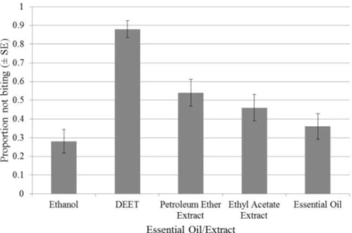
A. dahurica showed biting deterrent activity lower than the positive control, DEET (Figure 5). Petroleum ether extract and EtOAc extract showed activity higher than solvent control whereas activity of the essential oil was similar to ethanol. Pure compounds did not show any activity.
Table 4. In vitro antileishmanial activity of compounds isolated from A. dahurica against L. donovani,
thecausative agent for visceral leishmaniasis.
Sample name IC 50 (g/mL) IC 90 (g/mL)
suberosin 4.43 10.26
bergapten NA NA
alloimperatorin 20.43 NA
5-methoxy-8-hydroxypsoralen NA NA
Pentamidine 1.28 1.53
Amphotericine B 0.18 0.29
*NA = not active
3.5. Herbicide Bioassays
The EtOAc extract caused a phytotoxicity ranking at 1 mg/mL of 4 against both agrostis and lettuce (Table 5). The petroleum ether extract also showed phytotoxicity against agrostis with a ranking of 4 at 1 mg/ml, where 0 = no effect and 5 = no growth or no germination of the seeds (Table 5). Bergapten was reported in the literature to have strong activity against Lactuca sativa and Agrostis stolonifera [22], therefore further research in herbicide activity was not conducted.
Figure 5. Proportion not biting values of A. dahurica essential oil and extracts tested at a concentration of 10 µg/cm2
against female A. aegypti.
(*DEET at 25 nmol/cm2 was used a positive control while ethanol was the solvent control.)
Table 5. Herbicide Assay.
Ranking
Sample ID Tested conc. Solvent Day Lettuce Agrostis
70% EtOH extract 1 mg/mL water 7 0 0
PE extract 1 mg/mL EtOH 7 0 4
EtOAc extract 1 mg/mL EtOH 7 4 4
n-BuOH extract 1 mg/mL EtOH 7 0 0
residue 1 mg/mL water 7 0 0
* Ranking based on scale of 0 to 5 (0 = no effect, 5 = no growth)
3.6. Conclusion
In summary, extracts of A. dahurica, especially the EtOAc and the PE extracts, showed good activity in antifungal, antibacterial, and herbicidal bioassays and weak activity in mosquito bioassays. Bergapten showed significant antifungal activities against P. viticola and P. obscurans. In addition, potent herbicidal activity of bergapten has been previously reported [22]. Suberosin showed significant antifungal activities against P. obscurans, and it was the most effective compound against
L. donovani, the causative agent for visceral leishmaniasis. Alloimperatorin showed good antifungal activities against P. obscurans and can inhibit the growth of leshimania. 5-Methoxy-8-hydroxy-psoralen was determined to be antibacterial against the fish pathogenic bacterium S. iniae which has become a very important pathogen of certain freshwater fish cultured worldwide, including tilapias (Oreochromis spp.) and hybrid striped bass [Morone chrysops female x Morone saxatilis male (Percichthyidae)] [23]. Xanthotoxol displayed good antifungal activity against P. viticola. The compounds isolated from A. dahurica showed significant activity in biological assays including fungicidal, antibacterial, algicidal, herbicidal, opportunistic iinfectious pathogens, and antileishmanial bioassay,whichdemonstrates that TCM plants can provide a diverse and natural source of compounds with potential use as biopesticides and new pharmaceutical agents.
Acknowledgments
Project (2012TF1005), US Department of Defense Congressionally Directed Medical Research Program (Grant award #W81XWH-09-2-0093). United States Department of Agriculture ARS cooperative agreement (No. 58-6408-2-0009), USDA/ARS grant No. 56-6402-1-612 and Deployed War-Fighter Protection Research Program Grant funded by the U.S. Department of Defense through the Armed Forces Pest Management Board. We thank Dr. James J. Becnel, USDA-ARS, Gainesville, FL for supplying mosquito eggs.
References
[1] United States Department of Agriculture, Agricultural Research Service, Germplasm Resources Information Network, Beltsville, MD. USA. Angelica Dahurica, http://www.ars-grin.gov/cgi-bin/npgs/html/taxon.pl? 3418.
[2] J. P. Houand and Y.Y. Jin (2005). The healing power of Chinese herbs and medicinal recipes. Haworth Press. Binghampton, NY, pp: 312-315.
[3] S. H. Kim, S. S. Kang and C. M. Kim (1992). Coumarin glycosides from the roots of Angelica dahurica,
Arch. Pharm. Res. 15(1), 73-7.
[4] Y. S. Kwon, S. J. Shin, M. J. Kim and C. M. Kim(2002). A new coumarin from the stem of Angelica dahurica, Arch. Pharm. Res.25 (1), 53-56.
[5] Pharmacopoeia of the People' s Republic of China (2010), Chinese Pharmacopoeia Commission, People's Medical Publishing House. Beijing, China, Vol 1,pp: 97-98.
[6] R. M. Liu, A. F. Li and A. L. Sun (2004). Preparative isolation and purification of coumarins from Angelica dahurica (Fisch. ex Hoffm) Benth, et Hook. f (Chinese traditional medicinal herb) by high-speed counter-current chromatography, J. Chromatogr. A. 1052: 223-227.
[7] S. H. Lee, G. Li, H. J. Kim, J. Y. Kim, H. W. Chang, Y. D. Jahng, M. H. Woo, D. K. Song and J. K. Son (2003). Two new furanocoumarins from the roots of Angelica dahurica.Bull, Korean. Chem. Soc. 24, 1699-1701.
[8] N. Tabanca, D. E. Wedge, X. Wang, B. Demirci, S. J. Cutler, B. J. Smith, L. Zhou and K. H. C. Baser
(2008). Chemical composition and antifungal activity of Angelica sinensis essential oil against three Colletotrichum species, Nat. Prod. Commun. 3, 1073-1078.
[9] D. E. Wedge, J. Klun, N. Tabanca, B. Demirci, T. Ozek, K. H. C. Baser, Z. J. Liu, S. Zhang, C. L. Cantrell and J. Zhang (2009). Bioactivity-guided fractionation and GC-MS fingerprinting of Angelica sinensis and
A. archangelica root components for antifungal and mosquito deterrent activity, J. Agric. Food. Chem. 57, 464-470.
[10] A. Tosun (2006). Occurrence of coumarins in Seseli hartvigii growing in Turkey. Chem. Nat. Compd, 5, 608-609.
[11] J. Kang, L. Zhou, J. H. Sun, J. Han and D. A. Guo (2008). Chromatographic fingerprint analysis and characterization of furocoumarins in the roots of Angelica dahurica by HPLC/DAD/ESI-MSn technique, J. Pharmaceut. Biomed.47, 778-785.
[12] L. G. Avramenko, Y. E. Sklyar and M. G Pimenov (1975). Coumarins of Peucedanum baicalense, Khim. Prir. Soedin.11, 421-422.
[13] A. Espinel-Ingroff and T. M. Kerkering (1991). Spectrophotometric method of inoculum preparation for the in vitro susceptibility testing of filamentous fungi, J. Clin. Microbiol.29, 393-394.
[14] D. E. Wedge and J. M. Kuhajek (1998). A microbioassay for fungicide discovery, SAAS Bulletin Biochemistry and Biotechnology.11, 1-7.
[15] A. L. Homans and A. Fuchs (1970). Direct bioautography on thin-layer chromatograms as a method for detecting fungitoxic substances, J. Chromatogr. A.51, 327-329.
[16] D. E. Wedge and D. G. Nagle (2000). A new 2D-TLC bioautography method for the discovery of novel antifungal agents to control plant pathogens, J. Nat. Prod. 63, 1050-1054.
[17] K. K. Schrader and M. D. Harries (2006). A rapid bioassay for bactericides against the catfish pathogens
Edwardsiella ictaluri and Flavobacterium columnare, Aquacult. Res. 37, 928-937.
[18] W. Guerrant, S. C. Mwakwari, P. C. Chen, S. I. Khan, B. L. Tekwani and A. K. Oyelere (2010). A structure–activity relationship study of the antimalarial and antileishmanial activities of nonpeptide macrocyclic histone deacetylase inhibitors, Chem.Med.Chem.8, 1232–1235.
[20] Dethier, V. G., B. L. Browne, and C. N. Smith. (1960) The Designation of chemicals in terms of the responses they elicit from insects, J. Econ. Entomol.53: 134-136. M. R. Tellez, F. E. Dayan, K. K.
[21] J. W. Pridgeon, J. J. Becnel, G. G. Clark and K. J. Linthicum (2009). A high throughput screening method to identify potential pesticides for mosquito control, J. Med. Entomol. 46, 335-341.
[22] S. C. N. Queiroz, C. L. Cantrell, S. O. Duke, D. E. Wedge, V. K. Nandula, R. M. Moraes and A. L. Cerdeira (2012). Bioassay-directed isolation and identification of phytotoxic and fungitoxic acetylenes from
Conyza Canadensis, J. Agric. Food. Chem.60, 5893–5898.
[23] C. A. Shoemaker, P. H. Klesius and J. J. Evans (2001). Prevalence of Streptococcus iniae in tilapia, hybrid striped bass, and channel catfish on commercial fish farms in the United States, Am. J. Vet. Res. 62, 174-177.