Left Atrial Mechanical Function and Global
Strain in Hypertrophic Cardiomyopathy
Kyung-Jin Kim1,2☯, Hong-Mi Choi1,2☯, Yeonyee E. Yoon1,3
*, Hack-Lyoung Kim1,4, Seung-Pyo Lee1,2, Hyung-Kwan Kim1,2, Yong-Jin Kim1,2, Goo-Yeong Cho1,3, Joo-Hee Zo1,4, Dae-Won Sohn1,2
1Department of Internal Medicine, Seoul National University College of Medicine, Seoul, Republic of Korea,
2Department of Internal Medicine, Cardiovascular Center, Seoul National University Hospital, Seoul, Republic of Korea,3Division of Cardiology, Department of Internal Medicine, Seoul National University Bundang Hospital, Seongnam-si, Gyeonggi-do, Republic of Korea,4Division of Cardiology, Department of Internal Medicine, Seoul National University Boramae Medical Center, Seoul, Republic of Korea
☯These authors contributed equally to this work.
Abstract
Background
Atrial fibrillation is the most common arrhythmia and is associated with adverse outcomes in hypertrophic cardiomyopathy (HCM). Although left atrial (LA) remodeling and dysfunction are known to associate with the development of atrial fibrillation in HCM, the changes of the LA in HCM patients remain unclear. This study aimed to evaluate the changes in LA size and mechanical function in HCM patients compared to control subjects and to determine the characteristics of HCM associated with LA remodeling and dysfunction.
Methods
Seventy-nine HCM patients (mean age, 54±11 years; 76% were men) were compared to 79 age- and sex-matched controls (mean age, 54±11 years; 76% were men) and 20 young healthy controls (mean age, 33±5 years; 45% were men). The LA diameter, volume, and mechanical function, including global strain (ε), were evaluated by 2D-speckle tracking
echocardiography. The phenotype of HCM, maximal left ventricular (LV) wall thickness, LV mass, and presence and extent of late gadolinium enhancement (LGE) were evaluated with cardiac magnetic resonance imaging.
Results
HCM patients showed increased LA volume index, impaired reservoir function, and
decreased LAεcompared to the control subjects. When we divided the HCM group
accord-ing to a maximal LA volume index (LAVImax) of 38.7 ml/m2or LAεof 21%, no significant
dif-ferences in the HCM phenotype and maximal LV wall thickness were observed for patients with LAVImax>38.7 ml/m2or LAε21%. Conversely, the LV mass index was significantly
higher both in patients with maximal LA volume index>38.7 ml/m2and with LA
ε21% and was independently associated with LAVImaxand LAε. Although the LGE extent was
a11111
OPEN ACCESS
Citation:Kim K-J, Choi H-M, Yoon YE, Kim H-L, Lee S-P, Kim H-K, et al. (2016) Left Atrial Mechanical Function and Global Strain in Hypertrophic Cardiomyopathy. PLoS ONE 11(6): e0157433. doi:10.1371/journal.pone.0157433
Editor:Sakthivel Sadayappan, Loyola University Chicago, UNITED STATES
Received:December 6, 2015
Accepted:May 31, 2016
Published:June 23, 2016
Copyright:© 2016 Kim et al. This is an open access article distributed under the terms of theCreative Commons Attribution License, which permits unrestricted use, distribution, and reproduction in any medium, provided the original author and source are credited.
Data Availability Statement:Data cannot be made publicly available due to ethical restrictions set by the IRB of Seoul National University Bundang Hospital; i.e., public availability would compromise patient confidentiality and participant privacy. Please contact the corresponding author to request the minimal anonymized dataset.
increased in patients with LAε21%, it was not independently associated with either LAVImaxor LAε.
Conclusions
HCM patients showed progressed LA remodeling and dysfunction; the determinant of LA remodeling and dysfunction was LV mass index rather than LV myocardial fibrosis by LGE-magnetic resonance imaging.
Introduction
Atrial fibrillation is the most common arrhythmia in patients with hypertrophic cardiomyopa-thy (HCM), occurring in about one fifth of all HCM patients, which is four times the frequency expected in the general population [1–3], and causing substantial morbidity and mortality by promoting progressive heart failure and increasing the risk for embolic stroke [2–4]. Therefore, early recognition of susceptibility to atrial fibrillation would be advantageous for longitudinal surveillance and timely prophylactic intervention and management strategies in HCM patients. In such patients, the left atrium (LA) has been shown to have an increased size and decreased mechanical function, especially in the advanced stage [5,6]. Moreover, atrial fibrillation is more prevalent in patients who demonstrate LA remodeling and dysfunction [2,4,7]. Recently, there has been increasing interest in LA strain analysis using two-dimensional (2D) speckle-tracking echocardiography to quantify the magnitude of atrial deformation [8–11]. Previous studies have reported that the LA global longitudinal strain (ε) is decreased in patients with paroxysmal atrial fibrillation compared with normal control subjects [10] and that decreased LAεis associated with atrial fibrillation progression to a persistent or permanent stage [8] and with recurrence after catheter ablation [12]. However, little is currently known regarding the changes in LAεin patients with HCM.
Cardiac magnetic resonance imaging (MRI) has emerged as a useful adjunctive imaging modality for the diagnosis and risk stratification of HCM [13–15]. Cardiac MRI has the unique capability of acquiring tomographic images with high spatial and temporal resolution, and with excellent tissue contrast, but without limitations associated with either the imaging win-dow or imaging plane [13]. In addition, late gadolinium enhancement (LGE)-MRI allows non-invasive identification and quantification of myocardial fibrosis, which is associated with increased morbidity and mortality in HCM [15]. However, to date, little is known with regard to the cardiac MRI characteristics of HCM associated with LA remodeling and dysfunction. Accordingly, in the present study, we tried to determine the LA remodeling and functional changes, including LAε, in HCM by comparing HCM patients with age- and sex-matched control subjects and with young healthy subjects. Furthermore, we also evaluated the charac-teristics of HCM associated with increased LA size and decreased LAε.
Materials and Methods
Study population
The medical records of 182 consecutive adult patients with HCM and with sinus rhythm with-out a history of atrial fibrillation who underwent 2D speckle tracking echocardiography at Seoul National University Bundang Hospital between 2009 and 2013 were retrospectively reviewed. HCM diagnosis was established by the presence of left ventricular (LV) hypertrophy
(LV wall thickness15 mm) on echocardiography, associated with a non-dilated LV chamber, in the absence of other cardiac or systemic diseases explaining the observed hypertrophy [16]. Among these, 83 patients who underwent both 2D speckle tracking echocardiography and car-diac MRI within 3 months were assessed for eligibility. Subsequently, we excluded patients with newly diagnosed atrial fibrillation between echocardiography and cardiac MRI (n = 2) and patients with a prior coronary artery disease, defined as prior myocardial infarction, prior coronary revascularization, or coronary artery disease on prior catheterization (n = 2). Thus, the remaining 79 patients (mean age, 54 ± 11 years; 76% were men) formed the study cohort (Fig 1).
For comparison of the LA size and mechanical function in HCM patients to healthy popula-tion, we also retrospectively formed two control groups: age- and sex-matched control group and young healthy control group. The age- and sex-matched control group consisted of 79 healthy subjects with similar age and sex (mean age, 54 ± 11 years; 76% were men) who were randomly selected from the subjects who volunteered for general routine health evaluation and echocardiography. The young healthy control group consisted of 20 young healthy subjects (mean age, 33 ± 5 years; 45% were men) who volunteered for both echocardiography and car-diac MRI. None of the controls had any cardiovascular disease or systemic disease or any other
Fig 1. Flow chart of the study population.Abbreviations: HCM, hypertrophic cardiomyopathy; MRI, magnetic resonance imaging; CAD, coronary artery disease.
risk factors, and had sinus rhythm. The institutional ethics committee of Seoul National Uni-versity Bundang Hospital approved this retrospective study and waived of the requirement for both written and verbal informed consent from the entire study subjects including control sub-jects due to the retrospective nature of the evaluation without the risk of harm to study subsub-jects. Patient records/information was anonymized and de-identified prior to analysis.
Transthoracic echocardiography
A Vivid 7 ultrasound system (GE Vingmed Ultrasound AS, Horten, Norway) was utilized for the transthoracic echocardiographic examination. All images and measurements were acquired from the standard views, according to the guidelines of the American Society of Echocardiography [17–19] and were digitally stored for offline analysis. As described in detail previously [10], the LA maximum anterior-posterior (A-P) diameter was measured in the parasternal long-axis view. The following LA volumes were measured using a biplane area-length method from the apical 4-chamber and 2-chamber view and were indexed according to the body surface area: maximum LA volume index (before mitral valve opening) (LAVImax), pre-A LA volume index (before atrial
contraction) (LAVIpre-A), and minimum LA volume index (after atrial contraction) (LAVImin).
The LA expansion index (%) and active emptying fraction (%) were calculated as: [(LAVImax
−-LAVImin) / LAVImin] × 100% and [(LAVIpre-A−LAVImin) / LAVIpre-A] × 100% [10].
Global LA myocardial longitudinal strain (ε) during ventricular systole was measured by 2D speckle tracking echocardiography, as previously described [8,10]. Gray scale images of the apical 4-chamber view were obtained with frame rates of 50–80 Hz. All recordings were pro-cessed with speckle-tracking software (EchoPAC; GE Vingmed Ultrasound AS), allowing off-line semi-automated speckle-based strain analysis. Briefly, at the time of the end-systolic phase, the lines were traced manually along the LA endocardium. An additional epicardial line, which was generated automatically by the software, created a region of interest. After manually adjusting the shape of the region of interest, the LAεduring the whole cardiac cycle was calcu-lated [12,20].
Cardiac MRI
MR images were obtained by using a 1.5-T MR system (Intera CV release 10; Philips Health-care, Amsterdam, the Netherlands) with five-channel cardiac coils. All images were acquired with electrocardiographic gating and breath-holding. Steady-state free-precession cine-MR images were obtained for each patient, including vertical long-axis images, four-chamber view images, and a set of short-axis images covering the entire LV. The sequence parameters were as follows: field of view: 350–400 mm, repetition time/echo time: 3.0–3.6/1.5–1.8 ms, flip angle: 60°, slice thickness: 8 mm. Fifteen minutes after intravenous administration of 0.2 mmol/kg of gadodiamide (Omniscan; GE Healthcare), an inversion recovery-prepared, T1-weighted, gradi-ent-echo sequence was used to obtain LGE-MRI in the same planes as the cine images. The LGE imaging parameters were as follows: field of view: 350–400 mm, repetition time/echo time: 4.5–4.6/1.3–1.5 ms, flip angle: 15°, inversion time: 200–300 ms, slice thickness: 8 mm. The inversion time was adjusted to nullify the signal of the normal myocardium.
LGE mass of all slices yielded the total mass of the LGE, and the extent of LGE was expressed as a percentage of the total LV mass (the % LV mass with LGE). For statistical analysis, the LGE score and extent were divided into quartiles: LGE extent of 0%, 1–4%, 5–12%, and13%; LV mass index<56 g/m2, 57–71 g/m2, 72–83 g/m2, and84 g/m2.
Statistical analysis
The clinical, echocardiographic, and MRI parameters of the HCM patients, age- and sex-matched control subjects, and young healthy control subjects are reported. Continuous vari-ables are expressed as the means and standard deviations and categorical varivari-ables are expressed as proportions. Comparison of continuous variables was performed with the paired t-test between HCM patients and age- and sex-matched control subjects and with Student’s t-test between HCM patients and young healthy control subjects. Categorical variables were compared using theχ2 test or Fisher’s exact test, as appropriate. Between-group differences by HCM phenotype were compared by one-way analysis of variance (ANOVA) followed by Bon-ferroni’s post-hoc test. Further, we also evaluated the characteristics of HCM patients by two-group comparison according to the median values of LAVImaxand LAε. Univariate linear
regression analyses were performed to examine the effects of various characteristics of HCM on LAVImaxand LAε. Covariates obtaining aPvalue<0.2 in the univariate analyses were
included in the multivariate linear regression analyses. For all analyses, a two-sidedPvalue of less than 0.05 was considered to represent a statistically significant difference. All analyses were performed using SPSS version 20.0 (IBM, Chicago, IL)
Results
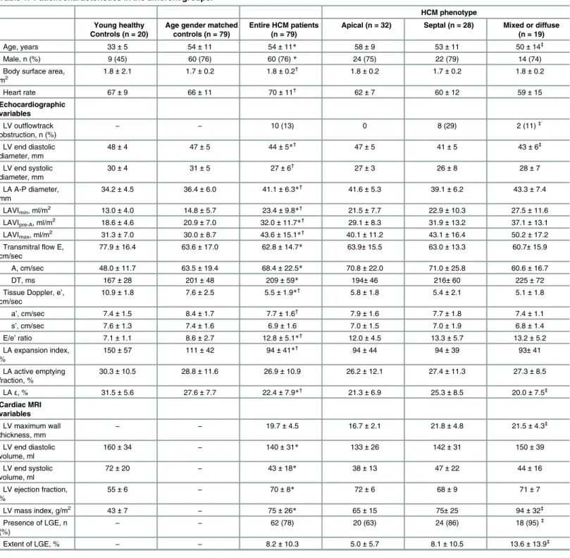
The baseline characteristics of the 79 HCM patients, 79 age- and sex-matched control subjects, and 20 young healthy control subjects are presented inTable 1. The young healthy control group comprised significantly younger subjects and less male subjects compared to the other two groups. However, there were no significant differences between the HCM group and age- and sex-matched control group with respect to age, sex, and body surface area. In the HCM group, 32 subjects (2%) had hypertension and 7 subjects (9%) had diabetes mellitus. Meanwhile, none of the patients in the age- and sex-matched group or young healthy control group had a history of hypertension or diabetes mellitus. When comparing the echocardiographic characteristics, the LA A-P diameter and LA volume indices, including LAVImax, LAVIpre-A, and LAVImin, were
sig-nificantly increased in the HCM group compared to in the control groups. The trans-mitral flow velocities were comparable, but the annular tissue velocity e’was significantly lower, and the E/e’ ratio was significantly higher in the HCM group compared to in both control groups. Further-more, while the reservoir function, as estimated by the LA expansion index, was significantly decreased in the HCM group, the contractile function, as estimated by the LA active emptying fraction, was not significantly different in the HCM group compared to in both control groups. On the other hand, LAεwas significantly decreased in the HCM group compared to in both control groups. When we compared the characteristics by HCM phenotype, there was no signifi-cant difference in the LA size and function. Although LAεwas highest in patients with septal HCM, Bonferroni post-hoc analysis did not demonstrate a significant difference between the groups (septal versus apical,P= 0.150; septal versus mixed,P= 0.067).Fig 2shows the changes in LAVImaxand LAεby HCM phenotype compared to in the control groups.
To define significant predictors of LA remodeling and dysfunction, we compared the char-acteristics of HCM patients according to the median values of LAVImaxand LAεin the HCM
group (Table 2). When we divided the HCM group by the median LAVImax,38.7 ml/m2,
Table 1. Patient characteristics in the different groups.
HCM phenotype
Young healthy Controls (n = 20)
Age gender matched controls (n = 79)
Entire HCM patients (n = 79)
Apical (n = 32) Septal (n = 28) Mixed or diffuse (n = 19)
Age, years 33±5 54±11 54±11* 58±9 53±11 50±14‡
Male, n (%) 9 (45) 60 (76) 60 (76)* 24 (75) 22 (79) 14 (74)
Body surface area, m2
1.8±2.1 1.7±0.2 1.8±0.2†
1.8±0.2 1.7±0.2 1.8±0.2
Heart rate 67±9 66±11 70±11†
62±7 60±12 59±15
Echocardiographic variables
LV outflowtrack obstruction, n (%)
− − 10 (13) 0 8 (29) 2 (11)
‡`
LV end diastolic diameter, mm
48±4 47±5 44±5*† 47±5 41±5 43±6‡
LV end systolic diameter, mm
30±4 31±5 27±6†
27±3 26±8 28±7
LA A-P diameter, mm
34.2±4.5 36.4±6.0 41.1±6.3*† 41.6±5.3 39.1±6.2 43.3±7.4
LAVImin, ml/m2 13.0±4.0 14.8±5.7 23.4±9.8*† 21.5±7.7 22.9±10.3 27.5±11.6
LAVIpre-A, ml/m2 18.6±4.6 20.9±7.0 32.0±11.7*† 29.1±8.3 31.9±13.2 37.1±13.1
LAVImax, ml/m2 31.3±7.0 30.0±8.7 43.6±15.1*† 40.1±11.2 43.1±16.4 50.2±17.2
Transmitralflow E, cm/sec
77.9±16.4 63.6±17.0 62.8±14.7* 63.9±15.5 63.0±13.3 60.7±15.9
A, cm/sec 48.0±11.7 63.5±19.4 68.4±22.5* 70.8±22.0 71.0±25.8 60.6±16.7
DT, ms 167±28 201±48 209±59* 194±46 216±60 225±72
Tissue Doppler, e’, cm/sec
10.9±1.8 7.6±2.5 5.5±1.9*† 5.8±1.8 5.4±2.1 5.1±1.8
a’, cm/sec 7.4±1.5 8.4±1.7 7.7±1.6† 7.9±1.6 7.7±1.8 7.4±1.1
s’, cm/sec 7.6±1.3 7.4±1.6 6.9±1.6 7.0±1.5 7.0±1.9 6.8±1.4
E/e’ratio 7.1±1.1 8.6±2.7 12.8±5.1*†
12.0±4.5 13.3±5.7 13.2±5.2
LA expansion index, %
150±57 111±42 94±41*† 94±44 94±39 93±41
LA active emptying fraction, %
30.3±10.5 28.8±11.6 26.9±10.9 26.2±12.1 27.4±11.3 27.3±8.5
LAε, % 31.5±5.6 27.6±7.7 22.4±7.9*† 21.3±6.9 25.3±8.5 20.0±7.5‡
Cardiac MRI variables
LV maximum wall thickness, mm
− − 19.7±4.5 16.7±2.1 21.8±4.8 21.5±4.3‡
LV end diastolic volume, ml
160±34 − 140±31* 133±26 142±31 150±39
LV end systolic volume, ml
72±20 − 43±18* 38±13 47±22 44±16
LV ejection fraction, %
55±6 − 70±8* 72±6 68±9 71±7
LV mass index, g/m2 43±7
− 75±26* 65±15 75±25 94±32‡
Presence of LGE, n
(%) − −
62 (78) 20 (63) 24 (86) 18 (95)‡
Extent of LGE, % − − 8.2±10.3 5.0±5.7 8.1±10.5 13.6±13.9
‡
Data are presented as the mean±SD or n (%). Abbreviations: LV, left ventricle; LA, left atrium; LA A-P diameter, LA maximum anterior-posterior diameter; LAVImin, minimum LA volume index; LAVIpre-A, LA volume index before atrial contraction; LAVImax, maximum LA volume index; DT, deceleration time;
LGE, late gadolinium enhancement
*p<0.05 by student’st-test (HCM group vs. young healthy control group)
†
p<0.05 by pairedt-test (HCM group vs. age- and sex-matched control group)
‡
p<0.05 by ANOVA.
increased E/e’ratio. Moreover, although the LA expansion index and active emptying fraction were not significantly different, the LAεwas significantly decreased in patients with LAVImax
>38.7 ml/m2. The LV maximal wall thickness, evaluated by cardiac MRI, was also not
signifi-cantly different according to the LAVImaxor LAε. Instead, the LV mass index, evaluated by
cardiac MRI, was significantly increased in patients with increased LAVImax, whereas the LGE
presence and extent were not. When we divided the HCM group by the median LAε, 21%, patients with decreased LAεof21% showed significantly increased LA A-P diameter and volume indices, including LAVImax, LAVIpre-A, and LAVImin. Among the MRI variables, the
HCM phenotype and maximal LV wall thickness were not significantly different in patients with decreased LAε. However, the LV mass index was increased, LGE was observed more fre-quently, and the LGE extent was significantly increased in patients with decreased LAε (Table 2).
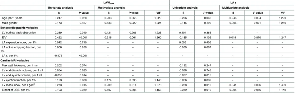
The associations of LAVImaxand LAεwith the LV variables evaluated by cardiac MRI are
demonstrated inTable 3. By univariate linear regression analysis, LAVImaxwas found to be
asso-ciated with age, presence of LV outflow track obstruction, E/e’ratio, LAε, and LV mass index, while LAεwas associated with the LV mass index and LGE extent. However, when we per-formed multivariate linear regression analyses, only the LV mass index was independently associ-ated with both LAVImaxand LAε. When we illustrated the relation between LAVImaxand LAε according to the LV mass index and the LGE extent by quartiles, both LAVImaxand LAεshowed
a graded association with the LV mass index, but not with the extent of LGE (Fig 3).
Discussion
The present study demonstrated the following findings: 1) HCM patients showed an increased LA size, impaired reservoir function, and decreased LAεcompared to control subjects. 2) LA Fig 2. Maximal left atrial volume index (A) and global longitudinal strain (B) in young healthy control subjects, age- and sex-matched control subjects, overall hypertrophic cardiomyopathy patients, and each group of hypertrophic cardiomyopathy by the phenotype. Abbreviations: LAVImax, maximum left atrial volume index; LAε, left atrial global longitudinal strain.*AllPvalues between HCM phenotypes by Bonferroni’s post-hoc test in ANOVA were greater than 0.05 (septal versus mixed,P= 0.067; septal versus apical,P= 0.150; mixed versus apical,P>0.999).
Table 2. Patient characteristics by LAVImaxand LAε.
LAVImax LAε
LAVImax38.7 ml/m2 (n = 39)
LAVImax>38.7 ml/m2 (n = 40)
LAε>21.0% (n = 39) LAε21.0% (n = 40)
Age, years 52±11 56±12 53±12 56±11
Men, n (%) 32 (82) 28 (70) 30 (77) 30 (75)
Body surface area, m2 1.8±0.2 1.7±0.2 1.8±0.2 1.8±0.2
Echocardiographic variables
LV end diastolic diameter, mm
45±5 43±6 44±5 44±6
LV end systolic diameter, mm
28±5 27±7 25±4 29±7*
LA A-P diameter, mm 38.8±5.4 43.4±6.4* 38.8±5.3 43.4±6.5*
LAVImin, ml/m2 17.2±4.6 29.6±9.7* 19.4±6.0 27.4±11.2*
LAVIpre-A, ml/m2 23.5±5.6 40.3±10.0* 26.6±7.9 37.4±12.4*
LAVImax, ml/m2 31.6±5.4 55.3±12.0* 37.6±11.6 49.5±15.9*
Transmitralflow E, cm/sec 61.7±11.1 63.8±17.7 65.1±13.1 60.6±16.0
A, cm/sec 64.8±20.1 72.0±24.3 69.5±20.8 67.4±24.3
DT, ms 196±47 221±66 199±48 219±67
Tissue Doppler, e’, cm/sec 6.1±1.9 4.8±1.8* 5.9±2.1 5.0±1.6*
a’, cm/sec 8.0±1.3 7.4±1.8 8.2±1.2 7.3±1.8*
s’, cm/sec 7.4±1.5 6.5±1.7* 7.2±1.7 6.6±1.6
E/e’ratio 10.8±3.1 14.7±6.0* 12.0±3.9 13.5±6.0
LA expansion index, % 91±36 96±46 98±38 90±45
LA active emptying fraction, %
26.6±10.3 27.2±11.6 26.4±10.5 27.4±11.4
LAε, % 25.0±7.9 19.8±7.0* 28.5±6.5 16.4±2.7*
Cardiac MRI variables
LV maximum wall thickness, mm
19.0±4.7 20.3±4.2 18.9±5.0 20.4±4.3
LV end diastolic volume, ml
139±33 141±31 138±34 143±29
LV end systolic volume, ml 45±14 41±21 41±15 44±20
LV ejection fraction, % 68±6 72±9 70±7 70±9
LV mass index, g/m2 69±25 82±25* 68±24 82±26*
Presence of LGE, n (%) 28 (72) 34 (85) 27 (69) 35 (88)*
Extent of LGE, % 6.5±9.7 9.8±10.8 4.8±5.4 11.5±12.8*
HCM phenotype
Apical type, n (%) 20 (51) 12 (30) 15 (39) 17 (43)
Septal type, n (%) 13 (33) 15 (38) 18 (46) 10 (25)
Mixed or diffuse type, n (%)
6 (16) 13 (32) 6 (15) 13 (32)
Data are presented as the mean±SD or n (%)
Abbreviations: LA, left atrium; LAVImax, maximum LA volume index; LAε, LA global longitudinal strain; LV, left ventricle; LA A-P diameter, LA maximum anterior-posterior diameter; LAVImin, minimum LA volume index; LAVIpre-A, LA volume index before atrial contraction; DT, deceleration time; LGE, late
gadolinium enhancement.
*P<0.05 by Student’st-test.
Table 3. Univariate and multivariate linear regression analyses of factors associated with LAVImaxand LAε.
LAVImax LAε
Univariate analysis Multivariate analysis Univariate analysis Multivariate analysis
ß Pvalue ß Pvalue VIF ß Pvalue ß Pvalue VIF
Age, per 1 years 0.247 0.028 0.203 0.065 1.229 -0.206 0.068 -0.246 0.034 1.229
Male gender 0.173 0.127 0.133 0.220 1.224 -0.146 0.199 -0.206 0.071 1.210
Echocardiographic variables
LV outflow track obstruction 0.289 0.010 0.121 0.266 1.226 0.104 0.366 − − −
E/e’ 0.422 <0.001 0.216 0.061 1.360 -0.185 0.102 0.019 0.870 1.247
LA expansion index, per 1% 0.042 0.710 − − − 0.095 0.406 − − −
LA active emptying fraction, per 1%
0.006 0.959 − − − -0.059 0.607 − − −
LAε, per 1% -0.473 <0.001 − − − - - − − −
Cardiac MRI variables
Max wall thickness, per 1 mm 0.202 0.074 − − − -0.132 0.247 − − −
LV end diastolic volume, per 1 ml 0.054 0.635 − − − -0.038 0.743 − − −
LV end systolic volume, per 1 ml -0.058 0.614 − − − -0.027 0.815 − − −
LV ejection fraction, per 1% 0.193 0.088 0.174 0.098 1.140 -0.026 0.839 − − −
LV mass index, per 1 g/m2 0.273 0.015 0.289 0.014 1.378 -0.288 0.010 -0.341 0.006 1.409
Extent of LGE, per 1% 0.193 0.089 0.107 0.308 1.153 -0.289 0.010 -0.205 0.066 1.149
Abbreviations: LA, left atrium; LAVImax, maximum LA volume index; LAε, LA global longitudinal strain; LV, left ventricle; LGE, late gadolinium enhancement; VIF, variance inflation factor.
doi:10.1371/journal.pone.0157433.t003
Left
Atrial
Size
and
Mechanical
Function
in
Hypertr
ophic
Cardiomy
|DOI:10.137
1/journal.p
one.0157433
June
23,
remodeling and dysfunction did not significantly differ according to the phenotype of HCM. 3) The LV mass index, evaluated by cardiac MRI, was independently associated with increased LA volume and decreased LAε, while the LGE extent was not.
Atrial fibrillation is the most common arrhythmia in HCM, occurring in 20% of patients and potentially impacting the prognosis of HCM [2]. Since atrial remodeling is recognized as a key feature in the pathogenesis and perpetuation of atrial fibrillation, evaluating the LA size and mechanical function in HCM patients has prognostic importance [2,4,22]. For that rea-son, there has been an increasing interest in noninvasive evaluations of LA size and mechanical function using 2D echocardiography. However, to date, data on LA enlargement and dysfunc-tion in HCM are sparse [5,6,23]. Yang et al. included 104 HCM patients and found that patients with HCM and LA enlargement (LAVImax>34 ml/m2) had more serious
cardiovascu-lar events and demonstrated greater LV hypertrophy and more diastolic dysfunction compared to HCM patients without LA enlargement [5]. However, HCM patients with atrial fibrillation were also included in the study, and there was no control group for comparison. Shin et al. per-formed 3D echocardiography on 26 HCM patients and 15 control subjects and found that the Fig 3. Maximal left atrial volume index (A, B) and global longitudinal strain (C, D) in young healthy control subjects, age- and sex-matched control subjects, and each group of hypertrophic cardiomyopathy stratified by left ventricular late gadolinium enhancement extent and left ventricular myocardial mass index. Abbreviations: LAVImax, maximum left atrial volume index; LGE, late gadolinium enhancement; LV, left ventricle; LAε, left
atrial global longitudinal strain.
LAVImaxwas increased and LA active emptying fraction was decreased in HCM patients [6].
However, their study included control subjects without age- and sex-matching, and, in fact, there was a significant age difference between the HCM and control groups, potentially limit-ing the value of the study findlimit-ings, as age is an important determinant of LA size and function. To our knowledge, only the study by Tigen et al. evaluated LA volume and function of HCM patients by using 2D echocardiography and compared HCM patients to age- and sex-matched control subjects [23]. The investigators demonstrated that the LAVI was significantly increased and LA reservoir function was significantly decreased in HCM patients compared to control subjects, similar to in the present study. Moreover, they also reported that the LA peak early and late diastolic longitudinal strains measured by 2D speckle-tracking echocardiography were significantly decreased in HCM patients. However, they did not perform cardiac MRI, and LA strain according to the HCM phenotype, LV mass index, LGE presence and extent were not evaluated.
Strain analysis with 2D speckle-tracking echocardiography can be applied to the LA to quantify the magnitude of LA deformation [8,10,12]. Although LA functions are traditionally estimated using 2D echocardiography and Doppler analysis of transmitral and pulmonary vein flow, 2D echocardiography is limited by the use of geometric models to determine the volume of a non-symmetric chamber and by errors due to foreshortening. In addition, the evaluation of LA function by Doppler analysis is indirect and therefore also limited. Meanwhile, strain analysis is expected to allow a more direct assessment of LA endocardial contractility and pas-sive deformation. Previous studies have already reported that LAεis significantly decreased in patients with paroxysmal atrial fibrillation compared to in normal control subjects [10] and that decreased LAεis associated with atrial fibrillation progression to the persistent or perma-nent stage [8] and with recurrence after catheter ablation [12]. In our previous study, LAεwas significantly decreased in patients with first paroxysmal atrial fibrillation (27.3 ± 7.2%) com-pared to age- and sex-matched controls (32.6 ± 7.0%) and was associated with the LA volume index and reservoir function [10]. In a subsequent multicenter study evaluating 313 patients with paroxysmal atrial fibrillation, decreased LAεwas an independent predictor of progression of atrial fibrillation and served as a good predictor even in patients without LA enlargement [8]. In the present study, we first reported a significantly decreased LAεin HCM patients (22.4 ± 7.9%) compared to age- and sex-matched control subjects (27.6 ± 7.7%) and young healthy control subjects (31.5 ± 5.6%), as well as a significantly increased LA diameter and vol-ume index. These findings support the hypothesis that HCM patients, even with sinus rhythm, might demonstrate LA remodeling and dysfunction before the development of atrial fibrilla-tion, and that LAεcould represent the progression of LA remodeling.
Although HCM patients are usually expected to have increased LA volume and decreased LA mechanical function, it has not been evaluated which characters of HCM determine the progression of LA remodeling and dysfunction. Therefore, in the present study, we also attempted to evaluate the association between HCM characteristics and LA remodeling and dysfunction. First, we defined the subtype of HCM and quantified the LV mass by using cine-MRI. Cine-MRI provides excellent contrast between the blood pool and myocardium, without limitation of either the imaging window or imaging plane, and therefore enables detailed char-acterization of the HCM phenotype. Further, cine-MRI is highly accurate and allows reproduc-ible quantification of the LV mass [24]. When we divided the HCM patients by the median values of LAVImaxand LAε, the phenotype of HCM was not significantly different in patients
with increased LAVImaxor in those with decreased LAε. The LV maximal wall thickness was
also not significantly different according to LAVImaxor LAε, whereas the LV mass index was
significantly higher in patients with increased LAVImaxand in patients with decreased LAε. In
associated with both LAVImaxand LAε. These observations raise a potentially important
con-sideration for risk stratification of HCM. Although the current risk stratification strategy for HCM uses the maximal LV wall thickness to represent the overall burden of hypertrophy, it has often been proven to be an unreliable estimate of the total LV mass index [24]. The present analysis supports the previous findings that the LV mass index rather than the maximal wall thickness may prove more relevant to the assessment of risk in HCM [24].
Lastly, we also evaluated the presence and extent of LV myocardial fibrosis by using LGE-MRI. LGE evaluated by cardiac MRI is known to correlate with LV wall thickening and to inversely correlate with the LV ejection fraction [25–27]. In addition, it has prognostic value in predicting adverse cardiovascular events, including sudden cardiac death, heart failure death, cardiovascular mortality, and all-cause mortality [15,28,29]. Therefore, the assessment of LGE by cardiac MRI is expected to have the potential to provide important information to improve risk stratification in HCM. However, in the present study, the LGE presence and extent were not significantly different in patients according to the LAVImax. Although LGE was more
fre-quently observed and the LGE extent was significantly increased in patients with decreased LA ε, the LGE extent failed to demonstrate a graded association with LAεand was not indepen-dently associated with either LAVImaxor LAεwhen adjusted for the LV mass index. Thus, in contrast to previous observations, these results suggest that the LV mass index rather than the LGE extent is an important determinant of LA remodeling and dysfunction. It is still conceiv-able that the association between LGE and LAVImaxor LAεmight bae alleviated because our
study population demonstrated relatively high prevalence of LGE of 78%. Considering that HCM is a heterogeneous cardiac disease with a diverse clinical presentation and course, further studies including larger number of patients are required to confirm our findings and to evalu-ate whether the LV mass index or LGE extent can predict the occurrence and progression of atrial fibrillation in HCM.
Study Limitations
First, the study cohort was not population-based, and a relatively small number of patients were enrolled. Thus, the generalizability of our findings might be limited. Although this study is the largest one to evaluate LAεof HCM patients compared to age- and sex-matched control subjects, we could not match control subjects by hypertension and diabetes which could affect LA size and function. Additionally, the absence of HCM with atrial fibrillation in the HCM group did not allow the evaluation of LA remodeling and dysfunction according to the occur-rence of atrial fibrillation. Second, this is a cross-sectional study; thus, we substituted LA enlargement and dysfunction for occurrence of atrial fibrillation. LA enlargement and dysfunc-tion were assessed by LAVImaxand LAε, respectively. Although LAVImaxand LAεare
well-known predictors of the occurrence, progression, and recurrence of atrial fibrillation [8,10,
significantly thinner compared with the LV wall, obtaining images with sufficient spatial reso-lution to detect atrial fibrosis in a quantifiable and objective manner remains challenging. Finally, in the present study, we aimed to evaluate LA volume and function, including LAε, by speckle tracking echocardiography in patients with HCM. Further studies are required to address these issues.
Conclusions
In this study, we demonstrated that patients with HCM have increased LAVI, impaired reser-voir function, and decreased LAεin comparison with age- and sex-matched control subjects and young healthy control subjects. LA remodeling and dysfunction, as evaluated by LAVImax,
and LAεwere associated with the LV mass index. In contrast prior studies, LV myocardial fibrosis by LGE-MRI was not independently associated with LA remodeling and dysfunction.
Acknowledgments
This study was supported by a research Grant (02-2013-028) from the Seoul National Univer-sity Bundang Hospital. We would like to acknowledge the Asian Society of Cardiovascular Imaging (ASCI) and Seoul National University Bundang Hospital medical research collaborat-ing center for their contribution to this study.
Author Contributions
Conceived and designed the experiments: KJK HMC YEY SPL. Performed the experiments: KJK HMC YEY. Analyzed the data: KJK HMC YEY GYC. Contributed reagents/materials/ analysis tools: KJK HMC YEY. Wrote the paper: KJK YEY HLK HKK YJK JHZ DWS. Interpre-tation of data: KJK HMC YEY SPL. Critical revision for important intellectual content: HLK SPL HKK YJK GYC JHZ DWS.
References
1. Maron BJ, Maron MS. Hypertrophic cardiomyopathy. Lancet. 2013; 381(9862):242–55. doi:10.1016/ S0140-6736(12)60397-3PMID:22874472.
2. Olivotto I, Cecchi F, Casey SA, Dolara A, Traverse JH, Maron BJ. Impact of atrial fibrillation on the clini-cal course of hypertrophic cardiomyopathy. Circulation. 2001; 104(21):2517–24. PMID:11714644.
3. Siontis KC, Geske JB, Ong K, Nishimura RA, Ommen SR, Gersh BJ. Atrial Fibrillation in Hypertrophic Cardiomyopathy: Prevalence, Clinical Correlations, and Mortality in a Large High‐Risk Population. Journal of the American Heart Association. 2014; 3(3):e001002. doi:10.1161/JAHA.114.001002PMID:
24965028
4. Maron BJ, Olivotto I, Bellone P, Conte MR, Cecchi F, Flygenring BP, et al. Clinical profile of stroke in 900 patients with hypertrophic cardiomyopathy. Journal of the American College of Cardiology. 2002; 39(2):301–7. PMID:11788223
5. Yang H, Woo A, Monakier D, Jamorski M, Fedwick K, Wigle ED, et al. Enlarged left atrial volume in hypertrophic cardiomyopathy: a marker for disease severity. J Am Soc Echocardiogr. 2005; 18 (10):1074–82. doi:10.1016/j.echo.2005.06.011PMID:16198885.
6. Shin M-S, Fukuda S, Song J-M, Tran H, Oryszak S, Thomas JD, et al. Relationship between left atrial and left ventricular function in hypertrophic cardiomyopathy: a real-time 3-dimensional echocardio-graphic study. Journal of the American Society of Echocardiography. 2006; 19(6):796–801. PMID:
16762759
7. Caselli S, Maron MS, Urbano-Moral JA, Pandian NG, Maron BJ, Pelliccia A. Differentiating left ventricu-lar hypertrophy in athletes from that in patients with hypertrophic cardiomyopathy. The American journal of cardiology. 2014; 114(9):1383–9. doi:10.1016/j.amjcard.2014.07.070PMID:25217454.
8. Yoon YE, Oh IY, Kim SA, Park KH, Kim SH, Park JH, et al. Echocardiographic Predictors of Progres-sion to Persistent or Permanent Atrial Fibrillation in Patients with Paroxysmal Atrial Fibrillation (E6P Study). J Am Soc Echocardiogr. 2015; 28(6):709–17. doi:10.1016/j.echo.2015.01.017PMID:
9. Saraiva RM, Demirkol S, Buakhamsri A, Greenberg N, PopovićZB, Thomas JD, et al. Left atrial strain measured by two-dimensional speckle tracking represents a new tool to evaluate left atrial function. Journal of the American Society of Echocardiography. 2010; 23(2):172–80. doi:10.1016/j.echo.2009. 11.003PMID:20152699
10. Yoon YE, Kim HJ, Kim SA, Kim SH, Park JH, Park KH, et al. Left atrial mechanical function and stiffness in patients with paroxysmal atrial fibrillation. Journal of cardiovascular ultrasound. 2012; 20(3):140–5. doi:10.4250/jcu.2012.20.3.140PMID:23185657; PubMed Central PMCID: PMC3498311.
11. Henein M, Zhao Y, Henein MY, Lindqvist P. Disturbed left atrial mechanical function in paroxysmal atrial fibrillation: a speckle tracking study. International journal of cardiology. 2012; 155(3):437–41. doi:10. 1016/j.ijcard.2011.10.007PMID:22088228
12. Hwang HJ, Choi E-Y, Rhee SJ, Joung B, Lee B-H, Lee S-H, et al. Left atrial strain as predictor of suc-cessful outcomes in catheter ablation for atrial fibrillation: a two-dimensional myocardial imaging study. Journal of interventional cardiac electrophysiology. 2009; 26(2):127–32. doi: 10.1007/s10840-009-9410-yPMID:19529886
13. To AC, Dhillon A, Desai MY. Cardiac magnetic resonance in hypertrophic cardiomyopathy. JACC: Car-diovascular Imaging. 2011; 4(10):1123–37. doi:10.1016/j.jcmg.2011.06.022PMID:21999873 14. Rickers C, Wilke NM, Jerosch-Herold M, Casey SA, Panse P, Panse N, et al. Utility of cardiac magnetic
resonance imaging in the diagnosis of hypertrophic cardiomyopathy. Circulation. 2005; 112(6):855–61. PMID:16087809
15. Chan RH, Maron BJ, Olivotto I, Pencina MJ, Assenza GE, Haas T, et al. Prognostic value of quantita-tive contrast-enhanced cardiovascular magnetic resonance for the evaluation of sudden death risk in patients with hypertrophic cardiomyopathy. Circulation. 2014; 130(6):484–95. doi:10.1161/ CIRCULATIONAHA.113.007094PMID:25092278
16. Gersh BJ, Maron BJ, Bonow RO, Dearani JA, Fifer MA, Link MS, et al. 2011 ACCF/AHA Guideline for the Diagnosis and Treatment of Hypertrophic CardiomyopathyA Report of the American College of Car-diology Foundation/American Heart Association Task Force on Practice Guidelines Developed in Col-laboration With the American Association for Thoracic Surgery, American Society of
Echocardiography, American Society of Nuclear Cardiology, Heart Failure Society of America, Heart Rhythm Society, Society for Cardiovascular Angiography and Interventions, and Society of Thoracic Surgeons. Journal of the American College of Cardiology. 2011; 58(25):e212–e60. doi:10.1016/j.jacc. 2011.06.011PMID:22075469
17. Cheitlin MD, Alpert JS, Armstrong WF, Aurigemma GP, Beller GA, Bierman FZ, et al. ACC/AHA Guide-lines for the Clinical Application of Echocardiography. A report of the American College of Cardiology/ American Heart Association Task Force on Practice Guidelines (Committee on Clinical Application of Echocardiography). Developed in collaboration with the American Society of Echocardiography. Circu-lation. 1997; 95(6):1686. PMID:9118558
18. Klues HG, Schiffers A, Maron BJ. Phenotypic spectrum and patterns of left ventricular hypertrophy in hypertrophic cardiomyopathy: morphologic observations and significance as assessed by two-dimen-sional echocardiography in 600 patients. Journal of the American College of Cardiology. 1995; 26 (7):1699–708. PMID:7594106
19. Quiñones MA, Otto CM, Stoddard M, Waggoner A, Zoghbi WA. Recommendations for quantification of Doppler echocardiography: a report from the Doppler Quantification Task Force of the Nomenclature and Standards Committee of the American Society of Echocardiography. Journal of the American Soci-ety of Echocardiography. 2002; 15(2):167–84. PMID:11836492
20. D'Andrea A, Caso P, Romano S, Scarafile R, Riegler L, Salerno G, et al. Different effects of cardiac resynchronization therapy on left atrial function in patients with either idiopathic or ischaemic dilated cardiomyopathy: a two-dimensional speckle strain study. Eur Heart J. 2007; 28(22):2738–48. doi:10. 1093/eurheartj/ehm443PMID:17959621.
21. Harrigan CJ, Peters DC, Gibson CM, Maron BJ, Manning WJ, Maron MS, et al. Hypertrophic cardiomy-opathy: quantification of late gadolinium enhancement with contrast-enhanced cardiovascular MR imaging. Radiology. 2011; 258(1):128–33. doi:10.1148/radiol.10090526PMID:21045187.
22. Maron BJ, Haas TS, Maron MS, Lesser JR, Browning JA, Chan RH, et al. Left atrial remodeling in hypertrophic cardiomyopathy and susceptibility markers for atrial fibrillation identified by cardiovascular magnetic resonance. The American journal of cardiology. 2014; 113(8):1394–400. doi:10.1016/j. amjcard.2013.12.045PMID:24589281.
23. Tigen K, Sunbul M, Karaahmet T, Dundar C, Ozben B, Guler A, et al. Left ventricular and atrial functions in hypertrophic cardiomyopathy patients with very high LVOT gradient: a speckle tracking echocardio-graphic study. Echocardiography. 2014; 31(7):833–41. doi:10.1111/echo.12482PMID:24341920.
American College of Cardiology. 2008; 52(7):559–66. doi:10.1016/j.jacc.2008.04.047PMID:
18687251.
25. Choudhury L, Mahrholdt H, Wagner A, Choi KM, Elliott MD, Klocke FJ, et al. Myocardial scarring in asymptomatic or mildly symptomatic patients with hypertrophic cardiomyopathy. Journal of the Ameri-can College of Cardiology. 2002; 40(12):2156–64. PMID:12505229
26. Olivotto I, Maron BJ, Appelbaum E, Harrigan CJ, Salton C, Gibson CM, et al. Spectrum and clinical sig-nificance of systolic function and myocardial fibrosis assessed by cardiovascular magnetic resonance in hypertrophic cardiomyopathy. The American journal of cardiology. 2010; 106(2):261–7. doi:10.1016/ j.amjcard.2010.03.020PMID:20599013.
27. Maron MS, Appelbaum E, Harrigan CJ, Buros J, Gibson CM, Hanna C, et al. Clinical profile and signifi-cance of delayed enhancement in hypertrophic cardiomyopathy. Circulation: Heart Failure. 2008; 1 (3):184–91.
28. Rubinshtein R, Glockner JF, Ommen SR, Araoz PA, Ackerman MJ, Sorajja P, et al. Characteristics and clinical significance of late gadolinium enhancement by contrast-enhanced magnetic resonance imag-ing in patients with hypertrophic cardiomyopathy. Circulation: Heart Failure. 2010; 3(1):51–8.
29. Green JJ, Berger JS, Kramer CM, Salerno M. Prognostic value of late gadolinium enhancement in clini-cal outcomes for hypertrophic cardiomyopathy. JACC: Cardiovascular Imaging. 2012; 5(4):370–7. doi: