quenching assay, inhibited intracellular fatty acid biosynthesis and growth of S. aureus, and increased the minimum inhibitory concentration forfabI-overexpressingS. aureus. The compounds that were not effective against the FabK isoform, however, inhibited the growth ofStreptococcus pneumoniaethat contained only the FabK isoform. Additionally no resistant mutant to the compounds was obtained. Importantly, fabK-overexpressing Escherichia coli was not resistant to these compounds, but was resistant to triclosan. These results demonstrate that the compounds inhibited another target in addition to FabI. Thus, meleagrin is a new class of FabI inhibitor with at least one additional mode of action that could have potential for treating multidrug-resistant bacteria.
Citation:Zheng CJ, Sohn M-J, Lee S, Kim W-G (2013) Meleagrin, a New FabI Inhibitor fromPenicillium chryosogenumwith at Least One Additional Mode of Action. PLoS ONE 8(11): e78922. doi:10.1371/journal.pone.0078922
Editor:Martin Pavelka, University of Rochester, United States of America
ReceivedApril 12, 2013;AcceptedSeptember 16, 2013;PublishedNovember 28, 2013
Copyright:ß2013 Zheng et al. This is an open-access article distributed under the terms of the Creative Commons Attribution License, which permits unrestricted use, distribution, and reproduction in any medium, provided the original author and source are credited.
Funding:This work was supported by Basic Science Research Program through the National Research Foundation of Korea (NRF) funded by the Ministry of Education, Science and Technology (2012R1A2A2A01014821) and the Intelligent Synthetic Biology Center of Global Frontier Project funded by the Ministry of Education, Science and Technology (2011-0031944). The funders had no role in study design, data collection and analysis, decision to publish, or preparation of the manuscript.
Competing Interests:The authors have declared that no competing interests exist. * E-mail: wgkim@kribb.re.kr
Introduction
Multidrug-resistant bacteria such as methicillin-resistant Staph-ylococcus aureus (MRSA), vancomycin-resistant Enterococci, and vancomycin-resistantS. aureus have become an important global health concern [1,2]. One approach to combat antibiotic resistance is to identify new drugs that can function through novel mechanisms of action. One such target is bacterial type 2 fatty acid synthesis (FASII), which is essential for bacterial cell growth [3–5]. FASII is conducted by a set of individual enzymes, whereas mammalian fatty acid synthesis is mediated by a single multifunctional enzyme-acyl carrier protein (ACP) complex referred to as type I. Enoyl-ACP reductase catalyzes the final and rate-limiting step of the chain-elongation process of the FASII. Four isoforms have been reported for enoyl-ACP reductase. FabI is highly conserved among most bacteria, including S. aureus and Escherichia coli. Streptococcus pneumoniae contains only FabK, whereas Enterococcus faecalis and Pseudomonas aeruginosa contain both FabI and FabK, and Bacillus subtilis contains both FabI and FabL. Recently, the FabV isoform was isolated fromVibrio cholera, Pseudomonas aeruginosa, andBurkholderia mallei [6,7]. No analogue protein is present in mammals for similar transformation; thus, FabI inhibitors should not interfere with mammalian fatty acid synthesis. Because of these properties, FabI is an attractive target for antibacterial drug development
[8,9]. As drugs with single targets such as rifampicin and fosfomycin are particularly vulnerable to mutational resistance [10], FabI-specific inhibitors also have a tendency to develop resistance in bacteria by mutations that alter the drug-binding site. FabI is known to be the main target for triclosan and isoniazid, which have been used in consumer products and for treating tuberculosis, respectively [11,12]. Triclosan-resistant bacteria and isoniazid-resistantM. tuberculosisare highly prevalent because of point mutations in their FabI genes [13–15]. In addition, rapid mutation development has been often reported in synthetic FabI inhibitors [16]. Thus, it has been recently emphasized that ideal antibiotics should bind to multiple targets [17].
more unique FabI inhibitors need to be obtained from microorganisms.
During our continued screening for FabI inhibitors from microbial metabolites, we found meleagrin (1) with a druggable structure during solid-state fermentation of a seashore slime-derived Penicillium chrysogenum, a penicillin-producing species (Figure 1). Here, we report the isolation and analog preparation of meleagrin, in addition to its inhibition of FabI isoforms and whole cells of various pathogenic bacteria, target validation, and its multitarget effect.
Materials and Methods
Bacterial strains
The bacterial strains used in the antibacterial activity assays were obtained from the Culture Collection of Antimicrobial Resistant Microbes of Korea and the Korean Collection for Type Cultures. The pump-negative (tolC) E. coli EW1b was obtained from the E. coli Genetic Stock Center of Yale University.
Screening and isolation of compound 1
Over 25,000 microbial extracts composed of actinomycetes and fungi were screened against S. aureus FabI and confirmed through a target-based whole cell assay by using fabI -overex-pressing S. aureus. This analysis led to the identification of compound1from fungal strain F717 (Fig. 1). Compound1was isolated from the fermented whole medium of the fungal strain F717, which was isolated from seashore slime collected at Daechun beach, Chungcheongnam-do, Korea. The strain was identified as Penicillium chrysogenum based on standard biological and physiological tests and taxonomic determination. Seed culture was conducted in a liquid culture medium containing 2% glucose, 0.2% yeast extract, 0.5% peptone, 0.05% MgSO4,
and 0.1% KH2PO4(pH 5.7 before sterilization). A sample of the
strain from a mature plate culture was inoculated into a 500-mL Erlenmeyer flask containing 80 mL of the above sterile seed liquid medium and cultured on a rotary shaker (150 rpm) at 28uC for 3 days. Subsequently, 5 mL of the seed culture was transferred into 500-mL Erlenmeyer flasks (54 flasks) containing 80 g of bran medium, which was cultivated for 7 days at 28uC to
detector. The column was eluted using MeOH: H2O (75:25) at a
flow rate of 5 mL/min to afford compound1with.99% purity at a retention time of 19.4 min. The chemical structure of compound 1 was determined to be meleagrin [23] by mass spectroscopy (MS) and nuclear magnetic resonance (NMR) spectra as follows: [a]D =296.7u (c= 0.04, MeOH);
HRESI-MS: m/z 434.18463 (M+H)+
, C23H23N5O4 requires 434.18228; 1
H-NMR (600 MHz, DMSO-d6): 8.30 (1H, s, NH-19), 8.17 (1H,
s, H-15), 7.77 (1H, s, H-20), 7.53 (1H, d,J= 7.5, H-4), 7.34 (1H, s, H-18), 7.25 (1H, t, J= 7.5, H-6), 7.03 (1H, t, J= 7.5, H-5), 6.96 (1H, d,J= 7.5, 7), 6.00 (1H, brs, 22), 5.25 (1H, s, H-8), 5.01 (1H, d, J= 17.1, Ha-23), 4.98 (1H, d, J= 9.0, Hb-23), 3.66 (3H, s, 1-OCH3), 1.19 (6H, s, CH3-24 and 25),13C-NMR
(150 MHz, DMSO-d6): 165.0 (C-13), 158.6 (C-10), 146.2 (C-7a),
143.3 22), 142.7 9), 137.6 20), 134.1 18), 127.8 (C-6), 126.0 (C-3a), 125.9 (C-1(C-6), 124.7 (C-4), 123.7 (C-12), 123.1 (C-5), 112.8 (C-23), 111.6 (C-7), 109.2 (C-8), 106.7 (C-15), 101.5 (C-2), 64.8 (1-OCH3), 52.2 (C-3), 41.8 (C-21), and 23.0 (C-24
and 25).
Preparation of derivatives of compound 1
Several derivatives of1 were obtained by chemical modifica-tion of funcmodifica-tional groups such as hydroxyl and amine groups (Fig. 1). Demethoxylation of compound1afforded glandicolin A (2) together with compound 7 as a byproduct. Methylation of compound1produced oxaline (3),N14-methylmeleagrin (4), and O,N14-dimethylmeleagrin (5). O,N14-dimethylglandicolin (6) was obtained by methylation of compound 2. Details regarding the
induction with isopropylthiogalactoside. The C-terminal His-tagged protein was purified as described previously [24]. Assays were conducted in half-area, 96-well microtiter plates. The compounds were dissolved in DMSO and evaluated in 100-mL assay mixtures containing components specific for each enzyme (see below). Reduction of thetrans-2-octenoyl N-acetylcysteamine (t-o-NAC thioester) substrate analog was measured spectrophoto-metrically following the utilization of NADH or NADPH at 340 nm at 30uC for the linear period of the assay.S. aureusFabI assays contained 50 mM sodium acetate (pH 6.5), 200mM t-o-NAC thioester, 200mM NADPH, and 150 nMS. aureusFabI.
NADH was used as a cofactor rather than NADPH for theE. coli FabI assay. Substrate concentrations used for the Line-weaver–Burk plot were 100, 200, 300, and 400mM, whereas the concentrations of the cofactor were 100, 200, 400, and 600mM. The rate of decrease in the amount of NADPH in each reaction was measured with a microtiter enzyme-linked immunosorbent assay (ELISA) reader by using the SOFTmax PRO software (Molecular Devices, Sunnyvale, CA, USA). The inhibitory activity was calculated according to the following formula: % of inhibition = 1006[12 (rate in the presence of compound/ rate in the untreated control)]. IC50 values were calculated by
fitting the data to a sigmoid equation. An equal volume of DMSO solvent was used for the untreated control. FabK assays contained 100 mM sodium acetate (pH 6.5), 2% glycerol, 200 mM NH4Cl, 50mM t-o-NAC thioester, 200mM NADH,
and 150 nMS. pneumoniae FabK.
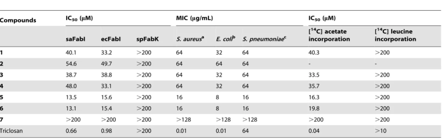
Table 1.Comparison of the inhibitory effects of meleagrin (1) and its derivatives againstStaphylococcus aureusandE. coliFabI, bacterial growth, and [14C] acetate and [14C] leucine incorporation into membrane fatty acids.
Compounds IC50(mM) MIC (mg/mL) IC50(mM)
saFabI ecFabI spFabK S. aureusa E. colib S. pneumoniaec
[14C] acetate
incorporation
[14C] leucine
incorporation
1 40.1 33.2 .200 64 32 64 40.3 .200
2 54.6 49.7 .200 64 64 64 -
-3 38.7 38.8 .200 64 32 64 33.5 .200
4 48.0 33.1 .200 64 32 64 35.7 .200
5 13.5 15.6 .200 16 8 16 16.3 .200
6 13.1 15.4 .200 16 8 16 19.8 .200
7 .200 .200 .200 .128 .128 .128 .200 .200
Triclosan 0.66 0.98 .200 0.01 0.01 64 0.04 .10
Fluorescence quenching assay
Fluorescence spectra were measured using a SHIMADZU fluorescence spectrophotometer (model RF-5310PC). S. aureus FabI (15 ng/ml) was incubated with different concentrations of triclosan (1, 2, 4, 8, and 16 nM in PBS buffer) and compounds1, 5, or 7 (10, 20, 40, 80, and 160 nM in PBS buffer). Protein quenching was monitored at 25uC by using 5-nm excitation and 5-nm emission wavelength. The excitation wavelength was
280 nm, and the emission spectra were measured between 290 and 430 nm.
Determination of minimum inhibitory concentrations (MICs)
Whole-cell antimicrobial activity was determined by broth microdilution as described previously [21]. The test strains except forS. pneumoniaewere grown to mid-log phase in Mueller–Hinton broth and diluted 1,000-fold in the same medium. Cells (105/mL)
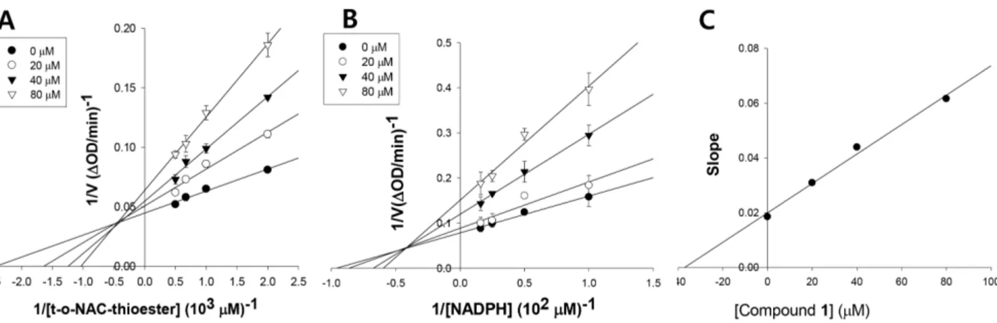
Figure 2. The mechanism of inhibition ofStaphylococcus aureusFabI by meleagrin respective to t-o-NAC thioester (A) and NADPH (B), and Kidetermination of meleagrin (C).
doi:10.1371/journal.pone.0078922.g002
Figure 3. Direct binding of the derivatives of meleagrin withStaphylococcus aureusFabI by fluorescence quenching assay.(A) The more
of [1-14C] acetate (50 mCi/mmol), [2-14C] thymidine (59.8 mCi/ mmol), [U-14C] uridine (539 mCi/mmol), L-[U-14C] leucine (306 mCi/mmol) or L-[U-14C] isoleucine (329 mCi/mmol), and N-acetyl-D-[1-14C] glucosamine (58.1 mCi/mmol) intoS. aureusand S. pneumoniaewere measured as described previously [21].S. aureus was exponentially grown to an A650 of 0.2 in Mueller–Hinton
broth.S. pneumoniaewas grown in tryptic soy broth supplemented with 5% sheep blood. Each 1-mL culture was treated with drugs at 2 times the MIC for 10 min. An equal volume of DMSO solvent was added to the untreated control. After incubation with the radiolabeled precursors at 37uC for 1 h, followed by centrifuga-tion, the cell pellets were washed twice with PBS buffer. After acetate incorporation, the total cellular lipids were extracted with chloroform-methanol-water. The incorporated radioactivity in the chloroform phase was measured by scintillation counting. For the other precursors, incorporation was terminated by adding 10% (w/v) TCA and cooling on ice for 20 min. The precipitated material was collected on Whatman GF/C glass microfiber filters, washed with TCA and ethanol, dried, and counted using a scintillation counter. The total counts incorporated at 1 h of incubation without inhibitors ranged from .7,000 for [U-14C] uridine to ,13,000 for [1-14C] acetate. The inhibition of radiolabeled precursor incorporation was calculated using the following formula: % inhibition = 1006[12(radioactivity values of the treated samples/control (no antibacterial) values)]. In all experiments, known antibacterial agents were included as positive controls.
Overexpression assay
An overexpression assay using S. aureus RN4220, S. aureus RN4220 (pE194), and S. aureus RN4220 (pE194-fabI) was conducted to perform target validation of FabI inhibitors as described previously [21]. Additionally, both fabI- and fabK -overexpressingE. coliwere constructed to test a multitarget effect of the compounds. The wild-type fabI gene from the genomic DNA ofE. coliW3110 was amplified by PCR by using the primers 59-ATGGGTTTTCTTTCCGGTAAGCGCA-39 and 59 -TTTCAGTTCGAGTTCGTTCATT-39. The wild-type fabK gene from the genomic DNA ofS. pneumoniae KCTC 5412 was amplified by PCR by using the primers 59 -ATGAAAACGCG-TATTACA-39and 59-GTCATTTCTTAC AACTCCTGTCCA-39. The resulting products were cloned into the pBAD-TOPO TA expression vector (Invitrogen, Carlsbad, CA, USA) to yield the pBAD-fabI and pBAD-fabK recombinant plasmids, which placed the expression of the genesfabIandfabK, respectively, under the control of the arabinose promoter [25]. Recombinant pBAD-fabI and pBAD-fabK were then introduced into the pump-negative (tolC)E. coliEW1b via electroporation to generateE. coliEW1b (pBAD-fabI) andE. coliEW1b (pBAD-fabK), respectively.
Results
Isolation of meleagrin as a new FabI inhibitor
A FabI inhibitor was isolated fromPenicillium chrysogenumF717, which is known as a penicillin-producing species. MS and NMR spectral analyses of the inhibitor revealed that it was meleagrin (1) (Fig. 1). Compound1inhibited bothE. coliandS. aureusFabI with
Table 2.Reduced susceptibility offabI-overexpressingStaphylococcus aureusto meleagrin (1) and its derivatives.
Compounds IC50(mM) MIC (mg/mL) Mode of action
saFabI Wild type S. aureus(pE194) S. aureus(pE194-fabI)
1 40.1 64 64 256 FabI
3 38.7 64 64 256 FabI
4 48.0 64 64 256 FabI
5 13.5 16 16 128 FabI
6 13.1 16 16 128 FabI
Triclosan 0.6 0.01 0.01 1.6 FabI
Erythromycin .100 0.5 64 64 Protein synthesis
Oxacillin .100 0.25 0.25 0.25 Cell wall
Norfloxacin .100 1 1 1 DNA synthesis
IC50 values of 33.2 and 40.1mM, respectively (Table 1). To
determine whether compound 1 selectively inhibited FabI, its effect on FabK, which is the enoyl-ACP reductase ofS. pneumoniae, was examined. Compound 1did not inhibit S. pneumoniaeFabK even at 200mM, which indicates that it is selective for FabI.
Mode of FabI inhibition
The FabI reaction mechanism requires the nucleotide cofactors NADH or NADPH as the first substrates [26]. The FabI inhibitor could bind to the free enzyme, the enzyme-substrate complex, or both to prevent catalysis. In the first case, the inhibition pattern with respect to the cofactor would be competitive; in the second, the inhibition pattern would be non-competitive; and in the third case, mixed-type inhibition would occur. Inhibition of S. aureus FabI by compound1was mixed with respect totrans-2-octenoyl N-acetylcysteamine, with aKivalue of 39.8mM (Fig. 2A and 2C). In
addition, compound1exhibited mixed inhibition with respect to NADPH, with aKivalue of 32.3mM (Fig. 2B). Thus, compound1
must bind to both the free enzyme and the FabI-NADPH complex to prevent binding of the nucleotide cofactor and the substrate, respectively.
Effects of structural changes in compound 1 on FabI and related activity
To determine whether structural changes in compound 1 influence its effects on FabI, compound1and its derivatives were tested against S. aureus and E. coli FabI and bacterial growth (Table 1). Compounds5and6, which were modified at both the 9-OH and 14-NH groups, produced a significant increase inS. aureus and E. coli FabI-inhibitory activity, and they enhanced antibacterial activity against S. aureus and E. coli. In contrast, compounds2,3, and4, which were modified at the 1-NH, 9-OH, and 14-NH groups, respectively, did not affect activity. Compound
7, which was brominated at the benzene ring of compound 2, totally lost its activity.
Effects on fluorescence quenching ofS. aureusFabI We examined whether active compounds directly bind with FabI by fluorescence quenching analysis.S. aureusFabI displayed strong maximal fluorescence at 307 nm after excitation at 270 nm (Fig. 3), whereas triclosan, kanamycin, 5, and 7 had no fluorescence at this wavelength (data not shown). WhenS. aureus FabI was incubated with increasing amounts of active compound 5, its fluorescence intensity decreased gradually (Fig. 3A), whereas the inactive compound7did not exhibit such an effect (Fig. 3B). Compound1showed the same pattern as compound5(data not shown). As a positive control, triclosan binding resulted in fluorescence quenching of S. aureus FabI (Fig. 3C), whereas kanamycin as a negative control did not (Fig. 3D). These data indicate that the active compounds1and5directly interact with S. aureusFabI, whereas compound7does not, thus explaining their effects on FabI.
Inhibition of cellular fatty acid synthesis
To evaluate whether the active compounds inhibit cellular fatty acid synthesis, we determined whether the compounds inhibited the incorporation of acetate into membrane fatty acidsin vivo. We measured their effects on the incorporation of [1-14C] acetate into membrane fatty acids in S. aureus. In agreement with their antibacterial activity and FabI-inhibitory activity, the more active compounds5and6indeed blocked incorporation of radioactively-labeled acetate into chloroform/methanol-extractable phospho-lipidsin vivoin a concentration-dependent manner, with approx-imately 2-fold higher activity than the less active compounds1,3, and4 (Table 1). The inactive compound7did not exhibit such fatty acid synthesis inhibition even at 200mM, as expected. As a positive control, triclosan inhibited fatty acid synthesis in a concentration-dependent manner (data not shown). In contrast,
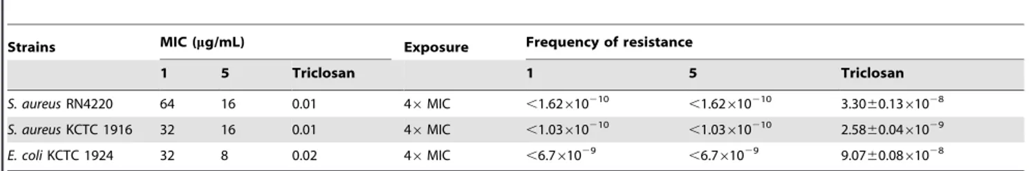
Table 3.Frequency of resistance to meleagrin (1) and its more active derivative.
Strains MIC (mg/mL) Exposure Frequency of resistance
1 5 Triclosan 1 5 Triclosan
S. aureusRN4220 64 16 0.01 46MIC ,1.62610210 ,1.62
610210 3.30
60.1361028
S. aureusKCTC 1916 32 16 0.01 46MIC ,1.03610210 ,1.03610210 2.5860.0461029
E. coliKCTC 1924 32 8 0.02 46MIC ,6.761029 ,6.761029 9.0760.0861028
doi:10.1371/journal.pone.0078922.t003
Table 4.Effects of meleagrin (1) on incorporation of radiolabeled precursors intoS. aureusandS. pneumoniae.
Strains Compounds Inhibition of precursor incorporation (%) [1-14C]
Acetate
[2-14C]
Thymidine
[U-14C]
Uridine
L-[U-14C]
Isoleucine
N-Acetyl-D-[1-14C]
Glucosamine
S. aureusa Reference antibacterialc 87 79 69 74 79
1 62 13 17 6 25
S. pneumoniaeb Reference antibacteriald 95 83 92 85 88
1 65 15 2 9 3
aS. aureusRN4220;bS. pneumoniaeKCTC 3932.cReference antibacterials used for inhibition of acetate, thymidine, uridine, isoleucine, and N-acetyl-D-glucosamine incorporation are triclosan, norfloxacin, rifampin, chlorampenicol, and vancomycin, respectively.dReference antibacterials inS. pneumoniaewere the same as inS. aureus, except cerulenin was used instead of triclosan for acetate inhibition.
the incorporation of leucine into proteins was not inhibited by the active compounds (Table 1), whereas the protein synthesis inhibitor, chloramphenicol, inhibited incorporation (data not shown).
Antibacterial activity
Consistent with their FabI-inhibitory activity, compounds5and 6showed 2–4 times higher antibacterial activity than compound1 against S. aureus RN4220 and the highly sensitive strain E. coli KCTC 1924 (Table 1), as expected. Interestingly, compounds that were inactive against the FabK isoform exhibited antibacterial activity againstS. pneumoniaeKCTC 3932, which contains only the FabK isoform. This finding suggests that the compounds inhibit not only FabI but also another target. Compounds5and 6also showed antibacterial activity against other gram-positive bacteria, including S. aureus503,S. aureus KCTC 1916, MRSA CCARM 3167, MRSA CCARM 3506, QRSA CCARM 3505, QRSA CCARM 3519, Staphylococcus epidermis KCTC 3958, B. subtilis KCTC 1021, andMicrococcus luteus KCTC 1056 with MIC values of 8–16mg/mL.
Effects onfabI-overexpressingS. aureus
The increase in the MIC for the fabI-overexpressing strain relative to the wild type is indicative of FabI being the mode of antibacterial action [27]. The antibacterial activity of the active compounds for the fabI-overexpressing strain was investigated to determine whether overexpression offabIshifted the MIC forS. aureus. The MICs for the fabI-overexpressing strain S. aureus RN4220 (pE194-fabI) were 4–8-fold higher than those of the wild-type strain S. aureus RN4220, or the vector-containing strain S. aureus RN4220 (pE194) (Table 2). The MIC for triclosan in the fabI-overexpressing strain increased, which was used as a positive control. Erythromycin, the selection marker for the vector pE194, increased the MICs for both thefabI-overexpressing strain and the vector-containing strain, which indicated that the engineered constructs functioned as expected. Antibiotics with different modes of action such as oxacillin and norfloxacin were applied as negative controls and did not change the MICs of the 3 strains, which indicates that altered expression offabIdoes not alter the sensitivity of cells to antibiotics in general. These results indicate that the active compounds inhibited the growth of S. aureus by inhibiting thefabI-encoded ENR.
Frequency of spontaneously resistant mutants
We isolated resistant mutants to determine which other gene or genes were targeted by the active compounds (Table 3). As a control, triclosan-resistant mutants were isolated at a frequency of 3.3060.1361028, 2.5860.0461029, and 9.0760.0861028from
S. aureus RN4220, S. aureus KCTC 1916, and the antibiotic-sensitiveE. coli KCTC 1942, respectively. However, no mutants resistant to compounds 1 and 5 were detected from the strains tested. These results suggest that compounds 1 and 5 inhibit multiple targets.
Effects on macromolecular biosynthesis
To identify other pathways inhibited by compound1, the effects of compound1on the incorporation of radiolabeled precursors of macromolecular synthesis in S. pneumoniae and in S. aureus were investigated. All reference antibacterial agents selectively inhibited the macromolecular synthesis pathway, which is consistent with their known mechanism of action (Table 4). Compound 1 inhibited the incorporation of acetate into lipids in bothS. aureus and S. pneumoniae by 62% and 65%, respectively, whereas the incorporation of thymidine, uridine, isoleucine, and N-acetylglu-cosamine, into DNA, RNA, protein, and the cell wall, respectively, was not inhibited. Because compound 1 is inactive against the FabK isoform, these data suggest that compound1inhibits at least one additional target in addition to FabI in the fatty acid pathway.
Effects onfabK-overexpressingE. coli
Discussion
We screened 25,000 microbial extracts consisting of actinomy-cetes and fungi to identify new FabI inhibitors. Meleagrin was isolated from the solid-state fermentation of the fungal strainP. chrysogenum F717. Meleagrin was previously isolated from P. meleagrinum[28] andP. chrysogenum[29], but its biological activity, including antimicrobial activity, has not been reported. Although its activity was weak, meleagrin clearly showed inhibition selective forS. aureusFabI overS. pneumoniaeFabK. Importantly, the binding of meleagrin with S. aureus FabI was demonstrated by the fluorescence quenching assay. Furthermore, its inhibition of FabI was supported by results obtained using its chemical derivatives, the intracellular fatty acid synthesis assay, and the fabI -overex-pressing assay. Interestingly, meleagrin and its more active derivatives showed antibacterial activity against S. pneumoniae, in which FabK is the sole enoyl-ACP reductase, and it did not produce spontaneously resistant mutants ofS. aureusorE. coli, in contrast to triclosan, which suggests that meleagrin inhibits multiple targets. Meleagrin inhibited the incorporation of radio-labeled acetate into lipids inS. pneumoniae andS. aureus, whereas incorporation of thymidine (DNA), uridine (RNA), isoleucine (protein), and N-acetylglucosamine (cell wall) was not inhibited, which indicates that these compounds inhibit fatty acid synthesis through one or more modes of action in addition to FabI inhibition. The multitarget effect was confirmed by the fabK -overexpression assay in E. coli. The multitarget effect is very important from the point of view of drug development because a single point mutation in one gene for a drug with a single target renders the strain resistant and the drug useless. Thus, when considering that one of the advantages of antibacterial agents having multiple targets is the reduced development of drug resistance [10], meleagrin and its derivatives hold promise for the development of new antibiotics that can treat infections caused by multidrug-resistant pathogens.
Several FabI inhibitors have been reported, and most were derived from compound libraries and were synthetically developed using structure-based approaches, including 1,4-disubstituted
imidazoles, aminopyridines, naphthyridinones, and thiopyridines [30]. Although synthetic inhibitors are potent, they have a disadvantage, as resistant mutants occur at relatively high frequency [16], A few natural FabI inhibitors have been reported, such as vinaxanthone [21], cephalochromin [31], kalimantacin/ batumin [22], EGCG, and flavonoids [32]. EGCG and flavonoids inhibit several targets such as FabG, FabZ, and FabI. The mode of action of vinaxanthone, cephalochromin, and kalimantacin/ batumin was demonstrated by FabI-overexpressing strains. To our knowledge, this is the first study on a multitarget effect of FabI inhibitors.
In summary, meleagrin is a new class of FabI inhibitor with antibacterial activity against multidrug-resistant bacteria such as MRSA and QRSA. Meleagrin is structurally unique, and it inhibits at least one more target in addition to FabI, thereby resulting in a no resistance mutant; thus, meleagrin may have potential as a useful lead compound for the development of a new anti-MRSA agent.
Supporting Information
Information S1 Preparation and spectral data of com-pounds 2–7.
(DOCX)
Acknowledgments
We thank the Culture Collection of Antimicrobial Resistant Microbes of Korea and theE. coliGenetic Stock Center of Yale University for providing the bacterial strains used in this study.
Also we express our thanks to Korea Basic Science Institute for the NMR measurements.
Author Contributions
Conceived and designed the experiments: WGK. Performed the experiments: CJZ MJS SL WGK. Analyzed the data: MJS WGK. Contributed reagents/materials/analysis tools: CJZ MJS. Wrote the paper: WGK.
References
1. Klein E, Smith DL, Laxminarayan R (2007) Hospitalizations and deaths caused by methicillin-resistant Staphylococcus aureus, United States, 1999–2005. Emerg Infect Dis 13: 1840–1846.
2. Levy SB, Marshall B (2004) Antibacterial resistance worldwide: causes, challenges and responses. Nat Med 10: S122–129.
3. Miesel L, Greene J, Black TA (2003) Genetic strategies for antibacterial drug discovery. Nat Rev Genet 4: 442–456.
4. Heath RJ, Rock CO (2004) Fatty acid biosynthesis as a target for novel antibacterials. Curr Opin Investig Drugs 5: 146–153.
5. Wang J, Soisson SM, Young K, Shoop W, Kodali S, et al. (2006) Platensimycin is a selective FabF inhibitor with potent antibiotic properties. Nature 441: 358– 361.
6. Massengo-Tiasse RP, Cronan JE (2008) Vibrio cholerae FabV defines a new class of enoyl-acyl carrier protein reductase. Journal of Biological Chemistry 283: 1308–1316.
7. Zhu L, Lin J, Ma J, Cronan JE, Wang H (2010) Triclosan resistance of Pseudomonas aeruginosa PAO1 is due to FabV, a triclosan-resistant enoyl-acyl carrier protein reductase. Antimicrobial Agents and Chemotherapy 54: 689– 698.
8. Zhang YM, White SW, Rock CO (2006) Inhibiting bacterial fatty acid synthesis. J Biol Chem 281: 17541–17544.
9. Lu H, Tonge PJ (2008) Inhibitors of FabI, an enzyme drug target in the bacterial fatty acid biosynthesis pathway. Acc Chem Res 41: 11–20.
10. Silver LL (2007) Multi-targeting by monotherapeutic antibacterials. Nat Rev Drug Discov 6: 41–55.
11. McMurry LM, Oethinger M, Levy SB (1998) Triclosan targets lipid synthesis. Nature 394: 531–532.
12. Rozwarski DA, Grant GA, Barton DH, Jacobs WR Jr, Sacchettini JC (1998) Modification of the NADH of the isoniazid target (InhA) from Mycobacterium tuberculosis. Science 279: 98–102.
13. Cardoso RF, Cooksey RC, Morlock GP, Barco P, Cecon L, et al. (2004) Screening and characterization of mutations in isoniazid-resistant Mycobacte-rium tuberculosis isolates obtained in Brazil. Antimicrob Agents Chemother 48: 3373–3381.
14. Hazbon MH, Brimacombe M, Bobadilla del Valle M, Cavatore M, Guerrero MI, et al. (2006) Population genetics study of isoniazid resistance mutations and evolution of multidrug-resistant Mycobacterium tuberculosis. Antimicrobial Agents and Chemotherapy 50: 2640–2649.
15. Chen Y, Pi B, Zhou H, Yu Y, Li L (2009) Triclosan resistance in clinical isolates of Acinetobacter baumannii. Journal of Medical Microbiology 58: 1086–1091. 16. Escaich S, Prouvensier L, Saccomani M, Durant L, Oxoby M, et al. (2011) The
MUT056399 Inhibitor of FabI Is a New Antistaphylococcal Compound. Antimicrob Agents Chemother 55: 4692–4697.
17. Silver LL (2011) Challenges of antibacterial discovery. Clin Microbiol Rev 24: 71–109.
18. Livermore DM (2011) Discovery research: the scientific challenge of finding new antibiotics. Journal of Antimicrobial Chemotherapy 66: 1941–1944. 19. Singh MP, Greenstein M (2000) Antibacterial leads from microbial natural
products discovery. Curr Opin Drug Discov Devel 3: 167–176.
20. Zheng CJ, Sohn MJ, Lee S, Hong YS, Kwak JH, et al. (2007) Cephalochromin, a FabI-directed antibacterial of microbial origin. Biochemical and Biophysical Research Communications 362: 1107–1112.
21. Zheng CJ, Sohn MJ, Kim WG (2009) Vinaxanthone, a new FabI inhibitor from Penicillium sp. Journal of Antimicrobial Chemotherapy 63: 949–953. 22. Mattheus W, Masschelein J, Gao LJ, Herdewijn P, Landuyt B, et al. (2010) The
kalimantacin/batumin biosynthesis operon encodes a self-resistance isoform of the FabI bacterial target. Chemistry and Biology 17: 1067–1071.