Regions Are Infrequently Somatically Mutated in Ovarian
Cancers
Georgina L. Ryland1,2., Jennifer L. Bearfoot1,3., Maria A. Doyle4
, Samantha E. Boyle1,
David Y. H. Choong1, Simone M. Rowley1, Australian Ovarian Cancer Study Group5, Richard W. Tothill6, Kylie L. Gorringe1,3", Ian G. Campbell1,3*"
1Victorian Breast Cancer Research Consortium Cancer Genetics Laboratory, Peter MacCallum Cancer Centre, East Melbourne, Victoria, Australia,2Centre for Cancer Research, Monash Institute of Medical Research, Monash University, Clayton, Victoria, Australia,3Department of Pathology, University of Melbourne, Parkville, Victoria, Australia,4Bioinformatics Core Facility, Peter MacCallum Cancer Centre, East Melbourne, Victoria, Australia,5Peter MacCallum Cancer Centre, East Melbourne, Victoria, Australia,6Molecular Genomics Core Facility, Peter MacCallum Cancer Centre, East Melbourne, Victoria, Australia
Abstract
MicroRNAs are key regulators of gene expression and have been shown to have altered expression in a variety of cancer types, including epithelial ovarian cancer. MiRNA function is most often achieved through binding to the 39-untranslated region of the target protein coding gene. Mutation screening using massively-parallel sequencing of 712 miRNA genes in 86 ovarian cancer cases identified only 5 mutated miRNA genes, each in a different case. One mutation was located in the mature miRNA, and three mutations were predicted to alter the secondary structure of the miRNA transcript. Screening of the 39-untranslated region of 18 candidate cancer genes identified one mutation in each ofAKT2,EGFR,ERRB2andCTNNB1. The functional effect of these mutations is unclear, as expression data available forAKT2andEGFRshowed no increase in gene transcript. Mutations in miRNA genes and 39-untranslated regions are thus uncommon in ovarian cancer.
Citation:Ryland GL, Bearfoot JL, Doyle MA, Boyle SE, Choong DYH, et al. (2012) MicroRNA Genes and Their Target 39-Untranslated Regions Are Infrequently Somatically Mutated in Ovarian Cancers. PLoS ONE 7(4): e35805. doi:10.1371/journal.pone.0035805
Editor:Austin John Cooney, Baylor College of Medicine, United States of America ReceivedJanuary 29, 2012;AcceptedMarch 22, 2012;PublishedApril 20, 2012
Copyright:ß2012 Ryland et al. This is an open-access article distributed under the terms of the Creative Commons Attribution License, which permits unrestricted use, distribution, and reproduction in any medium, provided the original author and source are credited.
Funding:This work was supported by the Victorian Breast Cancer Research Consortium, Australia, and the Australian National Health and Medical Research Council (NHMRC). GLR is supported by an Australian Postgraduate Award. The Australian Ovarian Cancer Study Group was supported by the U.S. Army Medical Research and Materiel Command under DAMD17-01-1-0729, The Cancer Council Tasmania, The Cancer Foundation of Western Australia and the NHMRC. The funders had no role in study design, data collection and analysis, decision to publish, or preparation of the manuscript.
Competing Interests:The authors have declared that no competing interests exist.
* E-mail: ian.campbell@petermac.org
.These authors contributed equally to this work.
"These authors also contributed equally to this work.
Introduction
MicroRNAs (miRNAs), a class of small non-coding RNA molecules, have important regulatory roles in diverse cellular pathways including proliferation, differentiation, senescence and metabolism [1]. This regulation is achieved through semi-complementary base paring with the 39-untranslated region (39 -UTR) of the target messenger RNA (mRNA) [1–3], as well as the 59-untranslated region or coding regions of mRNAs, which are subsequently degraded or post-transcriptionally silenced [4–6]. Accumulating evidence now demonstrates that miRNA expression is aberrant in cancer [7], leading to the hypothesis that alterations in miRNA pathways may be an important step in the initiation and progression to malignancy. Consistent with this hypothesis is the observation that miRNA genes are frequently localised in genomic regions commonly altered in cancer, including minimal regions of deletion, loss of heterozygosity and amplification as well as fragile sites [8–10]. Mutation is an alternative mechanism for miRNA deregulation in the cancer setting, whereby mutation may alter miRNA transcription, processing or miRNA-mRNA inter-actions. This mechanism was first described by Calinet al.[8], who
identified a germline variation inhsa-miR-16-1that was linked with susceptibility to chronic lymphocytic leukaemia. Since then, germline polymorphisms in miRNA genes have been associated with predisposition to other cancer types [11]. Despite the hypothesis that miRNAs may function as conventional oncogenes or tumor suppressors, several studies have suggested that somatic mutation within miRNAs are a rare occurrence and those that have been reported show little effect on miRNA activity [10,12– 14]. However, the majority of these studies have favoured a candidate gene approach and to date, un-biased assessment of the occurrence of somatic alterations in miRNA genes in any cancer type is lacking.
mechanism for repression or activation of a cancer-associated mRNA.
Aberrant miRNA activity is frequently associated with the pathogenesis and progression of epithelial ovarian cancer, the most common form of ovarian malignancy. MiRNA profiling studies consistently observe global silencing of miRNA expression in ovarian tumors, which is contributed to in part by genomic loss and epigenetic alterations [10,18–22]. Similarly, expression of many known and putative cancer genes is dysregulated in ovarian cancer, for exampleBRCA2, for which only a proportion of the observed loss of expression can be attributed to mutation, and promoter methylation is not observed [23]. Previously, we have demonstrated that the frequency of somatic mutations in 10 cancer-implicated miRNAs is low in ovarian tumors [24]. In the present study, we extend this analysis by comprehensively characterising somatic mutations in 712 miRNA genes using massively parallel targeted re-sequencing. In addition, we screened the 39-UTRs of 18 candidate cancer genes with the aim of identifying somatic mutations that alter predicted miRNA binding sites. Although these genes are frequently implicated in the tumorigenic process, coding mutations, methylation or copy number alterations only account for a subset of the expression differences seen in ovarian tumors.
Results and Discussion
Somatic mutations targeting microRNA genes are infrequent events in ovarian tumors
To investigate whether mutations in miRNA genes contribute to altered miRNA activity in ovarian cancer, 86 primary epithelial ovarian tumors were assessed for somatic mutations in genomic regions corresponding to precursor or mature miRNA sequences. Clinical characteristics of these cases are summarised in Table S1. Targeted next generation sequencing was used to assess 712 miRNA genes annotated in the Sanger miRNA database (version 13.0, March 2009). Following data alignment, 95% of targeted bases within the 712 miRNA genes had a minimum 10-fold sequence coverage, with a corresponding mean coverage of 92-fold. Filtering to remove germline variants detected in matched normal lymphocyte DNA and validation by conventional sequencing identified somatic mutations in 5 miRNA genes: hsa-miR-10a, hsa-miR-622, hsa-miR-767-5p, hsa-miR-888and hsa-miR-1280(Figure 1). Overall, somatic mutations were detected in 6% (5/86) of tumors and in less than 1% (5/712) of miRNA genes analysed, with no miRNA genes recurrently targeted by mutation. Consistent with previous reports, mutations within mature miRNAs were uncommon; only one mutation was located within the mature region of hsa-miR-767-5p (but external to the seed region), whilst the remaining four occurred within the precursor hairpin. This data is in agreement with previous smaller scale studies suggesting that somatic mutations in miRNA genes are an infrequent event in tumor samples [8,12,14,24,25].
MiRNA biogenesis is a multistep process initiated by RNA polymerase II-mediated transcription, followed by RNAse III-dependent trimming into a hairpin intermediate and subsequent cleavage into a functional mature miRNA [1,26]. This process is dependent on sequence motifs and RNA secondary structure elements within the primary and precursor miRNA molecules [26]. As such, mutations arising in precursor regions may alter RNA secondary structure and thereby block processing into mature miRNA. To determine if RNA structural changes may result from the somatic miRNA mutations identified, we used the RNAfold program [27] to predict the most stable secondary structure for both the wild-type and mutant sequences.
Confor-mational changes were predicted for mutanthsa-miR-622, hsa-miR-767-5p and hsa-miR-1280 (Figure S1). However, conformational changes predicted in silicorarely equate to a physiological effect [12] and the functional implication of mutations identified here requires further investigation within vitroassays.
In contrast to the frequent observation of tumor-specific variations contributing to the activation or repression of protein coding genes in cancer, these findings demonstrate that somatic mutations in miRNA genes are an infrequent event during ovarian pathogenesis and add to accumulating evidence from a range of tumor types suggesting that miRNAs are rarely dysregulated by this mechanism. Given the large number of mRNA targets predicted for a single miRNA and the diverse roles of those predicted target genes, any somatic mutation in a miRNA gene (and particularly those occurring within the seed sequence) are likely to impact many biological pathways [2,3], some of which may be involved in maintaining cell homeostasis. Consequently, even if a somatically mutated miRNA gene altered mRNA expression to positively affect tumor survival, it is likely that there would be a larger number of gene expression changes which would not be conducive to tumor cell survival. Conversely, in certain situations, mRNA transcriptional repression may result from the action of multiple miRNAs [28] and as such, the altered activity of a single miRNA may be insufficient to result in a biological effect. Finally, it is becoming increasingly recognised that miRNA alterations observed in cancer tissues may occur secondary to defects in components of the miRNA processing machinery, including transcription factors and chromatin remodelling genes regulating miRNA transcription, as well as components of miRNA post-transcriptional regulation [29,30]. In ovarian tumors, DIC-ER1 and EIF2C2 (Argonaute2) DNA copy number gains have been observed in 24.5% and 51.5% of tumors respectively [9] and median overall survival is reduced among women whose tumors have lower DICER1 and DROSHA mRNA expression [31]. Further investigation is needed establish the importance of alterations to these and other components of the miRNA biogenesis pathway in the pathogenesis of ovarian cancer.
Somatic mutations targeting 39-untranslated regions are infrequent events in ovarian tumors
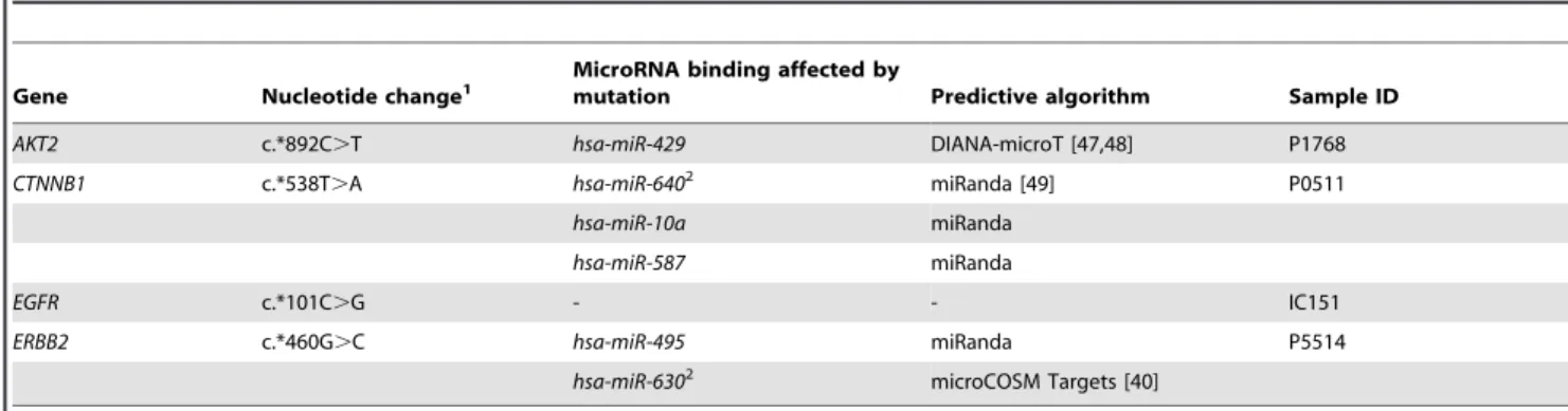
miRNA target prediction algorithms suggest that mutated loci in AKT2, CTNNB1 and ERBB2 may occur within the region of a predicted miRNA binding site, with two mutations, the c.*538T.A (CTNNB1) and c.*460G.C (ERBB2) substitutions, predicted to occur within the seed sequence ofhsa-miR-630and hsa-miR-640 binding respectively. RNA expression profiling was available for samples with mutations in AKT2 and EGFR and demonstrated that the transcript levels of these genes was not altered in samples with somatic 39-UTR mutations relative to other tumor samples of the same ovarian subtype (Figure S2).
Although it is recognised that miRNAs can also impart transcriptional repression through action on the 59-UTR of an mRNA target, this study provides preliminary evidence that somatic mutations altering miRNA binding sites within the 39 -UTR of common cancer genes are infrequent in epithelial ovarian tumors. Effective translational silencing may require synergistic action of miRNAs at multiple sites across a UTR, either by a single family of microRNAs or by a combination of unrelated microRNAs, and thus the single somatic mutations identified here are likely insufficient to silence the respective transcript [28,38]. The somatic mutation prevalence in 39-UTR regions of candidate cancer genes sequenced was 1.43 mutations per Mb. By comparison, the mutation prevalence in protein coding regions of known cancer genes in this tumor cohort is 570.57, 183.60 and 32.63 mutations per Mb forTP53,KRASandPIK3CArespectively, while the estimated mutation prevalence in the coding exome is 2.4 mutations per Mb in ovarian tumors [23]. Thus, it is likely that
the low rate of mutation in 39UTRs compared to exons indicates that the majority of mutations in 39-regulatory regions identified here occur as bystander events in tumor cell development.
In summary, somatic mutations in miRNA genes were infrequently observed in ovarian tumors and thus are unlikely to account for altered miRNA activity observed in this tumor type. In addition, we provide preliminary evidence that selection for somatic mutations within the 39-UTRs of candidate cancer genes, which would be hypothesised to interfere with miRNA dependent gene regulation, is unlikely to represent a common mechanism for altered mRNA expression in ovarian tumors.
Materials and Methods
Ethics statement
Accrual and use of patient material for this study was approved by the following Human Research Ethics Committees: South-ampton Hospital Human Research Ethics Committee, Peter MacCallum Cancer Centre Human Research Ethics Committee, Queensland Institute of Medical Research Human Research Ethics Committee, University of Melbourne Human Research Ethics Committee, Westmead Hospital Human Research Ethics Committee. All individuals gave written informed consent for the use of their tissue in research. This project was approved by the Peter MacCallum Cancer Centre Human Research Ethics Committee (Approval#09/29).
Figure 1. Somatic mutations identified in microRNA genes.Tumor specific mutations are marked as black bars relative to the mature microRNA (white box) and precursor microRNA (grey box) sequences. The positions of mutations are reported with respect to the following precursor microRNA transcripts:hsa-miR-10aNR_029608.1;hsa-miR-622NR_030754.1;hsa-miR-767-5pNR_030409.1;hsa-miR-888NR_030592.1;hsa-miR-1280 NR_031703.1.
doi:10.1371/journal.pone.0035805.g001
Table 1.Somatic mutations identified in 39-untranslated regions of candidate mRNAs.
Gene Nucleotide change1
MicroRNA binding affected by
mutation Predictive algorithm Sample ID
AKT2 c.*892C.T hsa-miR-429 DIANA-microT [47,48] P1768
CTNNB1 c.*538T.A hsa-miR-6402 miRanda [49] P0511
hsa-miR-10a miRanda
hsa-miR-587 miRanda
EGFR c.*101C.G - - IC151
ERBB2 c.*460G.C hsa-miR-495 miRanda P5514
hsa-miR-6302 microCOSM Targets [40]
1Nucleotide changes are described relative to the following sequences:AKT2NM_001626.3;CTNNB1NM_001904.3;EGFRNM_005228.3;ERBB2NM_004448.2. 2Indicates that the somatic mutation occurs within the microRNA-mRNA interaction at the seed region.
Ovarian tumor cohort
86 primary epithelial ovarian tumor tissue samples were obtained through the Peter MacCallum Cancer Centre Tissue Bank, Australia Ovarian Cancer Study or from patients presenting to hospitals in the south of England [39]. All tumor DNA samples were microdissected to ensure greater than 80% epithelial cell component. This tumor cohort comprised a mixture of serous (n = 45), endometrioid (n = 28), mucinous (n = 7) and clear cell (n = 6) subtypes. Matching peripheral blood samples were also collected from all patients at time of tumor collection and used as a source of germline DNA.
Candidate region identification for targeted next-generation sequencing
The 39-UTRs of 18 protein coding genes were selected for somatic mutation screening. Genome co-ordinates for selected 39 -UTRs were identified based on those annotated in the Ensembl database (release 54) for the following transcripts: AKT2 (ENST00000392038), BRAF (ENST00000288602), CCNE1 (ENST00000262643), CTNNB1 (ENST00000349496), EGFR (ENST00000275493 and ENST00000344576), ERBB2 (ENST00000269571), FGF1 (ENST00000359370), KRAS (ENST00000256078), MYC (ENST00000259523 and ENST00000377970), PIK3CA (ENST00000263967), RAB25 (ENST00000361084), BRCA1 (ENST00000309486), BRCA2 (ENST00000380152), CDKN2A (ENST00000304494), PTEN (ENST00000371953), RB1 (ENST00000267163), SPARC (ENST00000231061) andTP53(ENST00000269305). The geno-mic co-ordinates for human precursor miRNAs were obtained from the Sanger Institute miRBase (release 13.0, March 2009) [40,41], including 548 individual miRNA genes as well as 164 miRNAs within 62 miRNA clusters. All coding exons of TP53, KRASandPIK3CAwere included for sequence analysis.
Library preparation and target enrichment
200 ng of tumor or matched normal lymphocyte DNA was randomly fragmented to approximately 200 bp (Covaris, Woburn, MA) and end repair and A-tailing performed according to the Illumina genomic DNA library preparation protocol (Illumina, San Diego, CA). Following this, DNA was ligated with one of 7 custom multiplexing adapters compatible with Illumina single end sequencing. Indexed DNA samples were pooled equally prior to PCR enrichment. All reagents used during library preparation were obtained from New England Biolabs (NEB, Ipswich, MA). A boutique exon capture (SureSelect, Agilent Technologies, Santa Clara, CA) was used to specifically enrich for selected 39-UTRs, miRNAs and coding exons of cancer genes from genomic DNA libraries prior to next generation sequencing. Capture probes were designed using default parameters by submitting genomic co-ordinates to eArray (Agilent Technologies, Santa Clara, CA). Solution hybridization, washing, elution and amplification were performed according to the recommended protocol.
Somatic mutation analysis by Illumina GAIIx sequencing and capillary electrophoresis
Target enriched DNA libraries were sequenced on an Illumina GAIIx, generating 75 bp single end sequence reads. Image analysis and base calling was performed using the Genome Analyser Pipeline v1.6. Sequence reads were aligned to the human reference genome (GRCh37/hg19 assembly) using BWA and remaining unmapped reads were aligned with Novoalign software [42]. This was followed by local realignment with GATK [43]. Point mutations and insertions/deletions (indels) were identified
using GATK and Dindel [44] respectively and annotated with information from Ensembl release 56. Only mutations within miRNA transcripts annotated in miRBase were considered for further analysis.
Point mutations and indels were identified as somatic alterations only when (i) the variant was not called in the matched normal sample or identified as a germline alteration in another tumor/ normal pair (ii) the variant was not seen in.2% of reads in the matched normal sample following manual inspection of sequence reads using the Integrated Genomics Viewer [45] (iii) the variant was identified in at least four unique sequence reads with at least two mapping in the forward and two mapping in the reverse orientation.
All mutations which met the above criteria were subjected to validation by conventional PCR amplification and bidirectional capillary electrophoresis on the ABI3130 Genetic Analyser using BigDye Terminator v3.1 sequencing chemistry (Applied Biosys-tems, Foster City, CA).
Identification of miRNA-binding sites
The TargetScan (release 5.2) [46], microCOSM Targets (version 5) [40], DIANA-MicroT (version 3.0) [47,48] and miRanda (release August 2010) [49] predictive algorithms were used to determine whether the somatic mutations detected in mRNA 39UTRs occurred within miRNA binding sites. A mirSVR score threshold of less than 20.1 and minimum folding energy score threshold of less than or equal to216 kcal/mol were used for the miRanda algorithm. Default parameters were used for all other algorithms.
Supporting Information>
Figure S1 Predicted secondary structure changes as a result of somatic mutations in miRNA transcripts. Mature sequences are shadowed and the mutated base indicated by the arrowhead in (a)hsa-miR-622, (b)hsa-miR-1280and (c) hsa-miR-767-5p. The precursor miRNA sequence plus 50 bp flanking the precursor at the 59 and 39 ends was used to predict the secondary structure with the lowest free energy by the RNAfold program [29] using default parameters.
(PDF)
Figure S2 AKT2 and EGFR mRNA expression is not altered in the presence of 39-untranslated region somatic mutations relative to other ovarian samples of the same subtype. (a)AKT2expression in endometrioid tumors, including sample P1768 with an AKT2 c.*892C.T somatic mutation (indicated in red). mRNA expression profiling data was obtained from Tothillet al. [50].AKT2expression probe sets 225471_s_at and 226156_at are shown. (b)EGFRexpression in endometrioid tumors, including sample IC151 with an EGFR c.*101C.G somatic mutation (indicated in red). mRNA expression profiling data was obtained from Ramakrishnaet al.[51]. Error bars are representative of mean6SD.
(PDF)
Table S1 Clinical characteristics of ovarian tumors sequenced for somatic mutations in microRNA genes and candidate 39-untranslated regions.
(XLS)
Acknowledgments
assistants and all clinical and scientific collaborators and would like to thank all of the women who participated in AOCS. Members of the Australian Ovarian Cancer Study Group, collaborators and hospitals involved in AOCS can be found at http://www.aocstudy.org.
Australian Ovarian Cancer Study Group: David Bowtell (Peter MacCallum Cancer Centre, East Melbourne, Victoria, Australia), Georgia Chenevix-Trench (Queensland Institute of Medical Research, Brisbane, Queensland, Australia), Adele Green (Queensland Institute of Medical Research, Brisbane, Queensland, Australia), Penny Webb (Queensland Institute of Medical Research, Brisbane, Queensland, Australia), Anna
deFazio (Westmead Institute for Cancer Research, Westmead Millennium Institute, Westmead, New South Wales, Australia), Dorota Gertig (Victorian Cervical Cytology Registry, Carlton South, Victoria, Australia).
Author Contributions
Conceived and designed the experiments: GLR JLB KLG IGC. Performed the experiments: GLR SEB DYHC SMR. Analyzed the data: GLR MAD JLB. Contributed reagents/materials/analysis tools: AOCSG RWT. Wrote the paper: GLR JLB KLG IGC.
References
1. Bartel DP (2004) Micrornas: Genomics, biogenesis, mechanism, and function. Cell 116: 281–297.
2. Bartel DP (2009) Micrornas: Target recognition and regulatory functions. Cell 136: 215–233.
3. Rajewsky N (2006) Microrna target predictions in animals. Nat Genet 38 Suppl: S8–13.
4. Lytle JR, Yario TA, Steitz JA (2007) Target mrnas are repressed as efficiently by microrna-binding sites in the 59utr as in the 39utr. Proc Natl Acad Sci U S A 104: 9667–9672.
5. Lee I, Ajay SS, Yook JI, Kim HS, Hong SH, et al. (2009) New class of microrna targets containing simultaneous 59-utr and 39-utr interaction sites. Genome Res 19: 1175–1183.
6. Tay Y, Zhang J, Thomson AM, Lim B, Rigoutsos I (2008) Micrornas to nanog, oct4 and sox2 coding regions modulate embryonic stem cell differentiation. Nature 455: 1124–1128.
7. Calin GA, Croce CM (2006) Microrna signatures in human cancers. Nat Rev Cancer 6: 857–866.
8. Calin GA, Ferracin M, Cimmino A, Di Leva G, Shimizu M, et al. (2005) A microrna signature associated with prognosis and progression in chronic lymphocytic leukemia. N Engl J Med 353: 1793–1801.
9. Zhang L, Huang J, Yang N, Greshock J, Megraw MS, et al. (2006) Micrornas exhibit high frequency genomic alterations in human cancer. Proc Natl Acad Sci U S A 103: 9136–9141.
10. Zhang L, Volinia S, Bonome T, Calin GA, Greshock J, et al. (2008) Genomic and epigenetic alterations deregulate microrna expression in human epithelial ovarian cancer. Proc Natl Acad Sci U S A 105: 7004–7009.
11. Ryan BM, Robles AI, Harris CC (2010) Genetic variation in microrna networks: The implications for cancer research. Nat Rev Cancer 10: 389–402. 12. Diederichs S, Haber DA (2006) Sequence variations of micrornas in human
cancer: Alterations in predicted secondary structure do not affect processing. Cancer Res 66: 6097–6104.
13. Ramsingh G, Koboldt DC, Trissal M, Chiappinelli KB, Wylie T, et al. (2010) Complete characterization of the micrornaome in a patient with acute myeloid leukemia. Blood 116: 5316–5326.
14. Wu M, Jolicoeur N, Li Z, Zhang L, Fortin Y, et al. (2008) Genetic variations of micrornas in human cancer and their effects on the expression of mirnas. Carcinogenesis 29: 1710–1716.
15. Ratner E, Lu L, Boeke M, Barnett R, Nallur S, et al. (2010) A kras-variant in ovarian cancer acts as a genetic marker of cancer risk. Cancer Res 70: 6509–6515.
16. Wynendaele J, Bohnke A, Leucci E, Nielsen SJ, Lambertz I, et al. (2010) An illegitimate microrna target site within the 39utr of mdm4 affects ovarian cancer progression and chemosensitivity. Cancer Res 70: 9641–9649.
17. Chin LJ, Ratner E, Leng S, Zhai R, Nallur S, et al. (2008) A snp in a let-7 microrna complementary site in the kras 39untranslated region increases non-small cell lung cancer risk. Cancer Res 68: 8535–8540.
18. Iorio MV, Visone R, Di Leva G, Donati V, Petrocca F, et al. (2007) Microrna signatures in human ovarian cancer. Cancer Res 67: 8699–8707.
19. Wyman SK, Parkin RK, Mitchell PS, Fritz BR, O’Briant K, et al. (2009) Repertoire of micrornas in epithelial ovarian cancer as determined by next generation sequencing of small rna cdna libraries. PLoS One 4: e5311. 20. Yang H, Kong W, He L, Zhao JJ, O’Donnell JD, et al. (2008) Microrna
expression profiling in human ovarian cancer: Mir-214 induces cell survival and cisplatin resistance by targeting pten. Cancer Res 68: 425–433.
21. Nam EJ, Yoon H, Kim SW, Kim H, Kim YT, et al. (2008) Microrna expression profiles in serous ovarian carcinoma. Clin Cancer Res 14: 2690–2695. 22. Dahiya N, Sherman-Baust CA, Wang TL, Davidson B, Shih Ie M, et al. (2008)
Microrna expression and identification of putative mirna targets in ovarian cancer. PLoS One 3: e2436.
23. The Cancer Genome Atlas Research Network (2011) Integrated genomic analyses of ovarian carcinoma. Nature 474: 609–615.
24. Bearfoot JL, Choong DY, Gorringe KL, Campbell IG (2008) Genetic analysis of cancer-implicated microrna in ovarian cancer. Clin Cancer Res 14: 7246–7250. 25. Yang J, Zhou F, Xu T, Deng H, Ge YY, et al. (2008) Analysis of sequence variations in 59 micrornas in hepatocellular carcinomas. Mutat Res 638: 205–209.
26. Kim VN (2005) Microrna biogenesis: Coordinated cropping and dicing. Nat Rev Mol Cell Biol 6: 376–385.
27. Gruber AR, Lorenz R, Bernhart SH, Neubock R, Hofacker IL (2008) The vienna rna websuite. Nucleic Acids Res 36: W70–74.
28. Guttilla IK, White BA (2009) Coordinate regulation of foxo1 by 27a, mir-96, and mir-182 in breast cancer cells. J Biol Chem 284: 23204–23216. 29. van Kouwenhove M, Kedde M, Agami R (2011) Microrna regulation by
rna-binding proteins and its implications for cancer. Nat Rev Cancer.
30. Melo SA, Esteller M (2011) A precursor microrna in a cancer cell nucleus: Get me out of here! Cell Cycle 10: 922–925.
31. Merritt WM, Lin YG, Han LY, Kamat AA, Spannuth WA, et al. (2008) Dicer, drosha, and outcomes in patients with ovarian cancer. N Engl J Med 359: 2641–2650.
32. Futreal PA, Coin L, Marshall M, Down T, Hubbard T, et al. (2004) A census of human cancer genes. Nat Rev Cancer 4: 177–183.
33. Birrer MJ, Johnson ME, Hao K, Wong KK, Park DC, et al. (2007) Whole genome oligonucleotide-based array comparative genomic hybridization analysis identified fibroblast growth factor 1 as a prognostic marker for advanced-stage serous ovarian adenocarcinomas. J Clin Oncol 25: 2281–2287. 34. Sheach LA, Adeney EM, Kucukmetin A, Wilkinson SJ, Fisher AD, et al. (2009) Androgen-related expression of g-proteins in ovarian cancer. Br J Cancer 101: 498–503.
35. Cheng KW, Lahad JP, Kuo WL, Lapuk A, Yamada K, et al. (2004) The rab25 small gtpase determines aggressiveness of ovarian and breast cancers. Nat Med 10: 1251–1256.
36. Yiu GK, Chan WY, Ng SW, Chan PS, Cheung KK, et al. (2001) Sparc (secreted protein acidic and rich in cysteine) induces apoptosis in ovarian cancer cells. Am J Pathol 159: 609–622.
37. Said N, Najwer I, Motamed K (2007) Secreted protein acidic and rich in cysteine (sparc) inhibits integrin-mediated adhesion and growth factor-dependent survival signaling in ovarian cancer. Am J Pathol 170: 1054–1063. 38. Grimson A, Farh KK, Johnston WK, Garrett-Engele P, Lim LP, et al. (2007)
Microrna targeting specificity in mammals: Determinants beyond seed pairing. Mol Cell 27: 91–105.
39. Bryan EJ, Watson RH, Davis M, Hitchcock A, Foulkes WD, et al. (1996) Localization of an ovarian cancer tumor suppressor gene to a 0.5-cm region between d22s284 and cyp2d, on chromosome 22q. Cancer Res 56: 719–721. 40. Griffiths-Jones S, Saini HK, van Dongen S, Enright AJ (2008) Mirbase: Tools
for microrna genomics. Nucleic Acids Res 36: D154–158.
41. Kozomara A, Griffiths-Jones S (2011) Mirbase: Integrating microrna annotation and deep-sequencing data. Nucleic Acids Res 39: D152–157.
42. Novocraft Technologies.
43. McKenna A, Hanna M, Banks E, Sivachenko A, Cibulskis K, et al. (2010) The genome analysis toolkit: A mapreduce framework for analyzing next-generation DNA sequencing data. Genome Res 20: 1297–1303.
44. Albers CA, Lunter G, Macarthur DG, McVean G, Ouwehand WH, et al. (2010) Dindel: Accurate indel calls from short-read data. Genome Res.
45. Robinson JT, Thorvaldsdottir H, Winckler W, Guttman M, Lander ES, et al. (2011) Integrative genomics viewer. Nat Biotechnol 29: 24–26.
46. Friedman RC, Farh KK, Burge CB, Bartel DP (2009) Most mammalian mrnas are conserved targets of micrornas. Genome Res 19: 92–105.
47. Maragkakis M, Alexiou P, Papadopoulos GL, Reczko M, Dalamagas T, et al. (2009) Accurate microrna target prediction correlates with protein repression levels. BMC Bioinformatics 10: 295.
48. Maragkakis M, Reczko M, Simossis VA, Alexiou P, Papadopoulos GL, et al. (2009) Diana-microt web server: Elucidating microrna functions through target prediction. Nucleic Acids Res 37: W273–276.
49. Betel D, Wilson M, Gabow A, Marks DS, Sander C (2008) The microrna.Org resource: Targets and expression. Nucleic Acids Res 36: D149–153. 50. Tothill RW, Tinker AV, George J, Brown R, Fox SB, et al. (2008) Novel
molecular subtypes of serous and endometrioid ovarian cancer linked to clinical outcome. Clin Cancer Res 14: 5198–5208.