Received: Jan 2, 2012; Accepted: May 31, 2012 Vol. 2, No. 4, Autumn 2012, 233-242
Original Research Paper
Extract of
Adenanthera pavonina
L. seed reduces development of diabetic
nephropathy in streptozotocin-induced diabetic rats
Ramdas Pandhare1, 3*, Balakrishnan Sangameswaran2
Abstract
Objective: The aim of the present study was to investigate the renal protective effect of Adenanthera pavonina(A. pavonina) seed aqueous extract (APSAE), in streptozotocin (STZ)-induced diabetic rats. Materials and Methods:The renal protective effect of A. pavoninaseed aqueous extract (APSAE) was studied in STZ-induced diabetic rats. APSAE (50, 100 and 200 mg/kg per day) was given daily to diabetic rats for 13 weeks. Blood glucose, serum parameters such as albumin, creatinine, total protein, urea, lipid profile, glycated haemoglobin (HbA1c), and urine parameters such as urine protein and albumin were examined. Kidney histopathology was also done.
Results: After 13 weeks of treatment, in STZ-induced diabetic rats, severe hyperglycemia was developed, with marked increase in proteinuria and albuminuria. However, APSAE treatment significantly reduced proteinuria, albuminuria, lipid levels, and HbA1c deposition in diabetic rats.
Conclusion: These results suggested that APSAE has reduced development of diabetic nephropathy in streptozotocin-induced diabetic rats and could have beneficial effect in reducing the progression of diabetic nephropathy.
Keywords: Adenanthera pavonina,Albuminurea, Diabetic nephropathy, HbA1c, Proteinurea.
1- Department of Pharmacology, MES College of Pharmacy, Sonai, Newasa, Ahmednagar, Maharashtra-414105, India
2- Principal and Director Research Adesh Institute of Pharmacy and Biomedical Sciences Bathinda, Punjab, India
3- Research Scholar, Department of Pharmacy, Suresh Gyan Vihar University, Jaipur, Rajasthan, India *Corresponding author:Tel: +919881969052; Fax: +9102427-230848
Introduction
Diabetic nephropathy is an important complication of both type 1 and type 2 diabetic mellitus (white et al., 2000). The clinical hallmarks of diabetic nephropathy include progressive albuminuria followed by a gradual decline in renal function.
Glomerular basement thickening and
mesangial expansion have been identified as pathological precursors of these clinical changes (Mauer et al., 1984). The loss of glomerular podocytes precedes and predicts the onset of clinical nephropathy and may be an early pathological manifestation of diabetic nephropathy (Pagtalunan et al., 1997; Meyer et al., 1999). Podocytes are one of the important ingredients of filtration barrier which have special cytobiological characteristic and physiological function. The injury of podocytes can unavoidably lead to the occurrence of proteinuria (Zang et al., 2007). Advanced glycation end products (AGEs) are a complex, heterogeneous, and
sugar derived irreversible protein
modifications that have been implicated in the pathogenesis of diabetic complications (Brownlee, 1995; Singh et al., 2001). The irreversible formation of AGEs affects proteins and lipids, such as haemoglobin, collagen and lipoprotein, and causes damage to the kidney, eyes, and blood vessels (Brownlee, 2005). The levels of AGEs are much higher in patients with diabetes. Moreover, it was reported that AGEs induce apoptosis of murine cultured podocytes. AGEs have been proposed for being the potential causative factor of podocyte
damage (Chuang et al., 2007). Some
medicinal herbs have also been used widely for the treatment of diabetes and diabetic complications for hundreds of years (Alarcon et al., 1998; Li et al., 2004). In the past few years, many herbal extracts have been screened for possible AGEs inhibitory effects in vitro.
Adenanthera pavonina Linn. (Family: Leguminosae-Mimosaceae), is a deciduous tree, 18-24 m tall, bole erect and 60 cm in diameter (Bouquet et al.,1974). Many species of Adenanthera, including A. pavonina, have been used as traditional herbal medicine against a variety of diseases. The plant is reported to have a wide range of biological activities, such as astringent and styptic (used in diarrhoea, stomach haemorrhage, haematuria) and anti-inflammatory (in rheumatic affections, gout) actions (Khare, 2007). Seeds are anticephalgic and also used for the treatment of paralysis.
The seed contains an anti-inflammatory active principle, O-acetylethanolamine. The leaves contain octacosanol, dulcitol, glucosides of betasitosterol, and stigmasterol. The bark contains stigmasterol glucoside (Khare, 2007).Traditionally, the ground seed is widely used for the treatment of various human ailments such as treatment of boils, inflammation, blood disorders, arthritis, rheumatism, cholera, paralysis, epilepsy, convulsion, spasm, and indigestion (Burkill,
1966; Balogun et al., 2004).
Phytochemically, the seed and its pod contain glycosides, saponins, and steroids (Howes, 1974; Yadav et al., 1976). A new five-membered lactone ring compound, pavonin, was isolated from the methanol soluble part of A. pavonina(Muhammad et al., 2005). Oil extracted from the seed has been reported to have membrane stabilizing activity by reducing lytic effect on erythrocytes, exhibited by many intravenous drugs (Anna et al., 2007). The methanol seed extract has also been reported to demonstrate anti-inflammatory and analgesic activities (Olajide et al., 2004). However, the effect of
this herb on diabetes and diabetic
Materials and Methods
Collection of plant materialSeeds of A. pavonina were collected during March 2009 from the Mahatma Phule Krishi Vidyapeeth, Rahuri, Maharashtra, India. The leaves were identified by Dr. P.G. Diwakar, Joint Director, Botanical Survey of
India, Pune. A voucher specimen
(BSI/WRC/Tech/2010/463) has been kept in herbarium, Botanical Survey of India, Pune Maharashtra. These seeds were powdered and the powder was used for the extraction preparation.
Chemicals
Streptozotocin (STZ) was purchased from Sigma chemical company, Banglore. All other chemicals used in the experiments were purchased locally (Merck and S. D. fine Chemicals) and were of analytical grade.
Preparation of aqueous extract
The powdered seed material was
macerated with distilled water for 48 h at room temperature with occasional stirring. It was then filtered through Whatmann filter paper. The filtrate was air dried and stored in refrigerator for further use as APSAE (Adenanthera pavonina seed aqueous extract). The yield of the extract was 2.5% (w/w). During experiment the crude extract was diluted with distilled water just before oral administration to animals.
Induction of diabetes
Diabetes was induced in male Wistar albino rats aged 2–3 months (180–200 g
body weight) by intraperitoneal
administration of STZ (single dose of 55 mg/kg BW) dissolved in freshly prepared 0.01 M citrate buffer, pH 4.5 (Gupta et al., 2004). After 72 h, rats with marked hyperglycemia (fasting blood glucose≥250 mg/dl) were selected and used for the study. All the animals were allowed free access to
room temperature in plastic cages, as per the guidelines of institutional animal ethics committee.
Experimental design
To investigate the effects of APSAE, the animals were divided into six groups each consisting of six animals as Group 1: Untreated normal rats, Group 2: Untreated diabetic rats, Group 3: Diabetic rats treated with glibenclamide at 0.25 mg/kg BW, Group 4: Diabetic rats treated with APSAE at 50 mg/kg BW, Group 5: Diabetic rats treated with APSAE at 100 mg/kg BW, Group 6: Diabetic rats treated with APSAE at 200 mg/kg BW
After overnight fasting, A. pavonina seed aqueous extract suspended in distilled water was fed to the group 4, 5, and 6 rats by gastric intubation using a force feeding needle. Group 1 and 2 rats were fed with water alone and group 3 rats were fed with standard drug Glibenclamide daily orally for 13 weeks.
Metabolic and morphological analysis
After 13 weeks period, blood samples were collected from the tail vein after 16 h fast and blood glucose estimation was carried out by glucose oxidase–peroxidase method (Kesari et al., 2005). HbA1c was estimated by the method of Eross (Eross et al., 1984). The estimation of serum lipids was carried out by the method of Folch (Folch et al., 1957). Estimation of serum cholesterol was carried out by the method of Zlatkis (Zlatkis et al., 1953). Serum triglycerides were estimated by the method of Foster (Foster et
al., 1973) and HDL cholesterol was
creatinine, urea, and total protein were also estimated. Individual rats were placed in metabolic cages to obtain 24-h urine collections, and urinary protein, albumin, and glucose excretion levels were measured.
Immunohistochemical and
immunofluorescent staining
At 13 weeks, after a 24-h fast, the kidneys of all animals were collected for histopathological examination. In brief, the
kidneys were preserved in 4%
paraformaldehyde at room temperature for 24 h, embedded in paraffin, and sectioned (3
μm). Paraffin sections were deparaffinized,
hydrated with water, and stained with periodic acid Schiff (PAS) reagent and haematoxylin as a counterstain. Sections were observed under light microscope (450X) for number of mesangial cells, matrix of glomeruli, and hyaline thickening of arterioles (Sohn et al., 2007).
Statistical analysis
All values were expressed as Mean±SEM. The statistical analysis of the difference was carried out by using one way analysis of varience (ANOVA) followed by Dunnette’s multiple comparison test and significant level was assumed at p<0.05.
Results
Body weight and serum biochemical parameters
At the end of 13 weeks treatment with APSAE and Glibenclamide, the body weight of normal, APSAE, and glibenclamide-treated rats were significantly increased compared with the diabetic control group (Table 1). Similarly, animals treated with APSAE and glibenclamide showed a significant decrease in blood glucose and HbA1c level as compared with diabetic control rats (Table 1).
Assessment of long-term effect of APSAE on lipid profiles in normal and STZ-induced diabetic rats
In STZ-induced diabetic rats, after 13 weeks of treatment, the effect of aqueous extract on serum triglycerides (TG), total
cholesterol, LDL, VLDL, and HDL
cholesterol in all the experimental groups of rats were studied. The serum triglycerides,
total cholesterol, LDL, and VLDL
cholesterol levels were significantly increased, while the HDL cholesterol levels decreased in the diabetic control rats when compared with normal rats. Treatment of the diabetic rats with APSAE and glibenclamide caused a significant reduction in the serum
triglycerides, total LDL, and VLDL
cholesterol with a significant increase in HDL cholesterol levels when compared with diabetic control rats (Table 2).
Assessment of long-term effect of APSAE on serum parameters in normal and STZ-induced diabetic rats
In STZ-induced diabetic rats, after 13 weeks of treatment, the effect of aqueous extract on serum albumin, creatinine, urea, and total protein in all the experimental groups of rats were studied. The serum albumin, creatinine, urea, and total protein levels were significantly higher in diabetic control rats compared with those in normal rats. However, treatment of the diabetic rats
with APSAE produced a significant
reduction in serum albumin, creatinine, urea, and total protein levels when compared with diabetic control rats (Table 3).
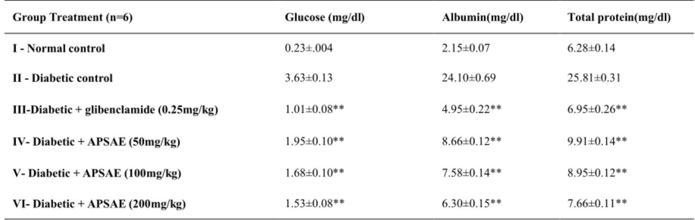
Assessment of long-term effect of APSAE on urine parameters in normal and STZ-induced diabetic rats
and total protein levels were significantly higher in diabetic control rats compared with those in normal rats. Treatment of the diabetic rats with APSAE produced a significant reduction in the albumin, creatinine, urea and total protein levels when compared with diabetic control rats (Table 4).
Immunohistochemical and
immunofluorescent study
Diabetic nephropathy resulted in
significant histopathological changes assessed in transverses section of the kidney. In transverse section nerve derangement,
axonal swelling, increase in number of mesangial cells, matrix of glomeruli, and hyaline thickening of arterioles were also noted. Administration of the APSAE (50, 100, and 200 mg/kg p.o.) significantly
attenuated diabetes induced fiber
derangement, swelling of nerve fibers, and increase in number of mesangial cells as a marker of histopathological alterations when compared with diabetic control group (Figure 1a-f). Microscopic examinations were
performed under 450X light microcopy, scale bar 35 μm.
Table 1. Effect of Adenanthera pavonina seed aqueous extract on blood glucose, glycosylated Hb and body weights
of normal and diabetic rats.
Change in body weight (gm) HbA1c mg/gm Hb
Blood glucose (mg/dl) Group Treatment (n=6)
186.17±1.70 6.45±0.17
103.17±4.57 I - Normal control
142.50±1.25 10.83±0.11
413.33±2.80 II - Diabetic control
170.17±1.70 ** 7.50±0.005**
106.33±1.28** III- Diabetic + glibenclamide (0.25mg/kg)
156.83±1.77** 8.40±0.38**
125.83±0.60* IV- Diabetic + APSAE (50mg/kg)
164.33±1.30** 8.16±0.04**
118.00±0.57** V- Diabetic + APSAE (100mg/kg)
170.67±1.20** 7.80±0.07**
113.00±1.03** VI- Diabetic + APSAE (200mg/kg)
*p<0.05, **p<0.01. Values are Mean±SEM, n=6, when compared with diabetic control by using one way ANOVA followed by Dunnette’s multiple comparison test.
Table 2. Effect of Adenanthera pavonina seed aqueous extract on serum lipid profile of normal and diabetic rats.
Triglycerides VLDL
HDL
in mg/dl LDL
Body Cholesterol Group (n=6)
66.00±0.57 16.66±0.30
11.50±0.42 23.33±0.42
66.83±0.47 I
113.17±0.60 23.00±0.36
8.00±0.51 95.50±0.76
94.00±0.73 II
75.50±0.76** 17.83±0.65**
12.16±0.30** 36.83±0.60**
69.66±0.49** III
82.50±0.76** 20.33±0.42*
12.33±0.33** 53.16±0.47**
81.16±0.47* IV
79.50±0.78** 18.83±0.40**
13.33±0.33** 46.66±0.49**
77.00±0.57** V
77.50±0.38** 18.66±0.21**
13.66±0.42** 38.83±0.60**
73.50±0.42** VI
Table 3. Effects of Adenanthera pavonina seed aqueous extract on serum parameters of normal and diabetic rats.
Total protein(mg/dl) Urea(mg/dl)
Creatinine (mg/dl) Albumin (mg/dl)
Group Treatment (n=6)
7.26±0.12 29.66±0.88
0.58±0.05 3.26±.004
I - Normal control
4.98±0.13 69.00±1.78
2.56±0.20 2.40±0.12
II - Diabetic control
6.96±0.07** 36.66±0.88**
0.93±0.07** 3.16±0.01**
III- Diabetic + glibenclamide (0.25mg/kg)
5.81±0.21* 52.16±1.35*
1.55±0.07* 2.75±0.08*
IV- Diabetic + APSAE (50mg/kg)
6.26±0.08** 40.33±0.88**
1.20±0.05** 2.98±0.02**
V- Diabetic + APSAE (100mg/kg)
6.75±0.07** 39.16±0.47**
1.07±0.04** 3.10±0.08**
VI- Diabetic + APSAE (200mg/kg)
*p<0.05, **p<0.01. Values are Mean±SEM, n=6, when compared with diabetic control by using one way ANOVA followed by Dunnette’s multiple comparison test.
Figure 1 (A-F).Histopathological changes in the kidney where, imagesA-Fshows transverse-section of kidney of normal, diabetic control, diabetic treated with APSAE (50, 100 and 200 mg/kg) and glibenclamide pretreated groups, respectively (A-F).
FiguresAand Fshows normal fiber arrangement with mesangial cells. Figure Bshows nerve derangement, axonal
swelling, increase in number of mesangial cells, matrix of glomeruli, and hyaline thickening of arterioles. Figure C,
D, and E shows attenuation of STZ-induced nerve derangement, axonal swelling and increase in number of
Table 4. Effect of Adenanthera pavonina seed aqueous extract on Urine parameters of normal and diabetic rats.
Total protein(mg/dl) Albumin(mg/dl)
Glucose (mg/dl) Group Treatment (n=6)
6.28±0.14 2.15±0.07
0.23±.004 I - Normal control
25.81±0.31 24.10±0.69
3.63±0.13 II - Diabetic control
6.95±0.26** 4.95±0.22**
1.01±0.08** III-Diabetic + glibenclamide (0.25mg/kg)
9.91±0.14** 8.66±0.12**
1.95±0.10** IV- Diabetic + APSAE (50mg/kg)
8.95±0.12** 7.58±0.14**
1.68±0.10** V- Diabetic + APSAE (100mg/kg)
7.66±0.11** 6.30±0.15**
1.53±0.08** VI- Diabetic + APSAE (200mg/kg)
*p<0.05, **p<0.01 Values are Mean±SEM, n=6, when compared with diabetic control by using one way ANOVA followed by Dunnette’s multiple comparison test.
Discussion
Diabetic nephropathy is one of the most common complications of diabetes and is characterized by increased urinary protein and loss of renal function. A number of studies have now definitely proven that improved metabolic control that achieves
near-normoglycemia can significantly
decrease the development and progression of diabetic nephropathy (Mogensen, 1984; Lee et al., 1995; Park et al., 1998). The metabolic factors such as AGEs, sorbitol beyond blood glucose level are also implicated in the pathogenesis of diabetic nephropathy (Schrijvers et al., 2004). Traditional plant remedies have been used for centuries in the treatment of diabetes (Akhtar et al., 1984; Kesari et al., 2005; Kesari et al., 2007; Rai et al., 2007), but only few of these plants have been scientifically evaluated. Therefore, we have investigated the effect of APSAE on glycemic and renal protection in STZ-induced diabetic rats. A. pavonina seed aqueous extract showed a dose dependent effect on fasting blood glucose in diabetic rats. The capacity of APSAE to decrease the elevated blood glucose to normal level is an
normal homeostasis during experimental diabetes. The possible mechanism by which APSAE exerts its hypoglycemic action in diabetic rats may be due to potentiating the insulin release, since the percentage of reduction in blood glucose levels was considerable. In uncontrolled or poorly controlled diabetes, there is an increased glycosylation of a number of proteins including haemoglobin and crystalline of lens (Alberti, 1982). HbA1c was found to increase in patients with diabetes mellitus and the amount of increase was directly proportional to the fasting blood glucose levels (Pari et al., 2002).
with the diabetic control rats which may be due to its protective effect in controlling
muscle wasting, i.e., reversal of
gluconeogenesis and also the improvement in glycemic control.
The results from the current study showed that APSAE reduces the development of diabetic nephropathy by increasing serum parameters such as albumin and total protein, but a reduction in serum creatinine and urea as well as urine albumin and total protein in treated rats as compared with diabetic control rats. Hence, current study confirmed that
APSAE-treated diabetic rats showed
significant improvement in renal functions
such as proteinuria and
albuminuria.Treatment of the diabetic rats with APSAE and glibenclamide caused a significant reduction in the serum triglycerides, LDL and VLDL cholesterol with a significant increase in HDL cholesterol levels when compared with diabetic control rats. Administration of the APSAE (50, 100 and 200 mg/kg p.o.) significantly attenuated diabetes induced fiber derangement, swelling of nerve fiber, and increase in number of mesangial cells as a marker of histopathological alterations when compared with diabetic control group. In conclusion, the present data clearly demonstrate that administration of APSAE reduces metabolic factors influencing diabetic nephropathy such as blood glucose, albumin, total protein creatinine and HbA1c in experimental diabetes. Taking together these results indicate that, APSAE has therapeutic or preventive effects on several pharmacological targets in the complicated
pathological mechanism of diabetic
nephropathy. Thus, it is worthwhile to be further investigated for its potential
pharmacological effects in diabetic
nephropathy.
Acknowledgements
The authors sincerely thank to Principal, M.E.S. College of Pharmacy, Prashant Patil
Gadakh Secretary, Mula Education Society, Sonai and Department of Pharmacy, Suresh Gyan Vihar University, Jaipur, Rajasthan, India for encouragement and availing of the laboratory facilities during the course of investigation.
Conflict of interest
The authors declare that there are no conflicts of interest.
References
Akhtar MS, Ali MR. 1984. Study of antidiabetic effect of a compound medicinal plant prescription in normal and diabetic rabbits. J Pak Med Assoc, 34: 239-244.
Alarcon-Aguilara FJ, Roman-Ramos R, Perez-Gutierrez S, Aguilar-Contreras A, Contreras-Weber CC, Flores-Saenz JL. 1998. Study of the anti-hyperglycemic effect of plants used as antidiabetics. J Ethnopharmacol, 61: 101-110. Alberti KGMM. 1982. M. Press, The
biochemistry and the complications of diabetes In: H. Keen, J. Jarrett (Eds.), Complications of Diabetes, vol.43, Edward Arnold Ltd., London pp. 231-270.
Anna J, Robert Z, Arkadiusz K. 2006. Emulsions of oil from Adenanthera pavonina L. seeds and their protective effect. J Cell Mol Biol, 3: 1425.
Balogun AM, Fetuga BL. 2004. Fatty acid composition of seed oils of some members of the leguminosae Family. Food Chem,17: 175-82.
Bouquet A, Debray M. 1974. Medicinal plants in Ivory Coast. Document Orstom France, 32: 1-4.
Brownlee M. 1995. Advanced protein glycosylation in diabetes and aging. Annual Rev Med, 46: 223-234.
Brownlee M. 2005. The pathobiology of diabetic complications: a unifying mechanism. Diabetes, 54: 1615-1625.
Burkill IH. 1966. A dictionary of the economic products of the Malay Peninsula Edited by: Ministry of Agriculture (Malaysia). Crown Agents for the colonies London pp. 839. Burstein M, Scholnichk HR, Morin R. 1970.
from human serum by precipitation with polyanions. J Lipid Res, 11: 583-595.
Chatterjea MN, Shinde R. 1976. Diabetes mellitus, Textbook of medical biochemistry, 5th ed, Jaypee Brothers Medical Publishers Ltd., New Delhi pp.1976.
Chuang PY, Yu Q, Fang W, Uribarri J, He JC. 2007. Advanced glycation end products induce podocyte apoptosis by activation of the FOXO4 transcription factor. Kidney Int, 72: 965-976.
Eross J, Kreutzman D, Jimenez M, Keen R, Rogers S, Cowell C, Vines R, Silink M. 1984. Colorimetric measurement of glycosylated protein in whole blood cells plasma and dried blood. Ann Clin Biochem, 21: 519-522. Folch J, Lees M, Solane SGH. 1957. A simple
method for isolation and purification of total lipids from animal tissues. J Biol Chem, 26: 497-509.
Foster JB, Dunn RT.1973. Stable reagents for determination of serum triglycerides by colorimetric Hantzsch condensation method. Clin Chem, 19: 338-340.
Friedwald WT, Levy RI, Fredrickson DS. 1972. Estimation of the concentration of LDL-cholesterol in plasma without the use of the preparative ultracentrifuge. Clin Chem, 18: 499-502.
Gupta S, Kataria M, Gupta PK, Murganandan S, Yashroy RC. 2004. Protective role of extracts of neem seeds in diabetes caused by Streptozotocin in rats. J Ethnopharmacol, 90: 185-189.
Howes FN. 1974. A dictionary of useful everyday plants and their common names. Cambridge University Press pp. 15.
Huebschmann AJ, Regensteiner JG, Vlassara H, Reusch JE. 2006. Diabetes and advanced glycoxidation end products. Diabetes Care, 29: 1420-1432.
Kaji Y, Amano S, Usui T, Oshika T, Yamashiro K, Ishida S, Suzuki K, Tanaka S, Adamis AP, Nagai Horiuchi R. 2003. Expression and function of receptors for advanced glycation end products in bovine corneal endothelial cells. Invest Ophthalmol Vis Sci, 44: 521-528. Kasper M, Roehlecke C, Witt M, Fehrenbach H, Hofer A, Miyata T, Weigert C, Funk RH, Schleicher ED. 2000. Induction of apoptosis
epithelial cell line L132. Am J Respir Cell Mol Biol, 23: 485-491.
Kesari AN, Gupta RK, Watal G. 2005. Hypoglycemic effects of Murraya koenigii on normal and alloxan diabetic rabbits. J Ethnopharmacol, 97: 247-251.
Kesari AN, Kesari S, Singh SK, Gupta RK, Watal G. 2007. Studies on the glycemic and lipidemic effect of Murraya koenigii in experimental animals. J Ethnopharmacol, 112: 305-311.
Khare CP. 2007. Indian medicinal plants - An illustrated Dictionary, Springer-Verlag; Berlin pp. 601.
Lee KU, Park JY, Kim SW, Lee MH, Kim GS, Park SK, Park JS. 1995. Prevalence and associated features of albuminuria in Koreans with NIDDM. Diabetes Care, 18: 793-799. Li HL, Zheng HC, Bukuru J, De Kimpe D. 2004.
Natural medicines used in the traditional Chinese medical system for therapy of diabetes mellitus. J Ethnopharmacol, 92: 1-21.
Mauer SM, Steffes MW, Ellis EN, Sutherland DE, Brown DM, Goetz FC.1984. Structural– functional relationships in diabetic nephropathy. J Clin Invest, 74: 1143-1155. Meyer TW, Bennett PH, Nelson RG.1999.
Podocyte number predicts long-term urinary albumin excretion in pima Indians with Type II diabetes and microabuminuria. Diabetologia, 42: 1341-1344.
Mogensen CE. 1984. Microalbuminuria predicts clinical proteinuria and early mortality in maturity-onset diabetes. The New England Journal of Medicine, 310: 356-360.
Muhammad SA, Farman A, Iqbal A, Muhammad KP. 2005. Pavonin: A new five membered lactone from Adenanthera pavonina Linn. (Mimosaceae). Nat prod res, 9: 37-40.
Olajide AO, Echianu CA, Adedapo AD, Makinde JM. 2004. Anti-inflammatory studies on Adenanthera pavonina seed extract. Inflammopharmacology, 3: 196- 202.
Pagtalunan ME, Miller PL, Jumping-Eagle S, Nelson RG, Myers BD, Rennke HG, Coplon NS, Singh R, Barden A, Mori T, Beilin L.2001. Advanced glycation end-products: a Review. Diabetologia 44: 129-146.
diabetes mellitus. Comp Biochem Physiol C: Pharmacol Toxicol Endocrinol, 131: 19-25. Park JY, Kim HK, Chung YE, Kim SW, Hong
SK, Lee KU. 1998. Incidence and determinants of microalbuminuria in Koreans with type 2 diabetes. Diabetes Care, 21: 530-534.
Rai PK, Rai NK, Rai AK, Watal G. 2007. Role of LIBS in elemental analysis of Psidium guajava responsible for glycemic potential. Instrum Sci Technol, 35: 507-522.
Sadasivam S, Manickam A.1996. Methods in Biochemistry. 2nd ed. New Delhi, New Age International Pvt. Ltd pp.
Schrijvers BF, De Vriese AS, Flyvbjerg A. 2004. From hyperglycemia to diabetic kidney disease: the role of metabolic, hemodynamic, intracellular factors and growth Factors/cytokines. Endocr Rev, 25: 971-1010. Sohn EJ, Kim CS, Kim YS, Jung DH, Jang DS,
Lee YM, Kim JS. 2007. Effects of magnolol (5,50-diallyl-2, 20-dihydroxybiphenyl) on diabetic nephropathy in type 2 diabetic Goto-Kakizaki rats. Life Sci, 80: 468-475.
Sun L, Meyer TW. 1977. Podocyte loss and progressive glomerular injury in type II diabetes. J Clin Invest, 99: 342-348.
Swanston-Flatt SK, Day C, Bailey CJ, Flatt PR.1990. Traditional plant treatment for diabetes: studies in normal and streptozotocin diabetic mice. Diabetologia, 33: 462-464. Tiwari AK, Madhusudanarao J. 2002. Diabetes
mellitus and multiple therapeutic approaches
of phytochemicals: present status and future prospects. Curr Sci, 83: 30-38.
White KE, Bilous RW. 2000. Type 2 diabetic patients with nephropathy show structural functional relationships those are similar to type 1 disease. J Am Soc Nephrol, 11: 1667-1673.
Yadav N, Misra G, Nigram SK.1976. Triterpenoids from Adenanthera pavonina bark. Plant Med, 29:176-178.
Yamagishi S, Takeuchi M, Matsui T, Nakamura K, Imaizumi T, Inoue H. 2005. Angiotensin II augments advanced glycation end product-induced pericyte apoptosis through RAGE overexpression. FEBS Lett, 579: 4265-4270. Yamagishi S, Inagaki Y, Amano S, Okamoto T,
Takeuchi M, Makita Z. 2002. Pigment Epithelium-derived factor protects cultured retinal pericytes from advanced glycation end product-induced injury through its antioxidative properties. Biochem Biophys Res Commun, 296: 877-882.
Zhang Y, Chen B, Hou XH, Guan GJ, Liu G, Liu HY, Li XG. 2007. Effects of mycophenolate mofetil, valsartan and their combined therapy on preventing podocyte loss in early stage of diabetic nephropathy in rats, Chin Med J (Engl.), 120: 988-995.