Periodontics
Ioanna Xynogala(a) Eudoxie Pepelassi(a) Despina Perrea(b) George Agrogiannis(c) Alkistis Pantopoulou(b) Efstratios Patsouris(c) Ioannis Vrotsos(a)
(a)Department of Periodontology, School of Dentistry, University of Athens, Athens, Greece.
(b)Laboratory of Experimental Surgery and Surgical Research “N. S. Christeas”, School of Medicine, University of Athens, Athens, Greece.
(c)First Department of Pathology, School of Medicine, University of Athens, Athens, Greece.
Corresponding author: Ioanna Xynogala
E-mail: [email protected]
Received for publication on Jul 30, 2011 Accepted for publication on Nov 16, 2011
Adiponectin and interleukin-6 levels
in insulin-treated diabetic rats with
experimental periodontitis
Abstract: The aim of the study was to compare the serum levels of ad-iponectin and interleukin-6 (IL-6) in insulin-treated diabetic rats with or without periodontitis. Forty male Wistar rats were randomly divided into 2 groups (20 rats each): a) insulin-treated diabetic group (control, DI) and b) insulin-treated diabetic periodontitis group (test, DIP). Dia-betes was induced, and insulin treatment was initiated on day 5. On day 16, periodontitis was induced in the DIP group. All rats were euthanized on day 77. Adiponectin and IL-6 were assessed on days 16 and 77. At the end of the experiment, 14 and 11 rats survived in the DI and DIP groups, respectively. Adiponectin levels were statistically signiicantly higher at the end of the experiment compared with levels on day 16 in the peri-odontitis group (p < 0.05), but not in the control group. At the end of the experiment, adiponectin levels were statistically signiicantly higher in the periodontitis group compared with the control group (p < 0.05). Within-group and between-group comparisons of IL-6 levels showed no statistically signiicant difference. In conclusion, serum adiponectin was increased in insulin-treated diabetic rats with periodontitis in compari-son with insulin-treated diabetic rats, while IL-6 levels did not differ be-tween groups.
Descriptors: Adiponectin; Rats; Diabetes Mellitus; Periodontitis; Insulin.
Introduction
Type 1 diabetes mellitus is characterized by a deiciency of insulin secretion, caused by a cellular-mediated autoimmune destruction of
pan-creatic b-cells.1 Periodontitis is a chronic inlammatory disease involving
tooth-supporting tissues.2 There is an increased severity of periodontitis
in individuals with poorly controlled diabetes.3 The periodontal
condi-tions, when diabetes is controlled, are comparable with those of the
gen-eral population.3
Adiponectin, a 30-kDa protein mainly secreted by adipocytes, has
anti-inlammatory, antidiabetic, and anti-atherogenic properties.4
Re-search indicates that it is involved in the regulation of insulin sensitiv-ity.5 Adiponectin levels are increased in type 1 diabetes6 and decreased
in type 2 diabetes7 and in obesity.8 Adiponectin seems to be involved in
bone metabolism. Adiponectin and adiponectin receptors, AdipoR1 and
AdipoR2, are expressed in osteoblastic and osteoclastic cells.9,10 Positive
as well as negative effects on bone formation10 and
osteoclast formation11-13 have been reported for
adi-ponectin. Adiponectin’s role in periodontal disease has not been fully elucidated.
Interleukin-6 (IL-6) is a pro-inlammatory cyto-kine that has been reported to be elevated in type 2 diabetes14 and decreased in type 1.15 It is described
as one of the osteoclast-inducing factors acting on
osteoblasts.16 The hypothesis of this study was that
adiponectin and IL-6 levels are different in insulin-treated diabetic rats with experimental periodontitis than in insulin-treated diabetic rats.
The purpose of the present study was to investi-gate the effect of experimental periodontitis on se-rum adiponectin and IL-6 levels in insulin-treated diabetic rats.
Methodology
Animals
Forty male Wistar rats (225-250 g) were ran-domly divided into 2 groups (20 rats each):
• diabetes induction and insulin administration
(control, DI); and
• diabetes induction, insulin administration, and
periodontitis induction (test, DIP).
Rats with probing depth > 0.5 mm were
exclud-ed.17 They were housed in stainless steel cages with
no bedding material (2 per cage, 12-hour light/dark,
room temperature 18-22 °C, relative humidity
55-65%). They were fed with a standard laboratory diet
in powder form,18 given tap water ad libitum and
ac-climatized to the new environment for 7 days.19 The
study protocol was in accordance with local laws and regulations, with the European Communities Council Directive of 24 November 1986 (86/609/ EEC), and with guidelines approved by the Coun-cil of the American Psychological Society (1980). The study was approved by the Ethics and Research Committee of the School of Dentistry, University of Athens, and by the Veterinary Directorate of the Prefecture of Athens (# K/3935/28-5-2008).
On day 1, the rats were anesthetized with an intramuscular injection of ketamine hydrochloride
solution (100 mg/kg body weight, Imalgene 1000,
MERIAL, Lyon, France) and xylazine (10 mg/kg
body weight, Rompun, Bayer HealthCare,
Leverku-sen, Germany). Type 1 diabetes was induced by the intravenous injection (into the tail vein) of strepto-zotocin (STZ) (Sigma, St. Louis, USA), 45 mg/kg
body weight,20 dissolved in citrate buffer (10 mM,
pH 4.5). Rats with glucose levels > 300 mg/dL19,21
up to the 5th day were included in the study. Body
weight and blood glucose were measured daily19
(glucometer Wellion Linus, AgaMatrix Inc., Salem,
USA) after a 12-hour nocturnal fast. The induction of diabetes was conirmed when glucose levels irst
exceeded 300 mg/dL during the irst 5 days (Gd).
On day 5, insulin (Protaphane, Novo Nordisk A/S,
Bagsværd, Denmark) treatment was initiated in both groups (subcutaneously, once/day). The dosage for each rat was adjusted daily.
On day 16, all rats were anesthetized, as men-tioned above. Blood samples were collected from the
tail vein and stored in Eppendorf tubes at -80 °C. In
the DIP group, a sterile 4/0 silk ligature (Medipac,
Kilkis, Greece) was placed subgingivally around the
maxillary right second molar.22 On day 77, blood
samples were drawn from the tail vein and stored
in Eppendorf tubes at -80 °C, and the rats were
euthanized with ketamine hydrochloride solution (200 mg/kg body weight) (intramuscularly) and pen-tothal solution 10% (intraperitoneally).
Biomedical evaluation
Serum samples were analyzed for adiponec-tin and IL-6 levels by an Enzyme-Linked Immu-nosorbent Assay (ELISA) and read with Luminex 100 (Multiplexed Biomarker Immunoassays for
Luminex Instrumentation/xMAP Technology -
Luminex Corporation, Austin, USA). Adiponectin
and IL-6 levels were determined with Milliplex
Map Kits (Millipore Corporation, Billerica, USA) (Adiponectin, Rat CVD Panel 3, # RCVD3-89K; and IL-6, Rat Cytokine / Chemokine, # RCYTO-80K).
Histometric evaluation
separated along the midline, and embedded in par-afin. The sections from the right side (6 mm thick mesio-distally parallel to the tooth axis, semi-serial, longitudinal) were stained with hematoxylin-eosin. One section was selected from each specimen from the middle area of the tooth. It was photographed by light microscopy (Nikon Eclipse 80i, Nikon, To-kyo, Japan) and a digital camera (Nikon DS-2 MW, Nikon, Tokyo, Japan) and analyzed with Image Pro Plus 5.1 program (Media Cybernetics, Bethesda, USA). The distance from the cemento-enamel
junc-tion to the alveolar crest22 was measured. For each
second right maxillary molar, the mesial and distal measurements were averaged (HR), and the median was calculated for each group. Measurements were repeated twice by the same examiner, blinded to the treatment groups, with 2 weeks between measure-ments. The average of these 2 scorings was docu-mented. In almost 10% of the sites, the difference between the 2 scorings was 2-3%, while in most sites, the scorings were identical.
Statistical analysis
For statistical analysis, mean values and stan-dard deviations or median (Q1-Q3) (when the nor-mality assumption was not met) were calculated for weight, glucose levels, histological parameters, and serum adiponectin and IL-6 levels at speciic
time-points. The t-test or Mann-Whitney test for
normal-ly or non-normalnormal-ly distributed continuous variables was used for comparison of the above-mentioned measurements between the 2 study groups at each time-point. Comparisons within groups between
different time-points were performed by t-test for
dependent variables or the Wilcoxon signed-ranks test. Results were signiicant if p < 0.05. Statistical analysis was performed with STATA 9.1 (Stata,
Col-lege Station, USA).
Results
At the end of the experiment, 14 rats (70%) in the DI and 11 rats (55%) in the DIP groups survived (p = 0.33).
There was no statistically signiicant difference
in mean baseline (W1) (p = 0.64) and inal weight
(W77) (p = 0.28) between groups. For both groups,
mean inal weight (W77) was statistically
signiicant-ly increased compared with baseline weight (W1)
(DI: p = 0.002 and DIP: p = 0.001) (Table 1).
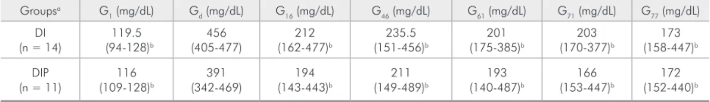
There was no statistically signiicant difference in median glucose levels between the groups on day 1 (G1), day of diabetes conirmation (Gd), and day 16 (G16) (p > 0.05). The same applied for median glucose levels on days 46 (G46), 61 (G61), 71 (G71),
and on the day of euthanasia (G77) (p > 0.05).
Me-dian Gd glucose levels were statistically signiicantly
higher compared with levels on days 16, 46, 61, 71, and 77 for both groups (for all occasions, p < 0.05) (Table 2).
Regarding the histometric evaluation, median values of HR were statistically signiicantly greater
for the DIP [1045.7 (850.4-2217.7) mm]than for the
DI [419.7 (360.6-459.1) mm] group (p < 0.0001).
Median adiponectin levels were not statistical-ly signiicantstatistical-ly different between the DI and DIP groups on day 16 (p = 0.14), while they were sta-tistically signiicantly higher for the DIP than the DI group on day 77 (p = 0.046). Concerning the DI group, median adiponectin concentrations did not differ statistically signiicantly between day 77 (i-nal) and day 16 (p = 0.4). In contrast, median inal adiponectin values were statistically signiicantly in-creased compared with those on day 16 for the DIP group (p = 0.04). Median IL-6 levels did not change
Table 1 - Mean weight ± SD for groups DI and DIP on days 1 (W1), 5 (W5), 16 (W16), and 77 (W77) and average weight for the
entire experimental period (Wm).
Groupsa W
1 (g) W5 (g) W16 (g) W77 (g) Wm (g)
DI (n = 14) 253.43 ± 5.60c 233.86 ± 22.67b 231.43 ± 23.51b 273.57 ± 25.28b,c 249.10 ± 23.64 DIP (n = 11) 257.82 ± 25.56c 237.64 ± 6.87b 237.64 ± 7.54b 286.36 ± 32.86b,c 257.82 ± 29.6
a No statistically significant difference between groups on days 1 (W
1), 5 (W5), 16 (W16), 77 (W77), and the average weight (Wm) (p > 0.05). b Statistically
statistically signiicantly between day 16 and day 77 in any group. Between-group comparisons also showed no statistically signiicant difference (Table 3).
Discussion
The present study compared serum adiponectin and IL-6 levels in insulin-treated diabetic rats with or without experimentally induced periodontitis.
Diabetes induction was conirmed in rats 219 and 321
days after STZ injection. In the present study, day 5 was selected as the cut-off day. Since glucose levels were measured daily, the authors noticed that even though diabetes was conirmed in all rats by day 5, some rats did not exhibit glucose levels > 300 mg/dL at days 2 and 3 (data not shown), suggesting that 2 or 3 days might be a rather short time for STZ to act completely.
Both groups had similar glycemic states prior to insulin treatment. Insulin treatment signiicantly re-duced glucose levels. However, glucose levels were not decreased to the levels documented in previous studies,19,21 despite daily adjustment of the insulin
dosage.
The present study demonstrated that adiponec-tin levels were increased in insulin-treated diabetic rats in the presence of periodontitis, while IL-6 lev-els did not show signiicant changes. IL-6 promotes
osteoclast differentiation.16 Furugen et al.23 found
no signiicant difference in serum IL-6 levels in indi-viduals with and without periodontitis, while higher
levels have also been reported in periodontitis.24
There is research showing that IL-6 inhibits adipo-nectin gene expression and secretion in 3T3-L1 adi-pocytes, indicating that it is implicated in
adiponec-tin regulation.25
The precise role of adiponectin in bone homeo-stasis has not been fully elucidated. Berner et al.9
showed that the addition of recombinant mouse adiponectin to the culture medium of murine
os-teoblasts enhanced their proliferation. Oshima et
al.26 noticed increased trabecular bone mass and
decreased numbers of osteoclasts when mice were injected with adenovirus-expressing adiponectin.
Their in vitro experiments showed that adiponectin
suppressed the macrophage-colony-stimulating fac-tor/receptor activator of nuclear factor-kB ligand (M-CSF/RANKL)-induced differentiation of mouse bone marrow stromal cells, as well as the differen-tiation of human CD14-positive peripheral blood mononuclear cells (PBMCs) into osteoclasts. Adipo-nectin treatment of CD14-positive cells also reduced the bone-resorption activity of osteoclasts. Their re-sults suggest that adiponectin enhances bone mass by activating osteoblastogenesis and suppressing osteoclastogenesis. Other studies also conirm the
Table 2 - Median glucose levels (Q1-Q3) for each group (DI, DIP) at day 1 (G1), day of diabetes confirmation (Gd), and days
16 (G16), 46 (G46), 61 (G61), 71 (G71), and 77 (G77).
Groupsa G
1 (mg/dL) Gd (mg/dL) G16 (mg/dL) G46 (mg/dL) G61 (mg/dL) G71 (mg/dL) G77 (mg/dL) DI
(n = 14) (94-128)119.5 b (405-477)456 (162-477)212 b (151-456)235.5 b (175-385)201 b (170-377)203 b (158-447)173 b DIP
(n = 11) (109-128)116 b (342-469)391 (143-443)194 b (149-489)211 b (140-487)193 b (153-447)166 b (152-440)172 b
a No statistically significant difference between groups for any of the time-points examined (p > 0.05). b Statistically significant difference when compared
with Gd (p < 0.05).
Adiponectin (mg/mL) IL-6 (pg/mL)
Groups Day 16 Day 77 Day 16 Day 77
DI (n = 14) 14.7 (11-21.5) 10.4 (9.1-15) 253.2 (24.4-1834.3) 48.9 (24.4-643.6) DIP (n = 11) 12.2 (8.9-13.3) 16.6 (13.1-33.1)a,b 656.2 (331.1-847.6) 902.3 (90.8-2030) a Statistically significant difference between groups on the specific day (p < 0.05). b Statistically significant
dif-ference within the group, day 16 versus day 77 (p < 0.05).
inhibitory effect of adiponectin on osteoclast forma-tion11,12 Another study,10 however, suggested both
positive as well as negative effects of adiponectin on bone formation. The positive effect on bone forma-tion was by locally produced adiponectin through the autocrine/paracrine pathway and by circulating adiponectin via enhancement of insulin signaling through the indirect pathway. A negative action was reported through the direct pathway by circulating adiponectin. Adding to this complicated picture is the fact that Luo et al.13 showed that adiponectin
increased osteoclast formation indirectly (via stimu-lation of RANKL and inhibition of osteoprotegerin production in osteoblasts).
The different actions of adiponectin may be par-tially explained by its various possible forms,
with-out a full exploration of their biological activities.9
There are studies23,27,28 that have not shown
statis-tically signiicant differences in serum adiponec-tin levels in periodontitis patients compared with healthy control individuals, while a recent article29
showed a lower ratio of high-molecular-weight adi-ponectin to total adiadi-ponectin in patients with peri-odontal pockets; this ratio was independently
asso-ciated with periodontal condition.
In addition, the expression of AdipoR1 and AdipoR2 in gingival tissues has been shown to be
reduced in periodontitis.30 A decrease in the
ex-pression of the receptors leads to a decrease in adi-ponectin binding and thereby in its effects
(adipo-nectin resistance).30
Conclusion
In conclusion, within its limitations, this study showed that the serum levels of adiponectin were increased in insulin-treated diabetic rats in the pres-ence of periodontitis, while serum IL-6 levels did not change. The role of adiponectin in periodontitis and its correlation with other factors needs to be further explored. Future studies are required to clarify the mechanisms by which adiponectin is implicated in periodontal diseases.
Acknowledgement
We express our gratitude to Mrs. K. Perrea for the assessment of the adiponectin and IL-6 levels.
References
1. American Diabetes Association. Diagnosis and classification of diabetes mellitus. Diabetes Care. 2011 Jan;34(Suppl 1):S62-69.
2. Lindhe J, Ranney R, Lamster I, Charles A, Chung CP, Flem-mig T, et al. Consensus report: chronic periodontitis. Ann Periodontol. 1999 Dec;4(1):38.
3. Kinane D, Bouchard P. Periodontal diseases and health: con-sensus report of the Sixth European Workshop on Periodon-tology. J Clin Periodontol. 2008 Sep;35(8 suppl):333-7. 4. Brochu-Gaudreau K, Rehfeldt C, Blouin R, Bordignon V,
Murphy BD, Palin MF. Adiponectin action from head to toe. Endocrine. 2010 Feb;37(1):11-32.
5. Maeda N, Shimomura I, Kishida K, Nishizawa H, Matsuda M, Nagaretani H, et al. Diet-induced insulin resistance in mice lacking adiponectin/ACRP30. Nat Med. 2002 Jul;8(7):731-7. 6. Maahs DM, Ogden LG, Snell-Bergeon JK, Kinney GL, Wadwa
RP, Hokanson JE, et al. Determinants of serum adiponectin in persons with and without type 1 diabetes. Am J Epidemiol. 2007 Sep 15;166(6):731-40.
7. Hotta K, Funahashi T, Arita Y, Takahashi M, Matsuda M, Okamoto Y, et al. Plasma concentrations of a novel,
adipose-specific protein, adiponectin, in type 2 diabetic patients. Ar-terioscler Thromb Vasc Biol. 2000 Jun;20(6):1595-9. 8. Arita Y, Kihara S, Ouchi N, Takahashi M, Maeda K,
Mi-yagawa J, et al. Paradoxical decrease of an adipose-specific protein, adiponectin, in obesity. Biochem Biophys Res Com-mun. 1999 Apr 2;257(1):79-83.
9. Berner HS, Lyngstadaas SP, Spahr A, Monjo M, Thommesen L, Drevon CA, et al. Adiponectin and its receptors are ex-pressed in bone-forming cells. Bone. 2004 Oct;35(4):842-9. 10. Shinoda Y, Yamaguchi M, Ogata N, Akune T, Kubota N,
Yamauchi T, et al. Regulation of bone formation by adipo-nectin through autocrine/paracrine and endocrine pathways. J Cell Biochem. 2006 Sep 1;99(1):196-208.
11. Williams GA, Wang Y, Callon KE, Watson M, Lin JM, Lam JB, et al. In vitro and in vivo effects of adiponectin on bone. Endocrinology. 2009 Aug;150(8):3603-10.
13. Luo XH, Guo LJ, Xie H, Yuan LQ, Wu XP, Zhou HD, et al. Adiponectin stimulates RANKL and inhibits OPG expression in human osteoblasts through the MAPK signaling pathway. J Bone Miner Res. 2006 Oct;21(10):1648-56.
14. Pradhan AD, Manson JE, Rifai N, Buring JE, Ridker PM. C-reactive protein, interleukin 6, and risk of developing type 2 diabetes mellitus. JAMA. 2001 Jul 18;286(3):327-34. 15. Dogan Y, Akarsu S, Ustundag B, Yilmaz E, Gurgoze MK.
Serum IL-1beta, IL-2, and IL-6 in insulin-dependent diabetic children. Mediators Inflamm. 2006;2006(1):59206. 16. Suda T, Udagawa N, Nakamura I, Miyaura C, Takahashi
N. Modulation of osteoclast differentiation by local factors. Bone. 1995 Aug;17(2 Suppl):87S-91S.
17. Verzeletti GN, Gaio EJ, Rösing CK. Effect of methotrexate on alveolar bone loss in experimental periodontitis in Wistar rats. Acta Odontol Scand. 2007 Nov;65(6):348-51. 18. Pontes Andersen CC, Flyvbjerg A, Buschard K, Holmstrup P.
Relationship between periodontitis and diabetes: lessons from rodent studies. J Periodontol. 2007 Jul;78(7):1264-75. 19. Mishima N, Sahara N, Shirakawa M, Ozawa H. Effect of
streptozotocin-induced diabetes mellitus on alveolar bone deposition in the rat. Arch Oral Biol. 2002 Dec;47(12):843-9. 20. Tesseromatis C, Kotsiou A, Parara H, Vairaktaris E, Tsamouri
M. Morphological changes of gingiva in streptozotocin dia-betic rats. Int J Dent. 2009;2009:725628. Epub 2009 Jan 27. 21. Holzhausen M, Garcia DF, Pepato MT, Marcantonio E
Jr. The influence of short-term diabetes mellitus and insu-lin therapy on alveolar bone loss in rats. J Periodontal Res. 2004 Jun;39(3):188-93.
22. Semenoff TA, Semenoff-Segundo A, Bosco AF, Nagata MJ, Garcia VG, Biasoli ER. Histometric analysis of ligature-induced periodontitis in rats: a comparison of histological section planes. J Appl Oral Sci. 2008 Jul-Aug;16(4):251-6.
23. Furugen R, Hayashida H, Yamaguchi N, Yoshihara A, Ogawa H, Miyazaki H, et al. The relationship between periodontal condition and serum levels of resistin and adiponectin in el-derly Japanese. J Periodontal Res. 2008 Oct;43(5):556-62. 24. Loos BG, Craandijk J, Hoek FJ, Wertheim-van Dillen PM,
van der Velden U. Elevation of systemic markers related to cardiovascular diseases in the peripheral blood of periodontitis patients. J Periodontol. 2000 Oct;71(10):1528-34.
25. Fasshauer M, Kralisch S, Klier M, Lossner U, Bluher M, Klein J, et al. Adiponectin gene expression and secretion is inhibited by interleukin-6 in 3T3-L1 adipocytes. Biochem Biophys Res Commun. 2003 Feb 21;301(4):1045-50.
26. Oshima K, Nampei A, Matsuda M, Iwaki M, Fukuhara A, Hashimoto J, et al. Adiponectin increases bone mass by sup-pressing osteoclast and activating osteoblast. Biochem Biophys Res Commun. 2005 Jun 3;331(2):520-6.
27. Saito T, Yamaguchi N, Shimazaki Y, Hayashida H, Yonemoto K, Doi Y, et al. Serum levels of resistin and adiponectin in women with periodontitis: the Hisayama study. J Dent Res. 2008 Apr;87(4):319-22.
28. Shimada Y, Komatsu Y, Ikezawa-Suzuki I, Tai H, Sugita N, Yoshie H. The effect of periodontal treatment on serum leptin, interleukin-6, and C-reactive protein. J Periodontol. 2010 Aug;81(8):1118-23.
29. Nagano Y, Arishiro K, Uene M, Miyake T, Kambara M, No-tohara Y, et al. A low ratio of high molecular weight adipo-nectin to total adipoadipo-nectin associates with periodontal status in middle-aged men. Biomarkers. 2011 Mar;16(2):106-11. 30. Yamaguchi N, Hamachi T, Kamio N, Akifusa S, Masuda K,