D I A B E T E S & M E T A B O L I S M J O U R N A L
his is an Open Access article distributed under the terms of the Creative Commons At-tribution Non-Commercial License (http://creativecommons.org/licenses/by-nc/3.0/) which permits unrestricted non-commercial use, distribution, and reproduction in any medium, provided the original work is properly cited.
Autophagy: A Novel herapeutic Target for Diabetic
Nephropathy
Shinji Kume1, Daisuke Koya2
1Department of Medicine, Shiga University of Medical Science, Otsu,
2Department of Diabetology & Endocrinology, Kanazawa Medical University, Kahoku, Japan
Diabetic nephropathy is a leading cause of end stage renal disease and its occurance is increasing worldwide. he most efective treatment strategy for the condition is intensive treatment to strictly control glycemia and blood pressure using renin-angioten-sin system inhibitors. However, a fraction of patients still go on to reach end stage renal disease even under such intensive care. New therapeutic targets for diabetic nephropathy are, therefore, urgently needed. Autophagy is a major catabolic pathway by which mammalian cells degrade macromolecules and organelles to maintain intracellular homeostasis. he accumulation of damaged proteins and organelles is associated with the pathogenesis of diabetic nephropathy. Autophagy in the kidney is activat-ed under some stress conditions, such as oxidative stress and hypoxia in proximal tubular cells, and occurs even under normal conditions in podocytes. hese and other accumulating indings have led to a hypothesis that autophagy is involved in the patho-genesis of diabetic nephropathy. Here, we review recent indings underpinning this hypothesis and discuss the advantages of tar-geting autophagy for the treatment of diabetic nephropathy.
Keywords: AMP-activated protein kinases; Autophagy; Caloric restriction; Diabetic nephropathy; Mechanistic target of ra-pamycin complex 1; Podocytes; Sirt1; Tubular cell
Corresponding author: Daisuke Koya
Department of Diabetology & Endocrinology, Kanazawa Medical University, Kahoku-Gun, Ishikawa 920-0293, Japan
E-mail: [email protected] Received: Oct. 19, 2015; Accepted: Nov. 24, 2015
INTRODUCTION
Diabetic nephropathy is a leading cause of end-stage renal dis-ease throughout the world. he establishment of novel, efec-tive therapeutic strategies is, therefore, urgently required. Pro-teinuria and/or albuminuria is a sign of glomerular lesions in diabetic nephropathy. hese lesions can subsequently develop into tubulointerstitial lesions that lead to renal dysfunction [1]. Clinically, therefore, reducing proteinuria is considered a principal therapeutic target to improve renal outcomes in pa-tients with diabetic nephropathy.
he pathogenesis of diabetic nephropathy involves altered intracellular metabolism associated with hyperglycemia, in-cluding the activation of protein kinase C, the accumulation of
advanced glycation end-products, increased lux of the polyol pathway and oxidative stress [2-6]. Moreover, hemodynamic changes such as systemic and glomerular hypertension related to hyperactivation of the renin-angiotensin system are involved in diabetic nephropathy [7]. he strong association of these al-terations with the pathogenesis of diabetic nephropathy has been supported by a number of large clinical trials such as the Diabetes Control and Complications Trial (DCCT), United Kingdom Prospective Diabetes Study (UKPDS), Kumamoto study and Reduction of Endpoints in NIDDM with the Angio-tensin II Antagonist Losartan (RENNAL) study, all of which showed that intensive control of glycemia and blood pressure with the blockades of renin-angiotensin system could success-fully prevent the progression of diabetic nephropathy [7-10]. http://dx.doi.org/10.4093/dmj.2015.39.6.451
Furthermore, interestingly, recent clinical studies have revealed that microalbuminuria was reversible and overt proteinuria partially reversible with strict glycemic and blood pressure controls [11,12]. hus, some aspects of diabetic nephropathy are becoming treatable. However, it is also an undeniable fact that some patients develop treatment-resistant proteinuria, such as nephrotic syndrome, resulting in end stage renal dis-ease. hus, additional therapeutic options are needed to further reduce proteinuria and/or to protect proximal tubular cells from proteinuria-related toxicity.
Compared with several proteinuric kidney diseases, the re-nal prognosis of patients with diabetic nephropathy is extreme-ly poor. his suggests that the diabetic condition makes various renal cells vulnerable to damage. Cells have evolved several mechanisms to cope with stress and to maintain cellular ho-meostasis, such as the anti-oxidative stress response [5] and the endoplasmic reticulum (ER) stress response [13]. In addition, autophagy is an intracellular catabolic processes, in which pro-teins and organelles are degraded via lysosomes to maintain in-tracellular homeostasis under certain cytotoxic stress condi-tions, including hypoxia and ER stress [14]. Recent reports have shown that autophagy activity declines with obesity or ag-ing in some organs, a decline that is associated with the patho-genesis of obesity- and age-related diseases [15,16]. he func-tional roles of autophagy in the kidney have been intensely in-vestigated [17]. For example, autophagy has been reported to play renoprotective roles during both normal aging and ater acute kidney injury in some animal models [18,19]. hese ind-ings allow us to hypothesize that diabetes can impair autopha-gic activity, making kidney cells vulnerable to diabetes-related metabolic stress and to consider autophagy as a new therapeu-tic target for diabetherapeu-tic nephropathy [20]. In this review, we sum-marize recent experimental indings regarding the role of au-tophagy in diabetic nephropathy and discuss its therapeutic potential.
AUTOPHAGY
he term “autophagy” is derived from Greek, and means self-eating. It is a bulk degradation process involved in the clearance of damaged proteins and organelles and is a highly conserved process from yeast to mammals [21]. Autophagy has two major physiological roles in cells. One is to recycle intracellular energy resources in response to conditions of nutrient depletion [22], and the other is to remove cytotoxic proteins and damaged
or-ganelles under various stress conditions [14]. hus, autophagy is recognized as an essential system to maintain cellular ho-meostasis. Several types of autophagy have been described, in-cluding macroautophagy, microautophagy, and chaperone-mediated autophagy, all of which differ in their mechanisms and functions. Of these three types, macroautophagy is the most prevalent and is hereater referred to as autophagy. During autophagy, de novo isolation membranes (phagoph-ores) elongate and fuse while enguling a portion of the cyto-plasm within double-membraned vesicles (autophagosomes) [21]. Autophagosomes can originate from the ER membranes. Four major steps are involved in the formation of autophago-somes: initiation, nucleation, elongation, and closure [21], each of which is strictly regulated by proteins encoded by au-tophagy-related genes (Atgs).
NUTRITION-RELATED REGULATORY
MECHANISM OF AUTOPHAGY
Autophagy is triggered by nutrient starvation conditions and functions to overcome such a life-threatening situation. hus, the signaling associated with autophagy is mostly understood in the context of coping with nutritional stress and maintain-ing cellular homeostasis. Many studies have concentrated on amino acid and insulin-dependent signaling involving mam-malian target of rapamycin complex 1 (mTORC1). Hyperacti-vation of mTORC1 due to amino acid load or insulin stimula-tion can inhibit autophagy at the step of autophagy initiastimula-tion
[26]. Upstream of mTORC1, 5′-adenosine monophosphate
(AMP)-activated protein kinase (AMPK) phosphorylates and activates tuberous sclerosis 2, an inhibitor of mTORC1, in
re-sponse to a low adenosine triphosphate/AMP ratio, leading to mTORC1 inactivation and autophagy induction [27]. Further-more, under glucose starvation, AMPK promotes autophagy by directly activating Ulk1 through phosphorylation of Ser317 and Ser777 [28]. In contrast, under nutrient suiciency, high mTORC1 activity prevents Ulk1 activation by phosphorylat-ing Ulk1 Ser757 and disruptphosphorylat-ing the interaction between Ulk1 and AMPK [28]. his coordinated phosphorylation is impor-tant for Ulk1 in autophagy induction. Thus, mTORC1 and AMPK play central and opposite roles in regulating the initia-tion of autophagy in response to alterainitia-tions in intra- and ex-tra-cellular nutrient levels (Fig. 1).
Sirt1 is a nicotinamide adenine dinucleotide (NAD)-depen-dent deacetylase that can also act as an intracellular nutrient sensing signal through its deacetylase activity on some
scriptional factors and cytosolic proteins [29]. Sirt1 is activat-ed under energy depletactivat-ed conditions in response to an in-crease in the ratio of NAD/NADH. Sirt1 can directly deacety-late some Atg proteins, such as Atg5, Atg7, and Atg8, and this process is required for starvation-induced autophagy activa-tion [30]. Furthermore, Sirt1 deacetylates forkhead box O3a (FOXO3a), which results in upregulation of Bnip3 gene tran-scription, resulting in Beclin-1-dependent initiation of au-tophagy [19]. hus, Sirt1 regulates autophagosome formation via multiple components of the autophagy machinery (Fig. 1).
AUTOPHAGY ACTIVITY IN RENAL CELLS
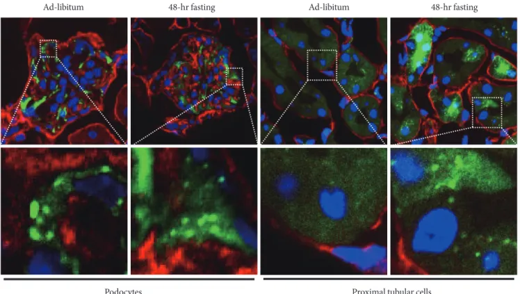
As mentioned above, once autophagy is activated, LC3 pro-teins localize to the autophagosome membrane. his can be observed in a transgenic mouse carrying a green luorescent protein (GFP)-LC3 fusion transgene as a green dot signal and, therefore, activation of autophagy can be detected in cells of the transgenic mouse [31]. his animal model is widely
em-ployed and we used it to examine autophagy activity in kid-neys. As previously reported [31], podocytes show active au-tophagy regardless of feeding condition (Fig. 2). In contrast, proximal tubular cells show autophagy activation only when they were exposed to starvation (Fig. 2). Using the GFP-LC3 mouse model, we did not ind autophagosome dots in renal cells other than podocytes and proximal tubular cells, for ex-ample in cells of the distal tubules and collecting duct. hese results are consistent with recent reports from other investiga-tors. hus, podocytes and proximal tubular cells are the focus of autophagy studies in the ield of kidney research.
PODOCYTE AUTOPHAGY IN DIABETIC
NEPHROPATHY
Proteinuria is a major clinical concern of diabetic nephropa-thy and is caused by disruption to the glomerular filtration barrier. Glomerular epithelial cells, also called podocytes, are predominantly responsible for maintaining the glomerular
il-Fig. 2. Autophagy activity determined using green fluorescent protein light chain 3 (GFP-LC3) transgenic mouse.
Autop-phagome can be detected as GFP-LC3 dots in tissues of this mouse model. Autophagosomes formation is constitutively observed in podocytes even under ad-libitum condition. In contrast, autophagy can be observed in proximal tubular cells exposed to 48-hour fasting. he white dotted line box indicates the area for each enlarged igure. Blue signal, DAPI stain to visualize nuclei. Red signal, nidogen stain to visualize basement membrane. Green signal, GFP signal indicating LC3 protein.
Podocytes
Ad-libitum 48-hr fasting Ad-libitum 48-hr fasting
tration barrier [32]. Podocytes are highly specialized, termi-nally differentiated and unable to proliferate. Podocyte loss due to cell death and podocyte foot process dysfunction result in massive proteinuria in diabetic nephropathy [33]. Thus, maintaining podocyte cell homeostasis is regarded as a thera-peutic target to prevent progression to nephrotic syndrome due to diabetic nephropathy.
The autophagy-lysosomal degradation pathway is likely to play an essential role in maintaining podocyte function. As mentioned above, podocytes exhibit active autophagy even un-der non-stress conditions, suggesting that podocytes require a high basal level of autophagy to maintain cellular homeostasis [31]. Podocyte-speciic autophagy-deicient mice, resulting from Atg5 gene deletion, have glomerular lesions accompanied by podocyte loss and albuminuria that increase with age [18]. Fur-thermore, the impairment of lysosomal function in podocytes by deletion of the mammalian target of rapamycin (mTOR), pro-renin receptor or mVps34 genes caused severe glomerular sclero-sis, massive proteinuria [34-36]. Since autophagy involves degra-dation by lysosomes, autophagosomal degradegra-dation was disturbed in the podocytes of these mouse models. hese results support the idea that the autophagy-lysosomal degradation pathway plays an essential role in maintaining podocyte cell homeostasis.
Despite this knowledge of the physiological role of podocyte autophagy, the role of autophagy in diabetic nephropathy has remained unclear. However, accumulating evidence now shows an association between autophagy and diabetic nephropathy. Autophagic activity in podocytes of streptozotocin-induced abetic mice was found to decline with increased duration of di-abetes [37]. Cultured podocytes exposed to high concentrations of glucose also showed lower autophagic activity, along with de-creased levels of autophagy-related proteins, such as beclin-1 and the Atg5-Atg12 complex [37]. Furthermore, suppression of beclin-1 expression in cultured podocytes decreased podocin expression resulting in albumin leakage [37]. These findings suggest that hyperglycemia reduces autophagy activity, leading to alterations in podocyte function and disturbance to the glo-merular iltration barrier.
Although many of these results support the hypothesis that autophagy is involved in the pathogenesis of diabetic nephrop-athy, direct evidence showing the involvement of autophagy in the pathogenesis of diabetic nephropathy using podocyte-spe-ciic autophagy deicient animals had not been reported until recently. However, two recent works, including our own, have clearly shown the renoprotective role of autophagy in diabetic
podocytes [38,39]. Podocyte-speciic autophagy-deicient mice developed podocyte loss and massive proteinuria when used in a high-fat diet (HFD)-induced obese type 2 diabetic model [38]. Furthermore, deletion of Atg5 speciically in podocytes resulted in accelerated diabetes-induced podocytopathy with a leaky glomerular iltration barrier in a streptozotocin-induced type 1 diabetic model [39]. Collectively, these indings suggest that autophagy is likely to play an essential role in coping with diabetic stress in podocytes, regardless of whether the diabetic nephropathy is type 1 or type 2.
What is the degradation target of podocyte autophagy? As mentioned previously, lysosomes are likely to be important in maintaining podocyte homeostasis. In our recent study, a mas-sive accumulation of lysosomes with abnormal morphology was observed in the podocytes of obese type 2 diabetic rodents with autophagy deiciency [38]. Although it remains unclear whether lysosomes are a unique target of podocyte autophagy under any pathogenic conditions, our results suggest that dam-aged lysosomes are an important degradation target of podo-cyte autophagy, at least under diabetic conditions.
AUTOPHAGY IN PROXIMAL TUBULAR
CELLS
Compared with most other glomerular diseases, the renal out-comes of patients with diabetic nephropathy are extremely poor and the underlying mechanisms remain unclear. he severity of proteinuria-induced tubulointerstitial lesions is strongly corre-lated with renal outcomes regardless of the cause of the glomer-ular disease [1,40,41]; therefore, diabetic conditions may exac-erbate proteinuria-induced tubulointerstitial lesions compared with other primary glomerular diseases [42]. If so, identifying the detailed molecular mechanisms underlying the diabetes- and/or obesity-mediated vulnerability of proximal tubular cells may contribute to the development of new therapies that can improve renal outcomes in obese type 2 diabetes patients with persistent proteinuria. Is autophagy involved in the mechanism of diabetes-related vulnerability in proximal tubular cells? In this section, we discuss this possibility based on several recent publications.
ischemia is becoming a serious health problem in clinical set-tings. A great number of recent animal studies have shown that autophagy in proximal tubular cells is enhanced during acute kidney injury caused by ischemic-reperfusion and cisplatin, a nephrotoxic anti-cancer drug [43-45]. Furthermore, mice lacking autophagy activity in proximal tubular cells, generated by deleting the Atg5 and Atg7 genes, showed progressive renal damage, suggesting that activation of autophagy during acute kidney injury is renoprotective [43,44,46]. These mice also showed premature renal aging, suggesting that a low level of basal autophagy is essential to keep cell homeostasis in proxi-mal tubular cells, or that autophagy induction is needed to cope with age-related extra- and intra-cellular stresses, such as hypoxia and ER stress.
Proteinuria creates strong nephrotoxic stress in a number of proteinuric kidney diseases, including diabetic nephropathy [1,40]. Based on our previous paper, an increased lux of pro-tein into the urinary lumen from the glomeruli was found to activate autophagy in proximal tubular cells, which reabsorb the protein in urinary lumen. Atg5 knockout mice, deicient in proximal tubular cell-specific autophagy, developed severe proteinuria-induced tubulointerstitial lesions, along with en-hanced proximal tubular cell apoptosis, similar to results ob-tained in animal models of acute kidney injury [47]. Collec-tively, proximal tubular cells can induce autophagy to cope with both acute and chronic nephrotoxic stresses. hus, au-tophagy in proximal tubular cells and podocytes plays a reno-protective role in various stages of proteinuric kidney diseases. Studies have also assessed the efects of obesity and diabetes on renoprotective autophagy in proximal tubular cells exposed to nephrotoxicity. Autophagy activity was shown to be signii-cantly suppressed in the kidneys of streptozotocin-induced di-abetic mice, HFD-induced obese mice and Wistar fatty rats [47,48]. his led to the accumulation of damaged molecules and organelles, including p62 protein and damaged mitochon-dria, which are normally degraded via the autophagy-lyso-somal pathway. Interestingly, autophagy insuiciency has been conirmed in renal biopsies of patients with obese type 2 dia-betes [47]. he proximal tubular cells of patients with type 2 diabetes showed the accumulation of p62 protein, suggesting that deicient autophagy also occurs in humans with obesity type 2 diabetes.
he mechanism involved in autophagy-deicient proximal tubular cells of obese animals and humans has also been inves-tigated. As mentioned in the previous section, autophagy is
regulated by intracellular nutrient signals, such as mTORC1, AMPK, and Sirt1. Of these signals, mTORC1 is likely to be in-volved in diabetes-related inhibition of stress-induced autoph-agy in proximal tubular cells [47]. Histological analysis showed that the proximal tubular cells of obese type 2 diabetic mice and humans were intensely positive for phosphorylated S6 protein, an indicator of mTORC1 activation, and strongly as-sociated with an obesity-related deiciency in autophagy [47]. These findings indicate that autophagy deficiency and the pathogenesis of diabetic nephropathy are closely associated. Obesity- or diabetes-mediated autophagy deiciency is likely to be involved in the vulnerability of proximal tubular cells (Fig. 3). Restoring autophagy activity may therefore be a new thera-peutic strategy for diabetic patients with overt proteinuria.
AUTOPHAGY IN OTHER RENAL CELLS
Diabetes primarily injures endothelial cells, which is strongly associated with the initiation of diabetic nephropathy. Lenoir et al. [39] have shown direct evidence of the association be-tween endothelial cell autophagy and diabetic nephropathy. hey showed that endothelial cell-speciic autophagy-deicient mice develop severe glomerular damage, indicating that au-tophagy in the endothelium also plays a renoprotective role against diabetic stress. hus, although autophagy activity was not apparent in these cells from studying GFP-LC3 transgenic mice, cell-speciic autophagy-deicient mice can provide
im-Podocytes Proximal tubular cells
Stress-responsible autophagy
Diabetes Obesity aging Basal autophagy
Disruption of cellular homeostasis
Massive proteinuria Tubular cell damage (apoptosis)
portant information regarding autophagy in diabetic nephrop-athy in renal cells other than podocytes and proximal tubular cells. his area is being actively researched and the role of dia-betic autophagy in mesangial cells, distal tubular cells and col-lecting duct is under scrutiny.
THERAPEUTIC STRATEGIES TARGETING
THE ACTIVATION OF AUTOPHAGY
he evolution of autophagy has enabled it to be activated un-der nutrient-depleted conditions, to overcome long-term pe-riods of starvation. For the past several decades, many investi-gators have tried to identify calorie restriction-mediated anti-aging efects in mammals. Activation of autophagy is essential for calorie restriction-mediated life span elongation and anti-aging efects in various organisms [49,50]. Autophagy in prox-imal tubular cells is activated by short-term starvation, sug-gesting that these cells possess a mechanism by which autoph-agy is induced in response to energy depletion. Calorie restric-tion has a renoprotective acrestric-tion against several kinds of renal injury [19,51]. A calorie restriction regimen, which activates autophagy, should therefore become a potent therapeutic strategy to prevent diabetic nephropathy. Indeed, calorie re-striction improves renal damage in type 2 diabetic Wistar fatty rats, as well as restoring autophagy activity in their proximal tubular cells [48]. hus, an agent that can mimic caloric re-striction may have potency to activate autophagy in mamma-lian cells, and become a therapy for diabetic nephropathy. Given that autophagy is regulated by nutrient-responsive intracellular signals such as mTORC1, AMPK, and Sirt1, agents that can modify the activity of these signals may have therapeutic potency to treat diabetic nephropathy. Indeed, an mTORC1 inhibitor, rapamycin, improved glomerular lesions in experimental diabetic nephropathy, and it can activate au-tophagy; therefore, autophagy may be involved in the reno-protective mechanism of rapamycin in diabetic nephropathy. However, excessive mTORC1 inhibition also led to podocyte dysfunction [52]. hus, whether mTORC1 inhibition is safe and efective for all patients with diabetic nephropathy is still under debate.
AMPK is a nutrient-sensing kinase that positively regulates autophagy. AMPK is activated under conditions of energy de-pletion and is likely suppressed in diabetic nephropathy [53]. Thus, AMPK-mediated induction of autophagy may be in-volved in its renoprotective mechanism. AMPK activation may
be linked to autophagy for maintaining renal homeostasis in diabetic kidneys. AMPK is inactivated by dephosphorylation in the glomeruli and tubules of both type 1 and type 2 diabetic animal models. his inactivation is reversed by agents such as metformin and resveratrol, along with the attenuation of dia-betic glomerular and tubular injury [53-57]. Decreases in AMPK activity may be involved in the pathogenesis of diabetic nephropathy by reducing autophagy, suggesting that AMPK activation may be a target for restoring autophagy activity, even in diabetic kidneys. Some agents, such as 5-aminoimidazole-4-carboxamide ribonucleotide and metformin, can improve renal lesions in experimental diabetic nephropathy models. hese agents might improve diabetic nephropathy through the restoration of autophagy activity in diabetic kidneys.
Sirt1 is another regulator of autophagy. A recent study has shown that Sirt1 deiciency is associated with the pathogenesis of both podocyte injury and proximal tubular cell damage in diabetic nephropathy, and that re-activation of this deacety-lase improved diabetic nephropathy in mice [58]. We previ-ously reported that an age-dependent decline of Sirt1 activity caused suppression of stress-induced autophagy in mouse kidneys, which accelerated the premature aging renal pheno-type [19]. hus, diabetes-related decline of Sirt1 activity may lead to inhibition of autophagy, leading to a severe renal phe-notype in diabetic nephropathy. Taken together, these indings suggest that nutrient-sensing signals are strong candidate tar-gets for modifying autophagy activity in kidney. However, di-rect evidence is still lacking and revealing the relationship among these signals, autophagy, and pathogenesis of diabetic nephropathy is a challenge that remains to be completed.
CONCLUSIONS
great number of issues remain to be resolved. Future studies will provide clear evidence to determine whether autophagy should be considered a novel therapeutic target for diabetic nephropathy, and we hope that this review can contribute to the study of autophagy in diabetic nephropathy.
CONFLICTS OF INTEREST
No potential conlict of interest relevant to this article was re-ported.
ACKNOWLEDGMENTS
his review manuscript supported by Grants-in-Aid for Scien-tiic Research (KAKENHI) from the Japan Society for the Pro-motion of Science (No. 25713033 to SK, and No. 25670414 and No. 70242980 to DK); from the Takeda Science Founda-tion (to SK); from the Banyu Life Science FoundaFounda-tion Interna-tional (to SK).
REFERENCES
1. Abbate M, Zoja C, Remuzzi G. How does proteinuria cause progressive renal damage? J Am Soc Nephrol 2006;17:2974-84. 2. Brownlee M. The pathobiology of diabetic complications: a
unifying mechanism. Diabetes 2005;54:1615-25.
3. Dunlop M. Aldose reductase and the role of the polyol path-way in diabetic nephropathy. Kidney Int Suppl 2000;77:S3-12. 4. Forbes JM, hallas V, homas MC, Founds HW, Burns WC,
Je-rums G, Cooper ME. he breakdown of preexisting advanced glycation end products is associated with reduced renal ibrosis in experimental diabetes. FASEB J 2003;17:1762-4.
5. Ha H, Hwang IA, Park JH, Lee HB. Role of reactive oxygen species in the pathogenesis of diabetic nephropathy. Diabetes Res Clin Pract 2008;82 Suppl 1:S42-5.
6. Koya D, King GL. Protein kinase C activation and the develop-ment of diabetic complications. Diabetes 1998;47:859-66. 7. Brenner BM, Cooper ME, de Zeeuw D, Keane WF, Mitch WE,
Parving HH, Remuzzi G, Snapinn SM, Zhang Z, Shahinfar S; RENAAL Study Investigators. Efects of losartan on renal and cardiovascular outcomes in patients with type 2 diabetes and nephropathy. N Engl J Med 2001;345:861-9.
8. he Diabetes Control and Complications Trial Research Group. The effect of intensive treatment of diabetes on the develop-ment and progression of long-term complications in
insulin-dependent diabetes mellitus. N Engl J Med 1993;329:977-86. 9. UK Prospective Diabetes Study (UKPDS) Group. Intensive
blood-glucose control with sulphonylureas or insulin com-pared with conventional treatment and risk of complications in patients with type 2 diabetes (UKPDS 33). Lancet 1998;352: 837-53.
10. Ohkubo Y, Kishikawa H, Araki E, Miyata T, Isami S, Motoyo-shi S, Kojima Y, FuruyoMotoyo-shi N, Shichiri M. Intensive insulin therapy prevents the progression of diabetic microvascular complications in Japanese patients with non-insulin-depen-dent diabetes mellitus: a randomized prospective 6-year study. Diabetes Res Clin Pract 1995;28:103-17.
11. Araki S, Haneda M, Sugimoto T, Isono M, Isshiki K, Kashiwagi A, Koya D. Factors associated with frequent remission of mi-croalbuminuria in patients with type 2 diabetes. Diabetes 2005; 54:2983-7.
12. Yokoyama H, Araki S, Honjo J, Okizaki S, Yamada D, Shudo R, Shimizu H, Sone H, Moriya T, Haneda M. Association between remission of macroalbuminuria and preservation of renal func-tion in patients with type 2 diabetes with overt proteinuria. Di-abetes Care 2013;36:3227-33.
13. Ron D, Walter P. Signal integration in the endoplasmic reticu-lum unfolded protein response. Nat Rev Mol Cell Biol 2007;8: 519-29.
14. Kroemer G, Marino G, Levine B. Autophagy and the integrat-ed stress response. Mol Cell 2010;40:280-93.
15. Singh R, Kaushik S, Wang Y, Xiang Y, Novak I, Komatsu M, Tanaka K, Cuervo AM, Czaja MJ. Autophagy regulates lipid metabolism. Nature 2009;458:1131-5.
16. Yoshizaki T, Kusunoki C, Kondo M, Yasuda M, Kume S, Mori-no K, Sekine O, Ugi S, Uzu T, Nishio Y, Kashiwagi A, Maegawa H. Autophagy regulates inlammation in adipocytes. Biochem Biophys Res Commun 2012;417:352-7.
17. Huber TB, Edelstein CL, Hartleben B, Inoki K, Jiang M, Koya D, Kume S, Lieberthal W, Pallet N, Quiroga A, Ravichandran K, Susztak K, Yoshida S, Dong Z. Emerging role of autophagy in kidney function, diseases and aging. Autophagy 2012;8: 1009-31.
18. Hartleben B, Godel M, Meyer-Schwesinger C, Liu S, Ulrich T, Kobler S, Wiech T, Grahammer F, Arnold SJ, Lindenmeyer MT, Cohen CD, Pavenstadt H, Kerjaschki D, Mizushima N, Shaw AS, Walz G, Huber TB. Autophagy inluences glomeru-lar disease susceptibility and maintains podocyte homeostasis in aging mice. J Clin Invest 2010;120:1084-96.
S, Sugimoto T, Haneda M, Kashiwagi A, Koya D. Calorie re-striction enhances cell adaptation to hypoxia through Sirt1-dependent mitochondrial autophagy in mouse aged kidney. J Clin Invest 2010;120:1043-55.
20. Kume S, homas MC, Koya D. Nutrient sensing, autophagy, and diabetic nephropathy. Diabetes 2012;61:23-9.
21. Mizushima N, Komatsu M. Autophagy: renovation of cells and tissues. Cell 2011;147:728-41.
22. Kuma A, Hatano M, Matsui M, Yamamoto A, Nakaya H, Yo-shimori T, Ohsumi Y, Tokuhisa T, Mizushima N. he role of autophagy during the early neonatal starvation period. Nature 2004;432:1032-6.
23. Liang XH, Jackson S, Seaman M, Brown K, Kempkes B, Hib-shoosh H, Levine B. Induction of autophagy and inhibition of tumorigenesis by beclin 1. Nature 1999;402:672-6.
24. Pattingre S, Espert L, Biard-Piechaczyk M, Codogno P. Regu-lation of macroautophagy by mTOR and Beclin 1 complexes. Biochimie 2008;90:313-23.
25. Komatsu M, Waguri S, Koike M, Sou YS, Ueno T, Hara T, Mizushima N, Iwata J, Ezaki J, Murata S, Hamazaki J, Nishito Y, Iemura S, Natsume T, Yanagawa T, Uwayama J, Warabi E, Yoshida H, Ishii T, Kobayashi A, Yamamoto M, Yue Z, Uchiya-ma Y, Kominami E, Tanaka K. Homeostatic levels of p62 con-trol cytoplasmic inclusion body formation in autophagy-dei-cient mice. Cell 2007;131:1149-63.
26. Hosokawa N, Hara T, Kaizuka T, Kishi C, Takamura A, Miura Y, Iemura S, Natsume T, Takehana K, Yamada N, Guan JL, Os-hiro N, Mizushima N. Nutrient-dependent mTORC1 associa-tion with the ULK1-Atg13-FIP200 complex required for au-tophagy. Mol Biol Cell 2009;20:1981-91.
27. Mihaylova MM, Shaw RJ. he AMPK signalling pathway co-ordinates cell growth, autophagy and metabolism. Nat Cell Biol 2011;13:1016-23.
28. Kim J, Kundu M, Viollet B, Guan KL. AMPK and mTOR regu-late autophagy through direct phosphorylation of Ulk1. Nat Cell Biol 2011;13:132-41.
29. Bordone L, Guarente L. Calorie restriction, SIRT1 and metab-olism: understanding longevity. Nat Rev Mol Cell Biol 2005;6: 298-305.
30. Lee IH, Cao L, Mostoslavsky R, Lombard DB, Liu J, Bruns NE, Tsokos M, Alt FW, Finkel T. A role for the NAD-dependent deacetylase Sirt1 in the regulation of autophagy. Proc Natl Acad Sci U S A 2008;105:3374-9.
31. Mizushima N, Yamamoto A, Matsui M, Yoshimori T, Ohsumi Y. In vivo analysis of autophagy in response to nutrient
starva-tion using transgenic mice expressing a luorescent autopha-gosome marker. Mol Biol Cell 2004;15:1101-11.
32. Kawachi H, Miyauchi N, Suzuki K, Han GD, Orikasa M, Shi-mizu F. Role of podocyte slit diaphragm as a iltration barrier. Nephrology (Carlton) 2006;11:274-81.
33. Pagtalunan ME, Miller PL, Jumping-Eagle S, Nelson RG, My-ers BD, Rennke HG, Coplon NS, Sun L, Meyer TW. Podocyte loss and progressive glomerular injury in type II diabetes. J Clin Invest 1997;99:342-8.
34. Chen J, Chen MX, Fogo AB, Harris RC, Chen JK. mVps34 de-letion in podocytes causes glomerulosclerosis by disrupting intracellular vesicle trafficking. J Am Soc Nephrol 2013;24: 198-207.
35. Cina DP, Onay T, Paltoo A, Li C, Maezawa Y, De Arteaga J, Ju-risicova A, Quaggin SE. Inhibition of MTOR disrupts autoph-agic lux in podocytes. J Am Soc Nephrol 2012;23:412-20. 36. Oshima Y, Kinouchi K, Ichihara A, Sakoda M, Kurauchi-Mito
A, Bokuda K, Narita T, Kurosawa H, Sun-Wada GH, Wada Y, Yamada T, Takemoto M, Saleem MA, Quaggin SE, Itoh H. Prorenin receptor is essential for normal podocyte structure and function. J Am Soc Nephrol 2011;22:2203-12.
37. Fang L, Zhou Y, Cao H, Wen P, Jiang L, He W, Dai C, Yang J. Autophagy attenuates diabetic glomerular damage through protection of hyperglycemia-induced podocyte injury. PLoS One 2013;8:e60546.
38. Tagawa A, Yasuda M, Kume S, Yamahara K, Nakazawa J, Chin-Kanasaki M, Araki H, Araki SI, Koya D, Asanuma K, Kim EH, Haneda M, Kajiwara N, Hayashi K, Ohashi H, Ugi S, Maegawa H, Uzu T. Impaired podocyte autophagy exacerbates protein-uria in diabetic nephropathy. Diabetes 2015 Sep 17 [Epub]. http://dx.doi.org/10.2337/db15-0473.
39. Lenoir O, Jasiek M, Henique C, Guyonnet L, Hartleben B, Bork T, Chipont A, Flosseau K, Bensaada I, Schmitt A, Masse JM, Souyri M, Huber TB, Tharaux PL. Endothelial cell and podocyte autophagy synergistically protect from diabetes-in-duced glomerulosclerosis. Autophagy 2015;11:1130-45. 40. Nath KA. Tubulointerstitial changes as a major determinant in
the progression of renal damage. Am J Kidney Dis 1992;20:1-17. 41. Risdon RA, Sloper JC, De Wardener HE. Relationship between
renal function and histological changes found in renal-biopsy specimens from patients with persistent glomerular nephritis. Lancet 1968;2:363-6.
protein-uria. J Am Soc Nephrol 2011;22:1122-8.
43. Kimura T, Takabatake Y, Takahashi A, Kaimori JY, Matsui I, Namba T, Kitamura H, Niimura F, Matsusaka T, Soga T, Raku-gi H, Isaka Y. Autophagy protects the proximal tubule from degeneration and acute ischemic injury. J Am Soc Nephrol 2011;22:902-13.
44. Liu S, Hartleben B, Kretz O, Wiech T, Igarashi P, Mizushima N, Walz G, Huber TB. Autophagy plays a critical role in kidney tubule maintenance, aging and ischemia-reperfusion injury. Autophagy 2012;8:826-37.
45. Yang C, Kaushal V, Shah SV, Kaushal GP. Autophagy is associ-ated with apoptosis in cisplatin injury to renal tubular epitheli-al cells. Am J Physiol Renepitheli-al Physiol 2008;294:F777-87.
46. Takahashi A, Kimura T, Takabatake Y, Namba T, Kaimori J, Kitamura H, Matsui I, Niimura F, Matsusaka T, Fujita N, Yo-shimori T, Isaka Y, Rakugi H. Autophagy guards against cispla-tin-induced acute kidney injury. Am J Pathol 2012;180:517-25. 47. Yamahara K, Kume S, Koya D, Tanaka Y, Morita Y, Chin-Kana-saki M, Araki H, Isshiki K, Araki S, Haneda M, Matsusaka T, Kashiwagi A, Maegawa H, Uzu T. Obesity-mediated autophagy insuiciency exacerbates proteinuria-induced tubulointerstitial lesions. J Am Soc Nephrol 2013;24:1769-81.
48. Kitada M, Takeda A, Nagai T, Ito H, Kanasaki K, Koya D. Di-etary restriction ameliorates diabetic nephropathy through anti-inlammatory efects and regulation of the autophagy via restoration of Sirt1 in diabetic Wistar fatty (fa/fa) rats: a model of type 2 diabetes. Exp Diabetes Res 2011;2011:908185. 49. Colman RJ, Anderson RM, Johnson SC, Kastman EK,
Kosmat-ka KJ, Beasley TM, Allison DB, Cruzen C, Simmons HA, Kem-nitz JW, Weindruch R. Caloric restriction delays disease onset and mortality in rhesus monkeys. Science 2009;325:201-4. 50. Fontana L, Partridge L, Longo VD. Extending healthy life span:
from yeast to humans. Science 2010;328:321-6.
51. Cherry, Engelman RW, Wang BY, Kinjoh K, El-Badri NS, Good RA. Calorie restriction delays the crescentic glomerulonephri-tis of SCG/Kj mice. Proc Soc Exp Biol Med 1998;218:218-22.
52. Inoki K, Mori H, Wang J, Suzuki T, Hong S, Yoshida S, Blattner SM, Ikenoue T, Ruegg MA, Hall MN, Kwiatkowski DJ, Rastal-di MP, Huber TB, Kretzler M, Holzman LB, Wiggins RC, Guan KL. mTORC1 activation in podocytes is a critical step in the development of diabetic nephropathy in mice. J Clin Invest 2011;121:2181-96.
53. Kume S, Uzu T, Araki S, Sugimoto T, Isshiki K, Chin-Kanasaki M, Sakaguchi M, Kubota N, Terauchi Y, Kadowaki T, Haneda M, Kashiwagi A, Koya D. Role of altered renal lipid metabo-lism in the development of renal injury induced by a high-fat diet. J Am Soc Nephrol 2007;18:2715-23.
54. Chang CC, Chang CY, Wu YT, Huang JP, Yen TH, Hung LM. Resveratrol retards progression of diabetic nephropathy through modulations of oxidative stress, proinlammatory cy-tokines, and AMP-activated protein kinase. J Biomed Sci 2011;18:47.
55. Takiyama Y, Harumi T, Watanabe J, Fujita Y, Honjo J, Shimizu N, Makino Y, Haneda M. Tubular injury in a rat model of type 2 diabetes is prevented by metformin: a possible role of HIF-1alpha expression and oxygen metabolism. Diabetes 2011;60: 981-92.
56. Ding DF, You N, Wu XM, Xu JR, Hu AP, Ye XL, Zhu Q, Jiang XQ, Miao H, Liu C, Lu YB. Resveratrol attenuates renal hyper-trophy in early-stage diabetes by activating AMPK. Am J Nephrol 2010;31:363-74.
57. Lee MJ, Feliers D, Mariappan MM, Sataranatarajan K, Mahi-mainathan L, Musi N, Foretz M, Viollet B, Weinberg JM, Choudhury GG, Kasinath BS. A role for AMP-activated pro-tein kinase in diabetes-induced renal hypertrophy. Am J Physiol Renal Physiol 2007;292:F617-27.