Cocos nucifera
(L.) (Arecaceae): A phytochemical
and pharmacological review
E.B.C. Lima
1, C.N.S. Sousa
1, L.N. Meneses
1, N.C. Ximenes
1, M.A. Santos Júnior
1,
G.S. Vasconcelos
1, N.B.C. Lima
2, M.C.A. Patrocínio
2, D. Macedo
1and S.M.M. Vasconcelos
11Laboratório de Neuropsicofarmacologia, Departamento de Fisiologia e Farmacologia, Faculdade de Medicina,
Universidade Federal do Ceará, Fortaleza, CE, Brasil
2Laboratório de Farmacologia, Curso de Medicina, Centro Universitário Christus-Unichristus, Fortaleza, CE, Brasil
Abstract
Cocos nucifera(L.) (Arecaceae) is commonly called the‘‘coconut tree’’and is the most naturally widespread fruit plant on Earth. Throughout history, humans have used medicinal plants therapeutically, and minerals, plants, and animals have traditionally been the main sources of drugs. The constituents ofC. nucifera have some biological effects, such as antihelminthic, anti-inflammatory, antinociceptive, antioxidant, antifungal, antimicrobial, and antitumor activities. Our objective in the present study was to review the phytochemical profile, pharmacological activities, and toxicology ofC. nuciferato guide future preclinical and clinical studies using this plant. This systematic review consisted of searches performed using scientific databases such as Scopus, Science Direct, PubMed, SciVerse, and Scientific Electronic Library Online. Some uses of the plant were partially confirmed by previous studies demonstrating analgesic, antiarthritic, antibacterial, antipyretic, antihelminthic, antidiarrheal, and hypoglycemic activities. In addition, other properties such as antihypertensive, anti-inflammatory, antimicrobial, antioxidant, cardioprotective, antiseizure, cytotoxicity, hepatoprotective, vasodilation, nephroprotective, and anti-osteoporosis effects were also reported. Because each part of C. nucifera has different constituents, the pharmacological effects of the plant vary according to the part of the plant evaluated.
Key words: Cocos nucifera(L.); Ethnopharmacology; Pharmacological properties; Biological activity; Review
Introduction
Cocos nucifera (L.) is an important member of the family Arecaceae (palm family) popularly known as coco-nut, coco, coco-da-bahia, or coconut-of-the-beach (1). The plant is originally from Southeast Asia (Malaysia, Indonesia, and the Philippines) and the islands between the Indian and Pacific Oceans. From that region, the fruit of the coconut palm is believed to have been brought to India and then to East Africa. After the discovery of the Cape of Good Hope, this plant was introduced into West Africa and, from there, dispersed to the American continent and to other tropical regions of the globe (2).
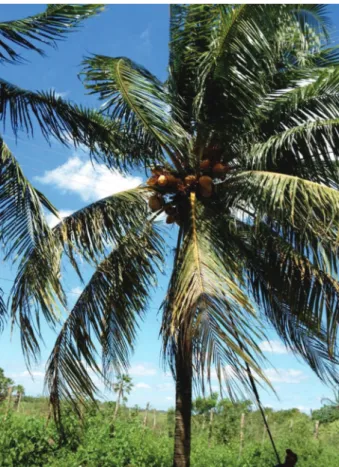
The plant is an arborescent monocotyledonous tree of around 25 m in height (giant coconut) with a dense canopy (Figure 1). The root of the coconut system is fasciculated. The stem is an unbranched type, and at its apex, a tuft of leaves protects a single apical bud. The pinnate leaves are feather-shaped, having a petiole, rachis and leaflets. Under favorable environmental condi-tions, the giant adult coconut emits 12-14 inflorescence
spikes per year, while the adult dwarf coconut can emit 18 spikes in the same period. The axillary inflorescence has globular clusters of female flowers. The plant is monoecious (male and female reproductive organs on the same plant) (3).
The coconut fruit comprises an outer epicarp, a mesocarp, and an inner endocarp. The epicarp, which is the outer skin of the fruit, and the mesocarp, which is heavy,fibrous, and tanned when dry, have many industrial uses. The endocarp is the hard dark core. Inside is a solid white albumen of varied thickness, depending on the age of the fruit, and with an oily pulp consistency and a liquid albumen called coconut water that is thick, sweet, and slightly acidic (3,4). The authors and the synonyms of the plant were confirmed using www.theplantlist.org (Table 1).
The present review highlights the traditional uses of C. nucifera, phytochemical compounds isolated from different parts of the plant, and the biological activity and toxicological studies to date.
Correspondence: S.M.M. Vasconcelos:<[email protected]> <[email protected]>.
Methods
Articles published in English were searched in the online databases Scopus, Science Direct, PubMed, SciVerse and Scientific Electronic Library Online (SciELO), with no time limits. Search terms included combinations of the following: ‘Cocos nucifera’, ‘C. nucifera and phytochemical profile’, ‘C. nuciferaand pharmacological properties’, and‘C. nucifera and toxicology’.
Traditional uses
All parts of the fruit of the coconut tree can be used. Both the green coconut water and solid albumen ripe fruits
are used industrially and in home cooking in many ways (5). Additionally, several parts of the fruit and plant have been used by people in different countries for the treatment of various pathological conditions (Table 2).
Currently, appreciation of natural coconut water is growing. Industry is using the huskfiber from the pith as raw material for carpets, car seat stuffing, and in agricultural as fertilizers. The hard core is used to make handcrafts. The stalk and leaves of the coconut tree are useful in construction, and sugar, vinegar, and alcohol can be extracted from the inflorescence (6).
In Brazil, extract from the huskfiber ofC. nuciferais used to treat diarrhea (7). In Papua New Guinea, the leaves and roots of young plants are chewed as treatment for diarrhea and stomachaches (8,9). In Fiji, coconut oil is used to prevent hair loss and coconut water is used to treat renal disease (10). In Ghana, people use coconut milk to treat diarrhea (11). In Guatemala, the huskfiber extract is used as an antipyretic, to reduce renal inflammation, and as a topic ointment for dermatitis, abscesses, and injuries (12). In Haiti, a decoction of the dry pericarp is used for oral treatment of amenorrhea, and the oil is applied as an ointment to burns (13); an aqueous extract from the husk fiber is also used for oral asthma treatment (14). In India, infusions made with the coconut inflorescence are used for the oral treatment of menstrual cycle disorders (15). In Indonesia, the oil is used as a wound ointment, the coconut milk is used as an oral contraceptive, and fever and diarrhea are treated with the root extract (16–18). In Jamaica, the husk fiber extract is
used to treat diabetes (19,20). In Mozambique, the fruit is consumed by men as an aphrodisiac (21). Peruvians use the aqueous extract of the fresh coconutfiber orally for asthma, as a diuretic, and for gonorrhea (22). In Trinidad, bark extract is used orally for amenorrhea and dysmenorrhea, and bark tea is used to treat venereal diseases (23). In Mexico, coconut is used to treat various disorders associated with urogenital tract infection byTrichomonas vaginalis(24). A decoction of the white flesh of the fruit is used in rural Malaysia to treat fever and malaria (25). In Kenya, the fruit is used to relieve skin rash caused by HIV infection (26).
Phytochemistry
Phytochemical studies of the coconut fiber (mesocarp) ethanolic extract revealed that the presence of phenols, tannins, leucoanthocyanidins,flavonoids, triterpenes, steroids,
Table 1.Synonyms ofCocos nucifera(L.)
Synonyms ofCocos nucifera(L.) References
Calappa nucifera(L.) Kuntze., Revis. Gen. Pl. 2: 982 (1891) Cocos indica Royle Ill. Bot. Himal. Mts.: 395 (1840) Cocos nana Griff Not. Pl. Asiat. 3: 166 (1851) Cocos nucifera var.synphyllica Becc Agric. Colon. 10: 586 (1916)
Palma cocos Miller Gard. Dict. ed. 8: n.° 2 (1768), nom. superfl. Cocos nucifera var.synphyllica Becc Agric. Colon. 10: 586 (1916)
and alkaloids (27), while a butanol extract recovered triterpenes, saponins, and condensed tannins (28). Notably, compounds like flavonoids having antioxidant action are widely distributed in edible vegetables, fruits, and many herbs (29–31). Condensed tannins are reported to possess
anti-helminthic activity by binding to proteins present in the cuticle, oral cavity, esophagus, and cloaca of nematodes, thus intensi-fying the physical and chemical damage in helminth (32).
The lyophilized extract and fractions, as well as ethyl acetate extracts, from the C. nucifera fiber are rich in polyphenols, compounds such as catechins, epicatechins, tannins, andflavonoids (7,33–35).
The constituents of the liquid albumen were identified as vitamin B, nicotinic acid (B3, 0.64mg/mL), pantothenic acid (B5, 0.52mg/mL), biotin (0.02mg/mL), riboflavin (B2, o0.01 ng/mL), folic acid (0.003mg/mL), with trace quantities of vitamins B1, B6, and C, pyridoxine, thiamine, folic acid, amino acids, L-arginine, plant hormones (auxin,
1,3-diphenylurea, cytokinin), enzymes (acid phosphatase, catalase, dehydrogenase, diastase, peroxidase, RNA polymerases), and growth-promoting factors (36–38).
Furthermore, oil extracted from the solid albumen is primarily lauric acid and alpha tocopherol (39,40). Root phenolic compounds were identified as flavonoids and saponins (41). Other compounds identified in leaf epicu-ticular wax were lupeol methylether, skimmiwallin, [3b-methoxy-25-ethyl-9,19-cyclolanost-24(241)-ene], and isoskimmiwallin [3b-methoxy-24-ethyl-9,19-cyclolanost-25 (251)-ene] (42) (Figure 2).
Pharmacological activities of extracts, fractions, and isolated constituents
Several studies have been conducted to identify the active molecules in coconut and their possible pharma-cological and biological activities. Various extracts, frac-tions, and isolated compounds from different parts of the
Table 2.Traditional uses ofCocos nuciferato treat different diseases.
Coconut parts Preparation Popular use Country References
Coconut shellfiber Tea Diarrhea treatment Brazil 7
Amenorrhea Haiti 13
Venereal diseases treatment Trinidad 23
Extract Antipyretic, kidney inflammation
Guatemala 12
Diuretics, gonorrhea treatment Peru 22
Urogenital inflammation caused byTrichomonas
vaginalis
Mexico 24
Amenorrhea, dysmenorrhea Trinidad 23
Diabetes treatment Jamaica 19,20
Asthma treatment Haiti, Peru 14,22
Cream Abscesses, dermatitis treatment and injuries
Guatemala 12
Burns Haiti 13
Root Tea Diarrhea and stomach pains Papua
New Guinea
8,9
Extract Antipyretic, diarrhea treatment Indonesia 16 Solid albumen (pulp)
of coconut
Oil Preventing hair loss, wound healing
Fiji, Indonesia
10,17
Milk Diarrhea treatment Ghana 11
Oral contraceptive Indonesia 18
Pulp Aphrodisiac Mozambique 21
Relief to rashes caused by HIV-AIDS infections
Kenya 26
Decoction of the pulp
Treatment of fever and malaria
Malaysia 25
Coconut water Water Treatment of renal diseases Fiji 10
Inflorescence Tea Treatment of changes in the
menstrual cycle
coconut fruit were tested, showing different activities, including antihypertensive; analgesia; vasodilation; pro-tection of kidney, heart, and liver functions; propro-tection against ulcers; and anti-inflammatory, oxidant, anti-osteoporosis, antidiabetes, antineoplastic, bactericidal, antihelminthic, antimalarial, leishmanicidal, antifungal, and
antiviral activities (43–47). These effects are described
below and also listed in Supplementary Table S1.
Analgesic activity
Crude husk-fiber extract and two aqueous extract fractions of molecular weights less than (F1) and greater
than (F2) 1 kDa were studied for their analgesic activity by acetic acid-induced abdominal writhing, tail-flick, and hot plate tests in mice (44). All three extracts induced peripheral and central antinociceptive activity. Oral admin-istration of the crude extract (50, 100, or 150 mg/kg) significantly inhibited writhing by 24%, 34%, and 52.4%, respectively, when compared with a control group. Fractions F1 and F2 reduced total writhing at 10 and 50 mg/kg. In the tail-flick test, oral pre-treatment with crude extract (100 and 150 mg/kg), F1 (10 and 50 mg/kg), or F2 (10 and 50 mg/kg) produced effects better or similar to morphine (5 mg/kg) until 80 min. However, with the exception of F1 (50 mg/kg, 60 min after administration), neither crude extract (150 mg/kg) nor F2 (50 mg/kg) significantly increased the latency of mice response to thermal stimulation in the hot-plate test. The mechanism of action of the extracts were also evaluated using the opioid antagonist naloxone (5 mg/kg), which inhibited the antinociceptive effect of the crude extract, F1, and F2, indicating a probable action on opioid receptors.
In another study, an ethanol extract of the huskfiber (40, 60, or 80 mg/kg) showed significant analgesic properties, as indicated by a reduction in the number of writhes and stretches induced in mice by 1.2% acetic acid (41). The results were similar to those in animals that received aspirin (68 mg/kg), paracetamol (68 mg/kg), or morphine sulfate (1.15 mg/kg). Furthermore, administra-tion of the ethanol extract along with morphine or pethidine not only produced analgesia in mice but also potentiated the analgesic effect of these two drugs.
These studies were performed using coconut husk
fiber extracts, suggesting that this part of the plant is a highly potent analgesic. Cocos nucifera may enable the production of new low-cost medicines for several ailments and may provide a very inexpensive source of new analgesic drugs. Further investigations are warranted. Further bioassay-guided fractionation and isolation of specific molecules are highly recommended so that the chemical moiety responsible for the activity can be identified and its mechanism of action established. Anti-inflammatory activity
Aqueous crude extracts of huskfiber ofC. nuciferaare used to treat arthritis and other inflammatory ills in Northeastern Brazil’s traditional medicine (7).
A study using animal models of inflammation (formalin test and subcutaneous air pouch model) showed that aqueous crude extracts ofC. nuciferavar. typica(50, or 100 mg/kg) significantly inhibited (Po0.05) the time that animals spent licking their formalin-injected paws and reduced inflammation induced by subcutaneous carra-geenan injection by reducing cell migration, extravasation of protein, and TNF-aproduction (45)
Huskfiber extracts were also tested on rat paw edema induced by carrageenan, histamine, and serotonin (44). Animals were pre-treated by oral administration of crude
extract (50, 100 or 150 mg/kg), F1 or F2 (1, 10, or 50 mg/kg), promethazine (30 mg/kg), or methysergide (5 mg/kg). The crude extract significantly (Po0.05) reduced histamine (at 150 mg/kg) and serotonin-induced rat paw edema (at 100 and 150 mg/kg). Even when mice were treated with 1 mg/kg of F1, a significant inhibitory effect was observed in histamine and serotonin-induced edema. However, F2 did not inhibit the edema induced by any pro-inflammatory agent.
Animal tests revealed significant activity supporting the use of these husk fiber extracts in traditional medicine (35). The chemical constituents responsible for their activity should be isolated, identified, and researched to establish safety doses.
Anti-bacterial, antifungal, and anti-viral activities Brushing the teeth with fibrous coconut husks is a common oral hygiene practice among rural people of South India (46). In this context, the antimicrobial proper-ties of alcoholic extracts of the husk against common oral pathogens were analyzed by the agar well diffusion method (47). There was significant concentration-depen-dent antimicrobial activity, expressed as a zone of inhibition with respect to all tested organisms except Actinomyces species. However, the effect of the C. nuciferaextract was less than that of chlorhexidine.
Ethanolic (cold and hot percolation), dry-distilled, and aqueous extracts of coconut endocarp were compared with gentamicin and ciprofloxacin for their antibacte-rial activities against methicillin-resistant Staphylococcus aureus (MRSA), methicillin-sensitive S. aureus, Pseudo-monas aeruginosa,Escherichia coli,Klebsiella pneumonia, Acinetobacter baumannii,Citrobacter freundii, Enterococ-cus,Streptococcus pyrogens,Bacillus subtilis, and Micro-coccus luteususing the Kirby-Bauer disc diffusion method. The endocarp extracts showed strong antimicrobial activity againstB. subtilis,P. aeruginosa,S. aureus, andM. luteus but had no effect onE. coli (26). The dry-distilled extract (1 mg/mL and 200mg/mL) could inhibit the growth of B. subtilis and Aspergillus spp. but was inactive against R. oligosporus at all concentrations (48). The crude aqueous extract of husk fiber andfive fractions obtained by thin layer chromatography (TLC) were also tested (10, 50, and 100 mg/kg) againstE. coli,S. aureus, and MRSA via agar diffusion; they were active only againstS. aureus and MRSA, with a minimum inhibitory concentration (MIC) of 1024 mg/mL for both (45).
Thein vitroantilisterial activities and time kill regimes of crude aqueous andn-hexane extracts of the huskfiber of C. nucifera were tested (50). The aqueous extracts were active against 29 of 37Listeria isolates examined, while then-hexane extracts were active against 30 (both at 25 mg/mL). The diameters of the zones of inhibition were 12-17 mm and 12-24 mm, respectively, while those of the control antibiotics were 20-50 mm for ampicillin and 22-46 mm for tetracycline. The MICs of the susceptible bacteria were 0.6-2.5 mg/mL for the aqueous fraction and 0.6-5.0 mg/mL for the n-hexane extract. The mean reduction in viable cell count in the time kill assay with the aqueous extract ranged from 0.32 to 3.2 log10CFU/mL after 4 h of interaction and from 2.6 to 4.8 log10CFU/mL after 8 h at 1 and 2 MIC. With then-hexane extract, the values were 2.8-4.8 log10CFU/mL after 4 h of inter-action and 3.5-6.2 log10 CFU/mL after 8 h in 1 and 2 MIC. For the aqueous extract, bactericidal activity was observed against three of the tested Listeria strains at a concentration of 2 MIC after 8 h exposure, while the n-hexane fraction was bactericidal against all five test bacteria at both MICs after 8 h.
In studies with crude extract and five TLC fractions (I-V) of fiber mesocarp of C. nucifera fruit, in vitro antimicrobial activity was seen in all trial strains of S. aureustested with fractions II-V (7). Antifungal activity was demonstrated as growth inhibition of Candida albicans,Cryptococcus neoformansorFonsecaea pedro-soi. Antiviral action was only seen with the crude extract and fraction II. The antifungal, antimicrobial, and antiviral effects were attributed to condensed tannins and cate-chins present in the crude extract and fractions II-V, especially fraction II, which had a higher concentration of these compounds.
Studies with alcohol extract of ripe dried coconut shell have demonstrated action against Microsporum canis, M. gypseum,M. audouinii,Trichophyton mentagrophytes, T. rubrum, T. tonsurans, and T. violaceum (51). This activity was attributed mainly to the high content of phenolic compounds. In another study, virgin oil from coconut pulp prevented growth ofC. albicans(52).
Coconut oil is very effective against a variety of viruses with lipid capsules, such as visna virus, cytomegalovirus, and Epstein-Barr virus (53). The medium chain saturated fatty acids from coconut oil destroy and break the membranes and interfere with viral maturation.
These reports indicate that various parts ofC. nucifera should be further tested for antibacterial, antifungal, and antiviral activities in different animal models. Future studies should consider formulations and exact dose levels suitable for use in humans to treat various strains of bacteria, viruses, and fungi.
Antioxidant activity
There is considerable interest in the consumption of certain foods to prevent the onset of diseases. Evidence
suggests that diets rich in phenolic compounds can significantly enhance human health because of the effects of phenolic antioxidants (54). Studies with virgin coconut oil (VCO) indicated that the total phenolic content was almost seven times that of commercial coconut oil, because the process of obtaining refined oil destroys some of the biologically active components (55). In the 1,1-diphenyl-2-picrylhydrazyl (DPPH) test, VCO had higher antioxidant activity compared to refined coconut oil (56).
The antioxidant activity of C. nucifera endocarp extracts was evaluated by DPPH radical scavenging, nitric oxide radical scavenging, and alkaline dimethyl sulfoxide (DMSO) methods. The DPPH analysis demon-strated that ethanolic (cold and hot percolation), dry-distilled, and aqueous extracts of endocarp had significant antioxidant activity (4.1828, 3.31, 20.83, 1.0179mg/mL, respectively) comparable with that of standard ascorbic acid (48).
In another study, the antioxidant potential of four varieties of coconut (green dwarf, yellow dwarf, red dwarf, and Malaysian yellow) were evaluated and compared with industrialized and lyophilized water of the green dwarf variety (57). All varieties were effective at eliminating DPPH (50% inhibition concentration (IC50) 73 mL) and nitric oxide (0.1 mL; inhibition percent (IP) 29.9%) as well as the in vitroproduction of thiobarbituric acid (1 mL; IP 34.4%). The green dwarf variety, which is commonly used, was especially potent compared with another variety of coconut. In cell culture, green dwarf water protected against oxidative damage induced by hydrogen peroxide. Micronutrients, such as inorganic ions and vitamins present in coconut water, play vital roles in helping the antioxidant defense system of the human body (58). Some evidence points toward an antioxidant action of coconut water. Thus, administering coconut water (6 mL/100 g of body weight) to female rats intoxicated with carbon tetrachloride recovered the action of antioxidant enzymes (superoxide dismutase and catalase levels) and decreased lipid peroxidation (59). Coconut water is also rich in L-arginine (30 mg/dL), which significantly reduces the generation of free radicals (60) and has antioxidant activity (61), as well as ascorbic acid (15 mg/100 mL), which decreases lipid peroxidation in rats (62).
In summary, many parts of C. nucifera plants have proven to contain phenolic compounds andflavonoids that support antioxidant activity.
Antineoplastic activity
and anti-MDR activity of Lucena 1 cells. In both varieties, the antitumoral activity was concentrated in fractions with molecular weights between 1 and 10 kDa (63).
There is great potential for future research on antineoplastic activity, as only one study has been reported. Because coconut is extensively cultivated in Brazil and its fiber is often discarded, it may offer an inexpensive source for new antineoplastic drugs.
Antiparasitic activity
The antihelminthic activity of liquid extract of the bark of the green coconut (LBGC), as well as butanol extract obtained from LBGC, was tested on mouse intestinal nematodes (28). Thirty-six naturally infected mice were distributed into 6 treatment groups as follows: group I, 1000 mg/kg of LBGC; group II, 2000 mg/kg of LBGC; group III, 500 mg/kg of butanol extract; group IV, 1000 mg/kg of butanol extract; group V, 0.56 mg/kg febendazole; and group VI, 3% dimethylsulfoxide. The LBGC did not show antihelminthic activity against the mouse nematodes compared with the negative control group (P40.05). However, the butanol extract at 500 and 1000 mg/kg had mean efficacy of 62.72% and 98.36%, respectively (Po0.05).
The ovicidal and larvicidal activity of the liquid from the coconut husk (LCCV) and butanolic LCCV extract were also tested against Haemonchus contortus (28). In egg hatching and larval development tests, 2.5 mg/mL LCCV and 10 mg/mL butanolic extract showed 100% ovicidal activity. Their larvicidal effects were 81.30% and 99.80% at 65 and 80 mg/mL, respectively.
These results suggest that coconut extracts can be used to control gastrointestinal nematodes and that more studies are needed to evaluate their use in humans.
Anti-Leishmaniaactivity
The in vitro leishmanicidal effects of C. nucifera on Leishmania amazonensis were evaluated (33). The polyphenolic-rich extract obtained from coconut huskfiber completely inhibited the cellular growth of L. amazonensis promastigote forms (MIC 10mg/mL) and killed 100% of both developmental stages of the parasite after 60 min (at 10 and 20mg/mL). In addition, pretreatment of mouse peritoneal macrophages with 10mg/mL ofC. nuciferapolyphenolic-rich extract reduced by approximately 44% their rate of associa-tion withL. amazonensispromastigotes with a simultaneous increase of 182% in nitric oxide production by macrophages compared with untreated macrophages.
Ethyl acetate extract (EAE) from husk fiber water was tested againstL. braziliensisinfected hamsters (35). Admin-istering EAE (0.2 mL, 300 mg/kg) for 21 consecutive days did not reduce edema of infected footpad nor the weight of lymph node drainage but reduced skin lesions after 14 days.
These results offer new promise for the development of drugs against leishmaniasis from coconut extracts because of their potent effects and the absence of in vivo allergic
reactions orin vitrocytotoxic effects in mammalian systems. Further studies with these and other species of the parasite are necessary to elucidate the role of C. nucifera in eliminating this etiological agent and its healing activity.
Depressant and anticonvulsant activity
Ethanol extract of root ofC. nucifera(EECN) at 40, 60, and 80 mg/kg, ip, significantly enhanced the duration of sleep induced by pentobarbital (40 mg/kg, ip), diazepam (3 mg/kg,ip), and meprobamate (100 mg/kg,ip) in mice, suggesting a probable depressive action on the central nervous system (41). The anticonvulsant action of EECN was also observed in pentylenetetrazole-induced seizure models. In the animals treated with 25 mg/kg,ip, EECN, 60.7% had seizures and died 30 min later. In the group that received EECN at 80 mg/kg, ip, no animals had seizures or died, even after 24 h. The components responsible for this depressant activity need to be identified, as well as the mechanism involved in this action. Research on the toxicity of these extracts is also warranted to guarantee the safety of possible future treatments.
Renal protective activity
Coconut water had prophylactic action against nephro-lithiasis in an experiment with a Wistar rat model (64). Rats were divided into three groups. Group I (control) was fed standard rat diet. Group II was administered 0.75% ethylene glycol in drinking water to induce nephrolithiasis. Group III was given coconut water in addition to ethylene glycol. All treatments lasted 7 weeks. Analysis of urine samples revealed a drastic decrease in the number of calcium oxalate crystals in group III compared with group II. Coconut water also significantly lowered the levels of creatinine and urea in group III animals, significantly reduced lipid peroxidation (group II: 38.99±3.36 mol MDA/mg protein/15 min; group III: 27.68±2.45 mol MDA/mg protein/15 min) and decreased the enzyme activities of superoxide dismutase and catalase. These results demonstrate that coconut water has important properties against urolithiasis and therefore must be investigated as a potential treatment for this condition.
Antimalarial activity
These results suggest that the Malaysian folkloric medic-inal application of C. nucifera has a pharmacological basis; however, chloroquine was much more effective at suppression and curing, and the extract did not increase the survival time of infected mice, indicating the need for additional studies to elucidate how C. nucifera can be used to treat malaria.
In another study, the antimalarial and toxicity potentials of husk fiber extracts of five Nigerian varieties of C. nuciferawere evaluatedin vitro. The results showed that only the West African Tall ethyl acetate extract fraction (WATEAEF) was active againstP. falciparumW2 strain with a selectivity index of 30.3. The phytochemicals present in the WATEAEF were alkaloids, tannins, and flavonoids. The same extract fraction was activein vivoagainst P. berghei NK65, causing more than 50% reduction in parasitemia on days 4 and 6 after inoculation at various doses. However, parasitemia varied on days 8 and 10, and results with WATEAEF were no better than with chloroquine. Addition-ally, treatment with 250 and 500 mg/kg body weight WATEAEF significantly increased (Po0.05) plasma creati-nine concentration compared with controls (65). Despite the reduction in parasitemia, the extract cannot yet be con-sidered an appropriate treatment for malaria. More studies are needed to clarify the adverse effects and effectiveness of the coconut extracts, especially in infections caused by P. falciparum(the main agent in humans).
Antitrichomonal activity
The crude methanol extracts of 22 plants used in Mexican folk medicine were tested in vitro against Trichomonas vaginalis. The susceptibility tests were performed using a previously described subculture method (66). Trophozoites (4104) were incubated for 48 h at 37°C in the presence of different concentrations (2.5-200 mg/mL) of the crude extracts in DMSO. Each test included metronidazole as a positive control, a negative control (culture medium plus trophozoites and DMSO), and a blank (culture medium). The experiments were performed in duplicate and repeated at least three times. The crude methanol extract of C. nucifera husk fiber demonstrated excellent antitrichomonal activity (IC50 value of 5.8mg/mL), standing out among the other tested plants, although the activity was less than that of metronidazole. Further research is needed to isolate the substances responsible for this activity and to test appropriate doses so that they can be used in the treatment of trichomoniasis (24).
Cardioprotective activity
An important biological action of coconut was demon-strated using an experimental model of myocardial infarction induced by isoproterenol in rats (67). Feeding these animals with tender coconut water (West Coast Tall variety) protected against the induction of myocardial infarction and decreased mitochondrial lipid peroxidation.
In another study, dietary coconut sprout (West Coast Tall variety) was tested on isoproterenol-induced myocar-dial infarction in rats (68). There was a decrease in the levels of cardiac markers (CK-MB and troponin-T) in serum of the group pretreated with coconut sprout (50, 100, or 200 mg/100 g body weight) orally for 45 days. Rats fed with 100 mg/100 g body weight showed better results than other treatment groups. In addition, pretreat-ment with coconut sprout decreased oxidative stress in the heart and increased antioxidant status. Biochemical analyses showed that sprouts contains bioactive compo-nents, such as vitamins, alkaloids, and polyphenols.
Tender coconut water could also reduce total choles-terol, very-low density lipoprotein, low density lipoprotein, and triglyceride levels in serum (69). Administering coco-nut water (4 mL/100 g body weight) in male rats counter-acted the increases in these substances promoted by cholesterol feeding.
The results presented here support the cardioprotective effects of coconut water. Its administration could reduce oxidative stress and cell damage in animals with induced myocardial infarction and reverse increases in cholesterol levels in animals fed high-fat diets. Therefore, further research is warranted on its potential use to prevent a second ischemic event or in the treatment of dyslipidemic states.
Hepatoprotective activity
The hepatoprotective effect of tender coconut water was investigated in carbon tetrachloride (CCl4)-intoxicated female rats. The animals were divided into three groups: normal control rats, CCl4-treated control rats, and tender coconut water pretreated rats intoxicated with CCl4. Carbon tetra-chloride caused elevated serum glutamate oxaloacetate transaminase and glutamate pyruvate transaminase levels and also lead to liver necrosis and fatty liver, while rats pretreated with coconut water showed decreased activities of these enzymes (59). With only one report describing these effects, there remains room to develop studies to define the real role ofC. nuciferain this action.
Antidiabetic activity
The effects of mature coconut water were also evalu-ated and compared with glibenclamide in alloxan-induced diabetic rats (71). Treatment of diabetic rats with lyophilized mature coconut water (1000 mg/kg body weight) or glibenclamide (0.6 mg/kg body weight) reduced blood glucose levels (129.23±1.95 and 120±2.3 mg/dL, respectively) when compared with the untreated control (275.32±4.25 mg/dL). Coconut water also increased insulin levels and liver glycogen concentrations and reduced glycated hemoglobin levels in diabetic rats. In addition, elevated levels of liver function enzymes markers like alkaline phosphatase, serum glutamate oxaloacetate transaminase, and serum glutamate pyruvate transaminase in diabetic rats were significantly reduced upon treatment with mature coconut water. It was also observed that diabetic rats showed altered levels of blood urea, serum creatinine, and albumin, and the albumin/globulin ratio was significantly reversed by treatment with mature coconut water and glibenclamide.
Administering immature coconut inflorescence metha-nol extract (West Coast Tall variety) to diabetic rats significantly reduced fasting glucose levels and increased insulin levels compared with a diabetic control (72). The 200 mg/kg body weight dose showed better antihypergly-cemic effects than other doses.
These studies demonstrated that different parts of C. nucifera can benefit diabetic rats, similar to oral hypoglycemic agents currently used clinically to control diabetes. Therefore, clinical trials and biochemical analyzes are recommended to isolate the compounds responsible for these actions and to establish them as drugs.
Effects on bone structure
VCO was investigated in postmenopausal osteoporo-sis rats to determine its effects on bone microarchitecture (73). Rats were supplemented with VCO (8% mixed with the standard rat chow diet) for 6 weeks. VCO administra-tion significantly increased bone volume, prevented a reduction in trabecular number, and lowered the trabe-cular separation compared with the ovariectomized con-trol. Bone histology revealed that the trabecular bones of the ovariectomized group appeared to be sparser and less dense than in the group treated with VCO. Treatment of ovariectomized rats with VCO seemed to reverse the effects of estrogen deficiency on bone structure. With only one report on the anti-osteoporosis activity ofC. nucifera available, there is a need for further studies.
Antihypertensive activity
The anti-hypertensive activity of an ethanolic extract of C. nucifera endocarp (EEC) using the deoxycorticosterone acetate (DOCA) salt-induced model of hypertension was observed. Administering EEC significantly reduced the mean systolic blood pressure in DOCA salt-induced hypertensive rats (from 185.3±4.7 to 145.6±6.1 mm Hg). This effect was attributed to the direct activation of the nitric oxide/guany-late cyclase pathway as well as stimulation of muscarinic
receptors and/or the cyclooxygenase pathway. These activities can be explained by the presence of phenolic compounds andflavonoids in the extract used (74). Based on these results,C. nuciferashould be studied further for its potential use against cardiovascular diseases.
Toxicity
Several studies have investigated the toxicological properties ofC. nucifera.One paper verified the effect of ethyl acetate extract ofC. nuciferafiber on physiological parameters and on topical inflammation induced by xylene in animal models (75). Regarding the physiological parameters and macroscopic aspects of the lymphoid organs in this study, neither mortality nor any symptom of toxicity was observed in the animals.
The possible toxic effects of crude extract, F1, and F2 (see above) of C. nucifera mesocarp were evaluated in mice (44). The oral treatment of mice over 5 days with a single dose (500 mg/kg) caused no behavioral changes. No injury or bleeding stomachs were observed.
Another study evaluated the toxicity of a methanol extract ofC. nuciferaendocarp (25). Five female and five male mice received a single dose orally (5000 mg/kg) of this extract. All male and female rats were observed for signs of toxicity and mortality on the day of dosing; at 1, 3, and 4 h after administration; and then twice daily for 14 days. No signs of toxicity and mortality were recorded, and all animals gained weight during the observation period.
Acute, subchronic, and chronic toxicity from liquid mesocarp of green coconut (LMGC) and butanol extract obtained from the LMGC were tested in mice and rats (28). No acute lethal effects were observed in mice receiving a dose of 3000 mg/kg orally of either extract. In contrast, when LMGC and butanol extract were administered intraperitone-ally at doses of 500 and 700 mg/kg, no animal survived. In subchronic toxicity tests, the rats treated with LBGC had significantly higher white blood cell, neutrophil, red blood cell, hematocrit, and platelet counts. In the chronic toxicity test, the group treated with LBGC showed higher values for neutrophils, white blood cells, basophils, and platelets (Po0.05). However, in the subchronic and chronic toxicity tests, no hematological parameters differed significantly in the group treated with butanol extract (P40.05). Only triglycerides were higher (Po0.05) in the group treated with LBGC during the chronic toxicity test. Rats treated with both extracts had no histopathological changes related to toxicity, nor did weight gain differ between treated and control groups (P40.05). In conclusion, both extracts showed low toxicity for those parameters.
Conclusion
appeal, since this plant is widely used in the food industry and use of discarded plant parts will reduce waste and pollution. The pharmacological effects of the plant differ according to the part of the plant or fruit used. Antioxidant activity predominated in the constituents of the endocarp and coconut water. In addition, thefiber showed antibacterial, antiparasitic, and anti-inflammatory activities. Only the ethanolic extract of the root had depressant and anticonvulsant action on the central nervous system. Coconut water seems to have protective effects, e.g., on the kidney and heart, and antioxidant activity, as well as a hypoglycemic effect.
Some limitations of the studies onC. nuciferamust be acknowledged. First, the studies have focused on the effects of different parts of the plant but without demon-strating the mechanisms underlying these actions. Sec-ond, formulations based on parts of the plant must be developed to conduct clinical trials.
Considering the diversity of pharmacological properties, future research into C. nucifera should be encouraged.
The main goals should be to isolate specific compounds, to clarify the mechanisms involved in the pharmacological effects, and to investigate possible toxic effects to produce safe phytotherapies.
Several factors may limit such studies. Geographical and seasonal variations among countries and regions can influence the chemical composition of the studied mate-rial. Therefore, standardized procedures for collecting samples and quantifying compounds should be used to assure the reproducibility of results.
Supplementary Material
Click here to view [pdf].
Acknowledgments
This study was supported by CNPq, CAPES, and the Ceará Foundation for the support of scientific and technological development (FUNCAP).
References
1. Aragão WM. Côco: pós-colheita. Série frutas do Brasil. Brasília: Embrapa Informac¸ão Tecnológica; 2002. http:// livraria.sct.embrapa.br/liv_resumos/pdf/00070000.pdf. 2. Purseglove JW.Tropical crops: monocotyledons. London:
Longman; 1972.
3. Passos EEM.Morfologia do coqueiro. A cultura do coqueiro no Brasil. 2nd edn. Brasília: Embrapa–Servic¸o de Produc¸ão de Informac¸ão; 1998.
4. Andrade AM, Passos PRA, Marques LGC, Oliveira LB, Vidaurre GB, Roch JDS. Pirólise de resíduos do coco-da-baía (Cocos nuciferaLinn) e análise do carvão vegetal.Rev Árvore 2004; 28: 707–714. http://www.scielo.br/pdf/rarv/v28n5/23409. pdf.
5. Rosa M de F, Santos FJ de S, Montenegro AAT, Abreu FAP, Correia D, Araújo FBS, et al.Caracterizac¸ão do pó da casca do coco verde usado como substrato agrı´cola. Fortaleza: Embrapa Agroindústria Tropical, Comunicado Técnico, 54; 2001. http:// www.ceinfo.cnpat.embrapa.br/arquivos/artigo_2459.pdf. 6. Fontenele RES. Cultura do côco no Brasil: caracterizac¸ão do
mercado atual e perspectivas futuras.Anais do Congresso da Sociedade Brasileira de Economia e Sociologia Rural. Instituic¸ões, eficiência, gestão e contratos no sistema agroindustrial. Ribeirão Preto: 2005. p 1–20. http://www. sober.org.br/palestra/2/168.pdf.
7. Esquenazi MD, Wigg MM, Miranda, Rodrigues HM, Tostes JBF, Rozental S, et al. Antimicrobial and antiviral activities of polyphenolics fromCocos nuciferaLinn. (Palmae) huskfiber extract.Res Microbiol 2002; 153: 647–652, doi: 10.1016/ S0923-2508(02)01377-3.
8. Holdsworth D, Wamoi B. Medicinal plants of the Admiralty Islands, Papua New Guinea. Part I.Int J Crude Drug Res 1982; 20: 169–181.
9. Holdsworth D. Medicinal plants of the Gazelle peninsula, New Britain Island, Papua New Guinea, Part I.Int J Pharmacog 1992; 30: 185–190, doi: 10.3109/13880209209053992.
10. Singh YN. Traditional medicine in Fiji: some herbal folk cures used by Fiji Indians. J Ethnopharmacol1986; 15: 57–88, doi: 10.1016/0378-8741(86)90104-2.
11. Yartey J, Harisson EK, Brakohiapa LA, Nkrumah FK. Carbohydrate and electrolyte content of some home-availablefluids used for oral rehydration in Ghana.J Trop Pediatr1993; 39: 234–237, doi: 10.1093/tropej/39.4.234. 12. Caceres A, Giron LM, Alvarado SR, Torres MF. Screening
of antimicrobial activity of plants popularly used in Guatemala for the treatment of dermatomucosal diseases. J Ethnopharmacol1987; 20: 223–237, doi: 10.1016/0378-8741(87)90050-X.
13. Weniger B, Rouzier M, Daguilh R, Henrys D, Henrys JH, Anton R. [Traditional medicine in the Central Plateau of Haiti. 2. Ethnopharmacologic inventory]. J Ethnopharmacol 1986; 17: 13–30, doi: 10.1016/0378-8741(86)90070-X.
14. Hope BE, Massey DG, Fournier-Massey G. Hawaiian materia medica for asthma.Hawaii Med J1993; 52: 160–166. 15. Bhandary MJ, Chandrashekar KR, Kaveriappa KM. Medical
ethnobotany of the Siddis of Uttara Kannada district, Karnataka, India. J Ethnopharmacol 1995; 47: 149–158, doi: 10.1016/0378-8741(95)01274-H.
16. Brondegaard VJ. Contraceptive plant drugs. Planta Med 1973; 23: 167–172, doi: 10.1055/s-0028-1099428. 17. Sachs M, von Eichel J, Asskali F. [Wound management with
coconut oil in Indonesian folk medicine].Chirurg2002; 73: 387–392, doi: 10.1007/s00104-001-0382-4.
18. Hirschhorn HH. Botanical remedies of the former Dutch East Indies (Indonesia). Part I: Eumycetes, Pteridophyta, Gymnospermae, Angiospermae (Monocotyledones only). J Ethnopharmacol 1983; 7: 123–156, doi: 10.1016/0378-8741(83)90016-8.
20. Mitchell SA, Ahmad MH. A review of medicinal plant research at the University of the West Indies, Jamaica, 1948–2001.West Indian Med J2006; 55: 243–269. 21. Amico A. Medicinal plants of Southern Zambesia.
Fitoter-apia1977; 48: 101–139.
22. Ramirez VR, Mostacero LJ, Garcia AE, Mejia CF, Pelaez PF, Medina CD et al. Vegetales empleados em medicina tradicional Norperuana. Trujillo: Banco Agrario del Peru & NACL Univ Trujillo; 1988.
23. Wong W. Some folk medicinal plants from Trinidad.Econ Bot1976; 30: 103–142, doi: 10.1007/BF02862958. 24. Calzada F, Yepez-Mulia L, Tapia-Contreras A. Effect of
Mexican medicinal plant used to treat trichomoniasis on Trichomonas vaginalis trophozoites. J Ethnopharmacol 2007; 113: 248–251, doi: 10.1016/j.jep.2007.06.001. 25. Al-Adhroey AH, Nor ZM, Al-MekhlafiHM, Amran AA, Mahmud
R. Evaluation of the use of Cocos nuciferaas antimalarial remedy in Malaysian folk medicine.J Ethnopharmacol2011; 134: 988–991, doi: 10.1016/j.jep.2011.01.026.
26. Nagata JM, Jew AR, Kimeu JM, Salmen CR, Bukusi EA, Cohen CR. Medical pluralism on Mfangano Island: use of medicinal plants among persons living with HIV/AIDS in Suba District, Kenya.J Ethnopharmacol2011; 135: 501–509, doi: 10.1016/j.jep.2011.03.051.
27. Matos FJA.Introduc¸ão àfitoquı´mica experimental. 2nd edn. Fortaleza: Imprensa Universitária; 1997.
28. Costa CT, Bevilaqua CM, Morais SM, Camurca-Vasconce-los AL, Maciel MV, Braga RR, et al. Anthelmintic activity of Cocos nuciferaL. on intestinal nematodes of mice.Res Vet Sci2010; 88: 101–103, doi: 10.1016/j.rvsc.2009.05.008. 29. Chao J, Lee MS, Amagaya S, Liao JW, Wu JB, Ho LK, et al.
Hepatoprotective effect of shidagonglao on acute liver injury induced by carbon tetrachloride.Am J Chin Med2009; 37: 1085–1097.
30. Huang X, Kojima-Yuasa A, Xu S, Kennedy DO, Hasuma T, Matsui-Yuasa I. Combination ofZizyphus jujubaand green tea extracts exerts excellent cytotoxic activity in HepG2 cells via reducing the expression of APRIL.Am J Chin Med2009; 37: 169–179.
31. Yook HS, Kim KH, Park JE, Shin HJ. Antioxidative and antiviral properties offlowering cherry fruits (Prunus serrulata L. var. spontanea).Am J Chin Med2010; 38: 937–948. 32. Hoste H, Jackson F, Athanasiadou S, Thamsborg SM,
Hoskin SO. The effects of tannin-rich plants on parasitic nematodes in ruminants.Trends Parasitol2006; 22: 253–261, doi: 10.1016/j.pt.2006.04.004.
33. Mendonca-Filho RR, Rodrigues IA, Alviano DS, Santos AL, Soares RM, Alviano CS, et al. Leishmanicidal activity of polyphenolic-rich extract from huskfiber ofCocos nucifera Linn. (Palmae). Res Microbiol 2004; 155: 136–143, doi: 10.1016/j.resmic.2003.12.001.
34. Rodrigues S, Pinto GAS. Ultrasound extraction of phenolic compounds from coconut (Cocos nucifera) shell powder. J Food Eng2007; 80: 869–872.
35. Freitas JCC, Nunes-Pinheiro DCS, Pessoa AWP, Silva LCR, Girão VCC, Lopes-Neto BE, et al. Effect of ethyl acetate extract from huskfiber water ofCocos nuciferainLeishmania braziliensisinfected hamsters.Rev Bras Farmacogn2011; 21: 1006–1011, doi: 10.1590/S0102-695X2011005000138. 36. Magda RR. Coco soft drink: health beverage from coconut
water.Food Market Technol1992; 6: 22–23.
37. Yong JW, Ge L, Ng YF, Tan SN. The chemical composition and biological properties of coconut (Cocos nucifera L.) water. Molecules 2009; 14: 5144–5164, doi: 10.3390/ molecules14125144.
38. Solangih A, Iqbal ZA. Chemical composition of meat (kernel) and nut water of major coconut (Cocos nuciferaL.) cultivars at coastal area of Pakistan. Pak. Am J Bot 2011; 43: 357–363.
39. Tangwatcharin P, Khopaibool P. Activity of virgin coconut oil, lauric acid or monolaurin in combination with lactic acid against Staphylococcus aureus. Southeast Asian J Trop Med Public Health2012; 43: 969–985.
40. Arlee R, Suanphairoch S, Pakdeechanuan P. Differences in chemical components and antioxidant-related substances in virgin coconut oil from coconut hybrids and their parentes. Int Food Res J2013; 20: 2103–2109.
41. Pal D, Sarkar A, Gain S, Jana S, Mandal S. CNS depressant activities of roots of Coccos nucifera in mice. Acta Pol Pharm2011; 68: 249–254.
42. Erosa FE, Gamboa-León MR, Lecher JG, Arroyo-Serralta GA, Zizumbo-Villareal D, Oropeza-Salín C, et al. Major components from the epicuticular wax of Cocos nucifera. Rev Soci Quı´mica México2002; 46: 247–250.
43. Akinpelu DA, Alayande KA, Aiyegoro OA, Akinpelu OF, Okoh AI. Probable mechanisms of biocidal action of Cocos nucifera Husk extract and fractions on bacteria isolates. BMC Complement Altern Med 2015; 15: 116, doi: 10.1186/s12906-015-0634-3.
44. Rinaldi S, Silva DO, Bello F, Alviano CS, Alviano DS, Matheus ME, et al. Characterization of the antinociceptive and anti-inflammatory activities from Cocos nucifera L. (Palmae). J Ethnopharmacol 2009; 122: 541–546, doi: 10.1016/j.jep.2009.01.024.
45. Silva RR, Oliveira e Silva, Fontes HR, Alviano CS, Fernandes PD, Alviano DS. Anti-inflammatory, antioxidant, and antimicrobial activities of Cocos nucifera var. typica. BMC Complement Altern Med2013; 13: 107.
46. Jose M, Sharma BB, Shantaram M. Ethnomedicinal herbs used in oral health and hygiene in coastal Dakshina Kannada.J Oral Health Comm Dent2011; 5: 107–111. 47. Jose M, Cyriac MB, Pai V, Varghese I, Shantaram M.
Antimicrobial properties ofCocos nucifera(coconut) husk: An extrapolation to oral health.J Nat Sci Biol Med2014; 5: 359–364, doi: 10.4103/0976-9668.136184.
48. Singla RK, Jaiswal N, Bhat GV, Jagani H. Antioxidant and antimicrobial activities ofCocos nucifera Linn.(Arecaceae) endocarp extracts.Indo Global J Pharm Sci2011; 1: 354–361. 49. Verma V, Bhardwaj A, Rathi S, Raja R.B. Potential anti-microbial agent fromCocos nuciferamesocarp extract.Int Res J Biol Sci2012; 1: 48–54.
50. Akinyele TA, Okoh OO, Akinpelu DA, Okoh AI. In-vitro antibacterial properties of crude aqueous and n-hexane extracts of the husk ofCocos nucifera.Molecules2011; 16: 2135–2145, doi: 10.3390/molecules16032135.
51. Venkataraman S, Ramanujam TR, Venkatasubbu VS. Antifungal activity of the alcoholic extract of coconut shell -Cocos nuciferaLinn.J Ethnopharmacol1980; 2: 291–293, doi: 10.1016/S0378-8741(80)81007-5.
53. Arora R, Chawla R, Marwah R, Arora P, Sharma RK, Kaushik V, et al. Potential of complementary and alternative medicine in preventive management of novel H1N1 flu (Swine flu) pandemic: thwarting potential disasters in the bud. Evid Based Complement Alternat Med 2011; 2011: 586506, doi: 10.1155/2011/586506.
54. Naczk M, Shahidi F. Extraction and analysis of phenolics in food. J Chromatogr A2004; 1054: 95–111, doi: 10.1016/ j.chroma.2004.08.059.
55. Seneviratne KN, Dissanayake DMS. Variation of phenolic content in coconut oil extracted by two conventional methods.Inter J Food Sci Technol2008; 43: 597–602. 56. Marina AM, Che Man YB, Nazimah SAH, Amin I. Chemical
properties of virgin coconut oil.J Am Oil Chem Soc2009; 86: 301–307, doi: 10.1007/s11746-009-1351-1.
57. Santos JL, Bispo VS, Filho AB, Pinto IF, Dantas LS, Vasconcelos DF, et al. Evaluation of chemical constituents and antioxidant activity of coconut water (Cocus nuciferaL.) and caffeic acid in cell culture.An Acad Bras Cienc2013; 85: 1235–1247, doi: 10.1590/0001-37652013105312. 58. Evans P, Halliwell B. Micronutrients: oxidant/antioxidant
status.Br J Nutr2001; 85 (Suppl 2): S67–S74.
59. Loki AL, Rajamohan T. Hepatoprotective and antioxidant effect of tender coconut water on carbon tetrachloride induced liver injury in rats. Indian J Biochem Biophys 2003; 40: 354–357.
60. Boger RH, Bode-Boger SM, Mugge A, Kienke S, Brandes R, Dwenger A, et al. Supplementation of hypercholesterolaemic rabbits with L-arginine reduces the vascular release of superoxide anions and restores NO production. Atherosclero-sis1995; 117: 273–284, doi: 10.1016/0021-9150(95)05582-H. 61. Salil G, Rajamohan T. Hypolipidemic and antiperoxidative effect of coconut protein in hypercholesterolemic rats. Indian J Exp Biol2001; 39: 1028–1034.
62. Das KK, Das SN, Dasgupta S. The influence of ascorbic acid on nickel-induced hepatic lipid peroxidation in rats. J Basic Clin Physiol Pharmacol2001; 12: 187–195. 63. Koschek PR, Alviano DS, Alviano CS, Gattass CR. The husk
fiber of Cocos nucifera L. (Palmae) is a source of anti-neoplastic activity.Braz J Med Biol Res2007; 40: 1339–1343, doi: 10.1590/S0100-879X2006005000153.
64. Gandhi M, Aggarwal M, Puri S, Singla SK. Prophylactic effect of coconut water (Cocos nucifera L.) on ethylene glycol induced nephrocalcinosis in male wistar rat. Int Braz J Urol2013; 39: 108–117.
65. Adebayo JO, Balogun EA, Malomo SO, Soladoye AO, Olatunji LA, Kolawole OM, et al. Antimalarial activity of Cocos nucifera husk fibre: further studies. Evid Based Complement Alternat Med 2013; 2013: 742476, doi: 10.1155/2013/742476.
66. Cedillo-Rivera R, Chavez B, Gonzalez-Robles A, Tapia A, Yepez-Mulia L. In vitro effect of nitazoxanide against Enta-moeba histolytica, Giardia intestinalis and Trichomonas vaginalistrophozoites.J Eukaryot Microbiol2002; 49: 201–208. 67. Rajamohan T, Anurag P. Cardioprotective effect of tender coconut water in experimental myocardial infarction.Plant Food Hum Nutr2003; 58: 1–12.
68. Chikku A, Rajamohan T. Dietary coconut sprout beneficially modulates cardiac damage induced by isoproterenol in rats. Bangladesh J Pharmacol2012; 7: 258–265.
69. Sandhya VG, Rajamohan T. Beneficial effects of coconut water feeding on lipid metabolism in cholesterol-fed rats. J Med Food2006; 9: 400–407, doi: 10.1089/jmf.2006.9.400. 70. Salil G, Nevin KG, Rajamohan T. Argenine rich coconut kernel protein modulates in alloxan treated rats. Chemico-Biol Interact2001; 89: 107–111.
71. Preetha PP, Girija Devi V, Rajamohan T. Antihyperlipidemic effects of mature coconut water and its role in regulating lipid metabolism in alloxan-induced experimental diabetes. Comp Clin Pathol 2013; 23: 1331–1337, doi: 10.1007/ s00580-013-1784-7.
72. Renjith RS, Chikku AM, Rajamohan T. Cytoprotective, antihyperglycemic and phytochemical properties ofCocos nucifera(L.) inflorescence.Asian Pac J Trop Med2013; 6: 804–810.
73. Hayatullina Z, Muhammad N, Mohamed N, Soelaiman I-N. Virgin coconut oil supplementation prevents bone loss in osteoporosis rat model. Hindawi Publishing Corporation; 2012.
74. Bankar GR, Nayak PG, Bansal P, Paul P, Pai KS, Singla RK, et al. Vasorelaxant and antihypertensive effect of Cocos nuciferaLinn. endocarp on isolated rat thoracic aorta and DOCA salt-induced hypertensive rats. J Ethnopharmacol 2011; 134: 50–54, doi: 10.1016/j.jep.2010.11.047.