Natural Guided Genome Engineering Reveals
Transcriptional Regulators Controlling
Quorum-Sensing Signal Degradation
Abbas El Sahili1☯, Anthony Kwasiborski1☯, Nicolas Mothe1, Christophe Velours1,
Pierre Legrand2, Solange Moréra1
*, Denis Faure1
*
1Institute for Integrative Biology of the Cell (I2BC), CNRS, CEA, Univ. Paris-Sud, Université Paris-Saclay, 91198 Gif-sur-Yvette Cedex, France,2Synchrotron SOLEIL, L’Orme des Merisiers, Saint Aubin BP48, Gif-sur-Yvette 91198, France
☯These authors contributed equally to this work.
*[email protected](SM);[email protected](DF)
Abstract
Quorum-quenching (QQ) are natural or engineered processes disrupting the quorum-sens-ing (QS) signallquorum-sens-ing which controls virulence and persistence (e.g. biofilm) in numerous bac-teria. QQ involves different enzymes including lactonases, amidases, oxidases and reductases which degrade the QS molecules such as N-acylhomoserine lactones (NAHL).
Rhodococcus erythropolisknown to efficiently degrade NAHL is proposed as a biocontrol agent and a reservoir of QQ-enzymes for biotechnology. InR.erythropolis, regulation of QQ-enzymes remains unclear. In this work, we performed genome engineering onR. ery-thropolis, which is recalcitrant to reverse genetics, in order to investigate regulation of QQ-enzymes at a molecular and structural level with the aim to improve the QQ activity. Deep-sequencing of theR.erythropolisenhanced variants allowed identification of a punctual mutation in a key-transcriptional factor QsdR (Quorum sensing degradation Regulation) which regulates the sole QQ-lactonase QsdA identified so far. Using biophysical and struc-tural studies on QsdR, we demonstrate that QQ activity can be improved by modifying the regulation of QQ-enzymes degrading QS signal. This modification requiring the change of only one amino-acid in a transcriptional factor leads to an enhancedR.erythropolisin which the QS-signal degradation pathway is strongly activated.
Introduction
Anti-virulence paradigm sustains the development of treatments which are alternative or com-plementary to antibiosis-based agents [1]. The regulatory pathways such as quorum-sensing (QS) which control bacterial behaviors are attractive targets of anti-virulence treatments [2]. In numerous bacteria, QS-signals are master regulators of a wide variety of behaviors (secretion of virulence factors, motility, horizontal gene transfer, biofilm development) which contribute to adaptation, proliferation and aggressiveness [3,4] The natural or engineered processes which
OPEN ACCESS
Citation:El Sahili A, Kwasiborski A, Mothe N, Velours C, Legrand P, Moréra S, et al. (2015) Natural Guided Genome Engineering Reveals Transcriptional Regulators Controlling Quorum-Sensing Signal Degradation. PLoS ONE 10(11): e0141718. doi:10.1371/journal.pone.0141718
Editor:Ya-Wen He, Shanghai Jiao Tong University, CHINA
Received:July 21, 2015
Accepted:October 12, 2015
Published:November 10, 2015
Copyright:© 2015 El Sahili et al. This is an open access article distributed under the terms of the
Creative Commons Attribution License, which permits unrestricted use, distribution, and reproduction in any medium, provided the original author and source are credited.
Data Availability Statement:Coordinates and structure factors of R. erythropolis transcriptional repressor QsdR from TetR family are available at the Protein Data Bank (PDB) under accession code 4ZA6.
disturb QS are called quorum-quenching [5]. Quorum-quenching (QQ) strategies encompass several molecular actors: chemical compounds (called QS-inhibitors) which inhibit synthesis, transport or perception of the signals, antibodies which recognize and could hydrolyze QS-signals, as well as enzymes which cleave the QS-signals [6]. Moreover, entire organisms which exhibit QQ-capacity may be directly used as biocontrol agents [7]. The QQ investigations con-cern human, plant and animal health, as well as water engineering and anti-biofouling [6,8–11].
The N-acylhomoserine lactones (NAHLs) are QS-signals mainly produced by alpha-, beta-, and gamma-proteobacteria, including the pathogensAgrobacterium tumefaciens,Burkholderia glumae,Pectobacterium atrosepticum,Pseudomonas aeruginosa,Pantoea stewartii[3,4]. The QQ-enzymes degrading NAHL have been discovered in several species of Archaea, Eukarya and Bacteria [11]. These are lactonases which open the NAHL lactone ring, amidases which cleave NAHL molecules into homoserine lactone and fatty acids, and NAHL-modifying enzymes such as oxidases and reductases which alter the acyl chain [7]. In some QS-emitting pathogens such asA.tumefaciensandP.aeruginosa, QQ-lactonases and QQ-amidases are involved in the clearing and recycling of their own NAHL-signals [12–14]. Other bacteria, such
asRhodococcus erythropolis,Bacillus thuringiensisandB.cereus, do not produce NAHLs but are able to degrade them efficiently [15–17]. These QQ-organisms are proposed as biocontrol
agents as well as reservoirs of quorum-quenching enzymes for biotechnology [8,9].
R.erythropolisis a unique actinobacterium in which three quorum-quenching activities using lactonase, amidase and reductase, have been discovered [18,19]. Consequently, several applied developments of theR.erythropolisquorum-quenching have been proposed in plant protection, anti-biofouling and water engineering [20–22]. InR.erythropolis, the lactonase
QsdA has been identified as the only lactonase [19]. This enzyme belongs to the phosphotries-terase-like lactonases family and cleaves a broad spectrum of NAHLs [19]. To date, no genetic and biochemical information are available about the regulation of QQ-enzymes inR. erythro-polis. To our knowledge, the only known transcriptional factor controlling QQ-enzyme expres-sion is BlcR (AttJ) inA.tumefaciens[12].
In this work, genome engineering is proposed for improving quorum-quenching capabili-ties ofR.erythropolis, and to access functional and structural characterization of transcriptional regulators controlling the QQ-pathway. Using directed evolution, we selectedR.erythropolis
derivatives in which QS-signal degradation capability were improved in comparison with the parental strainR.erythropolisR138. We then combined deep-sequencing, molecular and struc-tural biology for identifying and characterizing the incriminated mutations. This study high-lights that a single nucleotide variation in key-transcriptional factors is enough for improving functional properties of QS-signal degrading organisms and that directed evolution may be used to understand regulatory pathways of interest in bacteria which are recalcitrant to genetic manipulations.
Materials and Methods
Selection of the
R
.
erythropolis
variants with an enhanced QS-signal
assimilation
The wild type strainR.erythropolisR138 [23] was cultivated at 30°C in a synthetic AB medium [24], which is supplemented with ammonium chloride (1 g/L) and mannitol (2 g/L) as nitrogen and carbon source (AB-man).N-octanoylhomoserine lactone (C8HSL) and 3-oxo-octanoylho-moserine lactone (OC8HSL) from Sigma-Aldrich (St-Louis, MO, USA) were used as alterna-tive carbon sources at 1 mM in AB-C8HSL and AB-OC8HSL media, respecalterna-tively. A single pre-culture of the wild type strainR.erythropolisR138 was used for starting the propagation of three independent lineages in AB-OC8HSL. Twice a week, a fresh AB-OC8HSL medium was Competing Interests:The authors have declared
that no competing interests exist.
subsequently inoculated up to 7 weeks. After 7 and 14 subcultures, a single clone was isolated from each of the three lineages. The strainR.erythropolisR138 and its evolved derivatives were stored at -80°C.
C8HSL and OC8HSL assimilation assay
The parental strainR.erythropolisR138 and its evolved derivatives (M7.1, M7.2, M7.3, M14.1, M14.2 and M14.3) were cultivated at 30°C in AB-man medium for 24 h, then cells were washed in NaCl (0.8%) and suspended in AB-C8HSL and AB-OC8HSL media. The OD600
measure-ments were carried out every day. At the end of the bacterial growth, C8HSL and OC8HSL were extracted and quantified according to a procedure adapted from Chaet al. [25]. Briefly, bacterial cell cultures were centrifuged for 10 min at 15,000g, and NAHLs were extracted from
the supernatant by addition of one volume of ethyl acetate and by further air-drying the organic fraction. The extracted NAHLs were dissolved in 20μL of ethyl acetate, of which 5μL
was spotted on TLC (Thin Layer Chromatography) silica plates (Macherey-Nagel, Düren, Ger-many). TLC plates were overlaid with the NAHL-biosensor strainA.tumefaciensNT1(pZLR4) in AB medium supplemented with agar (15 g/L) and X-gal (40μg/mL). For quantification,
cali-bration curves were obtained with pure C8HSL or OC8HSL.
Variant search
Genome sequencing of theR.erythropolismutants M7.1, M7.2 and M7.3 was performed at the IMAGIF sequencing platform (CNRS, Gif-sur-Yvette, France) using Illumina Genome Analy-serIIx (paired-end, 2×74 bp reads) as described by Kwasiborskiet al. [26]. Sequence reads obtained for the OC8HSL consumer mutants were mapped on the annotated reference genome ofR.erythropolisR138. Mappings were carried out using the CLC Genomics Workbench v7.5 (CLC bio, Aarhus, Denmark) with a read length (90%) and similarity (95%). Genomic variant detection was processed using CLC Genomics Workbench with a variant occurrence of 100%. Characteristics of the evolved derivatives are described inTable 1.
Quantitative RT-PCR
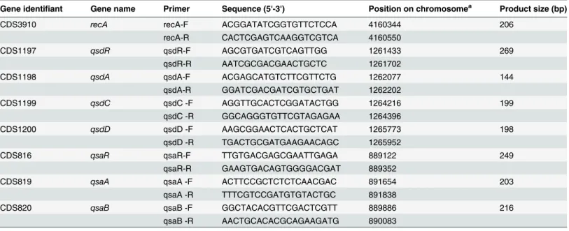
Gene expression was quantified by RT-qPCR using biological triplicates. Sequences and char-acteristics of the primers are presented inTable 2. Reverse transcriptions were carried out using the protocol for high GC content bacteria from the Revert Aid Reverse Transcriptase (Fermentas, Whaltham, USA). A Light Cycler 480 (Roche Applied Science, Penzberg, Ger-many) and Light Cycler 480 SYBR Green I Master (Roche Applied Science) were used for quantitative PCR. The 15μL final volume mix contained SYBR Green I Master (1x), forward
and reverse primers (1μM) and 0.01μg of cDNA samples. After denaturation at 95°C for 10
min, the amplification and quantification program was repeated 45 times as follows: 95°C for 15 s, 60°C for 15 s, 72°C for 20 s, with a single fluorescence measurement, followed by the melt-ing curve program (65°C-95°C with a heatmelt-ing rate of 0.1°C/s and a continuous fluorescence measurement) and a final cooling step at 45°C. The recombinase A (recA) gene was used as a
reference gene in order to normalize gene expression.
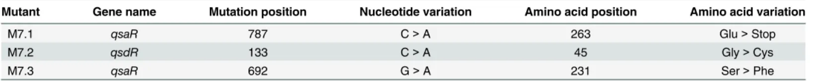
Table 1. Characteristics of bacterial derivatives M7.1, M7.2 and M7.3.
Mutant Gene name Mutation position Nucleotide variation Amino acid position Amino acid variation
M7.1 qsaR 787 C>A 263 Glu>Stop
M7.2 qsdR 133 C>A 45 Gly>Cys
M7.3 qsaR 692 G>A 231 Ser>Phe
Expression and purification of
QsdR
wtand
QsdR
G45CQsdRwtand QsdRG45Cnucleotide sequences were chemically synthesized using codon
optimi-zation for expression inE.coliand inserted into pET29b expression plasmid using NdeI and
SacI restriction enzymes (Genscript, Piscataway, NJ).E.coliBL21 competent cells transformed with pET29b-QsdRwtwere grown in 2TY media at 37°C (initial OD600of 0.1) until an OD600of
0.6 reached within 3 hours. Expression was induced for 4h by addition of 0.5 mM of isopropyl β-D-1-thiogalactopyranoside. The cells were pelleted by centrifugation at 8000 g for 20 min at 4°C and stored at -20°C before being resuspended in buffer A (50 mM Tris-HCl pH 8, 150 mM NaCl) and 20 mM imidazole and sonicated. After centrifugation at 25000 g for 45 minutes, the filtered supernatant was injected on a nickel affinity column (HiTrap 5 mL, GE Healthcare). After a washing step with buffer A and 35 mM imidazole, the protein is eluted with buffer A and 300 mM imidazole before its injection on a gel filtration Superdex 200 26/60 (GE Health-care) using buffer A. The protein fractions are pooled, concentrated using a 5,000 MWCO Vivaspin (GE healthcare) and stored at -80°C.
E.coliC41 cells transformed with the plasmid pET29b-QsdRG45Cwere grown at 37°C in LB
media until an OD600of 0.5. The pelleted cells were resuspended in fresh LB media
supple-mented with 4% (v/v) of ethanol and grown for 1 h at 20°C before inducing the expression with 0.5 mM isopropylβ-D-1-thiogalactopyranoside for 16 h. The cells were pelleted by centri-fugation at 8000 g for 20 min at 4°C and stored at -20°C. The purification protocol was the same as for QsdRwtin presence or in absence of Dithiothreitol (DTT).
Crystallization and data collection
Crystallization conditions for QsdRwtat 16 mg/mL were screened using Qiagen kits (Valencia,
CA, USA) with a Cartesian nanodrop robot (Genomic solutions). Two conditions manually optimized in hanging drops composed of a 1:1 volume ratio of protein solution and crystalliza-tion solucrystalliza-tion (20% 2-Methyl-2,4-pentanediol (MPD) or 20% Isopropanol, 0.2 M CaCl2, 0.1 M
Table 2. Sequences and characteristics of primers used in quantitative RT-PCR.
Gene identifiant Gene name Primer Sequence (5'-3') Position on chromosomea Product size (bp)
CDS3910 recA recA-F ACGGATATCGGTGTTCTCCA 4160344 206
recA-R CACTCGAGTCAAGGTCGTCA 4160550
CDS1197 qsdR qsdR-F AGCGTGATCGTCAGTTGG 1261433 269
qsdR-R AATCGCGACGAACTGCTC 1261702
CDS1198 qsdA qsdA-F ACGAGCATGTCTTCGTTCTG 1262077 144
qsdA-R GGATCGACGATCGTGCTGAT 1262202
CDS1199 qsdC qsdC -F AGGTTGCACTCGGATACTGG 1264216 199
qsdC -R GGCAGGGTGTTCGTAGAGAA 1264396
CDS1200 qsdD qsdD -F AAGCGGAACTCACTGCTCAT 1265773 198
qsdD -R TGACTGCGATGAAGAACAGC 1265952
CDS816 qsaR qsaR-F TTGTGACGAGCGAATTGAGA 889122 249
qsaR-R GAAGTGACAGTGGGGACGAT 889352
CDS819 qsaA qsaA -F ACTTCCGCTCTCTCAACGAC 891654 203
qsaA -R TTTCGTCCGATGTGTACTGC 891838
CDS820 qsaB qsaB -F GGCTACACGTTCGACTCGTT 889886 216
qsaB -R AACTGCACACGCAGAAGATG 890083
aNucleotide position is given according to genome sequence ofR.erythropolisR138 (NCBI ASKF00000000).
Na Acetate pH 4.5) led to crystals. Crystals from MPD conditions were directly flash-frozen in liquid nitrogen while those from isopropanol condition were transferred into mother liquor supplemented with 25% PEG 400 before. X-ray diffraction datasets were collected at 100 K on Proxima 1 beamline (SOLEIL synchrotron, Saint-Aubin, France). The datasets used for sulphur phasing were collected atλ= 1.7712 Å wavelength (7 keV) with an oscillation range of 0.1° and 0.1 s of exposure per image. Five datasets were collected: 360° aroundφwithκ= 0 andω= 0,
180° aroundωatφ= 0° andκ= 15°, 180° aroundωatφ= 180° andκ= 15°, 180° aroundωat φ= 0° andκ= -15° and 180° aroundωatφ= 180° andκ= -15°. Data were processed with XDS
package [27] and all datasets were then merged using XSCALE [27].
Structure determination and refinement
The crystal structure of QsdRwtwas determined at 2.4 Å resolution by SAD method from
sul-phurs contained in the protein. Solvent content analysis using CCP4 (Collaborative Computa-tional Project, Number 4) indicated the presence of one monomer in the asymmetric unit (AU). The positions of 8 sulphur atoms were found using SHELX suite program [28]. Phases were calculated using PHASER [29] and density modification was performed by PARROT (CCP4 suite). An initial model covering 90% of the QsdRwtsequence was automatically built
using BUCCANEER [30]. This initial model was used as a search model for molecular replace-ment to solve the structure of the higher resolution dataset (1.9 Å resolution) collected from a different crystal form. An iterative process of manual building in COOT [31] combined with refinement using BUSTER-2.10 [32] with NCS restraints and TLS groups (two molecules in asymmetric unit) was performed. Refinement details of the highest resolution structure are shown inTable 3. Molecular graphics images were generated using PyMOL (http://www. pymol.org).
Circular dichro
ï
sm experiments
Circular dichroïsm in the far-UV region was performed using a spectropolarimeter (Jasco J-810) equipped with a water-cooled Peltier unit (Jasco circular dichroïsm spectrometer model J810). QsdR was concentrated at 8 mg.ml-1(wild type), 9 mg.ml-1(QsdR
G45C) or 11 mg.ml-1
(QsdRG45C+DTT) in 50 mM Tris pH 8 and 150 mM NaCl Spectra were recorded in a cell
width of 0.01-mm path length (121.QS, Hellma) from 185 to 260 nm at 20°C. Three consecu-tive scans from each sample were merged to produce an averaged spectrum; the spectra were corrected using buffer baselines measured under the same conditions. Data were recorded in mdeg and converted as delta epsilon (Δε, M−1.cm−1). Secondary structure estimates were derived from the normalized spectra using the CDSSTR, SELCON3, CONTIN of the DICHROWEB server, or K2D3 [33,34].
Mass spectrometry protein identification
The presence of the protein in bothR.erythropolisQsdRwtand QsdRG45Cstrains was checked
by mass spectrometry. 50 ml of LB was inoculated with a colony ofR.erythropoliswild type or
R.erythropolisM7.2 mutant. Bacteria were grown at 28°C for 48 h. The volume of culture cor-responding to 1 OD600(1.250 and 1.430μL is centrifuged then the pellet is resuspended in
20μL of protein loading dye) was loaded on a SDS-PAGE. Bands corresponding to the apparent
LC-MS/MS analyses were performed with the Triple-TOF 4600 mass spectrometer (ABSciex) coupled to the nanoRSLC system (Thermo Scientific) equipped with a trap column (Acclaim PepMap100C18, 75μmi.d.× 2 cm, 3μm) and an analytical column (Acclaim
Pep-MapRSLCC18, 75μmi.d.× 25 cm, 2μm, 100 Å). Peptides were eluted at a flow rate of 300 nl/
min from the reverse phase C18 column using a 5–35% CH3CN gradient for 40 min. MS/MS
spectra were acquired with a Data Dependent acquisition method by selecting the 20 most intense precursors for CID fragmentation. Raw data were analysed with PeakView software (ABSciex) and processed with MS Data Converter software for generating.mgf data files. Pro-tein identification searches were performed using the MASCOT algorithm and nrNCBI data-base considering cysteine carbamidomethylation as complete modifications and oxidation (methionine and tryptophan) as variable modifications; peptide and fragment tolerance were respectively set at 10 ppm and 0.01 Da. Only ions with a score higher than the identity thresh-old at less than 1% of false positive discovery rate (<1% false discovery rate using the decoy
option in Mascot) were considered.
Isothermal titration microcalorimetry measurements
Isothermal titration microcalorimetry experiments were performed with an ITC200 isothermal titration calorimeter from MicroCal (GE Healthcare). The experiments were carried out at 20°C. Protein concentration in the microcalorimeter cell (0.2 ml) was 25μM. 19 injections of
2μl of putative effectors solution (OC8-HSL, C8-HSL, 4-hydroxybutanoic acid lactone and
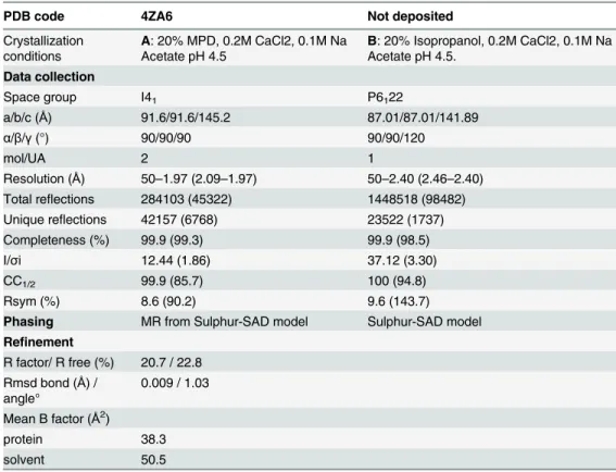
Table 3. Crystallographic data and refinement parameters.
PDB code 4ZA6 Not deposited
Crystallization conditions
A: 20% MPD, 0.2M CaCl2, 0.1M Na Acetate pH 4.5
B: 20% Isopropanol, 0.2M CaCl2, 0.1M Na Acetate pH 4.5.
Data collection
Space group I41 P6122
a/b/c (Å) 91.6/91.6/145.2 87.01/87.01/141.89
α/β/γ(°) 90/90/90 90/90/120
mol/UA 2 1
Resolution (Å) 50–1.97 (2.09–1.97) 50–2.40 (2.46–2.40) Total reflections 284103 (45322) 1448518 (98482) Unique reflections 42157 (6768) 23522 (1737)
Completeness (%) 99.9 (99.3) 99.9 (98.5)
I/σi 12.44 (1.86) 37.12 (3.30)
CC1/2 99.9 (85.7) 100 (94.8)
Rsym (%) 8.6 (90.2) 9.6 (143.7)
Phasing MR from Sulphur-SAD model Sulphur-SAD model Refinement
R factor/ R free (%) 20.7 / 22.8 Rmsd bond (Å) /
angle°
0.009 / 1.03
Mean B factor (Å2)
protein 38.3
solvent 50.5
Values in parenthesis are those for the last shell; MR means Molecular replacement. CC1/2= percentage of
correlation between intensities from random half‐dataset (P. A. Karplus, K. Diederichs, Science 2012, 336, 1030–1033).
gamma-caprolactone) with a concentration of 250μM were performed at intervals of 180 s
while stirring at 1000 rpm.
Results
Directed evolution improved OC8HSL degradation capability of
R.
erythropolis
R138
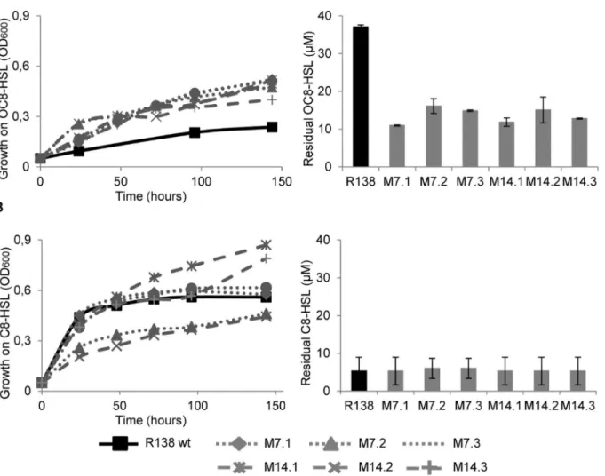
R.erythropolisR138 wild-type grows much better in a minimal medium supplemented with
C8HSL than with OC8HSL (Fig 1), suggesting that the degradation and assimilation of QS-sig-nal exhibiting a keto substitution at the carbon-3 in the acyl chain is limited.
Sub-cultures of the parentalR.erythropolisR138 in three parallel lineages on the minimal medium supplemented with OC8HSL led to the isolation of six clones M7.1, M7.2, M7.3, M14.1, M14.2 and M14.3. These clones were named according to the sampling time (7thand
14thsubcultures) and the lineage (1, 2 and 3). Growth of the parental
R.erythropolisR138 and
its derivatives was compared. After 144 h of incubation in the AB-OC8HSL medium, all
Fig 1. Assimilation and degradation of quorum-sensing signals.Growth ofR.erythropolisR138 wt and its evolved mutants M7.1, M7.2, M7.3, M14.1, M14.2 and M14.3 in the presence of OC8HSL (A) and C8HSL (B) as a sole carbon source. Left panels show growth curves (OD600), right panels indicate
concentration of residual quorum-sensing signals at the end of the growth (140 hours post-inoculation).
evolved derivatives reached a higher culture density (OD600= 0.4–0.5) compared with that
(OD600= 0.2) of the parental strain R138 (Fig 1A). This increased growth which is correlated
with a decreased of the residual OC8HSL in the culture medium (Fig 1A) indicates that a better assimilation of OC8HSL occurs in all evolved derivatives. Residual NAHLs are those which are not altered by QQ-enzymes, irrespectively of their use as a nutrient.
In contrast, the evolved derivatives (except M7.2 and M14.2) and their parentR.erythropolis
R138 grow similarly on C8HSL as a sole carbon source (Fig 1B). At the end of the growth assay (140 hours), the concentration of residual C8-HSL was similar in all culture media (Fig 1B).
Genomic characterization of evolved derivatives M7.1, M7.2 and M7.3.
As no improvement of the OC8HSL-assimilation was observed in the clones collected after the 14thsubculture compared with clones of the 7th(Fig 1), we focused on the earliest derivatives
M7.1, M7.2 and M7.3. Their total DNA was extracted and sequenced by Illumina technology using libraries of 300 bp fragments of which both extremities were sequenced. The number of filtered reads reached 20 267 452, 18 155 476 and 22 655 016 for clones M7.1, M7.2 and M7.3, respectively. All of the reads were mapped on the genome sequence of the parental strainR.
erythropolisR138 [26] with a mean coverage ranging from 193 to 240.
Using the CLC software and a selective filter at 100%, only three independent non-synony-mous substitutions were identified on the circular chromosome (Table 1). In the M7.2 deriva-tive, the mutation 7.2 is located in the gene CDS1197 which is adjacent to theqsdAgene (CDS1198) encoding the known NAHL cleaving lactonase QsdA [19]. We named the incrimi-nated geneqsdR(quorum-sensing degradation regulation) which codes for a transcriptional regulator of the TetR/FabR family. In the M7.1 and M7.3 derivatives, the distinctive mutations belong to the same gene CDS816, encoding a transcriptional regulator of the RipR family. We call this geneqsaR(quorum-sensing assimilation regulation).
qsd and qsa clusters were overexpressed in the evolved derivatives
—
In
R. erythropolis
R138 wild-type, the geneqsaRis divergently transcribed from two adjacent genes, that we namedqsaAandqsaB, coding for an amidohydrolase (CDS819) and a transporter (CDS820) of the Major Facilitator Superfamily (MFS), respectively. In the wild-type strain R138 and its derivatives M7.1 and M7.3, the expression of genesqsaR,qsaAandqsaBwas monitored by RT-qPCR in the presence of mannitol or OC8HSL as a sole carbon source (Fig 2). All the genes exhibited a higher transcription level in the evolved backgrounds as compared to that observed in the wild-type strain whatever the culture medium.
InR.erythropolisR138 wild-type, the geneqsdRis divergently transcribed fromqsdA(the lactonase-coding gene) which is adjacent to two other genes coding for a long-chain fatty acid CoA ligase (CDS1199) and a MFS transporter (CDS1200). We called these two genesqsdCand
qsdD, respectively. We did not use the nameqsdBwhich was previously proposed for a QS-sig-nal degrading amidohydrolase [35]. Expression of theqsdgenes was compared between the wild type strain and the clone M7.2 in the presence of mannitol or OC8HSL as a sole carbon source. All theqsdgenes were over-expressed in the clone M7.2 whatever the culture medium (Fig 3). In the wild-type and clone M7.2, theqsdRexpression decreased by 90% in the course of the culture. As the lactonase QsdA is the best known QS-signal degrading enzyme inR. ery-thropolis[19,36], we thereafter studied its transcriptional regulator QsdRwtand its variant
Structure and overall fold of QsdR
QsdR shares low sequence identity (around 20%) with regulatory proteins with known three dimensional structures. Thus the structure of QsdR at 2.4 Å resolution was solved using sulfur SAD method. One molecule was present in the asymmetric unit. A better resolution structure at 1.9 Å of QsdR from a different crystallization condition was determined by molecular replacement (using the sulfur-SAD structure as model) with two identical molecules in the asymmetric unit (root mean square deviation (rmsd) of 0.07 Å for 181 Cαatoms). Each mole-cule of QsdRwtcomprises 186 residues (Fig 4A) and is composed of 11αhelices, 3 of them
cor-responding to the binding domain, and the others to the regulatory domain. The DNA-binding domain (residues 2 to 41) forms the N terminus domain while the regulatory domain (residues 47 to 186) constitutes the C terminus. These two domains are connected by two resi-dues, Glycine 45 and Asparagine 46. Each monomer in the asymmetric unit forms a dimer by the crystallographic symmetry (Fig 4B). The dimer interface covers 995 A2per subunit
involv-ing 19 residues located in helicesα7,α9 andα10 and two loops, one betweenα7 andα8 and the other betweenα9 andα10. Therefore, the dimeric structure of QsdRwtis the functional
form in solution in line with results from gel filtration chromatography (molecular mass esti-mates at 44 kDa).
Although QsdR protein belongs to the Helix-Turn-Helix superfamily of regulatory proteins, a structural comparison of QsdRwtwith all PDB entries using SSM-EBI (http://www.ebi.ac.uk/ mrd-srv/ssm) [37] shows a very low structural similarity with known structures. The lowest rmsd value of 2.81 Å was obtained with theThermus thermophilusfatty acid degradation
tran-scriptional repressor FadR, of which the structure was solved in presence of a bound dodecyl-CoA [38]. Therefore, we cannot infer any putative regulatory molecule for QsdR based on structural similarity. However, a bound MPD molecule from the precipitant solution well defined in electron density maps indicates the entrance of a protein cavity and forms three hydrogen bonds with the OH side chains of Tyr18 and Thr59 and the CO main chain of Leu55. The cavity contains 3 deeply buried water molecules (Fig 4B). While two water cules directly interact with protein residues, the third one is bound to one of these water mole-cules. The cavity which is surrounded byαhelices is formed by 21 residues: Tyr18 fromαhelix 1 (H1), Leu55, Thr59 and Tyr63 belonging to H4, Phe 82, Val85, Met86, Ser88, Val89 from H5 Ser92 from the loop between H5 and H6, Leu95 from H6, Phe106 and Ala110 from H7, Ile116, Glu117, Ser120 from H8 Val151, Cys154 and Asp155 from H9 Leu158 and Tyr159 from the loop between helices 9 and 10. This deep cavity interior is mainly polar while the entrance con-tains hydrophobic residues. The size of the putative regulator binding site excludes the accom-modation of an effector bound to a CoA like the effector of FadR.
Determination of NAHL/QsdR possible affinity
We used isothermal titration microcalorimetry to measure a possible affinity between QsdRwt
and several putative effectors: OC8HSL, C8HSL, gamma-butyrolactone and gamma-caprolac-tone. All these compounds exhibit a gamma-lactone ring and are substrates of QsdA [39]. No interaction was detected between QsdRwtand any of these four molecules.
Fig 2. Expression of theqsaRABgenes.RT-qPCR monitoring of theqsaRABexpression inR.erythropolis
R138 wt and its mutants M7.1 and M7.3 grown in the presence of mannitol (AB-man) and OC8HSL (AB-OC8HSL) as a sole carbon source. Expressions were normalized using therecAgene as a reference gene. Experiments were done in triplicate.
Fold characteristics of the QsdR
G45Cof the derivative M7.2
QsdRG45Cprotein was purified using the same protocol as for QsdRwt. However, the size
exclu-sion chromatography elution profile was different because the major fraction of the protein was eluted in the exclusion volume of the column indicating that QsdRG45Cwas mostly
aggre-gated. Nevertheless, a small remaining protein fraction was eluted (Data not shown). Despite several crystallization attempts, obtaining crystals of QsdRG45Cwas unsuccessful. In contrast to
QsdRwt, QsdRG45Cseems instable.
Fig 3. Expression of theqsdRACDgenes.RT-qPCR monitoring of theqsdRACDexpression inR.
erythropolisR138 wt and its mutant M7.2 grown in the presence of mannitol (AB-man) and OC8HSL (AB-OC8HSL) as a sole carbon source. Expressions were normalized using therecAgene as a reference gene. Experiments were done in triplicated.
doi:10.1371/journal.pone.0141718.g003
Fig 4. Structure of QsdRwt.(A) Ribbon representation of QsdRwtfold. Its N-domain (residues 2–44) and C-domain (residues 47–186) are shown in blue and
pink respectively. The linker between the two domains composed of Gly45 and Asn46 is in green.αhelices (H) are numbered.(B) The putative binding pocket of QsdRwt. The protein is represented as trace and coloured blue for the N-domain (DNA binding domain) and pink for the C domain (effector binding
domain). The cavity surface is shown in its electrostatic surface potential map. Red, blue and white colours correspond to negative, positive and neutral charged regions, respectively. The MPD molecule bound to QsdRwt and the bound water molecules are shown in cyan sticks and as red spheres respectively.
In order to determine and compare the secondary structures of QsdRwtand QsdRG45C, each
protein was analyzed by circular dichroïsm. QsdRwtdisplays a high proportion ofαhelices (~
91%) in line with what is observed in the crystal structures. In contrast, QsdRG45Cpresents
only 25% ofα-helix secondary structures (Fig 5AandTable 4). One more analysis was per-formed with QsdRG45Cpurified in presence of 1 mM DTT to prevent the formation of a
disul-phide bond between the unique cysteine of the protein and that additional introduced by the M7.2 mutation. The drastic loss ofα-helix secondary structures was not recovered in presence of DTT proving that the 7.2 mutation is responsible for an improper folding of QsdRG45C.
Comparative accumulation of QsdR in
R. erythropolis
R138 wt and its
derivative M7.2
The presence of QsdRwtand QsdRG45CinR.erythropolisR138 and its derivative M7.2
respec-tively was checked by mass spectrometry analysis from the whole cells proteins content. The same amount of bacterial cells for wild-type and clone M7.2 was loaded on a SDS-PAGE. Pro-teins in the gel area corresponding to the molecular weight of QsdR were analyzed by LC-MS/ MS after trypsine digestion and fragments corresponding to QsdRwtand QsdRG45Cwere
searched. As shown inFig 5B, fragments of QsdRwtwere detected in wild type strain whereas
none was detected in the M7.2 derivative. This suggests that the entire QsdRG45C
transcrip-tional factor is absent in the M7.2 bacterial cells.
Discussion
In this work, we used genome engineering as a tool to improve the QS-signal degradation capacity of the bacteriumR.erythropolis, which is a biocontrol and anti-biofouling agent [20– 22,40]. The selective process was successful as it generated three evolved derivatives (M7.1, M7.2 and M7.3) exhibiting a higher assimilation of 3-oxo-subsituted QS-signals compared with their ancestor. This acquired function is of primary interest as the 3-oxo-subsituted QS-signals are produced by several pathogenic and biofilm-forming bacteria [3,4]. In previous works, directed mutagenesis and directed evolution were used for increasing NAHL produc-tion by NAHL-synthase LuxI [41], for modifying selectivity of the NAHL-sensor LuxR [42] as well as that of the NAHL-degrading amidase PvdQ [43]. Our work demonstrates that NAHL-degradation metabolic network may be enhanced in an entire organism by selecting mutations in key-regulators, even if they were previously uncharacterized. In addition, our attempts to construct mutants by reverse genetics inR.erythropolisR138, were unsuccessful, hence the approach based on natural selection was a helpful alternative way for identifying and studying the key-regulatory transcription factors involved in NAHL-degradation.
All the identified mutations are single nucleotide polymorphisms (SNPs) in genes coding for two transcriptional factors, revealing them as master-regulators of QS-signal degradation in
than in the wild type ancestor. All together, these observations suggest that the level of expres-sion rather than the catalytic properties of the lactonase QsdA seems a limiting factor for OC8HSL assimilation inR.erythropolis. Genome engineering allowed to overcome this limita-tion by selecting a bacterial derivative containing a single point mutalimita-tion, G45C, in QsdR, exhibiting a QsdR-independent expression of QsdA. Transcriptional regulators appeared as recurrent targets for improving metabolic properties of microbes, including resistance to toxic compounds such as alcohols [46], production of metabolites such as fatty acids [47], and assimilation of metabolites [48].
QsdR protein possesses a DNA-binding domain at the N-terminus and a regulatory domain at the C-terminus. The DNA-binding domain permits the protein to bind DNA inducing genes activation or repression under its control. The two domains are linked by two residues including glycine 45 which was the mutated residue. Glycine is known to be highly flexible and its presence in this short linker can help the mobility between the two domains. Replacing this glycine by a cysteine (G45C) would have made this linker rigid. A model of this mutation shows no clash or steric hindrance preventing the stability or correct folding of such mutant protein. Unexpectedly, we show here that this single mutation G45C is responsible for QsdR misfolding and the lack of the mutant protein in the total protein pool of the bacteria although its corresponding gene is transcribed. These findings suggest that QsdR is rapidly degraded due to stability and fold problems, resulting in the constitutive expression of QsdA protein.
The regulatory domain of QsdR presents a half hydrophobic/half polar cavity suggesting that QsdR can bind molecules having both hydrophobic and polar groups such as NAHL. However, our results from the isothermal titration microcalorimetry did not reveal any affinity with the tested lactone ring-based molecules such as C8HSL or OC8HSL. Despite several co-crystallization attempts with NAHL and soaking crystals of QsdRwt, obtaining crystals of
QsdRwtin complex with NAHL was unsuccessful. Moreover, the cavity structure of the QsdRwt
regulatory domain has no similarity with that of the known structures of the TetR/FabR family such as FadR, a fatty acid degradation transcriptional repressor inThermus thermophilus[38]. Therefore, we expect a difference in nature and size of the effector between these two transcrip-tional repressors. FadR is regulated with a CoA-link molecule whereas QsdR should be acti-vated by a molecule without any CoA extension.
In conclusion, this work reveals that a single modification of only one amino-acid in a tran-scriptional factor leads to the creation of a new targeted genetic circuit inR.erythropolis. Hence, genome engineering based on natural selection appeared a powerful approach for iden-tifying master-regulators in QS-signal degradation pathway, as well as for improving this path-way in QS-signal degrading organisms.
Fig 5. In vitro and in vivo stability of QsdRG45C.(A) CD analysis spectra of QsdR (black), QsdRG45C(red)
and QsdRG45Cin presence of DTT (blue). (B) Mass spectrometry analysis from a 12.5% SDS PAGE. Lane 1
control: purified QsdR protein; lane 2: whole protein content of wild typeR.erythropolisR138; lane 3: whole protein contentR.erythropolismutant M7.2. Protein bands around the corresponding gel area of pure QsdR were digested by trypsin and identified by LC-TOF/TOF peptide mass fingerprinting searching for matching fragments (Matched peptides are shown in red).
doi:10.1371/journal.pone.0141718.g005
Table 4. Secondary structure estimations from CD experiments.
QsdRwt QsdRG45C QsdRG45C+DTT
α-helices 91% 29% 26%
β-sheets 2% 21% 22%
Random coil 6% 50% 52%
Acknowledgments
We acknowledge SOLEIL for provision of synchrotron radiation facilities (proposal ID 20130869) in using beamline Proxima 1. This work has benefited from the facilities and exper-tise of crystallization platform and the SICaPS platform of IMAGIF (Centre de Recherche de Gif—www.imagif.cnrs.fr). We are thankful to David Cornu and Laila Sago for their help in the
analysis of MS results. We also thank Armelle Vigouroux for advices in protein crystallisation.
Author Contributions
Conceived and designed the experiments: SM DF. Performed the experiments: AES AK NM CV PL. Analyzed the data: AES AK SM DF. Wrote the paper: AES AK SM DF.
References
1. Gadakh B, Van Aerschot A. Renaissance in Antibiotic Discovery: Some Novel Approaches for Finding Drugs to Treat Bad Bugs. Curr Med Chem. 2015;
2. Tay S, Yew W. Development of Quorum-Based Anti-Virulence Therapeutics Targeting Gram-Negative Bacterial Pathogens. Int J Mol Sci. 2013; 14: 16570–16599. doi:10.3390/ijms140816570PMID:
23939429
3. Fuqua WC, Winans SC, Greenberg EP. Quorum sensing in bacteria: the LuxR-LuxI family of cell den-sity-responsive transcriptional regulators. J Bacteriol. 1994; 176: 269–75. PMID:8288518
4. Whitehead NA, Barnard AM, Slater H, Simpson NJ, Salmond GP. Quorum-sensing in Gram-negative bacteria. FEMS Microbiol Rev. 2001; 25: 365–404. PMID:11524130
5. Dong YH, Wang LH, Xu JL, Zhang HB, Zhang XF, Zhang LH. Quenching quorum-sensing-dependent bacterial infection by an N-acyl homoserine lactonase. Nature. 2001; 411: 813–7. PMID:11459062
6. Zhang L-H, Dong Y-H. Quorum sensing and signal interference: diverse implications. Mol Microbiol. 2004; 53: 1563–71. PMID:15341639
7. Uroz S, Dessaux Y, Oger P. Quorum sensing and quorum quenching: the yin and yang of bacterial communication. Chembiochem. 2009; 10: 205–16. doi:10.1002/cbic.200800521PMID:19072824
8. Dong Y, Wang L, Zhang L-H. Quorum-quenching microbial infections: mechanisms and implications. Philos Trans R Soc Lond B Biol Sci. 2007; 362: 1201–11. PMID:17360274
9. Faure D, Dessaux Y. Quorum sensing as a target for developing control strategies for the plant patho-genPectobacterium. Eur J Plant Pathol. 2007; 119: 353–365.
10. Lade H, Paul D, Kweon JH. Quorum quenching mediated approaches for control of membrane biofoul-ing. Int J Biol Sci. 2014; 10: 550–65. doi:10.7150/ijbs.9028PMID:24910534
11. Grandclément C, Tannières M, Moréra S, Dessaux Y, Faure D. Quorum quenching: role in nature and applied developments. DOI:http://dx.doi.org.gate1.inist.fr/10.1093/femsre/fuv038fuv038 First pub-lished online: 2 October 2015 PMID:26432822
12. Zhang H-B, Wang L-H, Zhang L-H. Genetic control of quorum-sensing signal turnover inAgrobacterium tumefaciens. Proc Natl Acad Sci U S A. 2002; 99: 4638–43. PMID:11930013
13. Lang J, Faure D. Functions and regulation of quorum-sensing inAgrobacterium tumefaciens. Front Plant Sci. 2014; 5: 14. doi:10.3389/fpls.2014.00014PMID:24550924
14. Wahjudi M, Papaioannou E, Hendrawati O, van Assen AHG, van Merkerk R, Cool RH, et al. PA0305 of
Pseudomonas aeruginosais a quorum quenching acylhomoserine lactone acylase belonging to the Ntn hydrolase superfamily. Microbiology. 2011; 157: 2042–2055. doi:10.1099/mic.0.043935-0PMID:
21372094
15. Uroz S, D’Angelo-Picard C, Carlier A, Elasri M, Sicot C, Petit A, et al. Novel bacteria degrading N-acyl-homoserine lactones and their use as quenchers of quorum-sensing-regulated functions of plant-patho-genic bacteria. Microbiology. 2003; 149: 1981–9. PMID:12904538
16. Dong Y-H, Zhang X-F, Xu J-LJ-L, Zhang L-HL-H. Insecticidal Bacillus thuringiensis silences Erwinia carotovora virulence by a new form of microbial antagonism, signal interference. Appl Environ Micro-biol. 2004; 70: 954–60. PMID:14766576
17. Dong Y-H, Gusti AR, Zhang Q, Xu J-L, Zhang L-H. Identification of quorum-quenching N-acyl homoser-ine lactonases from Bacillus species. Appl Environ Microbiol. 2002; 68: 1754–9. PMID:11916693
19. Uroz S, Oger PM, Chapelle E, Adeline M-T, Faure D, Dessaux Y. ARhodococcusqsdA-encoded enzyme defines a novel class of large-spectrum quorum-quenching lactonases. Appl Environ Microbiol. 2008; 74: 1357–66. doi:10.1128/AEM.02014-07PMID:18192419
20. Oh H-S, Yeon K-M, Yang C-S, Kim S-R, Lee C-H, Park SY, et al. Control of membrane biofouling in MBR for wastewater treatment by quorum quenching bacteria encapsulated in microporous membrane. Environ Sci Technol. 2012; 46: 4877–84. doi:10.1021/es204312uPMID:22471519
21. Cirou A, Mondy S, An S, Charrier A, Sarrazin A, Thoison O, et al. Efficient biostimulation of native and introduced quorum-quenchingRhodococcus erythropolispopulations is revealed by a combination of analytical chemistry, microbiology, and pyrosequencing. Appl Environ Microbiol. 2012; 78: 481–92. doi:10.1128/AEM.06159-11PMID:22081576
22. Oh H-S, Kim S-R, Cheong W-S, Lee C-H, Lee J-K. Biofouling inhibition in MBR by Rhodococcus sp. BH4 isolated from real MBR plant. Appl Microbiol Biotechnol. 2013; 97: 10223–10231. doi:10.1007/ s00253-013-4933-7PMID:23644749
23. Cirou A, Raffoux A, Diallo S, Latour X, Dessaux Y, Faure D. Gamma-caprolactone stimulates growth of quorum-quenchingRhodococcuspopulations in a large-scale hydroponic system for culturing Sola-num tuberosum. Res Microbiol. 2011; 162: 945–50. doi:10.1016/j.resmic.2011.01.010PMID:
21288487
24. Chilton MD, Currier TC, Farrand SK, Bendich AJ, Gordon MP, Nester EW.Agrobacterium tumefaciens
DNA and PS8 bacteriophage DNA not detected in crown gall tumors. Proc Natl Acad Sci U S A. 1974; 71: 3672–6. PMID:4530328
25. Cha C, Gao P, Chen YC, Shaw PD, Farrand SK. Production of acyl-homoserine lactone quorum-sens-ing signals by gram-negative plant-associated bacteria. Mol Plant Microbe Interact. 1998; 11: 1119– 29. PMID:9805399
26. Kwasiborski A, Mondy S, Beury-Cirou A, Faure D. Genome Sequence of the Quorum-Quenching Rho-dococcus erythropolisStrain R138. Genome Announc. 2014; 2.
27. Kabsch W. XDS. Acta Crystallogr D Biol Crystallogr. 2010; 66: 125–32. doi:10.1107/ S0907444909047337PMID:20124692
28. Sheldrick GM. A short history of SHELX. Acta Crystallogr A. 2008; 64: 112–22. PMID:18156677
29. McCoy AJ, Grosse-Kunstleve RW, Adams PD, Winn MD, Storoni LC, Read RJ. Phaser crystallographic software. J Appl Crystallogr. 2007/08/01 ed. 2007; 40: 658–674. PMID:19461840
30. Cowtan K. The Buccaneer software for automated model building. 1. Tracing protein chains. Acta Crys-tallogr D Biol CrysCrys-tallogr. 2006; 62: 1002–11. PMID:16929101
31. Emsley P, Cowtan K. Coot: model-building tools for molecular graphics. Acta Crystallogr D Biol Crystal-logr. 2004/12/02 ed. 2004; 60: 2126–2132. PMID:15572765
32. Blanc E, Roversi P, Vonrhein C, Flensburg C, Lea SM, Bricogne G. Refinement of severely incomplete structures with maximum likelihood in BUSTER-TNT. Acta Crystallogr D Biol Crystallogr. 2004; 60: 2210–21. PMID:15572774
33. Sreerama N, Woody RW. Estimation of protein secondary structure from circular dichroism spectra: comparison of CONTIN, SELCON, and CDSSTR methods with an expanded reference set. Anal Bio-chem. 2000; 287: 252–60. PMID:11112271
34. Whitmore L, Wallace BA. DICHROWEB, an online server for protein secondary structure analyses from circular dichroism spectroscopic data. Nucleic Acids Res. 2004; 32: W668–73. PMID:15215473
35. Tannières M, Beury-Cirou A, Vigouroux A, Mondy S, Pellissier F, Dessaux Y, et al. A metagenomic study highlights phylogenetic proximity of quorum-quenching and xenobiotic-degrading amidases of the AS-family. PLoS One. 2013; 8: e65473. doi:10.1371/journal.pone.0065473PMID:23762380
36. Barbey C, Crépin A, Cirou A, Budin-Verneuil A, Orange N, Feuilloley M, et al. Catabolic pathway of gamma-caprolactone in the biocontrol agentRhodococcus erythropolis. J Proteome Res. 2012; 11: 206–16. doi:10.1021/pr200936qPMID:22085026
37. Krissinel E, Henrick K. Secondary-structure matching (SSM), a new tool for fast protein structure align-ment in three dimensions. Acta Crystallogr D Biol Crystallogr. 2004/12/02 ed. 2004; 60: 2256–68. PMID:15572779
38. Agari Y, Agari K, Sakamoto K, Kuramitsu S, Shinkai A. TetR-family transcriptional repressorThermus thermophilusFadR controls fatty acid degradation. Microbiology. 2011; 157: 1589–601. doi:10.1099/ mic.0.048017-0PMID:21349973
39. Afriat L, Roodveldt C, Manco G, Tawfik DS. The latent promiscuity of newly identified microbial lacto-nases is linked to a recently diverged phosphotriesterase. Biochemistry. 2006; 45: 13677–86. PMID:
17105187
41. Kambam PKR, Sayut DJ, Niu Y, Eriksen DT, Sun L. Directed evolution of LuxI for enhanced OHHL pro-duction. Biotechnol Bioeng. 2008; 101: 263–72. doi:10.1002/bit.21901PMID:18428113
42. Collins CH, Leadbetter JR, Arnold FH. Dual selection enhances the signaling specificity of a variant of the quorum-sensing transcriptional activator LuxR. Nat Biotechnol. 2006; 24: 708–12. PMID:
16715074
43. Koch G, Nadal-Jimenez P, Reis CR, Muntendam R, Bokhove M, Melillo E, et al. Reducing virulence of the human pathogenBurkholderiaby altering the substrate specificity of the quorum-quenching acylase PvdQ. Proc Natl Acad Sci U S A. 2014; 111: 1568–73. doi:10.1073/pnas.1311263111PMID:
24474783
44. Latour X, Barbey C, Chane A, Groboillot A, Burini J-F.Rhodococcus erythropolisand Itsγ-Lactone Catabolic Pathway: An Unusual Biocontrol System That Disrupts Pathogen Quorum Sensing Commu-nication. Agronomy. 2013; 3: 816–838.
45. Kwasiborski A, Mondy S, Chong T-M, Barbey C, Chan K-G, Beury-Cirou A, et al. Transcriptome of the quorum-sensing signal-degrading Rhodococcus erythropolis responds differentially to virulent and avir-ulentPectobacterium atrosepticum. Heredity (Edinb). 2015;
46. Chong H, Geng H, Zhang H, Song H, Huang L, Jiang R. EnhancingE.coliisobutanol tolerance through engineering its global transcription factor cAMP receptor protein (CRP). Biotechnol Bioeng. 2014; 111: 700–8. doi:10.1002/bit.25134PMID:24203355
47. Zhang F, Ouellet M, Batth TS, Adams PD, Petzold CJ, Mukhopadhyay A, et al. Enhancing fatty acid production by the expression of the regulatory transcription factor FadR. Metab Eng. 2012; 14: 653– 60. doi:10.1016/j.ymben.2012.08.009PMID:23026122
48. Aguilar C, Escalante A, Flores N, de Anda R, Riveros-McKay F, Gosset G, et al. Genetic changes dur-ing a laboratory adaptive evolution process that allowed fast growth in glucose to anEscherichia coli