SUMMARY:
The aim of the study was to compare the effective-ness of eight endodontic ultrasonic tips in removing stain-less steel fragments from the curve of simulated root canals. Methods: Each of the instruments – K-files 25 (EMS), ET25 (Satelec), Redo2 (VDW), RT3 (EMS), CPR8 (Obtura Spartan), Proultra8 (Maillefer), E7 (NSK) and ENDO E3 (W&H) was used to remove 10 stainless steel fragments from the curve of simulated root canals (Dentsply-Maillefer) under magnification 10x and 16x with a dental microscope (OPMI Pico, Carl Zeiss). Success rate, working time and root canal enlargement were recorded and compared.
Results: Success rates were as follows: K-files – 80%, ET25 – 90%, Redo 2 – 80%, CPR8 – 70%, Proultra8 – 80%, RT3 – 70%, Endo E3 – 60%, E7 – 50%. The dif-ferences were not statistically significant.
Working time – mean values: K-files - 8,44 min, ET25 – 9,28 min, Redo 2 - 9,53, CPR8 – 11,01 min, Proultra8 – 10,31 min, RT3 – 11,57 min, Endo E3 – 15,34 min, E7 – 21,45 min. Endo E3 and E7 showed significantly longer working time, the differences between the other tips were not significant.
Mean values of canal diameters were - K-files – 1,11 mm, ET25 – 1,29 mm, Redo 2 – 1,31 mm, CPR8 – 1,54 mm, Proultra8 – 1,51 mm, RT3 – 1,61 mm Endo E3 – 1,68 mm and E7 – 1,72 mm. The differences in canal enlarge-ment between CPR8, Proultra8, RT3, Endo E3 and E7 were not statistically significant.
Conclusion: Endodontic ultrasonic tips with smaller diameters and sharp working points worked faster and pre-served root canal better.
Key words: endodontic ultrasonic tips, ultrasonic technique, broken instruments removal,
INTRODUCTION
Fractured instruments can prevent proper cleaning, shaping and sealing of the root canal system and thus com-promise the treatment outcome [1, 2]. This is especially true when canals are infected and apical radioluscency present. After good assessment of indications and risks, an attempt to remove the broken instrument can be made [3]. One
pos-EFFECTIVENESS IN THE CURVE OF EIGHT TYPES
OF ENDOSONIC TIPS FOR BROKEN
INSTRUMENTS REMOVAL
Kalin K. Shiyakov, Radosveta I. Vasileva,
Department of Conservative Dentistry, Faculty of Dental Medicine, Medical University – Sofia, Bulgaria
Journal of IMAB - Annual Proceeding (Scientific Papers)2014, vol. 20, issue 5
sibility is to use the so called ultrasonic technique, suggested by Ruddle [4, 5, 6, 7], and assessed by a number of differ-ent teams [8-16]. The technique includes removing of root canal dentin around the fragment to loosen and retrieve it. The procedure is performed under dental operating micro-scope. Special endodontic ultrasonic (endosonic) tips were developed for the purpose. All of them are thin, fine, long, sharp pointed instruments to be used with piezoelectric ul-trasonic scalers. The first instruments were developed by Ruddle himself and are now produced as ProUltra Endo tips (Dentsply-Maillefer), and also as CPR-tips (Obtura Spar-tan). Many other tips for the same technique appeared on the market after that. Currently they are not standardized and can have different parameters. Information on their length, taper, diameter is not available neither in the scien-tific literature, nor in the corresponding product catalogues. Most of them have not been studied. The studies examine success rates and complications during removing of frag-ments with different locations [8, 9, 14, 15, 16], and tem-perature rise on the external root surface due to ultrasonic friction [10, 11, 13]. The vast majority of endosonic tips have never been compared. This makes it very difficult for the clinician to choose a proper instrument for a specific clinical situation.
The aim of the present study is to compare the ef-fectiveness of eight endosonic tips in removing stainless steel fragments from the curves of simulated root canals. To complete the aim, the following tasks have been formu-lated: 1. to measure the tip diameter of the eight studied endosonic instruments; 2. to record and compare success rates for complete fragment removal with the eight tips; 3. to measure and compare the time, necessary for complete removal of the fragments; 4. to measure and compare the diameters of the simulated root canals after fragments re-moval.
MATERIALS AND METHODS
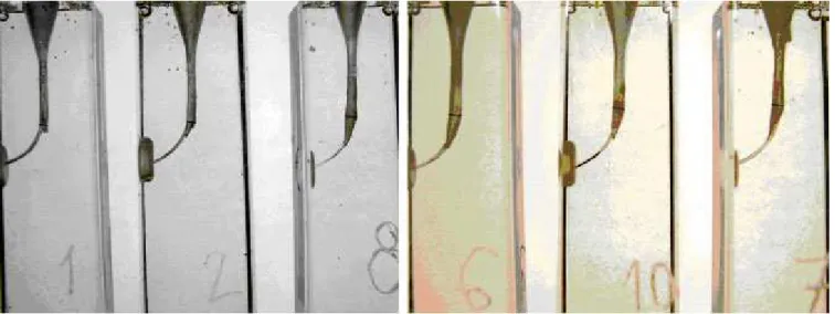
Eighty simulated curved root canals in clear resin blocks (Dentsply-Maillefer, figure 1) were used for the study. All canals had the following characteristics – 18.5 mm of length, diameter and taper equal to ISO instrument number 15. The curve had a 50 degree angle and a radius of 6.5 mm.
Journal of IMAB
ISSN: 1312-773X
http://www.journal-imab-bg.org
Fig. 1. Simulated root canals (Dentsply-Maillefer). Fig. 2. A fragment in the curve of the canal.
Fig. 3.
Stainless steel K-files number 20 (2% taper) were purposely fractured in the curve of the root canals, all frag-ments having the same length (4 mm) and location – inside the root canal curve, the head of the fragment lying in the straight portion of the root canal (figure 2).
RT3, Proultra8 and K-files 25 were used with the ul-trasonic scaler Woodpecker HW-3H (GWMI), Endo E3 was used with the scaler Pyon 2 led (W&H), and ET25, Redo 2, CPR8 and E7 were used with Varios 550 (NSK). The low-est possible power settings of the scalers were adjusted, with which the instruments could effectively remove material from the simulated root canal.
First Gates Glidden drills 1, 2 and 3 were used to en-large the canal and achieve visibility of the fragment. Then tip-modified Gates Glidden drills 1, 2 and 3 were rotated at the level of the fragment’s head to create a “staging plat-form”. Then fragments were treated ultrasonically as de-scribed by Ruddle [4, 5, 6, 7] using the vibrating tip, mate-rial was removed from the wall of the canal surrounding the fragment, and then going around it in a contra-clockwise rotation, the fragment was unscrewed and evacuated from the canal. A Stropko irrigator (Sybron Endo) was used to blow away the plastic dust, generated form the friction. All procedures were conducted under magnification 10x and 16x with a dental operating microscope (OPMI Pico, Carl Zeiss).
The following measurements were performed: 1. The diameter of the tip of the endosonic instru-ments was measured with an electronic calliper (Mitutoyo 500-455, Japan, 0.02mm); This was necessary, because no other source of such information was available, and the data was required for completeness of the discussion; 2. Success rates of complete fragment removal were recorded; 3. Re-corded was also the time, necessary for complete removal; 4. The largest diameter of the simulated root canals at the level of the fragment was measured after its removal, us-ing digital photographs of the canals in clear resin blocks and a software product (Klonk-Image Measurement) – level of measurements is presented on figure 5.
Then recorded data for the different ultrasonic
instru-ments was analyzed and compared.
All data was statistically analyzed (Chi-square inde-pendence test, p>0.05, ANOVA single factor, p>0.05, Stu-dent’s T-test, p>0.05).
RESULTS
The measured diameters of the tips of the endosonic instruments were as follows (table 1): K-files 25 - 0.25 mm, ET25 (Satelec) – 0.30 mm, Redo 2 (VDW) – 0.30 mm, RT3 (EMS) – 0.30 mm, Endo E3 (W&H) – 0.40 mm, E7 (NSK) – 0.42 mm, Proultra8 (Maillefer) – 0.44 mm, CPR8 (Obtura Spartan) – 0.44 mm. Data was used to analyze the depend-ence between the size of the instrument and the degree of root canal enlargement during fragment’s retrieval (root ca-nal diameter at the level of the removed fragment).
Success rates were (table 1): K-files – 80%, ET25 – 90%, Redo 2 – 80%, CPR8 – 70%, Proultra8 – 80%, RT3 – 70%, Endo E3 – 60%, E7 – 50%. The differences are not statistically significant (Chi-square independence test, p>0.05).
Working time (table 1) – mean values: K-files - 8,44 min, ET25 – 9,28 min, Redo 2 - 9,53, CPR8 – 11,01 min, Proultra8 – 10,31 min, RT3 – 11,57 min, Endo E3 – 15,34 min, E7 – 21,45 min. Endo E3 and E7 showed significantly longer working time, the differences between the other tips were not significant (ANOVA single factor, p>0.05, Stu-dent’s T-test, p>0.05).
Mean values of canal diameters at the level of the fragment were (table 1)- K-files – 1,11 mm, ET25 – 1,29 mm, Redo 2 – 1,31 mm, CPR8 – 1,54 mm, Proultra8 – 1,51, RT3 – 1,61 mm Endo E3 – 1,68 mm and E7 – 1,72 mm. The differences in canal enlargement between CPR8, Proultra8, RT3, Endo E3 and E7 were not statistically sig-nificant (ANOVA single factor, p>0.05, Student’s T-test, p>0.05).
Table 1.
K-files #25 ET25 Redo 2 Proultra8 CPR8 RT3 Endo E3 E7
(EMS) (Satelec) (VDW) (Maillefer) (Obt Sp) (EMS) (W&H) (NSK) Tip
0.25mm 0.3mm 0.3mm 0.44mm 0.44mm 0.3mm 0.4mm 0.42mm 1
diameters Success
80% 90% 80% 80% 70% 70% 60% 50% 2
rates
Working 8.44 min 9.28 min 9.53 min 10.31 min 11.01 min 11.57 min 15.34 min 21.45 min 3
time
*
*
Canal 1.11 mm 1.29 mm 1.31 mm 1.51 mm 1.54 mm 1.61 mm 1.68 mm 1.72 mm 4
diameters
•
•
•
1 – no statistical analysis was performed
2 – differences are not statistically significant (Chi square independence test, p>0.05)
DISCUSSION Working time
Currently no investigations comparing working time between endodontic ultrasonic tips exist, so we can not com-pare our results to such of other studies. Suter et al. [15] commented on working time in clinical conditions and claimed that after 30 min of work the risk of complications such as perforations significantly increased. In the present study all successful removals took less than 30 minutes.
In the present study K-files (EMS), ET25 (Satelec), Redo 2 (VDW), CPR8 (Obtura Spartan), Proultra8 (Maillefer) and RT3 (EMS) showed no statistically signifi-cant differences in working time (table 1), while Endo E3
(W&H) and E7 (NSK) had significantly longer working time (ANOVA single factor, p>0.05, T-test, p>0.05). Tak-ing in consideration the measured diameters of the ultra-sonic instruments (table 1), outside of statistics, smaller tips with sharp working points (figure 4) work faster. The only exception is RT3 (EMS), which has a smaller diameter than CPR8 (Obtura Spartan) and Proultra8 (Maillefer), but shows longer working time. We think this is due to the differences in the configurations of the tips. RT3 is diamond coated and has a rounded working point while the other two are non-diamond coated and have needle-sharp working points (fig-ure 4).
Figure 4.
Success rates
Shen et al. [17], using different techniques and no magnification in their clinical study, reported 60% success rates when the fragments were located at the curve and 31% when the fragments were beyond the curve. Suter et al [15] in their clinical study did not find statistical differences in success rates depending on the location of the broken in-strument in relation to the curve. Souter et al. [14] in their in vitro and clinical study concluded that due to very low success rates in removal of fragments beyond the curve and risk of perforation, the procedure should not be routinely attempted. Ward et al [16] using simulated canals and ex-tracted teeth discovered that success rates significantly de-creased when fragments were located entirely around the curve, and major canal damage often occurred. Ward et al [16] and Souter et al [14] used CPR-tips (Obtura Spartan), and Suter et al [15] used ultrasonic K-files. In the present study fragments were located inside the curve, the head of the instrument lying in the straight portion of the root ca-nal (figure 2). Our success rates (72.5% mean value, table 1) are a little higher than those cited above, but the differ-ence in fragment location should be taken into considera-tion. Although statistically the examined endosonic tips in the present study performed equally (Chi-square
independ-ing greater diameters and/or rounded workindepend-ing points (fig-ure 4 and table 1). At the moment no other studies compar-ing success rates between different endosonic tips exist.
Root canal diameter at the level of the fragment after its removal
1. Crump MC, Natkin E. Relation-ship of broken root canal instruments to endodontic case prognosis: a clini-cal investigation. J Am Dent Assoc. 1970; 80:1341–1347. [PubMed]
2. Feldman G, Solomon C, Notaro P, Moskovitz E. Retrieving broken en-dodontic instruments. J Am Dent. Assoc 1974 Mar;88(3):588 -91. [PubMed]
3. Strindberg LZ. The dependence of the results of pulp therapy on cer-tain factors. Acta Odontol Scand. 1956; 14(Suppl 21):1-156.
4. Ruddle CJ. Nonsurgical retreatment. J Endod. 2004 Dec; 30(12): 827-845. [PubMed] [CrossRef] 5. Ruddle CJ. Broken instrument removal. The endodontic challenge.
Dent Today. 2002 Jul;21(7):70-72, 74, 76 pasim. [PubMed]
6. Ruddle CJ. Nonsurgical
retreatment. In: Cohen S, Burns RC, eds. Pathways of the pulp, 8th ed. St Louis: Mosby; 2002:875–930.
7. Ruddle CJ. Micro-endodontic non-surgical retreatment. Dent Clin North Am. 1997 Jul;41(3):429-454. [PubMed]
8. D’Arcangelo C, Varvara G, De Fazio P. Broken instrument removal – two cases. J Endod. 2000 Jun;26(6): 368-370. [PubMed] [CrossRef]
9. Gencoglu N, Helvacioglu D. Comparison of the different techniques to remove fractured endodontic instru-ments from root canal systems. Eur J Dent. 2009 Apr;3(2):90-5. [PubMed]
10. Hashem AA. Ultrasonic vibra-tion: temperature rise on external root surface during broken instrument re-moval. J Endod. 2007 Sep;33(9):1070-1073. [PubMed] [CrossRef]
11. Madarati AA, Qualtrough AJ, REFERENCES:
Watts DC. Efficiency of a newly de-signed ultrasonic unit and tips in reduc-ing temperature rinse on root surface during removal of fractured files. J Endod. 2009 Jun;35(6):896-899. [PubMed] [CrossRef]
12. Madarati AA, Qualtrough AJ, Watts DC. A microcomputed tomogra-phy scanning study of root canal space: changes after the ultrasonic removal of fractured files. J Endod. 2009 Jan; 35(1):125-128. [PubMed] [CrossRef]
13. Madarati AA, Qualtrough AJ, Watts DC. Factors affecting tempera-ture rinse on the external rîît surface during ultrasonic retrieval of intracanal separated files. J Endod. 2008 Sep;34(9):1089-1092. [PubMed] [CrossRef]
14. Souter NG, Messer HH. Com-plications associated with fractured file removal using an ultrasonic technique. and their points are rounded (although Endo E3 is
diamond-coated), (figure 4). The differences between CPR8, Proultra8, RT3, Endo E3 and E7 are not statistically signifi-cant, (ANOVA single factor, p>0.05, T-test, p>0.05). Be-cause of risks of substantial removal of material and con-siderable root canal aberration (figure 5), we would
recom-mend using endosonic tips with the smallest diameter pos-sible in the curve of the root canals for broken instruments removal. No other studies exist comparing different endosonic tips in root canal enlargement during broken in-struments removal.
Figure 5. The largest diameter of the canals at the zone of the fragment was measured
CONCLUSION
Endodontic ultrasonic tips with smaller diameters and sharp working points worked faster and preserved root canal better during removal of fragments located inside the
J Endod. 2005 Jun;31(6):450-452. [PubMed]
15. Suter B, Lussi A, Sequeira P. Probability of removing fractured in-struments from root canals. Int Endod J. 2005 Feb;38(2):112-123. [PubMed] 16. Ward JR, Parashos P, Messer
HH. Evaluation of an ultrasonic tech-nique to remove fractured rotary nickel-titanium endodontic instruments from root canals: an experimental study. J Endod. 2003 Nov;29(11):756-63. [PubMed] [CrossRef]
17. Shen Y, Peng B, Cheung GS.
Address for correspondence:
Dr. Kalin Shiyakov
Department of Conservative Dentistry and Endodontics, Faculty of Dental Factors associated with the removal of fractured NiTi instruments from root canal systems. Oral Surg Oral Med Oral Pathol Oral Radiol Endod. 2004 Nov;98(5):605-610. [PubMed] [CrossRef]
Please cite this article as: Shiyakov KK, Vasileva RI, EFFECTIVENESS IN THE CURVE OF EIGHT TYPES OF ENDOSONIC TIPS FOR BROKEN INSTRUMENTS REMOVAL. J of IMAB. 2014 Oct-Dec;20(5):595-600.
DOI: http://dx.doi.org/10.5272/jimab.2014205.595