Copyright © 2017 The Korean Movement Disorder Society 59
Suspected Perinatal Depression Revealed to be
Hereditary Diffuse Leukoencephalopathy with
Spheroids
Josefine Blume, Robert Weissert
Department of Neurology, University of Regensburg, Regensburg, Germany
ABSTRACT
Early motor symptoms of neurodegenerative diseases oten appear in combination with psychiatric symptoms, such as depres-sion or personality changes, and are in danger of being misdiagnosed as psychogenic in young patients. We present the case of a 32-year-old woman who presented with rapid-onset depression, followed by a hypokinetic movement disorder and cognitive decline during pregnancy. Genetic testing revealed a mutation in the colony-stimulating factor 1 receptor gene, which led to the diagnosis of hereditary difuse leukoencephalopathy with spheroids. Hereditary difuse leukoencephalopathy with spheroids (HDLS) is probably an under-recognized disease. HDLS should be considered in patients with rapidly progressing parkinsonian symptoms and dementia accompanied by white matter lesions.
Key WordsaaHereditary difuse leukoencephalopathy with spheroids; parkinsonism; leukoencephalopathy; colony-stimulating factor 1 receptor.
CASE REPORT
https://doi.org/10.14802/jmd.16050 / J Mov Disord 2017;10(1):59-61 pISSN 2005-940X / eISSN 2093-4939
Received: October 10, 2016 Revised: November 1, 2016 Accepted: November 18, 2016
Corresponding author: Joseine Blume, MD, PhD, Department of Neurology, University of Regensburg, Universitaetsstrasse 84, Regensburg 93053, Germany / Tel: +49-0941-9410 / Fax: +49-0941-9413105 / E-mail: joblume.rb@gmail.com
cc his is an Open Access article distributed under the terms of the Creative Commons Attribution Non-Commercial License (http://creativecommons.org/ licenses/by-nc/3.0) which permits unrestricted non-commercial use, distribution, and reproduction in any medium, provided the original work is properly cited.
JMD
Hereditary difuse leukoencephalopathy with spheroids (HDLS) is a rare, autosomal dominant inherited disorder with adult on-set that leads to progressive cognitive decline and varying neuro-logic features, including ataxia, parkinsonism, dystonia and spasticity.1 Psychiatric symptoms, including personality changes,
apathy, drug abuse and depression, may proceed these symp-toms.2 he median age of onset is 45 years, and the median life
expectancy ater diagnosis is six years, although both parameters vary among afected patients,3 who carry mutations in the
colo-ny-stimulating factor 1 receptor (CSF1R) gene on chromosome 5.4 All known mutations lead to disturbed autophosphorylation
ater ligand binding.5 CSF1R is the cell surface receptor for
cyto-kine macrophage colony-stimulating factor 1 (CSF1) and IL-34, both of which play a role in regulating mononuclear phagocytic cells, including microglia.6 herefore, impaired microglia
surviv-al, proliferation and diferentiation are assumed to be causative for HDLS. The typical MRI findings of HDLS are confluent
FLAIR hyperintensities in the subcortex and deep white matter.7
hese MRI changes may be mischaracterized as ischemic small vessel disease, Cerebral Autosomal Dominant Arteriopathy with Subcortical Infarcts and Leukoencephalopathy (CADASIL) or demyelinating diseases, such as primary progressive multiple sclerosis, especially early in the disease. Bifrontal spotty calciica-tions have been described on CT in a small number of cases.8
CASE REPORT
60
J Mov Disord 2017;10(1):59-61
JMD
of 24 points on the Mini Mental State Examination. Perinatal depression was suspected, and she was treated by the Department of Psychiatry for six months without significant improvement in her symptoms. Upon completion of her treatment, the patient displayed an unusual wide-based, shuling, very slow and highly luctuating gait. She walked in small steps and sometimes staggered severely, but her symptoms were variable, and she did not fall. Therefore, her gait disturbances were classified as psychogenic. However, cerebral MRI showed conlu-ent white matter lesions suspicious for CADASIL. he patient had been treated for mild hypertension since the age of 29 but had otherwise been healthy. Her family history was negative for any hereditary diseases, but her reported history was fragmented because she had broken of all contact with her fa-ther at the age of 18.
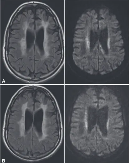
The patient subsequently presented to us ten months after symptom onset and six months after she had given birth to a healthy girl. She presented with conspicuous global bradykinesia with severe slowing and hesitation in her fine motor skills and symmetric rigidity in all her extremities, but without tremor. She also exhibited reduced spontaneous speech with slight amnestic aphasia and ataxic dysar-thria, with loss of modulation. Her gait disturbances had worsened, as she could walk only short distances independently, and she had diiculty liting her feet of the ground without external instruction but showed no typical freezing behaviors. Apraxia was an impor-tant inding, as it was evident in both her ine motor skills and her gait. Another MRI revealed the pres-ence of increasingly symmetrical, confluent FLAIR hyperintensities with partly restricted diffusion, but without contrast enhancement (Figure 1). Wide-ranging blood and CSF analyses, as well as electro-physiological tests, were not suggestive of a diagnosis. In particular, there was no evidence of an infectious or autoimmune cause of her symptoms.
The marked parkinsonian features, which im-proved slightly on levodopa, combined with the pro-gressive leukoencephalopathy and spotty frontal cal-ciications demonstrated by CT (Figure 2) led us to test for HDLS. Genetic testing revealed the presence of a heterozygous mutation (c.2541G>C) in the CS-F1R gene leading to a change in the corresponding amino acid sequence (p.E847D). This mutation was irst described in a patient who presented with
cog-Figure 2. Cerebral CT at the irst visit. Generalized supratentorial atrophy incon -sistent with an age of 32 years and multiple spotty calciications in the frontal white matter (arrow).
Figure 1.Cerebral MRI at the irst visit (A) and nine months later (B). A: Cere -bral MRI at the irst visit. Conluent hyperintensities in the periventricular and deep white matter (FLAIR, left) with partly restricted diffusion (diffusion-weight -ed, right). B: Cerebral MRI nine months later: increasing hyperintensities affect -ing almost the entire white matter (FLAIR, left). FLAIR: luid-attenuated inver -sion recovery.
A
Perinatal Depression Revealed to be Hereditary Diffuse Leukoencephalopathy with Spheroids Blume J, et al.
www.e-jmd.org 61 nitive decline and spastic paraparesis at the age of 44.3
he patient exhibited signs of progressive pyrami-dal as well as extrapyramipyrami-dal motor dysfunction and rapidly progressing dementia during the following months (Supplementary Video 1 in the online-only Data Supplement). Eighteen months ater symptom onset, the patient was admitted to a nursing home. By that time, she was not able to sit, stand, commu-nicate or recognize faces. She presented with a com-bination of rigid-spastic muscle tonus, pyramidal signs and primitive reflexes. The patient died 28 months ater symptom onset.
DISCUSSION
his young woman’s case was highly suspicious for infection or autoimmune disease due to its subacute onset and rapid progression during her pregnancy and shortly ater her irst childbirth. HDLS is caused by mutations in the CSF1R gene. CSF1R and its li-gands, CSF1 and IL-34, are required for placental development.9 We therefore hypothesize that the
ex-tensive adaptations of the maternal immune system that occur during pregnancy contribute to the clini-cal manifestations of the disease. Further research is needed to prove this theory.
In addition to the parkinsonian features, the spotty calcifications that were noted in the affected frontal white matter on CT were a hint to the diagnosis. hese indings were irst described by Fujioka et al.,10
who reported the case of a female patient with a CS-F1R mutation in 2013, and Konno et al.,8 who
pre-sented the results pertaining to a set of patients in 2014. CSF1R signaling is known to be necessary for osteoclast cytoskeletal reorganization. herefore, a di-rect pathogenic relationship between CSF1R signaling and calcification is conceivable. The calcifications seem to be speciic for HDLS, but this speciicity is not yet common knowledge. CT should be performed in suspected cases to conirm the diagnosis and to inves-tigate the speciicity of this inding further.
Early motor symptoms of neurodegenerative dis-eases, which oten appear in combination with psy-chiatric symptoms, such as depression or personality changes, are in danger of being misdiagnosed as psy-chogenic in young patients, especially during and shortly ater pregnancy. HDLS is probably an under-recognized disease. HDLS should be considered in patients with rapidly progressing parkinsonian
symp-toms and dementia accompanied by white matter le-sions.
Supplementary Video Legend
Video 1. The video shows the patient at 16 months after symptom onset. She is distinctly bradykinetic and rigid, and her fine motor skills are slow and apraxic. She is not able to stand up without the help of two nurses and cannot walk due to spasticity and severe gait apraxia.
Supplementary Materials
he online-only Data Supplement is available with this arti-cle at https://doi.org/10.14802/jmd.16050.
Conflicts of Interest
he authors have no inancial conlicts of interest.
REFERENCES
1. Lynch DS, Jaunmuktane Z, Sheerin UM, Phadke R, Brand-ner S, Milonas I, et al. Hereditary leukoencephalopathy with axonal spheroids: a spectrum of phenotypes from CNS vasculitis to parkinsonism in an adult onset leukodystro-phy series. J Neurol Neurosurg Psychiatry 2016;87:512-519. 2. Hofmann S, Murrell J, Harms L, Miller K, Meisel A, Brosch
T, et al. Enlarging the nosological spectrum of hereditary difuse leukoencephalopathy with axonal spheroids (HDLS). Brain Pathol 2014;24:452-458.
3. Guerreiro R, Kara E, Le Ber I, Bras J, Rohrer JD, Taipa R, et al. Genetic analysis of inherited leukodystrophies: geno-type-phenotype correlations in the CSF1R gene. JAMA Neurol 2013;70:875-882.
4. Rademakers R, Baker M, Nicholson AM, Rutherford NJ, Finch N, Soto-Ortolaza A, et al. Mutations in the colony stimulating factor 1 receptor (CSF1R) gene cause hereditary diffuse leukoencephalopathy with spheroids. Nat Genet 2011;44:200-205.
5. Chitu V, Gokhan S, Gulinello M, Branch CA, Patil M, Basu R, et al. Phenotypic characterization of a Csf1r haploinsuf-ficient mouse model of adult-onset leukodystrophy with axonal spheroids and pigmented glia (ALSP). Neurobiol Dis 2015;74:219-228.
6. Chitu V, Gokhan Ş, Nandi S, Mehler MF, Stanley ER. Emerging roles for CSF-1 receptor and its ligands in the nervous system. Trends Neurosci 2016;39:378-393. 7. Sundal C, Jönsson L, Ljungberg M, Zhong J, Tian W, Zhu T,
et al. Diferent stages of white matter changes in the origi-nal HDLS family revealed by advanced MRI techniques. J Neuroimaging 2014;24:444-452.
8. Konno T, Tada M, Tada M, Koyama A, Nozaki H, Hariga-ya Y, et al. Haploinsuiciency of CSF-1R and clinicopatho-logic characterization in patients with HDLS. Neurology 2014;82:139-148.
9. Pampfer S, Daiter E, Barad D, Pollard JW. Expression of the colony-stimulating factor-1 receptor (c-fms proto-onco-gene product) in the human uterus and placenta. Biol Re-prod 1992;46:48-57.