DOI: 10.5562/cca2685
Insight of the Iron Binding and Transport in Dke1 -
A Molecular Dynamics Study
Hrvoje Brkić
Faculty of Medicine, J. Huttlera 4, HR-31000 Osijek, Croatia Author’s e-mail address: hbrkic@mefos.hr
RECEIVED: June 18, 2015 REVISED: November 20, 2015 ACCEPTED: November 25, 2015
Abstract: Acetylacetone dioxygenase from Acinetobacter johnsonii (Dke1) is a non-heme Fe2+ dependent enzyme which catalyzes the oxidative degradation of β-dicarbonyl compounds. It is a homotetramer with four active sites, each containing single metal ion. Since the active site is buried, knowledge on transport of the metal ion and reactants (products) is essential for understanding the enzyme mechanism. The goal of this study was to assess the influence of several point mutations on the enzyme activity. The point mutations of hydrophilic amino acid residues (Tyr70, Arg80 and Glu98) that were shown to be important for metal binding and reactants stabilization were of the particular interest. Com-putational study enabled us to determine the preferred metal ion binding sites as well, as the pathways it utilizes to enter the enzyme active site. Besides, influence of the point mutations on the hydrogen bond network within enzyme was determined.
Keywords: metaloenzyme, non-heme, iron, molecular dynamics.
INTRODUCTION
PPROXIMATELY one quarter to one third of all pro-teins require metals to carry out their functions.[1] One of such proteins is the Acetylacetone dioxygenase from Acinetobacter johnsoii (Dke1), which belongs to the family of Fe2+ dependent dioxygenases. It is a homo-tetramer[2] with each subunit organized in a single-domain β-barrel fold, characteristic for the cupin superfamily of proteins. Dke1 catalyzes the oxidative degradation of the β-dicarbonyl compounds[3,4] and for this purpose it needs Fe2+ ion. In the crystallographically determined structure the metal ion is coordinated with three histidines, while the molecular dynamics study showed that beside the his-tidines one glutamate could also take part in the metal ion coordination.[5] Although the Dke1 active site, besides the Fe2+ ion, can host several other metal ions, such as Co(II), Ni(II), Cu(II) and Zn(II), the enzyme is unable to catalyze the oxidative conversion of acetylacetone in their pres-ence.[4] Until now only the effects of hydrophilic residues, placed in the second coordination sphere of the metal, have been investigated computationally, using the
mole-cular dynamics (MD) simulations.[5] The main purpose of this study was to get insights into molecular mechanisms of Dke1, which haven’t been demystified by now. Using computational methods the specific and non-specific metal ion binding sites are revealed and amino acid resi-dues responsible for metal transport in and out of the Dke1 active site are identified. Additionally, the influence of hydrophilic residues mutations on the metal ion affinity and stabilization of ligand, i.e. 2,4-pentandione (PD), is in-vestigated. Since the experimental data had suggested that hydrophilic residues in the active site pocket, Tyr70, Arg80 and Glu98, play a crucial role in the metal ion and substrate stabilization,[6] the point mutations of these amino acids were computationally prepared, and their in-fluence to the protein structure and dynamics as well as on ligands binding and transport were investigated by computational methods. Previously published data re-ported that Dke1 has 2 metal binding sites,[7] but until now only one of them has been specified. In this study the al-ternative metal ion binding site(s) was determined and mechanism of the metal ion circulation within the protein was elucidated.
Croat. Chem. Acta 2015, 88(3), 297–306 DOI: 10.5562/cca2685
METHODS
System Preparation
The crystal structure of the substrate free Dke1 obtained from the PDB database[8] (pdb_id 3BAL) was used as the in-itial structure for the simulations. Several systems were prepared, as follows.
The point mutants Glu98Gln, Arg80Ala and Tyr70Ala, were prepared by editing the PDB file and using the module
tleap (part of the AMBER12 package).[9] Water molecules de-termined in the crystal structure were removed, and zinc was replaced with the Fe2+ ion. Systems were parameterized us-ing the AMBER[9] ff10[10] force field, GAFF[11] (General Amber force field), and the parameters for Fe2+ ion that were derived and tested earlier.[5,12] Parameters development was achiev-ed with ab initio quantum mechanical calculations (using UHF method).[5] Equilibrium values for the bond distances, angles, and dihedrals, were determined, as well as their corres-ponding force constants. According to the experimental conditions,[2–5] the all simulations were performed at pH 7.5. Histidines were uncharged (singly protonated), aspartic and glutamic acids were negatively, and arginines and lysines positively charged. All other amino acid residues were neutral except the N and C terminal residues which were pos-itively and negatively charged, respectively. Hydrogen atoms were also added by tleap, while protonation of the histidine imidazole ring (either Nδ1 or Nε2) was adjusted manually de-pending on the possibilities of hydrogen bond formation. The complexes with PD (2,4 pentandione) were constructed using the earlier determined Dke1-PD complex[5] as a template. For the complexes, besides the already mentioned force fields, the parameters derived earlier for the Fe2+ – substrate (PD) interactions were used.[5]
MD Simulations
Systems were placed in the truncated octahedron box filled with TIP3P[13] water molecules, with minimal distance of 9 Ǻ between the solute atoms and the edge of the box. Neu-tralization was accomplished by adding Na+ ions at the ap-propriate places on the protein surface.
Minimization of the systems was done in four, and equilibration in seven cycles, for details see previously con-ducted studies.[5,12] After minimization and equilibration each of the constructed mutants, (Arg80Ala, Glu98Gln and Tyr70Ala) as well as their complexes with PD, were submit-ted to 30 ns of unconstrained MD simulation. The time step during pre-equilibration, equilibration and the first 2 ns of the productive MD simulations was 1 fs, and remaining sim-ulations were accomplished using the SHAKE algorithm[14] and 2 fs time step.
Besides the simulations where Fe2+ was described with both bonding and non-bonding parameters, several sets of MD simulations with the metal ion placed at different positions and described with non-bonding parameters solely were also performed. For the wild type and mutated proteins several sets of MD simulations were performed: a) with the Fe2+ ion initially placed into the active site (crystallographically determined pose), two sets of simulations, 16 ns and 30 ns each, b) with Fe2+ placed at the entrance of the water trafficking tunnels, 3 sets of simulations for the wild type enzyme (WT; 16, 6 and 3 ns long), 2 sets for the Tyr70Ala (16 and 5 ns) mutant and one for each the other two variants (16 ns)). Starting structures for these simulations were the final snapshots of the 5 ns long simulations with Fe2+ placed at (or close to) the entrance of the water trafficking tunnels, where one water molecule was replaced a with Fe2+. Table ST1 (in Supplemental) contains list of all the preformed simulations with its respective duration.
The Binding Free Energy Calculations for
Fe
2+Initially Placed in a Water Tunnel
The free energy for the hydrated Fe2+ binding to the enzyme was calculated using the MMPBSA[15] method as imple-mented in the AMBER package. Calculations were done on the 2-ns long final parts of the MD simulation trajectories. The concentration of the singly charged counterions was 0.1 M. Poisson–Boltzmann method was used to calculate the polar component of solvation, and non-polar component was determined using equation (1):
sol nonpolar
Δ H γSASA (1)
where γ is the surface tension with value of 0.0072 kcal mol−1 Ǻ−2. Solvent accessible surface area (SASA) was calculated using the MolSurf program.[16] The calculations were accomplished for the enzyme immersed into the solvent utilizing the solute dielectric constant of 4.0. As a ligand Fe2+ ion hydrated with two water molecules (the Fe2+ + two water molecules cluster) was considered, while the receptor was apo variant.
RAMD Simulations
DOI: 10.5562/cca2685 Croat. Chem. Acta 2015, 88(3), 297–306 than 0.01 Å during this period, a new direction was chosen
randomly; otherwise the same force was applied for the next period of 40 time steps.
The structures were sampled every 1.0 ps and trajec-tories were analysed in details. Root mean square devia-tion, amino-acid fluctuations, water population of the enzyme active site, hydrogen bonds between amino acids of interest and other interesting features were monitored and analysed using the program ptraj[18] available within the Amber program suite.
Secondary Structure Analysis
The secondary structure of the simulated variants was de-termined by STRIDE WEB server.[19]RESULTS AND DISCUSSION
In order to get insights into the molecular mechanisms of Fe2+ trafficking in and out of the enzyme active site, a set of MD simulations for the wild type protein and its single point mutants Tyr70Ala, Arg80Ala Glu98Gln and were performed: (i) of the Fe2+ free protein (apo enzyme), (ii) of the enzyme with Fe2+ bound in the active site, without and with applying random force, (iii) of the proteins with the metal ion located at the entrance of the water tunnel. The obtained trajecto-ries were analysed and the Fe2+ migration paths, in and out of the protein, were determined.(i) Simulations on APO Enzymes
A short, only 6 ns long, MD simulations of Dke1 wild type and its variants Tyr70Ala, Arg80Ala Glu98Gln and in their metal free form were performed in order to monitor influence of the point mutations on the overall protein structure and par-ticularly on the active site structure. According to the calcu-lated RMSD between the structure of the wild type enzyme and the mutants (1.7 Tyr70Ala, 2.4 Arg80Ala, 1.9 Glu98Gln) and the visual inspection it is clear that the mutations neither change the overall protein fold nor the secondary structure elements, ß sheets they are part of. However, due to differ-ences in size, hydropobicity and other physico-chemical properties of the amino acid residues in the native enzyme and mutants their local environment, as well as protein flexibility, have changed. A comparison of the resulting trajectories revealed that Glu98 was more than 50 % of the simulation time H-bonded to His104 in the WT and Tyr70Ala mutant,while in the other two mutants this interaction is significantly weaker and it has disappeared during simulation, and both Glu98 and His104 were more flexible (see Table 1). Another carboxylate residue that was found rather close to the metal binding motif, Glu11 (from neighbour subunit), was generally not H-bonded to any of the metal binding histidines or hydro-philic triad residues (Tyr70Ala, Arg80Ala, Glu98Gln) and pointed away from the active site in all variants (see Table 1). A Figure 1 shows the optimized structure of the active site in different variants obtained after 6 ns of MD simulations.
(ii) MD Simulations with the Fe
2+Ion
Bound into the Protein Active Site
Defined by 3His
During MD simulations with the Fe2+ cofactor bound to the metal binding motif, which is according to the crystallo-graphically determined Dke1 structure characterized with 3 histidines, Glu98 entered the metal ion coordination sphere
Table 1. Percentage of the simulation time during which the hydrogen bonds between Glu98(OE1/2)-His104(NE2) and Glu11(OE1/2)-His104(NE2) exists, Simulations were performed on APO variants of Dke1
WT Tyr70Ala Arg80Ala Glu98Gln
Glu11(OE1/2)-His104(NE2) 0.07 0 7.6 0.2
Glu98(OE1/2)-His104(NE2) 56.3 73.8 19.1 5.4
Croat. Chem. Acta 2015, 88(3), 297–306 DOI: 10.5562/cca2685 and most of the simulated time coordinated the metal ion
either monodentately or bidentately in all variants (see Table 2). A typical orientation of Glu98 during the simu-lations is shown in Figure 2a. Most of the time during which Glu98 monodentately coordinated Fe2+ its other carboxyl oxygen was H-bonded to Arg80, in both the WT protein[5] and in the Tyr70Ala mutant (64 % and 92.5 % of the time, respectively).
Intriguingly, increased bidentate coordination of metal ion lead to reorientation of Glu11, and formation of the stable H-bond between Glu11 and His104 in the resting WT protein and the Arg80Ala mutant (during 84 % and 100 % of the simulation time, respectively, see Table 2 and Table 3). Glu11 changed its orientation and, showed a notable propensity of H-bonding to His104 in variants Glu98Gln and Tyr70Ala (Table 3). Differently, in the ligand
Table 2. Interactions between the metal ion and Glu/Gln98 during the simulations with the iron ion described with both bonding and non-bonding parameters. First two rows show the percentage of the time for Glu/Gln98-Fe2+ type of interaction. In last
two rows distance (Å) between the iron and the carboxyl Glu/Gln side chain oxygens is given
WT Tyr70Ala Arg80Ala Glu98Gln
Monodentate coordination 73.5 65.2 32.6 45.4
Bidentate coordination 16.7 8.4 33.3 -
Average distance of closer oxygen 2.5 2.6 2.5 3.1
Average distance of farther oxygen 3.7 4.5 3.1 -
Table 3. Hydrogen bond analyses, percentage of the each bond persistent time is given. “Cplx“ extension denotes complex with PD, „free“ denotes ligand free protein with a metal ion in the active site and “T“ denotes systems with Fe2+ initially placed into
one of the water tunnels. X-not found; One letter notation for aminoacids is used to reduce space in table *data already published by Brkić et al.[5]
E98Q
cplx E98Q free E98QT Y70A cplx Y70A free Y70AT R80A cplx R80A free R80AT WT* cplx WT* free WTT GLY68 (O) GLU98 (N) 99.9 99.5 9.9 74.6 75.0 X 99.5 99.9 99.9 74.0 100 X
GLY68 (O) GLU98 (C) 63.5 X 13.9 2.9 13.0 X X X X X 5.0 X
GLU11 (OE12) HIE104 (NE2) 43.5 X 4.9 24.4 X X X 100.0 23.5 3.0 84.0 11.1 ARG80 (NE) GLU98 (OE1/2) X X 6.4 36.9 15.3 69.9 X X X 34.0 63.0 21.3 ARG80 (NH2) GLU98 (OE1/2) X X 25.0 58.8 92.5 81.0 X X X 20.0 64.0 61.4
GLU98 HB(2,3) GLU11 (OE1/2) 100.0 X 63.8 X X X X X 17.4 3.6 7.2 X
GLU98 (OE12) HIE104 (NE2) 14.2 61.5 10.2 27.2 X 36.0 68.8 X 2.6 49.4 X 27.9
DOI: 10.5562/cca2685 Croat. Chem. Acta 2015, 88(3), 297–306 free protein this H-bond was mostly present in the WT
protein and the Arg80Ala mutant. Further on it was noticed that in the complexes Tyr70Ala-PD and Glu98Gln-PD the residue Arg80 is H-bonded to His104 during about 20−30 % of the simulation time (see Table 4). Interactions between Glu11, His104 and Glu98 in the simulated complexes are shown in Figure 2b.
(iii) Simulations of the Systems with
Substrate Bound to Dke1
The substrate, PD, binding to the active site induced expul-sion of the Glu98 from the Fe2+ coordination sphere in all variants. During the simulations PD coordinated the metal ion bidentately, while Glu98 reoriented preserving its inter-actions with Arg80 in the WT protein (either with Ne, 34 %, or with NH2, 20 % of the simulation time) and in the Tyr70Ala mutant (with Ne, 37 %, and with NH2 59 % of the simulation time). In the variants Glu98Gln and Arg80Ala in-teractions between Arg80 and Glu98 were not established during the simulations. Arg80 NH2 interacted with Cδ of His104 during 28 % and 26 % of the simulation time in the Glu98Gln and Tyr70Ala mutants, respectively, while in other systems there was no such interaction. The interac-tion between Glu98 and His104 was the strongest in the Arg80Ala variant, follows WT, see Table 3.
To summarize, in the substrate bound complexes, the coordinate bond between Glu98/Gln98 and Fe2+ ion was lost in all variants, leading to a 3-His and diketonate li-gated Fe(II) center. Instead, Glu98 interacted with His104 while intensity of this interaction varied among Dke1 vari-ants (see text above and Table 3). Glu11 changed its orien-tation and, showed a notable propensity of H-bonding to His104 in variants Glu98Gln and Tyr70Ala (Table 3). Differ-ently, in the ligand free protein this H-bond was mostly pre-sent in the WT protein and the Arg80Ala mutant. Further on it was noticed that in the complexes Tyr70Ala-PD and Glu98Gln-PD the residue Arg80 is H-bonded to His104 dur-ing about 20-30 % of the simulation time (see Table 4). In-teractions between Glu11, His104 and Glu98 in the simulated complexes are shown in Figure 2b.
(iv) Fe
2+Detachment from the Active Site
In order to trace the putative trajectories of Fe2+ when mi-grating out of the active site set of MD simulations was per-formed in which the metal ion was described by non-bonding parameters only. The simulations were performed
a) without and b) with an additional random force applied to the metal ion (details of the simulations are given in Ma-terials and Methods).
(iv a) Fe
2+Migration out of the Metal
Binding Site - no Additional Force
During MD simulations at room temperature with Fe2+ de-scribed only with its charge (1.5 e+) and the van der Waals parameters migration of the metal ion out of the active site in all variants was noticed. At the end of the first 5 ns of MD simulations, about 2/3 of the metal binding sites were al-ready empty in the Arg80Ala and the Glu98Gln variants, see Table 5.The analysis of trajectories sampled for the WT pro-tein and the Tyr70Ala mutant revealed that during 46 ns of MD simulations Fe2+ spent, on average, 30-35% of the sim-ulation time in the active site (data averaged over all subu-nits). However, resistance of the metal ion to leave His62, His64 and His104 was significantly lower in the Arg80 and Glu98 variants (see Table 5). Such behaviour of metal ion is consistent with the binding free energies (precisely, the binding enthalpies) calculated using MM_PBSA method (see Table 6).
The metal ion migration can be traced in Figure 1S (see supplementary material) where its distance from the initial (3His, Glu) position is given. While in the Arg80Ala mutant Fe2+ ions left the metal binding site very fast, in the WT protein they migrated from and back to the active sites and rarely moved more than 3.5 Ǻ away from their initial positions (Figure 1S, supplementary material).
In order to understand/rationalize mechanism of Fe2+ detachment from the active site metal coordination and the His104-Glu98-Arg80 interactions (see Figures 2S and 3S in supplementary material) were analysed. During the simulations Glu98 (in the case of the Glu98Gln variant,
Table 5. Percentage (%) of simulation time (averaged over all subunits) during which Fe2+ was present in the active site,
i.e. coordinated with tree histidines and Glu98
Simulation time (ns)
Enzyme variant
WT Arg80Ala Glu98Gln Tyr70Ala
first 5 69.1 36.9 32.0 55.1
46 34.8 9.1 16.9 28.3
Table 4. Persistence (% of the simulation time is given) of hydrogen bonds between residues Arg80 – His104, and Met117 – His 64 in simulations of Dke1-PD complexes. The iron ion was described with both bonding and nonbonding parameters
WT Tyr70Ala Arg80Ala Glu98Gln
Arg80(NH2) – His104(Ne2) - 20 - 27
Croat. Chem. Acta 2015, 88(3), 297–306 DOI: 10.5562/cca2685 Gln98) coordinates the metal ion either monodentately or
bidentately. Ratio of bi/mono coordination is highest in the Arg80Ala mutant, follows the Tyr70Ala one (see Table 7). During simulations of the WT protein Glu98 was coordinat-ing the metal ion in the active site (Fe2+ bound to three his-tidines) monodentately most of the time. At the same time the other carboxyl oxygen of the Glu98 was interacting via H-bond with Arg80. The metal migration out of the active site was accompanied with the Arg80-Glu98 separation. Due to reorientation of Arg80 (moving away from the metal centre), the H-bond between Arg80 and Glu98 broke result-ing with increased mobility of Glu98. Subsequently Glu98 moved away from the metal binding site, and pulled the Fe2+ ion with itself (green and purple line in Figure 3S in sup-plementary material (WT) graphs). After that Glu11 reori-ented, and took part in the metal ion coordination. The transient coordination of the metal ion by Glu11 and Glu98 was proposed to be important for Fe2+ transport in and out of the protein (see Figure 3). Since in the case of the Arg80Ala there is no Arg80 to constrain Glu98 motions it
almost instantaneously (see Table 8), i.e. at the very beginning of MD simulation, pulled the metal ion out of the 3His binding site. Glu98 remained either bidentantely coordinated to the Fe2+ or it established hydrogen bond with His104 (see Figure 3S in supplementary material).
In the Glu98Gln variant no H-bond between Gln98 and either His104 or Arg80 was present during the simula-tion while the Fe2+ ion was in the active site. However, when the Fe2+ left the active site Gln98 hydrogen bonded to His104 (see Figures 1S and 2S i.e. subunit D in supplemen-tary material).
Table 6. Relative energies (kcal/mol) for the binding of the hydrated Fe2+ (cluster of two water molecules and metal ion) to
different Dke1 variants. Energy was calculated for each chain (A – D) using MM_PBSA approach. Energy for the metal binding to the C subunit of the WT enzyme was used as a referent
Variant A B C D Average
WT 24.4 6.7 0.0 32.0 15.8
Tyr70Ala 19.4 46.9 42.4 35.4 36.0
Arg80Ala 44.5 21.0 27.4 37.2 32.5
Glu98Gln 60.2 43.6 45.3 31.0 45.0
Figure 3. The Glu98(OE1) – Fe2+ and Glu11(OE1) – Fe2+ distances during 10 ns of MD simulations (the iron ion was described
by nonbonding parameters only). The figure supports assumption that both Glu residues, Glu11 and Gu98, are important in the metal ion transportation, into and out of, the active site.
0 2 4 6 8 10 12 14 16
0 2000 4000 6000 8000 10000
Dis
ta
nc
e
/ Å
t/ ps
Glu11 and Glu98 distance from Fe
2+GLU98(OE1) GLU11(OE1)
Table 7. Percentage of simulation time during which Glu/Gln98 coordinatesFe2+monodentately/bidentately. The
iron ion was described by nonbonding parameters only
Coordination WT Tyr70Ala Arg80Ala Glu98Gln Monodentate 97.5 87.5 77.3 70.8
Bidentate 2.5 4.8 22.7 0
DOI: 10.5562/cca2685 Croat. Chem. Acta 2015, 88(3), 297–306
(iv b) The Detachment of Fe
2+from the
Active Site by Applying Random Force
In another approach Fe2+ expulsion from the active site en-hanced by the random force was simulated. In the presence of random force the metal ion detachment from the active site appeared in the much shorter time scale than noticed during ‘regular MD’ simulations (previous paragraph). In-stead of one, a several simulations for the same ‘computer cost’ could be performed, result of which was the statistical distribution of the Fe2+ detachment pathways. The fate of the Fe2+ over a time range of 250 ps was monitored, and the end positions of the ions are summarized in Table 8.In most cases the metal ion expelled from the 3His metal binding site ended close to Glu11 in all enzyme vari-ants. In short, results showed that the mechanism of pri-mary Fe2+ detachment out of the enzyme active site found by RAMD simulations is comparable to that described in the previous section. In the starting structure the Fe2+ was co-ordinated with 3 histidines and Glu98 (Figure 4S(A) in sup-plementary material), during the simulations the Fe2+ ion was pulled out of the active site by Glu98 but its coordina-tion with His104 was still preserved (Figure 4S(B)). In the third step Glu98 moved Fe2+ away from His104 (Figure 4S(C)) in the direction of Glu11 from the neighbor subunit
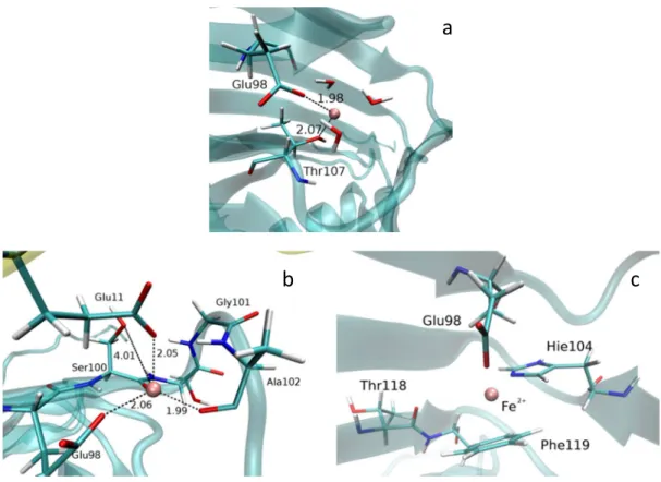
Figure 4. Positions of Fe2+ (pink ball) at the end of RAMD calculations: a) in the neighborhood of Thr107; b) close to Glu11
(from the neighbor subunit – the actually analyzed unit is given in transparent cyan, and neighbor subunit in yellow cartoon representation); c) next to Phe119.
Table 8. Fe2+ positions (and their 'population') at the end of RAMD simulations. For each variant 16 RAMD simulations were
performed. In the cases when the metal ion was expelled from the protein the number of expulsions through tunnel T2 (entrance defined by Glu11, see Figure 4) is given in square brackets
Fe2+ location WT Tyr70Ala Arg80Ala Glu98Gln
Out of protein [near Glu11] 2 [2] 4 [3] 5* 3 [2]
Glu11 neighbor sub. 3 8 4 6
Phe119 and Glu98 0 3 3 6
Thr107 and Glu78 7 0 0 0
Random 4 1 4 1
* in this mutants the water tunnels T1 and T2 cannot be distinguished (i.e. the Arg80 which is border between their entrances does not exist), however Glu11 still plays important role in of the metal ion expulsion
a
Croat. Chem. Acta 2015, 88(3), 297–306 DOI: 10.5562/cca2685 (Figure 4S(D)) where it became accessible to the bulky solvent.
Alternatively, Fe2+ can be expelled into direction of Thr107 (the preferable path in the WT protein) as it is shown in Figure 4S(E) in supplementary material), where pink spheres represents motion of Fe2+ during RAMD. Re-markably, the preferred path for the Fe2+ ion in the WT en-zyme was via a path that leads to Thr107 (Figure 4S(E)), where it rested, coordinated to the side chains of residues Glu78 and Thr107 while coordination to histidines was bro-ken (see Figure 4). Notable, Thr107 has been reported to be crucial for Fe2+ binding.[6] Thr107 and Arg80/Glu78 belong to two adjacent beta strands. The Glu78 and Thr107 side chains interact electrostatically and so has a crucial role in stabilization of these beta strands (see Figure 5S in supple-mentary material).
In the Tyr70Ala and Arg80Ala variants the scenario depicted in Figure 4S was additionally facilitated by the in-creased Glu98 mobility. Upon Tyr70 and Arg80 mutation, respectively, Glu98 was less constrained and it reoriented more easily than in the WT protein leading the Fe2+ ion in a distinct directions. Since during the simulations of the Glu98Gln variant the hydrogen bond between Arg80 and Gln98 was not established (Table 3) Gln98 was more flexi-ble than the Glu in the WT enzyme however, it was still aflexi-ble to guide Fe2+ out of the metal binding site, wherein in about 40 % of the cases metal ions were transferred to Glu11 of the neighbor subunit and in same amount of cases into di-rection of Phe119 (see Table 8 and Figure 4). Interestingly the Fe2+ coordination in the Glu98Gln mutant was not as tight as in the other variants.
In several cases the metal ion was expelled out of the protein. Expulsion of Fe2+ during simulations of the WT protein occurred through the water tunnel T2 (Table 8, see Figure S3 in previously published data[21] for definition of tunnels). As a consequence of lack of the Arg80 side chain a large exit tunnel, composed of exits of both water tunnels, was formed in the Arg80Ala mutant. As the result the Fe2+ ion ended in the bulk water, out of the protein more frequently than it was the case during simulations of the other Dke1 variants.
Data strongly suggest that the exit path for the metal ion is generally via tunnel T2, so passing beside Glu98 and Glu11. This tunnel was broadened during simulations of the all investigated variants. The alternative pathway, tunnel T1, comprising Thr107, was only occasionally used for the Fe2+ expulsion.
(v) Unrestricted Fe
2+Migration
into the Protein
In a complementary approach Fe2+ was placed into the water tunnels in vicinity (up to 2.5 Ǻ) of the Tyr70 side chain oxygen (which is part of both water trafficking tunnels)[5] and subjected to unrestricted MD simula-tions over 16 ns. In the wild type enzyme (apo form), Fe2+ was guided into/close to the 3-His 1-carboxylate metal binding site (defined as a <2.5 Ǻ distance to all 3 histidine coordinated nitrogens) in 8 of 12 cases (see Table 9). In two cases Fe2+ got stuck on the protein sur-face, and in two cases it accommodated in the vicinity of Glu11. When either an Fe2+ ion or both, Fe2+ and sub-strate, were already bound in the binding site, the
Table 9. The position of the metal ion, initially placed in the water trafficking tunnels within 2.5 Ǻ of the Tyr70 residue side chain oxygen, determined after 16 ns of MD simulations (this region is part of both water trafficking tunnels, T1 and T2).
System to which Fe was added Unit A Unit B Unit C Unit D
Position of the Fe dikation at the end of MD simulations
WT CA D A D2
WT** A1 D A S
WT* A1 D A1 S
WT+Fe D2 D2 D D2
WT + ACAC + Fe D D D D
Tyr70Ala D CA D D2
Tyr70Ala * D2 D D E
Arg80Ala E D CA D2
Glu98Gln CA D2 D2 D2
A = Fe2+ in the active site, coordinated with E98 and 3 His(s) ='ideal' position A1 = Fe2+ in the active site, coordinated with E98 and 1 or 2 His
CA = with respect to its initial position Fe2+ moved in the direction of the active site, and ended close to the 'ideal position' (within ca 5 Å of it), but the
coordination with E98 His(s) has not been established OR was established with only one of the 3His ligands D = Fe2+ in the 'ALTERNATIVE' BINDING site, coordinated with E98 AND E11 from neighbour subunit
D1 = Fe2+ almost (close to) the 'ALTERNATIVE' BINDING site, coordinated with either E98 or E11 only
D2 = Fe2+ moved from the initial to the 'ALTERNATIVE' BINDING site direction, but still have not accomodated in it (cordination with neither E11 or E98 was
established)
DOI: 10.5562/cca2685 Croat. Chem. Acta 2015, 88(3), 297–306 other Fe2+ ion predominantly ended at the nonspecific
binding site, bound to Glu11/Glu98 (see Table 9).
Secondary Structure Analysis
In order to quantify effect of the point mutation on the overall protein structure we analyzed their secondary structure before and after each MD simulation using the web server STRIDE. According to the results it seems that the introduced point mutations do not have induced protein refolding, i.e. no significant changes of the protein secondary structure during simulations were noticed, see Table 10.Discussion
The present study clearly speaks in evidence of biphasic binding of the metal ion to Dke1, which was predicted by Leitgeb at al.[7]
According to the results of RAMD simulations, the Fe2+ ion when expelled from the binding site could be trapped at different locations within the enzyme. However, the largest number the RAMD simulations ended close to Glu11 (3 times or (19 %) in WT; 4 in Arg80Ala (25 %); 6 in Glu98Gln (38 %); 8 in Tyr70Ala (50 %), Table 8 and Figure 4b), which can be considered as an alternative, low affinity binding site. The number of Fe2+ trajectories ending out of the protein was the smallest in the case of the Dke1-WT (only two of sixteen), indicating that the Dke1- Fe2+ complex is more stable than the mutated complexes.
In accord with the RAMD simulations the binding free energy calculations (see Table 6) revealed the neighborhood of Glu11 (form the neighbor subunit) as the second most favorable binding site for the Fe2+ ion (after the active site).
Detailed analysis of the metal ion coordination and His104-Glu98-Arg80 interactions during the simulations revealed importance of Glu98 in the ion transport. Further on, Arg80 and Tyr70, through their interactions with Glu98, also influenced the Fe2+ transport.
Results of the long unconstrained MD simulations for the enzyme variants (Table 5) indicated that presence of the metal ion in the active site is the shortest in Arg80 mutant.
Hydrogen bond analyses revealed relocation of the Gln98 side chain of the Glu98Gln variant in direction of the neighbor beta strand during the MD simulations. Such behavior of Gln98 is in favor of the metal ion translocation out of the active site in direction of Glu11 from the neighbor subunit. During the simulations of Tyr70Ala variant guanidine end of Arg80 established strong interaction with Glu98. As a result an ideal trap for the metal ion has been formed between the carboxyl groups of Glu98 and Glu11 from the neighbor unit. Arg80 to Ala mutation caused relocation of Glu11 from neighbor subunit closer to His104, enabling formation of the Glu11(OE1/2) – His104(NE2H/CD2H) H-bond (Table 1), while in the WT protein His104 was strongly interacting with Glu98, which was at the same time often interacting with Arg80, during the simulations. Figure 4S A-D indicate that Glu98, His104, Glu11 (from the neighbor subunit), and Arg80 interplay is crucial for the Fe2+ ion stabilization and transfer. Apparently Fe2+ affinity to bind to the glutamates carboxyl group is very high, since it spent the significant amount of simulations time coordinated either by Glu98 or Glu11, or both.
According to the computational results the metal ion could easier fluctuate between the active site and the alternative binding site next to Glu11 in the mutated (Tyr70Ala, Arg80Ala and Glu98Gln) enzymes than in the wild type. Yet, during the simulations of WT protein with Fe2+ initially placed in the water tunnel, the metal ion most often moved towards Glu11 (in 60 % simulations and in direction of the active site in 25 % simulations, see Table 9).
CONCLUSIONS
From the conducted computational analyses, mecha-nism of the Fe2+ trafficking in and out of the protein was elucidated. Interplay of Glu98, His104, Glu11 (from the neighbor subunit), and Arg80 residues is determined to be the most important for the Fe2+ transport. Also a place for the low affinity binding site is proposed, and it is in the vicinity of the Glu11 residue. Upon the point mutations of the hydrophilic residues Tyr70, Arg80 and Glu98 the metal ion is determined to be generally more flexible, so the role of the specified residues is shown to be important for metal stabilization in the active site.
Acknowledgment. The research was done in Physical Chemistry Department of Ruđer Bošković Institute in Zagreb, and Faculty of Medicine in Osijek. All MD simu lations were done using Isabella computing cluster (http://www.srce.unizg.hr/isabella/) and Croatian National Grid Infrastructure (www.cro-ngi.hr).
Table 10. The mean secondary structures determined for the variants after MD simulations, averaged over all subunits. Content of helices and sheets deviates up to 4 % while content of turns within the proteins deviates up to 8 %
Variant Helix (%) Sheet (%) Turn (%)
WT 7 48 21
Tyr70Ala 7 47 22
Glu98Gln 7 48 21
Croat. Chem. Acta 2015, 88(3), 297–306 DOI: 10.5562/cca2685
REFERENCES
[1] K. J. Waldron, N. J. Robinson, Nat. Rev. Microbiol.
2009, 7, 25.
[2] G. Stranzl, Ph. D. Thesis, Institute of Chemistry, Karl Franzens University Graz, Graz, Austria, 2002. [3] G. Straganz, L. Brecker, H.-J. Weber, W. Steiner, D.
W. Ribbons, Biochem. Biophys. Res. Commun. 2002,
297, 232.
[4] G. Straganz, A. Glieder, L. Brecker, D. Ribbons, W. Steiner, Biochem. J. 2003, 369, 573.
[5] H. Brkić, D. Buongiorno, M. Ramek, G. Straganz, S. Tomić, J. Biol. Inorg. Chem. 2012, 17, 801.
[6] G. D. Straganz, A. R. Diebold, S. Egger, B. Nidetzky, E. I. Solomon, Biochemistry 2010, 49, 996.
[7] S. Leitgeb, G. Straganz, B. Nidetzky, Biochem. J. 2009,
418, 403.
[8] S. Velankar, C. Best, B. Beuth, C. H. Boutselakis, N. Cobley, A. W. Sousa Da Silva, et al., Nucleic Acids Res.
2010, 38, D308.
[9] D. Case, T. Darden, T. Cheatham III, C. Simmerling, J. Wang, R. Duke, et al., AMBER 12, 2012, San Francisco. [10] Y. Duan, C. Wu, S. Chowdhury, M. C. Lee, G. Xiong,
W. Zhang, et al., J. Comput. Chem. 2003, 24, 1999.
[11] J. Wang, R. M. Wolf, J. W. Caldwell, P. A. Kollman, D. A. Case, J. Comput. Chem. 2004, 25, 1157.
[12] H. Brkic, B. Kovacevic, S. Tomic, Mol. Biosyst. 2015,
11, 898.
[13] M. W. Mahoney, W. L. Jorgensen, J. Chem. Phys. 2000, 112, 8910.
[14] J.-P. Ryckaert, G. Ciccotti and H. J. Berendsen, J. Comput. Phys. 1977, 23, 327.
[15] B. R. Miller III, T. D. McGee Jr, J. M. Swails, N. Homeyer, H. Gohlke, A. E. Roitberg, J. Chem. Theory Comput. 2012, 8, 3314.
[16] M. L. Connolly, J. Appl. Crystallogr. 1983, 16, 548.
[17] S. Lüdemann, C. Lounnas, R. Wade, J. Mol. Biol. 2000,
303, 797.
[18] D. R. Roe, T. E. Cheatham III, J. Chem. Theory Comput. 2013, 9, 3084.
[19] M. Heinig and D. Frishman, Nucleic Acids Res. 2004,
32, W500.
[20] W. Humphrey, A. Dalke and K. Schulten, J. Mol. Graphics 1996, 14, 33.
WT
Fe
ion initially placed into the active site
2
30
3
16
Fe
2+placed at the entrance of the water trafficking tunnels
4
6
5
3
6
5
7
16x0.25
RAMD
8
6
APO-enzyme
9
6
Second Fe
2+placed at the water trafficking
10
8
Second Fe
2+placed at the water trafficking and ligand bound
11
30
With bonding parameters
12
16
Tyr70Ala
Fe
2+ion initially placed into the active site
13
30
14
16
Fe
2+placed at the entrance of the water trafficking tunnels
15
5
16
5
17
16x0.25
RAMD
18
6
APO-enzyme
19
30
With bonding parameters
20
16
Arg80Ala
Fe
2+ion initially placed into the active site
21
30
22
16
Fe
2+placed at the entrance of the water trafficking tunnels
23
5
24
16x0.25
RAMD
25
6
APO-enzyme
26
30
With bonding parameters
27
16
Glu98Gln
Fe
2+ion initially placed into the active site
28
30
29
16
Fe
2+placed at the entrance of the water trafficking tunnels
30
5
31
16x0.25
RAMD
32
6
APO-enzyme
33
30
With bonding parameters
Fig. 2S The Glu/Gln98 – Fe2+ distances in one representative subunit (D) during first 30 ns of MD simulations at room
temperature
Fig. 3S Distances between Glu98 and Arg80 (and between His104 and Glu98) in four subunits during the first 15 ns of MD
Fig. 4S Representation of a Fe2+ detachment trajectory. The strands shown in green transparent representations, the amino
acid side chains in colored sticks, and Fe2+ as a pink ball (except in E, where it is represented by yellow sphere). Black
dashed lines represent bonds shorter 2.5 Ǻ.
![Figure is created using VMD. [20]](https://thumb-eu.123doks.com/thumbv2/123dok_br/18139955.326299/3.892.507.779.650.936/figure-is-created-using-vmd.webp)