08 artigo 460
ORIGINAL ARTICLE
1 – Lecturer in Orthopedics at the School of Medicine, University of Marília (UNIMAR), Marília, SP, Brazil. 2 –Titular Professor of Orthopedics and Traumatology, Marília Medical School (FAMEMA), Marília, SP, Brazil. 3 –Physician in the Clinical Staff at Marília University Hospital, Marília, SP, Brazil.
4 –Third-year Residents in the Orthopedics Service, Santa Casa de Misericórdia de Marília, Marília, SP, Brazil. Work performed at the Prof. Dr. Hilário Maldonado Orthopedics Clinic and Marília University Hospital, Marília, SP.
Correspondence: Vitor Barion Castro de Pádua, Av. das Esmeraldas 3023, 17516-000 Marília, SP. E-mail: [email protected] Received for publication: 11/23/2010, accepted for publication: 11/07/2011
COMPARATIVE STUDy OF ACL RECONSTRUCTION WITH ANATOMICAL
POSITIONING OF THE TUNNELS USING THE PATELLAR
TENDON VERSUS HAMSTRING TENDON
Vitor Barion Castro de Pádua1, Hilário Maldonado2, Júlio César Rodrigues Vilela3, Alexandre Ribeira Provenza4, Cleverson Monteiro4, Heleno Cavalcante de Oliveira Neto4
The authors declare that there was no conflict of interest in conducting this work
This article is available online in Portuguese and English at the websites: www.rbo.org.br and www.scielo.br/rbort ABSTRACT
Objective: To compare ACL reconstruction with ana-tomical positioning of the tunnels using the hamstring or patellar tendons. Methods: We prospectively evaluated 52 patients who underwent ACL reconstruction using the Chambat’s technique, with anatomical positioning of the tunnels drilled outside in. They were divided into group A, with 27 patients, using the patellar tendon as a graft, and group B, with 25 patients, using the hamstring. Results: In group A 26 patients were very satisfied or satisfied and 1 unhappy, in group B. 25 patients were very satisfied or satisfied with the procedure (p = 0.990). According to the Lysholm scale, group A had a mean score of 96.11 and group B, 95.32 (p=0.594). In relation to preoperative IKDC, 100% of the patients in group A and 92% of those in group B were IKDC C or D (p = 0.221); in the
assess-ment with a minimum of two-year follow-up, 96% of group A and 92% of group B were IKDC A or B (p = 0.256). The Lachman test, pivot shift, return to sports activities, and the comparative difference in anterior translation (Rolime-terTM)also showed no statistically significant difference. In
group A, 5 patients (18.5%) were unable to kneel on a hard surface, whereas no patient in group B had this complaint. Conclusion: The anterior cruciate ligament reconstruction presents similar results using the hamstring or patellar ten-don with anatomical positioning of the tunnels. Drilling the femoral tunnel outside in is a reproducible and accurate option in the correct placement the femoral tunnel.
Keywords - Anterior Cruciate Ligament; Patellar Liga-ment; Knee/anatomy & histology
INTRODUCTION
Anterior cruciate ligament (ACL) injuries are com-mon in athletes, incapacitating for certain sports ac-tivities and predispose towards meniscal and cartilage lesions that may evolve to arthrosis(1).
Since the studies by Clancy and Dejour, in the 1980s(2,3), and with the development of arthroscopic
techniques, there has been great evolution in ACL treatments.
ACL reconstruction leads to excellent results, ena-bling a return to sports activities, but the problem of rotational instability has still not been solved(4)
.
The patellar tendon was the graft material that was considered to be the gold standard for ACL recons-truction over the last decade(5,6). This tendon and the
flexor tendons are currently the graft materials that are used most(7,8), and each has its advantages and
disadvantages(9,10) .
big-51
THE PATELLAR TENDON HAMSTRING TENDON
gest cause of failure in ACL reconstruction is techni-cal failure, mainly with regard to tunnel positioning. This occurs especially with the femoral tunnel and usually consists of positioning that is too anterior(12).
According to the anatomy of the ACL, its origin is completely on the axial face of the lateral condyle, in its most proximal and posterior portion, below the lateral intercondylar crest(13).
The objective of this study was to compare ACL reconstructions with anatomical positioning of the tunnels, between using quadruple flexor tendons and using the patellar tendon.
MATERIALS AND METHODS
Between January 2007 and June 2008, 76 patients underwent ACL reconstruction according to the Cham-bat technique, with anatomical positioning of the tun-nels, which were both constructed independently and from outside to inside. A quadruple flexor tendon or patellar tendon graft was used, and all the procedures were performed by the same surgeon. Patients pre-senting associated peripheral lesions, those with iterative lesions undergoing revision and those with bilateral lesions were excluded. In addition, contact with some patients was lost, and thus 52 patients remained for prospective evaluation. These were divided in group A, consisting of 27 patients who received the patel-lar tendon as the graft, and group B, consisting of 25 patients who received the flexor tendon.
Group A comprised one female patient and 26 male patients, while group B comprised seven fe-male patients and 18 fe-male patients (p = 0.022). The mean age was 31 years (range: 18-43) in group A and 34 years (range: 21-50) in group B. The mean-time that elapsed between the injury and the surgery was 23 months (range: 1 to 120) in group A and 20 months (range: 2 to 160) in group B (Table 1). All the patients presented ACL lesions that were confirmed by magnetic resonance imaging and by physical examination (Lachman and pivot shift), and
they were reevaluated with a minimum follow-up of two years (range: 2 – 3.5 years ). With regard to the trauma mechanism, 23 patients (85.1%) in group A and 18 (72.0%) in group B had suffered their injury through playing soccer (Table 2).
Table 1 – Patient distribution in the groups.
Group A Group B
p
PT* FT**
Number of patients 27 25
Sex (Male/Female) 26/1 18/7 p = 0.022
Age 31 (18 - 43) 34 (21 - 50)
Time elapsed between injury
and surgery (months) 23 (1 - 120) 20 (2 - 160)
* PT: patellar tendon ** FT: flexor tendons
Table 2 – Injuries according to type of sport practiced.
Sport Group A Group B
n % n %
Soccer 23 85.18 18 72.00 Jiu-Jitsu 3 11.11
Basketball 1 3.70
Moto 2 8.00
Volleyball 2 8.00
Running 2 8.00
Taekwondo 1 4.00
Total 27 25
SURGICAL TECHNIqUE
The reconstruction was done using the Chambat technique(14) for the patellar tendon. By means of an
anterior incision above the patellar tendon, its central third (of 1 cm in thickness) was harvested together with a block of patellar bone of 9 X 20 mm and an-other from the tibia of trapezoidal shape, measuring 11 X 25 mm (Figure 1).
After performing arthroscopy and treatment of as-sociated lesions, the tunnels were constructed inde-pendently and from outside to inside, starting from the femur and using the Chambat (Phusis) femoral guide. This was brought in through the anteromedial portal and was attached to the most proximal edge of the axial face of the lateral condyle. By means of a 1.5 cm lateral access above the lateral epicondyle, the guidewire was introduced from outside to inside. It emerged intra-articularly, perpendicular to the axial face of the lateral condyle, at the anatomical location of the ACL on the femur.
By means of this guidewire, the initial hole was drilled was outside to inside, using a 6 mm cannulated bit, and this was progressively enlarged to a size of 10 mm (Figure 2).
tun-Figure 1 – Graft from patellar tendon: patellar cylinder (BP) and tibial cylinder (BT).
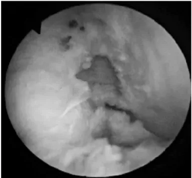
Figure 2 – Femoral tunnel at anatomical location.
Figure 3 – Flexor tendons with tibial insertion maintained. nel, and the trapezoidal tibial baguette was destined
for the femoral tunnel.
While the patellar baguette was being pulled through the tibial tunnel, the trapezoidal tibial ba-guette was attached to the femur, from outside to inside in “press fit” form. Following this, tibial fixa-tion was performed at 30 degrees of flexion, using an interference screw.
In the case of the flexor tendons, the tunnels were made in the same manner and with the same guides, up to a size of 8 mm or 9 mm, depending on the size of the quadruple graft from the flexor tendons. These were harvested by means of a small access above their tibial insertion, which was preserved (Figure 3).
The prepared graft was introduced from distal to
proximal, and was fixed using a first interference screw in the tibia, which thus generated a double fixa-tion, since the tibial insertion was maintained. Follow-ing this, it was introduced from outside to inside at 30 degrees of flexion for fixation in the femur, also using an interference screw.
No suction drain was used. The patients were dis-charged on the following day. Rehabilitation was con-ducted similarly in the two groups, starting after
53
THE PATELLAR TENDON HAMSTRING TENDON
pital discharge, with crutches and progressive partial weight-bearing for two weeks, achieving full range of motion after four weeks. They returned to day-to-day activities between the 4th and 12th weeks and started to
run again, with proprioception. They returned to their activities six months after the operation.
Postoperative evaluation
The patients included in this study were evaluated after a minimum follow-up of two years. The range of motion, Lachman test (hard, delayed hard or soft) and pivot shift (negative, +, ++, or +++) were assessed. The comparative differential anterior translation was measured by means of a RolimeterTM (Aircast®). For
the functional evaluation, the IKDC index, Lysholm scale, return to sports activities and patient satisfac-tion were used.
Data analysis
The results relating to qualitative variables were summarized by means of tables, absolute frequencies and percentages. Those relating to numerical varia-bles were summarized by means of tavaria-bles, means and standard deviations, minimum values and maximum values. Comparison between the patellar tendon tech-nique (A) and the flexor tendon techtech-nique (B), ac-cording to the RolimeterTM or Lysholm scale, was
done by means of Student’s t test for independent samples. Associations shown by techniques A and B with the variables of sex, pivot shift, Lachman, re-turn to activities, satisfaction and knee mobility were evaluated by means of Fisher’s exact test. Associa-tions shown by techniques A and B with meniscal lesions were investigated by means of the chi-square test, and with the IKDC by means of the G test. The level of 5% probability for rejection of the nullity hypothesis was used(15).
RESULTS
Seven medial meniscal lesions, five lateral menis-cal lesions and three lesions in both menisci were found in group A, while in group B there were six medial meniscal lesions and five lateral meniscal sions. Sutures were made in one medial meniscal le-sion and one lateral meniscal lele-sion in group A, and three medial meniscal lesions in group B (p = 0.609) (Table 3).
Regarding the range of motion, two patients (7.4%) in group A presented a deficit of less than 5º of ex-tension. In group B, two patients (8.0%) presented a
deficit of more than 5º of flexion, and one of them also presented a deficit of more than 5º of extension (p = 0.990).
In group A, two patients (7.4%) presented a de-layed hard result in the Lachman test and pivot shift +, while in group B there were six patients (24.0%) with delayed hard Lachman (p = 0.134) and four (16,0%) with pivot shift +, without any statistically significant difference (Table 4).
According to the Lysholm scale, group A attained a mean of 96.11 (SD 4.44; minimum 82) in the final evaluation, and group B attained a mean of 95.32 (SD 6.12; minimum 72) (p = 0.594).
The preoperative IKDC index showed that 100% of the patients in group A and 92% in group B were IKDC C or D (p = 0.221). In the postoperative eval-uation after at least two years of follow-up, 96% of group A and 92% of group B were IKDC A or B (p = 0.256) (Table 5), which was not statistically different.
The comparative differential in anterior translation according to the RolimeterTM (Aircast®) was 5.8 mm
(range: 4-7) in group A and 6.2 mm (range: 4-9) in group B before the operation. This evolved to 0.81
Table 3 – Meniscal lesions.
Medial Lateral Medial and Lateral
Medial suture
Lateral
suture p
n % n % n % n % n %
Group A 7 25.9 5 18.5 3 11.1 1 3.7 1 3.7 0.609
Group B 6 24.0 5 20.0 3 12.0
Table 4 – Lachman and pivot shift tests after the operation.
Lachman Pivot shift
Hard Delayed
hard Soft 0 + ++ +++
Group A 25 (92.6%) 2 (7.4%) 0 25 (92.6%) 2 (7.4%) 0 0
Group B 19 (76.0%) 6 (24.0%) 0 21 (84.0%) 4 (16.0%) 0 0
p = 0.134 p = 0.592
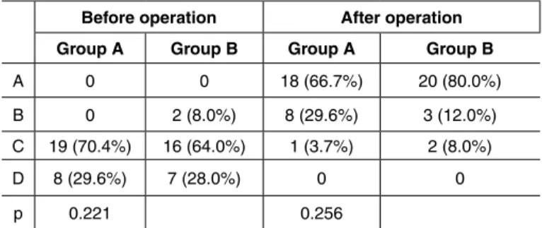
Table 5 - International Knee Documentation Committee (IKDC)
Before operation After operation
Group A Group B Group A Group B
A 0 0 18 (66.7%) 20 (80.0%)
B 0 2 (8.0%) 8 (29.6%) 3 (12.0%)
C 19 (70.4%) 16 (64.0%) 1 (3.7%) 2 (8.0%)
D 8 (29.6%) 7 (28.0%) 0 0
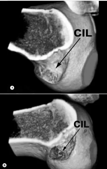
Figure 4 – Computed tomography scan showing the location of the femoral tunnel, posterior to the lateral intercondylar crest. (A) Before the operation. (B) After the operation.
A
B
mm (range: 0-3) (p = 0.314) and 1.12 mm (range: 0-4) (p = 0.289), respectively in groups A and B, also without any statistical difference (Table 6).
In group A, three patients (11.1%) changed their activities: one with a reduction in level and two with replacement activities. In group B, four patients (16.0%) changed their activities: three with reduc-tions and one with a replacement activity (p = 0.41). The patients were asked about their degree of sat-isfaction (very satisfied, satisfied, dissatisfied or very dissatisfied). In the patellar tendon group, 26 (96.3%) were very satisfied (24) or satisfied (2), while one patient was dissatisfied. In the flexor tendon group, all the patients were either very satisfied (20) or satisfied (5) (p = 0.99) (Table 7).
In group A, three patients (11.1%) presented com-plications: one case of neuropraxia of the sciatic nerve from using the tourniquet, which took around one year to recover from; one case of Cyclops syndrome that was operated nine months after the reconstruction; and one patient with anterior pain when running, and this was the dissatisfied patient. One patient in group B (4.0%) presented the complication of Sudeck’s at-rophy, with limitations on extension and flexion.
In relation to graft harvesting, five patients (18.5%) in the patellar tendon group were unable to kneel on the operated knee, whereas no one in the flexor group presented this type of complaint (p = 0.052).
One patient in group A suffered a new rupture two years after the reconstructed, and one patient in group B, 18 months after the surgery. In both cases, a new traumatic episode of spraining occurred.
Drilling of the femoral tunnel from outside to in-side reached the anatomical site of the ACL below the lateral intercondylar crest, as confirmed by tomogra-phy (Figure 4).
DISCUSSION
Over the last two decades, ACL reconstruction using the patellar tendon has been considered to be the gold standard(6). However, because of morbidity caused by
graft harvesting, there has been increasing use of the flexor tendons, which has given rise to a series of com-parative studies between the two types of graft(16-18).
Some authors have reported that there is a slight stability advantage in using the patellar tendon rather than the flexor tendons(19). However, a
meta-analy-sis by Biau et al(9) did not find any evidence that one
technique was superior to the other. It only found that harvesting the patellar tendon led to greater morbidity.
Some studies have shown that using the flexor tendons produced cases of slackness, in comparison with using the patellar tendon, because of the fixation
Table 7 – Level of satisfaction.
Very
satis-fied Satisfied Dissatisfied
Very
dissat-isfied p
n % N % n % n %
Group A 24 88.9 2 7.4 1 3.7 0 - p = 0.999
Group B 20 80.0 5 20.0 0 - 0
-Table 6 – Rolimeter™ (Aircast ®): comparison before and after the operation.
Before operation After operation
t test p Mean SD* (Min-Max) Mean SD* (Min-Max)
Group A 5.88 0.97 (4 - 7) 0.81 0.92 (0 - 3) 0.314
Group B 6.20 1.22 (4 - 9) 1.12 1.12 (0 - 4) 0.289
55
THE PATELLAR TENDON HAMSTRING TENDON
methods(8,20). Evolution of these cases made the results
similar. In another meta-analysis, Podromos et al(21)
confirmed that the superiority of the patellar tendon was due to failure of flexor tendon fixation, and that using suspended fixation led to the best results.
In our study, to attain secure fixation with the fle-xor tendons, we maintained the distal insertion in the tibia by means of an interference screw, thus leading to double tibial fixation. In the femur, because of the anatomical position of the tunnel (constructed from outside to inside), the graft made a curve of almost 90o,
thereby diminishing the traction force. In the cases with the patellar tendon, femoral fixation was achieved by means of the “press fit” technique: not only is this bio-logical and safe(22,23), but also it avoids screw deviation
or breakage of the posterior cortical boner(17).
Although some authors have reported that patients have returned to a higher activity level with the patellar tendon(24), Pinczewski(25) did not find any difference in
results between the two types of graft, in series with ten years of follow-up. However, because of the lower morbidity from harvesting the flexor tendons, and be-cause of the fewer radiographic abnormalities found in this group, these tendons were indicated as the first choice. This was in agreement with Prodromos(7), who
considered that ACL reconstruction using the flexor tendons was the current gold standard.
Our study did not show any significant differ-ence in comparisons of ACL reconstruction between using the patellar tendon and using the flexor ten-dons, according to the pivot shift, Lachman, IKDC, Lysholm, return-to-activity and patient satisfaction scales. Although our study was not randomized or ho-mogenous, its findings were in agreement with previous studies(8,18,25,26).
Persistent anterior pain has been considered by some authors to be a consequence of harvesting the patellar tendon(8,19,27). However, we only found one
patient in the patellar tendon group who presented this complaint, which was not significant. We agree with Shelbourne(6) that the loss of full extension might be the
cause of the anterior pain, since our patient presented an extension deficit. The morbidity that we found from harvesting the patellar tendon was that five patients (18.5%) complained that they were unable to kneel on hard surfaces, whereas none of the patients in group B presented this complaint.
In a study on evidence-based medicine, Spindler(28)
found that nine randomized studies comparing the patellar tendon and flexor tendons did not show any
significant difference between the groups, except for difficulty in kneeling in the patellar tendon group, thus concluding that the type of graft was not the main de-terminant of success in the operation.
Today, anatomical reconstruction of the ACL is sought, in an attempt to reproduce its structural and mechanical properties(29). In these attempts to come
close to the anatomy and seek better results in relation to rotational instability, double-band ACL reconstruc-tion has been greatly studied(29-31). This appears to be
promising, but still lacks results. The greatest contribu-tion of these studies has been the great attencontribu-tion given to the anatomy of the ACL, with raised awareness of the need for correct positioning of the tunnels(11).
Tunnel positioning is more important than the type of graft and fixation, and doing this correctly leads to better results and a lower failure rate(12,32).
Correct positioning in the femur can be attained by means of drilling through the medial accessory por-tal(33) or by drilling from outside to inside, following
the Chambat technique(14), which was our choice.
With independent drilling, from outside to inside, we consider that it is easier to achieve correct loca-tion in the femur(34,35), below the lateral intercondylar
crest(36), without difficulty. This was confirmed by the
tomography. In this manner, control over the rota-tional instability and the final funcrota-tional result were improved.
Studies comparing ACL reconstruction from outside to inside and from inside to outside have been conducted(37,38),
without showing any significant difference. However, O’Neil(17) found that a greater number of patients with
drilling from outside to inside returned to a higher com-petitive level, and that there was a greater percentage of patients with comparative KT-2000 less than 3 mm.
Drilling from outside to inside has been criticized because of the large lateral incision. However, using the Chambat technique, it is not greater than 2 cm, like in transverse fixation.
Our sample was neither homogenous nor randomi-zed, with a significantly higher percentage of female patients using flexor tendons. This occurred because we initially indicated flexor tendons for women because of the smaller incision and lower sports demands, which thus represents a bias in our study.
the patellar tendon to avoid greater medial slackness. We agree that independent of the type of graft and fixation, tunnel positioning is the factor leading to the best results(14,18,30,31-34).
CONCLUSION
ACL reconstruction with anatomical positioning of the tunnels presents similar results using flexor ten-dons or the patellar tendon as the graft. Independent
drilling of the femoral tunnel, from outside to inside, is a reproducible and precise option for correct posi-tioning of the femoral tunnel.
ACKNOWLEDGEMENT
To Dr. Sebastião Marcos Ribeiro de Carvalho, of the Extension Project (DPE/FFC), Unesp, Marília, SP, for help with statistics, which was an important collaboration for carrying out this work.
REFERENCES
1. Bray RC, Dandy DJ. Meniscal lesions and chronic anterior cruciate ligament-deficiency. Meniscal tears occurring before and after reconstruction. J Bone Joint Surg Br. 1989;71(1):128-30.
2. Clancy WG Jr, Nelson DA, Reider B, Narechania RG. Anterior cruciate ligament reconstruction using one-third of the patellar ligament, augmented by extra-articular tendon transfers. J Bone Joint Surg Am. 1982;64(3):352-9. 3. Dejour H. [Results of the treatment of anterior laxity of the knee]. Rev Chir
Orthop Reparatrice Appar Mot. 1983;69(4):255-302.
4. Tashman S, Collon D, Anderson K, Kolowich P, Anderst W. Abnormal rotational knee motion during running after anterior cruciate ligament reconstruction. Am J Sports Med. 2004;32(4):975-83.
5. Fu FH, Schulte KR. Anterior cruciate ligament surgery 1996. State of the art? Clin Orthop Relat Res. 1996;(325):19-24.
6. Shelbourne KD, Gray T. Results of anterior cruciate ligament reconstruction based on meniscus and articular cartilage status at the time of surgery. Five- to fifteen-year evaluations. Am J Sports Med. 2000;28(4):446-52.
7. Prodromos CC, Han YS, Keller BL, Bolyard RJ. Stability results of hamstring anterior cruciate ligament reconstruction at 2- to 8-year follow-up. Arthroscopy. 2005;21(2):138-46.
8. Harilainen A, Linko E, Sandelin J. Randomized prospective study of ACL recons-truction with interference screw fixation in patellar tendon autografts versus femoral metal plate suspension and tibial post fixation in hamstring tendon autografts: 5-year clinical and radiological follow-up results. Knee Surg Sports Traumatol Arthrosc. 2006;14(6):517-28.
9. Biau DJ, Tournoux C, Katsahian S, Schranz PJ, Nizard RS. Bone-patellar ten-don-bone autografts versus hamstring autografts for reconstruction of anterior cruciate ligament: meta-analysis. BMJ. 2006;332(7548):995-1001.
10. Poolman RW, Abouali JA, Conter HJ, Bhandari M. Overlapping systematic reviews of anterior cruciate ligament reconstruction comparing hamstring auto-graft with bone-patellar tendon-bone autoauto-graft: why are they different? J Bone Joint Surg Am. 2007;89(7):1542-52.
11. Steiner M. Anatomic single-bundle ACL reconstruction. Sports Med Arthrosc. 2009;17(4):247-51.
12. Harner CD, Poehling GG. Double bundle or double trouble? Arthroscopy. 2004;20(10):1013-4.
13. Giron F, Cuomo P, Aglietti P, Bull AM, Amis AA. Femoral attachment of the ante-rior cruciate ligament. Knee Surg Sports Traumatol Arthrosc. 2006;14(3):250-6. 14. Garofalo R, Mouhsine E, Chambat P, Siegrist O. Anatomic anterior cruciate
liga-ment reconstruction: the two-incision technique. Knee Surg Sports Traumatol Arthrosc. 2006;14(6):510-6.
15. Armitage P, Berry G. Estadística para la investigación biomédica. 3a.ed. Ma-drid: Harcourt Brace; 1997.
16. Matsumoto A, Yoshiya S, Muratsu H, Yagi M, Iwasaki Y, Kurosaka M, et al. A comparison of bone-patellar tendon-bone and bone-hamstring tendon-bone autografts for anterior cruciate ligament reconstruction. Am J Sports Med. 2006;34(2):213-9.
17. O’Neill DB. Arthroscopically assisted reconstruction of the anterior cruciate ligament. A prospective randomized analysis of three techniques. J Bone Joint Surg Am. 1996;78(6):803-13.
18. Shaieb MD, Kan DM, Chang SK, Marumoto JM, Richardson AB. A prospective randomized comparison of patellar tendon versus semitendinosus and gracilis tendon autografts for anterior cruciate ligament reconstruction. Am J Sports Med. 2002;30(2):214-20.
19. Eriksson K, Anderberg P, Hamberg P, Löfgren AC, Bredenberg M, West-man I, et al. A comparison of quadruple semitendinosus and patellar tendon grafts in reconstruction of the anterior cruciate ligament. J Bone Joint Surg Br. 2001;83(3):348-54.
20. Milano G, Mulas PD, Ziranu F, Piras S, Manunta A, Fabbriciani C. Comparison
between different femoral fixation devices for ACL reconstruction with doubled ha-mstring tendon graft: a biomechanical analysis. Arthroscopy. 2006;22(6):660-8.
21. Prodromos CC, Joyce BT, Shi K, Keller BL. A meta-analysis of stability after anterior cruciate ligament reconstruction as a function of hamstring versus patellar tendon graft and fixation type. Arthroscopy. 2005;21(10):1202. 22. Silva JLV, Tavares Filho GS, Namba MM, Pereira Filho FA, Barbosa M, Albano
M, et al: Estudo biomecânico, “in vitro”, em ovinos, da fixação femoral do tendão patelar na reconstrução do ACL: comparação entre parafusos metálicos de interferência e a fixação sob pressão com bloco ósseo cônico. Rev Bras Ortop. 2003;38(7):400-9.
23. Boszotta H. Arthroscopic reconstruction of anterior cruciate ligament using BTB patellar ligament in the press-fit technique. Surg Technol Int. 2003;11:249-53. 24. Laxdal G, Sernert N, Ejerhed L, Karlsson J, Kartus JT. A prospective com-parison of bone-patellar tendon-bone and hamstring tendon grafts for anterior cruciate ligament reconstruction in male patients. Knee Surg Sports Traumatol Arthrosc. 2007;15(2):115-25.
25. Pinczewski LA, Lyman J, Salmon LJ, Russell VJ, Roe J, Linklater J. A 10-year comparison of anterior cruciate ligament reconstructions with hamstring tendon and patellar tendon autograft: a controlled, prospective trial. Am J Sports Med. 2007;35(4):564-74
26. Jansson KA, Linko E, Sandelin J, Harilainen A. A prospective randomized study of patellar versus hamstring tendon autografts for anterior cruciate ligament reconstruction. Am J Sports Med. 2003;31(1):12-8.
27. Yunes M, Richmond JC, Engels EA, Pinczewski LA. Patellar versus hamstring tendons in anterior cruciate ligament reconstruction: A meta-analysis. Arthros-copy. 2001;17(3):248-257.
28. Spindler KP, Kuhn JE, Freedman KB, Matthews CE, Dittus RS, Harrell FE Jr. Anterior cruciate ligament reconstruction autograft choice: bone-tendon-bone versus hamstring: does it really matter? A systematic review. Am J Sports Med. 2004;32(8):1986-95.
29. Zantop T, Diermann N, Schumacher T, Schanz S, Fu FH, Petersen W. Anatomi-cal and nonanatomiAnatomi-cal double-bundle anterior cruciate ligament reconstruction: importance of femoral tunnel location on knee kinematics. Am J Sports Med. 2008;36(4):678-85.
30. Sonnery-Cottet B, Chambat P. Anatomic double bundle: a new concept inante-rior cruciate ligament reconstruction using the quadriceps tendon. Arthroscopy. 2006;22(11):1249.e1-4.
31. Yasuda K, Kondo E, Ichiyama H, Kitamura N, Tanabe Y, Tohyama H, et al. Anatomic reconstruction of the anteromedial and posterolateral bundles of the anterior cruciate ligament using hamstring tendon grafts. Arthroscopy. 2004;20(10):1015-25.
32. Fox JA, Nedeff DD, Bach Jr BR, Spindler KP. Anterior cruciate ligament recons-truction with patellar autograft tendon. Clin Orthop Relat Res. 2002;(402):53-63. 33. Zantop T, Kubo S, Petersen W, Musahl V, Fu FH. Current techniques in
ana-tomic anterior cruciate ligament reconstruction. Arthroscopy. 2007;23(9):938-47. 34. Domit Filho M, Monte APC, Nagai M, Ribeiro MV, Maciel LG. Estudo de posic-ionamento do enxerto na substituição do ligamento cruzado anterior. Rev Bras Ortop. 2002;37(4):141-50.
35. Khalfayan EE, Sharkey PF, Alexander AH, Bruckner JD, Bynum EB. The rela-tionship between tunnel placement and clinical results after anterior cruciate ligament reconstruction. Am J Sports Med. 1996;24(3):335-41.
36. Ferretti M, Ekdahl M, Shen W, Fu FH.Osseous landmarks of the femoral at-tachment of the anterior cruciate ligament: an anatomic study. Arthroscopy. 2007;23(11):1218-25.
37. Brandsson S, Faxén E, Eriksson BI, Swärd L, Lundin O, Karlsson J. Recons-truction of the anterior cruciate ligament: comparison of outside-in and all-inside techniques. Br J Sports Med. 1999;33(1):42-5.