Article
Comparison of Thyroglobulin Measurements Using Three
Different Immunoassay Kits: A BRAMHS Tg-Plus RIA
Kit, a BRAMHS hTg Sensitive Kryptor Kit, and a
Beckman Coulter ACCESS Immunoassay Kit
Mijin Kim1, Min Ji Jeon1, Won Gu Kim1, Jong Jin Lee2, Jin-Sook Ryu2, Eun-Jung Cho3, Dae-Hyun Ko3,
Woochang Lee3, Sail Chun3, Won-Ki Min3, Tae Yong Kim1, Young Kee Shong1, Won Bae Kim1
Departments of 1Internal Medicine, 2Nuclear Medicine, 3Laboratory Medicine, Asan Medical Center, University of Ulsan
College of Medicine, Seoul, Korea
Background: Second-generation thyroglobulin immunometric assays (Tg-IMAs) have been developed with improved sensitivi-ty. Our aim was to compare the diagnostic value of Tg-IMA measurements using a Kryptor (BRAHMS AG) kit (Tg-K) and an ACCESS (Beckman Coulter) kit (Tg-A) with that of the first-generation Tg measurement using a Tg-plus (BRAHMS AG) kit (Tg+).
Methods: We enrolled 82 differentiated thyroid cancer patients who underwent total thyroidectomy with radioactive iodine rem-nant ablation and who underwent diagnostic whole body scan using recombirem-nant human thyroid stimulating hormone (rhTSH). The Tg+, Tg-K, and Tg-A were measured before rhTSH administration during levothyroxine treatment (suppressed Tg) from the same sample. Serum Tg+ was measured after rhTSH stimulation (stimulated Tg).
Results: Suppressed Tg+ was more significantly correlated with suppressed Tg-K (R2=0.919, P<0.001) than with suppressed Tg-A (R2=0.536, P<0.001). The optimal cut-off values of suppressed Tg+, Tg-K, and Tg-A for predicting stimulated Tg+ of 1 ng/mL were 0.3, 0.2, and 0.2 ng/mL, respectively. The sensitivity, specificity, and accuracy of suppressed Tg+ were 67%, 100%, and 90%, respectively; those of suppressed Tg-K were 83%, 90%, and 88%; those of suppressed Tg-A were 96%, 82%, and 87%, respectively. The positive predictive and negative predictive values of Tg+ were 100% and 87%, respectively; those of Tg-K were 79% and 92%; and those of Tg-A were 73% and 98%.
Conclusion: We could not clearly demonstrate which kit had better diagnostic performance after comparison of first-generation Tg measurements with Tg-IMA measurements. Also, there were kit-to-kit variations between Tg-IMA kits. Suppressed Tg mea-sured by Tg-IMA was insufficient to completely substitute for a stimulated Tg measurement.
Keywords: Differentiated thyroid cancer; Thyroglobulin; Anti-thyroglobulin; Immunoassay
Received: 12 April 2016, Revised: 9 June 2016, Accepted: 5 July 2016
Corresponding author: Min Ji Jeon
Division of Endocrinology and Metabolism, Department of Internal Medicine, Asan Medical Center, University of Ulsan College of Medicine, 88 Olympic-ro 43-gil, Songpa-gu, Seoul 05505, Korea
Tel: +82-2-3010-1317, Fax: +82-2-3010-6962, E-mail: [email protected]
Copyright © 2016 Korean Endocrine Society
INTRODUCTION
Differentiated thyroid cancer (DTC) has a favorable prognosis with an 85% 10-year survival rate after primary treatment [1]. DTC patients need lifelong monitoring for recurrence of dis-ease because it can occur at any time during follow-up periods [2,3]. Serum thyroglobulin (Tg) is an useful biochemical tumor marker for detecting persistent or recurrent DTC [4,5]. Because the source of Tg is both normal remnant thyroid tissue and re-sidual cancer tissue, thyroid stimulating hormone (TSH) influ-ences the interpretation of serum Tg concentration. Serum Tg values during TSH suppression therapy (suppressed Tg) are not sensitive enough to detect small amounts of thyroid tissue or small changes in thyroid tissue. Serum Tg measurements dur-ing TSH stimulation (stimulated Tg) should be performed to maximize the diagnostic sensitivity with a negative predictive value (NPV) of 99% in absence of anti-thyroglobulin antibody (TgAb) [6,7].
Stimulated Tg should be evaluated 4 weeks after cessation of thyroid hormone or 48 to 72 hours after recombinant human TSH (rhTSH) intramuscular injection [8-10]. Cessation of thy-roid hormone treatments may induce severe hypothythy-roidism, and rhTSH injections may induce high cost or inconvenience due to frequent clinic visits [9,11].
Second-generation Tg immunometric assays (Tg-IMAs) with improved functional sensitivity (less than 0.1 ng/mL) have been newly developed and are now commercially available [12,13]. These assays show high NPVs, so it has been reported that they might substitute for stimulated Tg measurements in DTC patients with low recurrence risk [3,14,15].
The aim of the current study was to compare the diagnostic value of Tg-IMA measurements using a BRAMHS hTg sensi-tive Kryptor (BRAHMS AG, Henningsdorf, Germany) kit (Tg-K) and a Beckman Coulter ACCESS immunoassay (Beckman Coulter, Brea, CA, USA) kit (Tg-A) with the first-generation Tg measurements using a BRAMHS Tg-plus RIA (BRAHMS AG, Henningsdorf, Germany) kit (Tg+). Additionally, we as-sessed whether suppressed Tg-K or suppressed Tg-A measure-ments can obviate the need for stimulated Tg measurement.
METHODS
Patients
This study included a total of 82 DTC patients who underwent total thyroidectomy with radioactive iodine remnant ablation and who underwent diagnostic whole body scan using rhTSH
between March 2015 and November 2015 at a single center. Serum suppressed Tg and TgAb were measured using the same sample from each patient during levothyroxine suppression therapy by three different immunoassay kits: Tg+, TgAb+; Tg-K, TgAb-K; Tg-A, TgAb-A. Stimulated Tg+ was measured 24 hours after the second injection of 0.9 mg rhTSH, and all study samples had TSH values higher than 30 μU/mL. One patient with missing Tg-A was excluded in the final Tg-A analysis. Our study protocol was approved by the Institutional Review Board at Asan Medical Center, Seoul, Korea.
Serum Tg and TgAb measurement
Tg+ was measured using the BRAMHS Tg-plus RIA, a first-generation immunoassay kit. The functional sensitivity of Tg-plus kit was 0.2 ng/mL, and the analytical sensitivity was 0.08 ng/mL. The coefficients of variation (CV) within- and between assays were 1.5% to 5.6% and 2.2% to 9.9%, respectively. TgAb+ measurements were performed using the BRAHMS anti-Tg RIA kit (BRAHMS AG, Henningsdorf, Germany) with within- and between assay CVs of 2.0% to 7.5% and 3.1% to 5.5%, respectively. The status of TgAb+ was defined as posi-tive when the value of TgAb+ was higher than 60 U/mL.
Tg-K was measured by Tg-IMA using the BRAMHS hTg sensitive Kryptor kit, with a functional sensitivity of 0.15 ng/ mL and an analytical sensitivity of 0.09 ng/mL. The CV of Tg-K was 4.5% at low concentrations and 2.8% at high con-centrations. TgAb-K was measured using a BRAHMS anti-Tgn Kryptor kit (BRAHMS AG). The CV of TgAb-K was 6.0% at low concentrations and 4.2% at high concentrations. TgAb-K positivity was defined when the value of TgAb-K was higher than 33 U/mL.
Tg-A was measured using the Beckman Coulter ACCESS immunoassay kit, with functional and analytical sensitivities of 0.1 and 0.01 ng/mL, respectively. Within- and between Tg-A CVs were 1.3% to 2.7% and 1.7% to 4.9%, respectively. Serum TgAb was measured using a Beckman Access Antibody II Cal-ibrators (Beckman Coulter, Brea, CA, USA) and was named TgAb-A. TgAb-A positivity was defined when the value of TgAb-A was higher than 4 U/mL.
Statistical analysis
asso-ciations between suppressed Tg and stimulated Tg from the samples with Tg values higher than the analytical sensitivity of each kit. Receiver operating characteristics (ROC) curve analy-sis was performed to evaluate the optimal cut-off values of suppressed Tg+, Tg-K, and Tg-A for predicting a stimulated Tg+ of 1.0 ng/mL. Sensitivity, specificity, and accuracy were defined as ‘true positive/(true positive+false negative),’ ‘true negative/(true negative+false positive),’ and ‘(true positive+ true negative)/total,’ respectively. Positive predictive value (PPV) and NPV were defined as ‘true positive/(true positive+ false positive)’ and ‘true negative/(true negative+false nega-tive),’ respectively. Only P values less than 0.05 were consid-ered statistically significant. Statistical analysis was conducted using R version 3.10 (R Foundation for Statistical Computing, Vienna, Austria).
RESULTS
Baseline characteristics
Of the 82 patients enrolled in our study, 24 (29%) were male. The mean age of the study patients was 51.7 years, and the mean primary tumor size was 1.69 cm. According to the TNM (tumor, lymph node, metastasis) classification system of the 7th edition of the American Joint Committee on Cancer and the Union for International Cancer Control, except for three pa-tients whose histologic findings were unknown, 15 papa-tients (19%) were N0, 34 (43%) were N1a, and 30 (38%) were N1b; 59 (75%) had extrathyroidal extension. Distant metastasis was detected in three patients at the time of initial diagnosis.
Serum TgAb using three different immunoassay kits TgAb+, TgAb-K, and TgAb-A were positive in four (5%), eight (10%), and five (6%) of the 82 patients, respectively. All four TgAb+ positive patients were also positive for TgAb-K and TgAb-A. The values of TgAb+ for the four patients who were positive for TgAb-K but negative for TgAb+ were 25.7, 25.1, 58.8, and 17.1 U/mL. The value of TgAb+ for the one pa-tient who was positive for TgAb-A but negative for TgAb+ was 40.9 U/mL (Table 1).
Comparison of suppressed Tg+ with suppressed Tg-K and suppressed Tg-A in TgAb negative patients
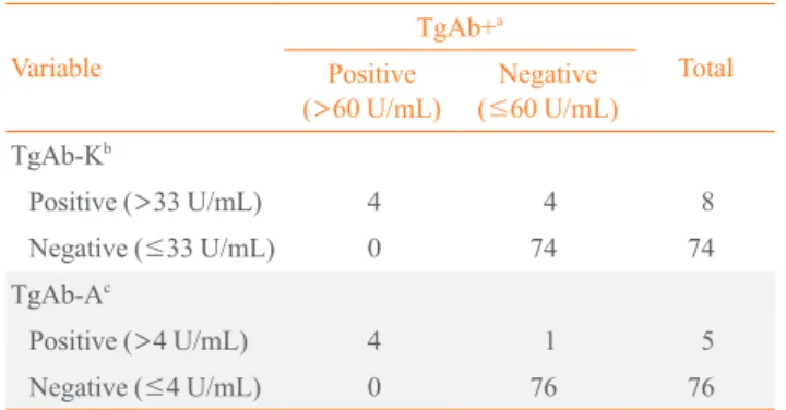
Suppressed Tg+ was highly correlated with suppressed Tg-K (R2=
0.919, P<0.001). As suppressed Tg+ increased by 1 ng/mL,
suppressed Tg-K increased by 0.93 ng/mL (Fig. 1A). Sup-pressed Tg+ was less correlated with supSup-pressed Tg-A than
with Tg-K and showed a linear relationship with a low concor-dance (R2=
0.536, P<0.001) (Fig. 1B).
Comparison of stimulated Tg+ with suppressed Tgs from different kits in TgAb negative patients
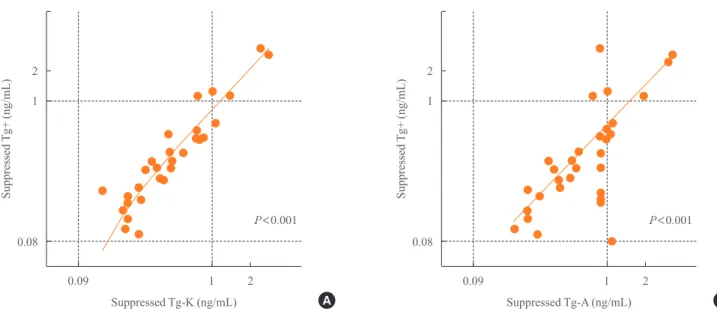
When we compared stimulated Tg+ with suppressed Tg+ in TgAb+ negative patients, there was a linear correlation (R2= 0.316) (Fig. 2A). There was also a linear correlation between stimulated Tg+ and suppressed Tg-K in TgAb-K negative pa-tients (R2=
0.214) (Fig. 2B). However, stimulated Tg+ was not associated with suppressed Tg-A (Fig. 2C).
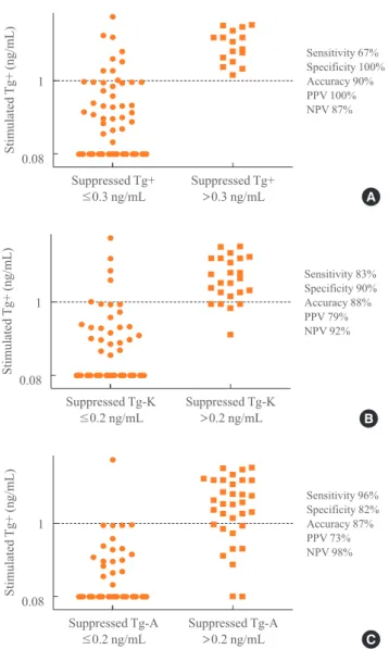
Sensitivity, specificity, and accuracy of suppressed Tg+, Tg-K, and Tg-A in predicting positivity of stimulated Tg+ We evaluated the sensitivity, specificity, and accuracy of sup-pressed Tg according to immunoassay kits for predicting posi-tivity of stimulated Tg. We performed ROC analysis to deter-mine the optimal cut-off values of suppressed Tg+, Tg-K, and Tg-A for predicting a stimulated Tg+ of 1 ng/mL. The appro-priate cut-off value of suppressed Tg+ was 0.3 ng/mL by ROC analysis, and the area under the curve (AUC) was 0.89 (P=
0.001). The most reasonable cut-off value of suppressed Tg-K and Tg-A was 0.2 ng/mL (AUC=0.92, P<0.001; AUC=0.92, P<0.001, respectively).
When we set the cut-off value of suppressed Tg positivity at 0.3 ng/mL for Tg+ and at 0.2 ng/mL for Tg-K/Tg-A, the sensi-tivity, specificity, and accuracy of suppressed Tg+ for predict-ing a stimulated Tg over 1 ng/mL were 67% (16/24), 100%
Table 1. Comparison of Anti-Thyroglobulin Antibody Status
of Study Subjects according to Assay Kit
Variable
TgAb+a
Total Positive
(>60 U/mL)
Negative (≤60 U/mL) TgAb-Kb
Positive (>33 U/mL) 4 4 8
Negative (≤33 U/mL) 0 74 74 TgAb-Ac
Positive (>4 U/mL) 4 1 5 Negative (≤4 U/mL) 0 76 76
TgAb, anti-thyroglobulin antibody.
aTgAb+, TgAb concentration measured with the BRAHMS anti-Tg
RIA kit (BRAHMS AG); bTgAb-K, TgAb concentration measured
with the BRAHMS anti-Tg Kryptor kit (BRAHMS AG); cTgAb-A,
(54/54), and 90% (70/78), respectively (P<0.001) (Table 2,
Fig. 3A). The sensitivity, specificity, and accuracy of sup-pressed Tg-K were 83% (19/23), 90% (46/51), and 88% (65/74) and those of suppressed Tg-A were 96% (24/25), 82% (42/51), and 87% (66/67), respectively (P<0.001 and P<0.001,
respectively) (Table 2, Fig. 3B, C).
Positive and negative predictive values of suppressed Tg+, Tg-K, and Tg-A in predicting positivity of stimulated Tg+ The PPVs of both Tg-K (79%, 19/24) and Tg-A (73%, 24/33)
Fig. 1. Concordance between (A) suppressed Tg+ and suppressed Tg-K and (B) suppressed Tg+ and suppressed Tg-A in
anti-thyroglob-ulin antibody negative patients. Tg, thyroglobanti-thyroglob-ulin; Tg+, Tg level measured with the BRAHMS Tg-plus RIA kit (BRAHMS AG); Tg-K, Tg level measured with the BRAMHS hTg sensitive Kryptor kit (BRAHMS AG); Tg-A, Tg level measured with the Beckman Coulter ACCESS immunoassay kit (Beckman Coulter).
2
1
0.08
Suppressed Tg+
(ng/mL)
Suppressed Tg-K (ng/mL)
0.09 1 2
A
P<0.001
2
1
0.08
Suppressed Tg+
(ng/mL)
Suppressed Tg-A (ng/mL)
0.09 1 2
B
P<0.001
Fig. 2. Concordance between stimulated Tg+ and (A) suppressed
Tg+, (B) suppressed Tg-K, and (C) suppressed Tg-A as measured with each of the three different immunoassay kits in anti-thyro-globulin antibody negative patients. Tg, thyroanti-thyro-globulin; Tg+, Tg level measured with the BRAHMS Tg-plus RIA kit (BRAHMS AG); Tg-K, Tg level measured with the BRAMHS hTg sensitive Kryptor kit (BRAHMS AG); Tg-A, Tg level measured with the Beckman Coulter ACCESS immunoassay kit (Beckman Coulter).
100
10
2 1
0.1
100
10
2 1
0.1
100
10
2 1
0.1
Stimulated
Tg+
(ng/mL)
Stimulated
Tg+
(ng/mL)
Stimulated
Tg+
(ng/mL)
Suppressed Tg+ (ng/mL)
Suppressed Tg-A (ng/mL)
Suppressed Tg-K (ng/mL)
0.08 1
0.01 1
0.09 1
A
C
B
R2=0.316
R2=0.039
were lower than that of Tg+ (100%, 16/16). The NPV of Tg+ was 87% (54/62), that of Tg-K was 92% (46/50), and that of Tg-A was 98% (42/43) (Table 2, Fig. 3).
DISCUSSION
We compared the diagnostic value of a first-generation Tg measurement with two different Tg-IMA measurements and evaluated whether the suppressed Tg-IMA measurement
obvi-ates the need for stimulated Tg measurement. In summary, Tg-IMA measurements presented higher sensitivity and NPV than the first-generation Tg measurement. However, Tg-IMA mea-surements presented poorer specificity, PPV, and accuracy than the first-generation Tg measurement.
Suppressed Tg+ was significantly more correlated with sup-pressed Tg-K than with supsup-pressed Tg-A. This discordance can be explained by the antigenic differences of the assay kits. Tg+ and Tg-K were measured with immunoassay kits from the same company (BRAHMS AG), while Tg-A was measured us-ing an immunoassay kit from another company (Beckman Coulter). In previous studies, different Tg assays on the same serum displayed 2-fold differences in the numeric Tg values [15-19]. The interassay variability can be explained by Tg mo-lecular heterogeneity. Tg is a large (660 kDa), highly glycosyl-ated dimeric protein that is heterogeneous with respect to dif-ferential thyroglobulin mRNA splicing, glycosylation, and de-gree of iodination. In addition, the processes involved in Tg maturation, dimerization, and molecular folding are complex and may become unregulated in thyroid tumor cells [3-5,11, 13,20,21]. These changes can lead to exposure or masking of epitopes and hence differences in Tg immunoreactivity. The different immunoassays, employing different epitopes, detect serum Tg isoforms with variable potency. This can result in variability in the measurement of different Tg isoforms in
pa-Table 2. Comparison of Stimulated Tg+ with Suppressed Tg+,
Suppressed Tg-K, and Suppressed Tg-A in TgAb Negative Pa-tients
Variable Stimulated Tg+
a
Total
≤1 ng/mL >1 ng/mL
Suppressed Tg+a
≤0.3 ng/mL 54 (87) 8 (13) 62 >0.3 ng/mL 0 16 (100) 16 Suppressed Tg-Kb
≤0.2 ng/mL 46 (92) 4 (8) 50 >0.2 ng/mL 5 (21) 19 (79) 24
Suppressed Tg-Ac
≤0.2 ng/mL 42 (98) 1 (2) 43 >0.2 ng/mL 9 (27) 24 (73) 33
Values are expressed as number (%).
Tg, thyroglobulin; TgAb, anti-thyroglobulin antibody.
aTg+, Tg level measured with the BRAHMS Tg-plus RIA kit (BRAHMS
AG); bTg-K, Tg level measured with the BRAMHS hTg sensitive
Kryptor kit (BRAHMS AG); cTg-A, Tg level measured with the
Beck-man Coulter ACCESS immunoassay kit (BeckBeck-man Coulter).
Fig. 3. Sensitivity, specificity, accuracy, and positive predictive
value (PPV) and negative predictive value (NPV) of (A) sup-pressed Tg+, (B) supsup-pressed Tg-K, and (C) supsup-pressed Tg-A in predicting positivity of stimulated Tg+. Tg, thyroglobulin; Tg+, Tg level measured with the BRAHMS Tg-plus RIA kit (BRAHMS AG); Tg-K, Tg level measured with the BRAMHS hTg sensitive Kryptor kit (BRAHMS AG); Tg-A, Tg level measured with the Beckman Coulter ACCESS immunoassay kit (Beckman Coulter).
1
0.08
Stimulated
Tg+
(ng/mL)
Suppressed Tg+
≤0.3 ng/mL
Suppressed Tg+
>0.3 ng/mL A
Sensitivity 67% Specificity 100% Accuracy 90% PPV 100% NPV 87%
1
0.08
Stimulated
Tg+
(ng/mL)
Suppressed Tg-A
≤0.2 ng/mL
Suppressed Tg-A
>0.2 ng/mL C
Sensitivity 96% Specificity 82% Accuracy 87% PPV 73% NPV 98%
1
0.08
Stimulated
Tg+
(ng/mL)
Suppressed Tg-K
≤0.2 ng/mL
Suppressed Tg-K
>0.2 ng/mL B
tient specimens and ultimately cause differences in Tg concen-trations depending on the assay [22,23]. In clinical practice, these kit-to-kit variations necessitate that Tg monitoring be performed using the same manufacturer kit [24].
In this study, we could not clearly demonstrate which kit had better diagnostic performance after comparison of first-genera-tion Tg measurements with second-generafirst-genera-tion Tg-IMA mea-surements. When we set the cut-off value of suppressed Tg positivity at 0.3 ng/mL for Tg+ and at 0.2 ng/mL for Tg-K/Tg-A, the sensitivities of suppressed Tg-K and Tg-A for predicting a stimulated Tg over 1 ng/mL were higher than that of Tg+, but the specificity and accuracy were not higher than those of Tg+. Our results are consistent with the previous findings that the NPV of suppressed Tg measured using the Tg-IMA kit is high-er than that measured using the first-genhigh-eration Tg kit [23,24]. However, the NPVs of Tg-K and Tg-A (92% and 98%, respec-tively) in this study were not higher than that of Tg-IMA mea-sured in a recent meta-analysis (99%) [25]. Also this study in-cluded samples with stimulated Tg+ positive but suppressed Tg-K or Tg-A negative. Therefore, we are unable to state that suppressed Tg measured by Tg-IMA can completely substitute for a stimulated Tg measurement.
The present study has several limitations. First, we only en-rolled patients who underwent follow-up at single institution. Second, we measured stimulated Tg using only the first-gener-ation immunoassay. Third, we used the functional sensitivity of the manufacturer for each immunoassay kit. Fourth, this study only compared the values of serum Tg before and after TSH stimulation but could not confirm the presence of recurrence.
In conclusion, the sensitivity and NPV of suppressed Tg-K and Tg-A were higher than those of Tg+, whereas the specifici-ty, accuracy, and PPV of suppressed Tg-K and Tg-A were low-er than those of Tg+. Furthlow-ermore, thlow-ere was great kit-to-kit variation between Tg-IMA kits. Suppressed Tg measured by Tg-IMA was insufficient to completely substitute for stimulat-ed Tg measurements. Further studies are warrantstimulat-ed to confirm the clinical utility of Tg-IMA measurement.
CONFLICTS OF INTEREST
No potential conflict of interest relevant to this article was re-ported.
ORCID
Mijin Kim http://orcid.org/0000-0002-1538-8859
Min Ji Jeon http://orcid.org/0000-0002-1297-107X
REFERENCES
1. Hundahl SA, Fleming ID, Fremgen AM, Menck HR. A
Na-tional Cancer Data Base report on 53,856 cases of thyroid carcinoma treated in the U.S., 1985-1995 see comments. Can-cer 1998;83:2638-48.
2. Tuttle RM, Tala H, Shah J, Leboeuf R, Ghossein R, Gonen
M, et al. Estimating risk of recurrence in differentiated thy-roid cancer after total thythy-roidectomy and radioactive iodine remnant ablation: using response to therapy variables to modify the initial risk estimates predicted by the new Amer-ican Thyroid Association staging system. Thyroid 2010; 20:1341-9.
3. McGrath RT, Preda VA, Clifton-Bligh P, Robinson B,
Sy-wak M, Delbridge L, et al. Is there a role for an ultrasensi-tive thyroglobulin assay in patients with serum antithyro-globulin antibodies? A large (Australian) cohort study in differentiated thyroid cancer. Clin Endocrinol (Oxf) 2015 Feb 5 [Epub]. http://dx.doi.org/10.1111/cen.12736.
4. Spencer C, Petrovic I, Fatemi S, LoPresti J. Serum
thyro-globulin (Tg) monitoring of patients with differentiated thyroid cancer using sensitive (second-generation) immu-nometric assays can be disrupted by false-negative and false-positive serum thyroglobulin autoantibody misclassi-fications. J Clin Endocrinol Metab 2014;99:4589-99.
5. American Thyroid Association (ATA) Guidelines Taskforce
on Thyroid Nodules and Differentiated Thyroid Cancer, Cooper DS, Doherty GM, Haugen BR, Kloos RT, Lee SL, et al. Revised American Thyroid Association management guidelines for patients with thyroid nodules and differenti-ated thyroid cancer. Thyroid 2009;19:1167-214.
6. Smallridge RC, Meek SE, Morgan MA, Gates GS, Fox TP,
Grebe S, et al. Monitoring thyroglobulin in a sensitive im-munoassay has comparable sensitivity to recombinant hu-man TSH-stimulated thyroglobulin in follow-up of thyroid cancer patients. J Clin Endocrinol Metab 2007;92:82-7.
7. Castagna MG, Brilli L, Pilli T, Montanaro A, Cipri C,
Fioravanti C, et al. Limited value of repeat recombinant hu-man thyrotropin (rhTSH)-stimulated thyroglobulin testing in differentiated thyroid carcinoma patients with previous negative rhTSH-stimulated thyroglobulin and undetectable basal serum thyroglobulin levels. J Clin Endocrinol Metab 2008;93:76-81.
Man-del SJ, Nikiforov YE, et al. 2015 American Thyroid Asso-ciation Management Guidelines for adult patients with thy-roid nodules and differentiated thythy-roid cancer: the Ameri-can Thyroid Association Guidelines Task Force on Thyroid Nodules and Differentiated Thyroid Cancer. Thyroid 2016; 26:1-133.
9. Schlumberger M, Pacini F, Wiersinga WM, Toft A, Smit
JW, Sanchez Franco F, et al. Follow-up and management of differentiated thyroid carcinoma: a European perspective in clinical practice. Eur J Endocrinol 2004;151:539-48.
10. Moon JH, Choi JY, Jeong WJ, Ahn SH, Lee WW, Kim KM,
et al. Recombinant human thyrotropin-stimulated thyro-globulin level at the time of radioactiveiodine ablation is an independent prognostic marker of differentiated thyroid carcinoma in the setting of prophylactic central neck dis-section. Clin Endocrinol (Oxf) 2016;85:459-65.
11. Evans C, Tennant S, Perros P. Thyroglobulin in
differentiat-ed thyroid cancer. Clin Chim Acta 2015;444:310-7.
12. Wunderlich G, Zophel K, Crook L, Smith S, Smith BR,
Franke WG. A high-sensitivity enzyme-linked immunosor-bent assay for serum thyroglobulin. Thyroid 2001;11:819-24.
13. Iervasi A, Iervasi G, Bottoni A, Boni G, Annicchiarico C,
Di Cecco P, et al. Diagnostic performance of a new highly sensitive thyroglobulin immunoassay. J Endocrinol 2004; 182:287-94.
14. Zophel K, Wunderlich G, Smith BR. Serum thyroglobulin
measurements with a high sensitivity enzyme-linked im-munosorbent assay: is there a clinical benefit in patients with differentiated thyroid carcinoma? Thyroid 2003;13: 861-5.
15. Iervasi A, Iervasi G, Ferdeghini M, Solimeo C, Bottoni A,
Rossi L, et al. Clinical relevance of highly sensitive Tg as-say in monitoring patients treated for differentiated thyroid cancer. Clin Endocrinol (Oxf) 2007;67:434-41.
16. Schlumberger M, Hitzel A, Toubert ME, Corone C, Troalen
F, Schlageter MH, et al. Comparison of seven serum thyro-globulin assays in the follow-up of papillary and follicular thyroid cancer patients. J Clin Endocrinol Metab 2007;92: 2487-95.
17. Spencer CA, Bergoglio LM, Kazarosyan M, Fatemi S,
LoPresti JS. Clinical impact of thyroglobulin (Tg) and Tg autoantibody method differences on the management of pa-tients with differentiated thyroid carcinomas. J Clin Endo-crinol Metab 2005;90:5566-75.
18. Ross HA, Netea-Maier RT, Schakenraad E, Bravenboer B,
Hermus AR, Sweep FC. Assay bias may invalidate decision limits and affect comparability of serum thyroglobulin as-say methods: an approach to reduce interpretation differ-ences. Clin Chim Acta 2008;394:104-9.
19. Giovanella L, Ceriani L, Ghelfo A, Maffioli M, Keller F.
Preoperative undetectable serum thyroglobulin in differen-tiated thyroid carcinoma: incidence, causes and manage-ment strategy. Clin Endocrinol (Oxf) 2007;67:547-51.
20. Spencer CA, Takeuchi M, Kazarosyan M. Current status
and performance goals for serum thyroglobulin assays. Clin Chem 1996;42:164-73.
21. Spencer CA, Lopresti JS. Measuring thyroglobulin and
roglobulin autoantibody in patients with differentiated thy-roid cancer. Nat Clin Pract Endocrinol Metab 2008;4:223-33.
22. Giovanella L. Highly sensitive thyroglobulin measurements
in differentiated thyroid carcinoma management. Clin Chem Lab Med 2008;46:1067-73.
23. Giovanella L, Clark PM, Chiovato L, Duntas L, Elisei R,
Feldt-Rasmussen U, et al. Thyroglobulin measurement us-ing highly sensitive assays in patients with differentiated thyroid cancer: a clinical position paper. Eur J Endocrinol 2014;171:R33-46.
24. Spencer C, LoPresti J, Fatemi S. How sensitive
(second-generation) thyroglobulin measurement is changing para-digms for monitoring patients with differentiated thyroid cancer, in the absence or presence of thyroglobulin autoanti-bodies. Curr Opin Endocrinol Diabetes Obes 2014;21:394-404.
25. Giovanella L, Treglia G, Sadeghi R, Trimboli P, Ceriani L,