HISTOPATHOLOGY OF MANGO - Ceratocystis fimbriata INTERACTION
Tese apresentada à Universidade Federal de Viçosa, como parte das exigências do Programa de Pós-Graduação em Fitopatologia, para obtenção do título de Doctor Scientiae.
VIÇOSA
Aos meus amados pais Elizabete M. Araujo e Valmir D. Araujo, à minha esposa Kamila, e à minha filha Ana Beatriz
AGRADECIMENTOS
À Deus.
À Universidade Federal de Viçosa, ao Departamento de Fitopatologia e ao Programa de Pós-graduação em Fitopatologia, por proporcionarem condições de realizar este trabalho.
Ao Núcleo de Microscopia e Microanálise da Universidade Federal de Viçosa pelo uso dos equipamentos.
Ao Conselho Nacional de Desenvolvimento Científico e Tecnológico (CNPq) pelo apoio financeiro.
A Vale S.A., pelo apoio financeiro para realização dos experimentos.
Aos representantes da Vale S.A., Camila Meireles, Domenica Blundi, Sandoval Carneiro pelo suporte técnico.
Aos funcionários do Departamento de Fitopatologia e Setor do Viveiro do Café, especialmente ao Senhor Mário e sua família, pela amizade e pelo apoio técnico.
Ao Professor Acelino Couto Alfenas e ao colega Leonardo Sarno Soares Oliveira por gentilmente terem fornecido os isolados de C. fimbriata utilizados neste estudo.
Aos professores do Departamento de Fitopatologia da Universidade Federal de Viçosa, pelos ensinamentos.
Ao professor Marciel João Stadnik por todo ensinamento e grandes conselhos dados que me ajudaram muito durante minha vida acadêmica.
Ao Professor Fabrício Ávila Rodrigues pela orientação pelo apoio, pela amizade e por seu exemplo de disciplina, competência e profissionalismo.
Aos meus pais, Elizabete e Valmir, meu padrinho Vilson, minha avó Egídia, minha esposa Kamila e minha filha Ana Beatriz pelo amor, pelo carinho, pela ajuda, pelo apoio incondicional e pelo referencial de dedicação e honestidade.
Aos integrantes do Laboratório de Interação Planta-Patógeno, André, Alessandro, Carlos, Daniel, Leandro, Maria, Patrícia, Renata, Sandro e Vinicíus pelo apoio e pela amizade e em especial para Isaías, Jonas, Maria Fernanda, Wiler e Wilka que além do companheirismo contribuíram diretamente para a realização deste trabalho.
A todos que direta ou indiretamente contribuíram para a realização deste trabalho.
BIOGRAFIA
SUMÁRIO
Pág
RESUMO ... vii
ABSTRACT ... ix
GENERAL INTRODUCTION ... ..1
REFERENCES ... 4
CHAPTER 1 ... 6
Histopathological aspects of mango resistance to the infection process of Ceratocystis fimbriata ABSTRACT ... 6
INTRODUCTION ... .8
MATERIALS AND METHODS ... 11
Plant material ... 11
Inoculation procedure... 11
Disease assessments ... 11
Processing infected stem tissues for light microscopy ... 12
Experimental design ... 13
RESULTS ... .14
Disease assessments ... 14
Light microscope observations ... 15
Colonization of the stem tissues of plants from susceptible and resistant cultivars by C. fimbriata ... .15
Colonization of collenchyma ... 15
Colonization of cortical parenchyma ... 15
Colonization of xylem vessels ... ..16
Colonization of pith parenchyma ... ..17
DISCUSSION ... 18
REFERENCES ... 22
CHAPTER 2 ... 44
Resistane in mango against infection by Ceratocystis fimbriata ABSTRACT ... 44
INTRODUCTION ... 46
MATERIALS AND METHODS ... 49
Plant material ... 49
Inoculation procedure... 49
Disease assessments ... 49
Processing the infected stem tissue for microscopic studies ... 50
Fluorescence microscopy ... 50
X-ray microanalysis ... 51
Scanning electron microscopy ... 51
Transmission electron microscopy ... 52
Experimental design and data analysis ... 52
RESULTS ... 53
Disease assessments ... 53
Fluorescence microscopy ... 53
X-ray microanalysis ... 53
Scanning electron microscopy ... 54
Transmission electron microscopy ... 54
DISCUSSION ... 56
LITERATURE CITED ... 60
LIST OF TABLE AND FIGURES ... 66
RESUMO
ARAUJO, Leonardo, D. Sc., Universidade Federal de Viçosa, Janeiro de 2014. Hitopatologia da interação mangueira - Ceratocystis fimbriata. Orientador: Fabrício Ávila Rodrigues. Coorientador: Gleiber Quintão Furtado.
do caule obtidas a partir dos pontos de inoculação foram utilizadas para observação por microscopia de fluorescência, microscopia eletrônica de varredura (MEV) associada à microanálise de raios-X e microscopia eletrônica de transmissão (TEM).
ABSTRACT
ARAUJO, Leonardo, D. Sc., Universidade Federal de Viçosa, January, 2014. Histopathology of mango - Ceratocystis fimbriata interaction. Adviser: Fabrício Ávila Rodrigues. Co-adviser: Gleiber Quintão Furtado.
Mango wilt, caused by Ceratocystis fimbriata, is one of the most important diseases affecting mango yields in Brazil. Information regarding the infection process of C. fimbriata in the stem tissues of mango from different cultivars and the mechanisms of host resistance against pathogen infection is scarcely available in the literature.
Thus, the general objective of this work was investigate through cuts histopathological possible defense mechanisms of different cultivars of mango plants formed after infection of C. fimbriata. In the first study, was investigated histopathologically how the infection process of two isolates of C. fimbriata (CEBS15 and MSAK16) in five mango plants cultivars and for it stem sections were obtained in the inoculation and prepared for histopathological observations in light microscopy. The factors mango cultivars and C. fimbriata isolates and their interaction were significant for all measures of disease development. Plants from the cultivars Espada, Haden and Palmer inoculated with two isolates of C. fimbriata were more susceptible, whereas plants from the cultivars Tommy and Ubá were moderately resistant and resistant, respectively. Histopathologically, fungal isolates intensively colonized the stem tissues of plants from the susceptible cultivars Espada, Haden and Palmer, starting from the collenchyma and moving in the direction of the cortical parenchyma, xylem vessels and pith parenchyma. By contrast, on the stem tissues of plants from the resistant cultivars Tommy Atkins and Ubá, most of the cells reacted to C. fimbriata infection by accumulating amorphous material which contributed to many fungal hyphae appearing dead. The results from the present study indicated the importance of phenolic-like compounds for resistance of mango
plants cultivars against the infection of C. fimbriata. In the second study was determined the response of two mango cultivars, Ubá (resistant) and Haden
microscopy, scanning electron microscopy (SEM) associated with X-ray microanalysis and transmission electron microscopy (TEM). Based on disease
progress, the Haden was more susceptible to mango wilt than Ubá. Tissue proximal to the necrotic areas on the stem sections from Ubá showed stronger autofluorescence compared with Haden. According to X-ray microanalysis, the peaks and depositions of sulfur (S) and calcium (Ca) on the stem tissue from Ubá were greater than for Haden. SEM observations, allowed note abundant fungal hyphae, chlamydospores and perithecia-like structures of C. fimbriata in the stem tissue from Haden. In contrast, the structures of C. fimbriata were barely observed in the stem tissue from Ubá. TEM observations showed that long and thickened hyphae of C. fimbriata colonized in the fiber and parenchyma cells as well as on the xylem vessels of the stem tissue from Haden. In contrast, from Ubá the fungal hyphae were thin, faint and often surrounded or trapped by dense amorphous material, with most hyphae with the appearance of dead. The parenchyma cell walls from Haden were completely degraded due to the colonization by C. fimbriata, while the parenchyma cell walls from Ubá rarely showed signs of degradation, primarily because they were protected by the accumulation of amorphous material. In Haden, fungal hyphae penetrated the pit membrane, reaching the xylem vessels of the stem tissue without apparently impediment, whereas on the stem tissue from Ubá, the penetration of fungal hyphae was often impeded by the presence of amorphous material. The results
GENERAL INTRODUCTION
Mango (Mangifera indica L.) is one of the most important tropical fruit worldwide. Asia and the Pacific region are the major mango producers, followed by Latin America, the Caribbean and Africa (Food and Agriculture Organization of the United Nations; FAO, 2013). Brazil is the seventh largest mango producer in the world (FAO, 2013) and the cultivars Espada, Haden, Palmer and Tommy Atkins are the most important for fresh consumption, whereas cultivar Ubá has been used for juice production (Carvalho et al., 2004; Ribeiro et al., 2008).
The adaptability of mango cultivars to different environmental conditions and their resistance to multiple diseases are among the factors that greatly improve yield (Carvalho et al., 2004). Although it is presently not quantified it is believed that mango wilt, caused by Ceratocystis fimbriata Ellis & Halst. affect mango production (Viegas, 1960; Ferreira et al., 2010). C. fimbriata causes the death of the entire tree either a few months after the fungus penetrates the roots or more slowly if the infection takes place on wounded branches of the canopy (Viegas, 1960; Ribeiro, 2005). Typical symptoms of mango wilt are wilting and browning of the leaves on single branches and gum exudation from the trunks (Viegas, 1960; Ribeiro, 2005). As the infection of C. fimbriata progresses, the internal and external stem tissue becomes dark brown due to intensive necrosis (Viegas, 1960; Ribeiro, 2005).
The fungus C. fimbriata have been introduced into many regions of the world by propagative materials from introductions by humans (CAB International, 2005). Agricultural practices such as pruning wounds are common entry points for C. fimbriata and often those are made by contaminated cutting tools (Viégas, 1960; CAB International, 2005). However, the fungus is soilborne, and root infections are common by chlamydospores (aleurioconidia) that have thick-walled and facilitates the survival in soil (CAB International, 2005; Ribeiro, 2005). Furthermore, many Ceratocystis spp. produce fruiting bodies and aromas that are believed to be adaptations for dispersal by insects (CAB International, 2005; Ribeiro, 2005; Al Adawi, et al., 2013).
populations of C. fimbriata that have limited diversity but are highly differentiated from other populations. However, the last years various new species of the C.
fimbriata complex were distinguished from C. fimbriata sensu stricto largely based on variation in ITS rDNA sequences (Harrington et al., 2014). But, recently intraspecific and intragenomic variability of ITS rDNA sequences reveals taxonomic problems in C. fimbriata sensu strict (Harrington et al., 2014). Therefore the taxonomic status of much species delineated primarily by ITS sequences needs further study, but they are considered doubtful species (Harrington et al., 2014).
In Brazil, the use of mango cultivars with high levels of resistance to mango wilt has been the most effective strategy adopted by farmers to control the disease, especially because the disease cannot yet be controlled by fungicide (Ribeiro et al., 1986; Rosseto et al., 1996; Ribeiro, 2005). However, due to the great genetic variability of C. fimbriata, resistance of mango cultivars may become overcome by other strains of the pathogen (Ribeiro et al., 1986; Rosseto et al., 1996; Ribeiro, 2005). The eradication of mango trees showing mango wilt symptoms can also be used as a strategy to reduce inoculum (Viegas, 1960; Ribeiro, 2005). Insecticide sprays to control insect dispersal are inefficient to minimize the impact of mango wilt (Al Adawi, et al., 2013).
A few studies have described the infection process of Ceratocystis spp. such as mango-C. manginecans (Al-Sadi et al., 2010), eucalyptus-C. fimbriata (Ferreira et
al., 2005) and American Elm-C. ulmi (Shigo and Tippett, 1981). The fungus Ceratocystis spp. has been reported as a typical vascular pathogen causing wilting and death of infected trees, due to the occurrence of internal necrosis of the stem tissues and the obstruction of the xylem vessels (Al-Sadi et al., 2010, CAB International, 2005). There are no studies to date explaining how the mango plants respond to C. fimbriata infection by examining the inducible defense mechanisms at the vascular system or in other tissues.
responses, such as the deposition of gels and the formation of tyloses in the vascular vessels; the formation of barrier zones; lignification and suberization of cell walls;
and the production of phenolics, phytoalexins and proteins related to the pathogenesis (Blanchette, 1992; Duchesne et al., 1992; Rioux and Baayen, 1997). Generally, defense mechanisms occurring upon wilt pathogen ingress are more efficient at containing the pathogen within a few cells within the host tissue in comparison to pre-formed mechanisms (Merrill, 1992).
REFERENCES
Al Adawi, A.O., Al Jabri, R.M., Deadman, M.L., Barnes, I., Wingfield, B., and Wingfield, M.J. 2013. The mango sudden decline pathogen, Ceratocystis manginecans, is vectored by Hypocryphalus mangiferae (Coleoptera: Scolytinae) in Oman. Eur. J. Plant Pathol. 135:243-251.
Al-Sadi, A. M., Al-Ouweisi, F. A., Al-Shariani, N. K., Al-Adawi, A. O., Kaplan, E. J., and Deadman, M. L. 2010. Histological changes in mango seedlings following infection with Ceratocystis manginecans, the cause of mango decline. J. Phytopathol.
158:738-743.
Biggs, A. R. 1992. Anatomical and physiological responses of bark tissues to mechanical injury. Pages 13-36 in: Defense Mechanisms of Woody Plants Against Fungi. R. A. Blanchette, and A. T. Biggs, eds. Springer-Verlag, Berlin.
Blanchette, R. A. 1992. Anatomical responses of xylem to injury and invasion by fungi. Pages 76-95 in: Defense Mechanisms of Woody Plants Against Fungi. R. A. Blanchette, and A. T. Biggs, eds. Springer-Verlag, Berlin.
CAB International, 2005. Ceratocystis fimbriata [original text prepared by T. C. Harrington]. In: Crop protection compendium. Wallingford, UK: CAB International. Carvalho, C. R. L., Rossetto, C. J., Mantovani, D. M. B., Morgano, M. A., Castro J. V., and Bortoletto N. 2004. Avaliação de cultivares de mangueira selecionadas pelo Instituto Agronômico de Campinas comparadas a outras de importância comercial. Rev. Bras. Frutic. 26:264-271.
Duchesne, L. C., Hubbes, M., and Jeng, R. S. 1992. Biochemistry and molecular biology of defense reactions in the xylem of angiosperm trees. Pages 133-142 in: Defense Mechanisms of Woody Plants Against Fungi. R. A. Blanchette, and A. T. Biggs, eds. Springer-Verlag, Berlin.
FAO, 2013. Medium-term prospects for agricultural Commdities. In: Food and
Agriculture Organization of the United Nations.
http://www.fao.org/docrep/006/y5143e/y5143e1a.htm.
Ferreira, E. M., Harrington, T. C., Thorpe, D. J., and Alfenas, A. C. 2010. Genetic diversity and interfertility among highly differentiated populations of Ceratocystis fimbriata in Brazil. Plant Pathol. 59:721-735.
Harrington, T. C., Kazmi, M. R., Al-Sadi, A. M., and Ismail, S. I. 2014. Intraspecific and intragenomic variability of ITS rDNA sequences reveals taxonomic problems in Ceratocystis fimbriata sensu stricto. Mycologia doi:10.3852/13-189.
Merrill, W. 1992. Mechanisms of resistance to fungi in woody plants: A historical perspective. Pages 1-11 in: Defense Mechanisms of Woody Plants Against Fungi. R. A. Blanchette, and A. T. Biggs, eds. Springer-Verlag, Berlin.
Ribeiro, I. J. A. 2005. Doenças da mangueira (Mangifera indica L.). Pages 457-465 in: Manual de Fitopatologia: Doenças das Plantas Cultivadas. H. Kimati, L. Amorim, A. Bergamin-Filho, L. E. A. Camargo, and J. A. M. Rezende, eds. Agronômica Ceres, São Paulo.
Ribeiro, S. M. R., Barbosa, L. C. A., Queiroz, J. H., Knodler, M., and Schieber, A. 2008. Phenolic compounds and antioxidant capacity of Brazilian mango (Mangifera indica L.) varieties. Food Chem.110:620-626.
Rioux, D., and Baayen, R. P. 1997. A suberized perimedullary reaction zone in Populus balsamifera novel for compartmentalization in trees. Trees 11:389-403. Rossetto, C. J., Ribeiro, I. J. A., Igue, T., and Gallo, P. B. 1996. Seca-da-mangueira XV. Resistência varietal a dois isolados de Ceratocystis fimbriata. Bragantia
55:117-121.
Shigo, A.L., and Marx, H.G. (1977) Compartmentalization of decay in trees. USDA For Serv Bull No 405, Washington, D.C.
Shigo, A., and Tippett J.T. 1981. Compartmentalization of American Elm tissues infected by Ceratocystis ulmi. Plant Disease 65:715-18.
CHAPTER 1
Accepted as original paper to Plant Pathology
Histopathological aspects of mango resistance to the infection process of Ceratocystis fimbriata
Leonardo Araujo, Wilka Messner Silva Bispo, Isaías Severino Cacique, Maria Fernanda Antunes Cruz and Fabrício Ávila Rodrigues
Viçosa Federal University, Department of Plant Pathology, Laboratory of Host-Pathogen Interaction, Viçosa, Minas Gerais State, Zip Code 36570-900, Brazil
Abstract
indicated the importance of phenolic compounds for mango cultivars against the infection of Brazilian C. fimbriata isolates.
Introduction
Mango (Mangifera indica L.) is one of the most important tropical fruit crops
worldwide. Asia and the Pacific region are the major mango producers, followed by Latin America, the Caribbean and Africa (Food and Agriculture Organization of the United Nations; FAO, 2013). Brazil is the seventh largest mango producer in the world (FAO, 2013) and the cultivars Espada, Haden, Palmer and Tommy Atkins are the most important for fresh consumption, whereas cultivar Ubá has been used for juice production (Carvalho et al., 2004; Ribeiro et al., 2008).
The ability of mango cultivars to adapt to different environmental conditions and to exhibit resistance to multiple diseases are among the factors that have greatly improve yield (Carvalho et al., 2004). Among the diseases affecting mango production, mango wilt, caused by Ceratocystis fimbriata Ellis & Halst. (Ferreira et al., 2010; Viégas, 1960) is one of the most important, especially in Brazil (Ribeiro, 2005; Batista et al., 2008). A very similar wilt disease of mango is known in Oman and Pakistan as sudden decline, but it is attributed to C. manginecans (Van Wyk et al. 2007). C. fimbriata causes the death of the entire tree a few months after root penetration or more slowly when the infection takes place on wounded branches of the tree canopy (Batista et al., 2008; Ribeiro, 2005; Viégas, 1960). Typical symptoms of mango wilt include wilting and browning of the leaves on single branches and gum exudation from the trunks (Ribeiro, 2005; Viégas, 1960). The
external and internal stem tissues become dark brown upon infection by C. fimbriata (Ribeiro, 2005; Viégas, 1960). Some populations of C. fimbriata infecting mango are highly differentiated from other geographically isolated populations (Ferreira et al., 2010), resulting in differences in fungal aggressiveness (Batista et al., 2008; Ribeiro, 2005; Rossetto et al., 1996).
Host resistance is the most common strategy to control mango wilt, especially because no fungicides have been registered to control this disease in Brazil (Batista et al., 2008; Ribeiro, 2005). However, due to the significant genetic variability of C. fimbriata, resistant cultivars may become susceptible over time (Ribeiro et al., 1986; Ribeiro, 2005). The eradication of mango trees showing mango wilt symptoms can also be used as a strategy to slow the progression of disease (Ribeiro, 2005; Viégas, 1960).
2010), eucalyptus-C. fimbriata (Ferreira et al., 2005) and American Elm-C. ulmi (Shigo & Tippett, 1981) pathosystems. The fungus Ceratocystis spp. has been
reported in the literature as a typical vascular pathogen causing wilting and death of infected trees due to the occurrence of internal necrosis of the stem tissues and the obstruction of the xylem vessels (Al-Sadi et al., 2010, CAB International, 2005). There are no studies to date explaining how the mango plants respond to C. fimbriata infection by examining the inducible defense mechanisms at the vascular system level or in other tissues.
In general, tree’s resistance to pathogens is based on its ability to confine them to a few cells (Duchesne et al., 1992). Suberized bark containing phenolic compounds, the composition of the xylem vessels and their diameter are the most common examples of pre-formed mechanisms of host resistance in response to wilt pathogen infection (Biggs, 1992; Duchesne et al., 1992; Merrill, 1992). In contrast, induced host defense mechanisms include anatomical responses such as the deposition of gels and/or tyloses in the vascular system; cell wall lignification and suberization; and the synthesis of phenolics, phytoalexins and proteins related to pathogenesis (Duchesne et al., 1992).
Generally, defense mechanisms occurring upon wilt pathogen ingress are more effective than pre-formed mechanisms at containing the pathogen within a few cells of host tissue (Merrill, 1992). Shigo and Tippett (1981) reported the formation of a
Information regarding the infection process of C. fimbriata in the stem tissues of mango, especially from different cultivars, and the basis of host resistance against
Materials and methods Plant material
Mango plants from the cultivars Espada, Haden, Palmer, Tommy Atkins and Ubá were obtained from a commercial orchard (Dona Euzébia city, Minas Gerais State, Brazil). The 1 ½ year-old plants were transplanted into plastic pots containing 8 kg of substrate consisting of a mixture of soil, sand and manure in the proportion of 2:1:1. The five mango cultivars were used as the canopy, whereas the cultivar Imbú was used as their rootstock. Plants were kept in the greenhouse (temperature of 30 ± 2°C and relative humidity of 70 ± 5%) for 2 months before the beginning of the experiments. Plants were irrigated as needed.
Inoculation procedure
C. fimbriata isolates, CEBS15 and MSAK16, obtained from symptomatic mango plants
collected in the cities of Brejo Santo and Aquidauana located in the States of Ceará and
Mato Grosso do Sul, respectively, in Brazil, were used to inoculate mango plants. The
isolates were preserved by Castellani's method (Dhingra & Sinclair, 1995). Plugs of
malt-extract-agar (MEA) medium containing fungal mycelia were transferred to Petri
dishes containing potato-dextrose-agar (PDA). After three days, PDA plugs containing
fungal mycelia were transferred to Petri dishes containing the same culture medium and
incubated at 25°C and 12 h photoperiod for 14 days.
The inoculation procedure was performed according to Al-Sadi et al. (2010) with a few modifications. Bark disks (10 mm in diameter and 2 mm height) were removed from the stems of plants from the five cultivars with the aid of a punch at approximately 5 cm above the graft scar. A PDA plug (10 mm in diameter) obtained from 14-day-old colonies of each C. fimbriata isolate was placed in the wound made with a punch. Each wound containing the PDA plug with fungal mycelia was carefully covered with a piece of moistened cotton and then covered with parafilm to maintain adequate moisture for fungal infection. PDA plug used to inoculate plants were taken from the middle portion of each fungal colony to make the inoculation as homogeneous as possible. Wound on the stems of plants receiving only plugs of PDA medium served as the control.
Disease assessments
Disease progress, including lesion development and wilting of the leaves of the
two isolates of C. fimbriata were evaluated by measuring the length (in cm) of the internal necrotic tissue using digital calipers. The upward relative lesion length
(URLL) and the downward relative lesion length (DRLL) were determined as the ratio between the length from the graft scar to the top of the stem (LGST) and the lesion length (LL) in the same interval (upward and downward) from the inoculation point according to the following formula: URLL or DRLL = LL × 100/LGST. The plants were standardized to a length of 20 cm (distance from the graft scar to the top of the stem). The radial fungal colonization (RFC) was determined as the length of the necrotic tissue in relation to the total stem diameter × 100. Symptoms of internal necrotic tissues in both longitudinal and transverse stem sections obtained from the five cultivars inoculated with the two isolates of C. fimbriata were photographed (6.5 × for the whole stem tissue and 40 × to show stem tissue-associated perithecia) under a stereomicroscope (Stemi 2000-C, Carl Zeiss, Germany) coupled with a digital camera (PowerShot A640, Canon). The percentage of wilted leaves from the total number of leaves per plant of each cultivar was determined according to Al-Sadi et al. (2010). Representative mango plants of the five cultivars infected with the two isolates of C. fimbriata were digitally photographed (Coolpix L110, Nikon) at each sampling time to record the pattern of wilting development. Data from URLL, DRLL, RFC and the percentage of wilted leaves were used, respectively, to calculate the area under the upward relative lesion length progress curve (AUURLLPC), area
under the downward relative lesion length progress curve (AUDRLLPC), area under the radial fungal colonization progress curve (AURFCPC) and area under the wilted leaves progress curve (AUWLPC) according to Shaner and Finney (1977).
Processing infected stem tissues for light microscopy
Nussloch, Heidelberg, Germany). During the preinfiltration and infiltration steps, samples were placed in a vacuum chamber for 2 h both in the morning and in the
afternoon for three weeks to allow better resin infiltration into the stem tissues. The samples were stored at 4°C after each vacuum procedure. A total of six blocks containing two stem fragments were obtained for each treatment at each sampling time. Longitudinal and transverse serial sections (1 μm thick) were cut from each block using a Leica RM 2245 rotary microtome (Leica Microsystems®, Nussloch, Germany) and stained with 1% toluidine blue in 2% sodium borate for 5 min. A total of 48 sections of infected stem fragments were obtained per block, which were randomly divided into four glass slides. Images of the details regarding fungal infection and host defense responses were acquired digitally (Axio Cam HR, Carl Zeiss) in bright-field mode with a Carl Zeiss Axio Imager A1 microscope (Carl Zeiss, Germany) and further processed with the software Axion Vision 4.8.1.
Experimental design
A 5 × 2 factorial experiment consisting of mango cultivars (Espada, Haden, Palmer,
Tommy Atkins and Ubá) and C. fimbriata isolates (CEBS15 and MSAK16) was
arranged in a completely randomized design with four replications. Each replication
consisted of a plastic plot containing one mango plant. The experiment was repeated
once. Data for AUWLPC was transformed to square root of x before statistical analysis.
The data were analyzed by an analysis of variance (ANOVA) and treatments means
comparisons by Tukey’s test (P ≤ 0.05) using SAS (Release 8.02 Level 02M0 for
Results
Disease assessments
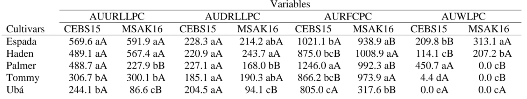
The factors mango cultivars and C. fimbriata isolates and their interaction were significant for AUURLLPC (P ≤ 0.01), AUDRLLPC (P ≤ 0.01), AURFCPC (P ≤ 0.01) and AUWLPC (P ≤ 0.01) (Table 1). Mango plants from the cultivars Espada, Haden and Palmer inoculated with C. fimbriata isolate CEBS15 were more susceptible than cultivars Tommy and Ubá based on the AUURLLPC (P ≤ 0.01) and AUWLPC (P ≤ 0.01) values (Table 1). There was no significant difference between the cultivars regarding the AUDRLLPC (P = 0.35) (Table 1). The AURRLLPC was significantly higher for cultivars Espada and Palmer compared to cultivar Ubá, whereas the Haden and Tommy cultivars showed intermediate values (P ≤ 0.05) (Table 1). Mango plants from the cultivars Espada and Haden inoculated with C. fimbriata isolate MSAK16 were more susceptible than Ubá based on the AUURLLPC (P ≤ 0.01), AUDRLLPC (P ≤ 0.05), AURFCPC (P ≤ 0.01) and AUWLPC (P ≤ 0.01) values (Table 1). The cultivars Palmer and Tommy showed variations in their level of resistance to MSAK16 according to the method of disease assessment (Table 1). Isolate CEBS15 was more aggressive compared to isolate MSAK16 on plants from cultivars Palmer (P ≤ 0.05) and Ubá (P ≤ 0.01) (Table 1). Isolate MSAK16 was more aggressive on plants of cultivar Haden (P ≤ 0.05) (Table 1). The two isolates of C. fimbriata showed variations in their level of aggressiveness
when inoculated on plants of cultivars Espada and Tommy based on the AURRLLPC and AUWLPC values (Table 1).
MSAK16 (Fig. 2F). On plants from cultivar Tommy, the wilting was mild when inoculated with isolate CEBS15 (Fig. 2G), but not exhibit symptoms when
inoculated with MSAK16. Light microscope observations
Colonization of the stem tissues of plants from susceptible and resistant cultivars by C. fimbriata
Examination of several longitudinal and transverse stem tissue sections at 22 dai using a light microscope confirmed the visual differences observed in the pattern of internal stem necrosis and wilting. The two isolates of C. fimbriata colonized the stem tissues of plants from the susceptible (Espada, Haden and Palmer), moderately resistant (Tommy Atkins) and resistant (Ubá) mango cultivars starting from the collenchyma and toward the cortical parenchyma, xylem vessels and pith parenchyma (Figs. 3, 4, 5, 6 and 7).
Colonization of collenchyma
Fungal hyphae of the two C. fimbriata isolates apparently did not grow well in the collenchyma of plants of any of the cultivars (Fig. 3, 4, 5, 6 and 7). Fungal hyphae that colonized the collenchyma cells were frequently surrounded by an amorphous granular material that stained densely with toluidine blue (Figs. 3G, 4C, 6C and 7C). In some cells, fungal hyphae appeared as empty shells (Figs. 4C, 6C and 7C).
Colonization of cortical parenchyma
of some fiber and parenchyma cells were detached, indicating possible cell death (Figs. 3E, 5I and 8K).
On plants from the moderately resistant (Tommy Atkins) and resistant cultivar (Ubá), most cells of the cortical parenchyma colonized by fungal hyphae of the two C. fimbriata isolates reacted strongly to fungal invasion by deposition of an amorphous granular material (Figs. 6 and 7) or were stained intense dark blue or purple (Figs. 6A, B, 7A and B). In these cells, fungal hyphae frequently appeared as empty shells (Figs. 6H and J; 7D, F, H, I and J) or were thin and faint (Figs. 6 and 7) and few chlamydospores were found (Figs. 7D). For both C. fimbriata isolates, fungal hyphae appearing as empty shells were more apparent in cultivar Uba than Tommy Atkins (Figs. 6 and 7). When comparing fungal isolates on plants from cultivar Uba, hyphae from isolate MSAK16, appearing as empty shells, was more commonly observed (Figs. 7D, E, F, H, I and K).
Colonization of xylem vessels
On the stem tissues of plants from susceptible cultivars Espada, Haden and Palmer, fungal hyphae of two isolates of C. fimbriata reached the xylem vessels, stimulating intense formation of polysaccharide gels and tyloses (Figs. 3, 4 and 5). Xylem vessels in the stem tissues of plants from cultivars Espada and Haden were observed to be more obstructed by deposition of polysaccharide gels, fungal hyphae, chlamydospores and tyloses when infected by isolate MSAK16 (Figs. 3H and I; 4J
and K) in comparison to CEBS15 (Figs. 3C and E; 4D and F). Chains of chlamydospores were observed in the xylem vessels of plants from cultivar Haden inoculated with isolate MSAK16 (Figs. 4H, I and K). In the stem tissues of plants from cultivar Palmer, xylem vessels were commonly obstructed when inoculated with isolate CEBS15 (Figs. 5C, E, F, H, I and J).
Colonization of pith parenchyma
The two C. fimbriata isolates extensively colonized the pith parenchyma in the radial
direction in the stem tissues of plants from the susceptible cultivars Espada, Haden and Palmer (Figs. 3, 4 and 5). Some cells reacted to fungal invasion by accumulating amorphous granular material (Figs. 3F and J; 4G and L; 5G and L). In the cells of pith parenchyma on the stem tissues of plants from cultivar Palmer, some fungal hyphae appeared as empty shells (Figs. 5G and L). Fungal hyphae of the isolates MSAK16 and CEBS15 grew in the pith parenchyma in the radial direction, reaching again the xylem vessels in the stem tissues of plants from cultivars Haden (Fig. 4B) and Palmer (Fig. 5A), respectively. Fungal hyphae of isolate CEBS15 were more frequently observed in the pith parenchyma in stem tissues of plants from cultivars Espada and Palmer compared with isolate MSAK16 (Figs. 3A and B; 5A and B). However, in the stem tissues of plants from cultivar Haden, fungal hyphae of isolate MSAK16 apparently grew more in the pith parenchyma (Figs. 4A and B).
Fungal hyphae of the two C. fimbriata isolates apparently scarcely colonized the pith parenchyma in the radial direction in the stem tissues of plants from the moderately resistant and resistant cultivars Tommy Atkins and Ubá (Figs. 6A, B, F and K and 7A, B, G and K). Most of the cells reacted to C. fimbriata infection, regardless of the isolate, by accumulating amorphous granular material that surrounded many fungal hyphae, which sometimes appeared thin and faint or as
Discussion
This study provides, to the best of our knowledge, the first direct evidence that
mango cultivars respond differently to the infection by C. fimbriata at the cellular level according to their basal level of resistance. The AURFCPC and AUWLPC provided the best separation of the five cultivars according to their levels of resistance to mango wilt. High values of AURFCPC and AUWLPC were associated with death of the susceptible mango plants cultivars. External and internal necrotic tissues in the stem of plants are the major symptoms of mango wilt prior to complete wilting (Ribeiro, 2005; Viégas, 1960). Park et al. (2013) showed that a high proportion of external necrotic tissues on the stem of hickory trees infected with C. smalleyi contributed to xylem dysfunction and resulted in wilting symptoms and decline of plants. In the present study, the lowest values for almost all measures of disease development were obtained for plants from cultivars Tommy Atkins and Ubá, which did not show intense wilting and death of plants until the end of the experiment.
A few studies have reported that Ceratocystis spp. colonizes mainly the vascular system of various plant species (Al Sadi et al., 2010; Ferreira et al., 2005; Park et al., 2013). According to Al Sadi et al. (2010), hyphae of C. manginecans were only found in the vascular system of inoculated mango seedlings. Ferreira et al. (2005) found hyphae of C. fimbriata in the vascular system and in the pith parenchyma of
Eucalyptus plants. In contrast, Viégas (1960) was the first to suggest that C. fimbriata is not an exclusive vascular pathogen because the xylem vessels were not completely obstructed with fungal hyphae, as in Verticillium-cotton pathossystem. Results from the present study clearly show that isolates of C. fimbriata obtained in Brazil colonized all stem tissues of mango cultivars, and therefore it, cannot be considered an exclusive vascular pathogen as reported in the literature.
et al., 2009; Hall et al., 2011; Kim et al., 2004; Rodrigues et al., 2003). Pre-formed or induced phenolic compounds play an important role in host defense against
pathogen infection (Nicholson & Hammerschmidt, 1992) such as in cell wall lignification (Benhamou & Bélanger, 1998), in antimicrobial activity (Rodrigues et al., 2003), as modulators of plant hormones in defense signaling and as scavengers of reactive oxygen species (Dixon & Paiva, 1995). In the present study, the scarcity of fungal hyphae in collenchyma cells may have been, due to the intense deposition of phenolic compounds in these cells.
Fungal hyphae of C. fimbriata apparently grew without any impedance in the cortical parenchyma and xylem vessels of stem tissues of susceptible cultivars. By contrast, in stems of moderately resistant and resistant cultivars, most cells reacted to C. fimbriata infection by accumulating amorphous material that contributed to many fungal hyphae appearing as empty shells. Several other studies have also reported the occurrence of empty fungal hyphae of pathogens surrounded by amorphous material in the cells of many plant species (Bélanger et al., 2003; Benhamou & Bélanger, 1998; Ouellette et al., 1992; Rodrigues et al., 2003). Eynck et al. (2009) observed intense deposition of phenolics and lignin compounds in the cells of roots and hypocotyls of resistant oilseed rape plants in response to infection by V. longisporum compared to the tissues of susceptible plants. In the present study, intense deposition of amorphous material surrounding fungal hyphae of both fungal in the cortical
parenchyma and xylem vessels helped to explain limited disease development on plants from cultivars Tommy Atkins and Ubá.
barrier that impeded the greater colonization of the vascular system on the stems of plants from the moderately resistant and resistant cultivars in comparison to tissues
of plants from the susceptible cultivars.
In the stem tissues of plants from the susceptible cultivars, hyphae of C. fimbriata reached the xylem vessels and stimulated the formation of intense polysaccharide gels and many tyloses. By contrast, in moderately resistant and resistant cultivars, vessels were free of any occlusions, including fungal hyphae, or were fully obstructed by many large tyloses and phenolic compounds. Gels and tyloses are good examples of occluding structures produced by plants that serve to impede tissue colonization by wilt pathogens (Duchesne et al., 1992; Rioux et al., 1998). These occluding structures can be effective whether produced in advance of fungal ingress or during the colonization process (Ouellette & Rioux, 1992). Williams et al. (2002) and Eynck et al. (2009) reported a positive relationship between the abundance of vascular occlusions (gels and tyloses) and the increased level of resistance on the stem tissues of tomato and oilseed rape plants to infection by Verticillium spp. In contrast, Park et al. (2013) observed that the accumulation of gels and tyloses in the xylem vessels contributed to reduced sap flow at the margin of the necrotic sapwood of bitternut hickory infected by C. smalleyi. According to Al Sadi et al. (2010), tyloses and hyphae of C. manginecans in the vascular system were responsible for the wilting and the death of mango seedlings. In the present study, differences in the
susceptibility of the mango cultivars to C. fimbriata could be attributable to differences in the ability of plants to prevent rapid pathogen colonization of the xylem vessels by quick deposition of tyloses impregnated with phenolic compounds.
probably due to the degradation of cell wall of stem tissues of plants from the susceptible cultivars by the action of pectolytic enzymes released by C. fimbriata.
Many chlamydospores of C. fimbriata were observed in the xylem vessels and parenchyma cells of stem tissues from plants of the susceptible cultivars. Ceratocystis fimbriata has specialized conidiophores that give rise the chlamydospores also called aleurioconidia that are pigmented, thick-walled, and filled with nutrient reserves substance (Viegas, 1960; CAB International, 2005; Paulin-Mahady et al., 2002). Chlamydospores are durable structures that allow the pathogen to survive in the absence of a susceptible host (CAB International, 2005; Paulin-Mahady et al., 2002). Ferreira et al. (2005) also found most chlamydospores, simple or in chains, of C. fimbriata in the xylem vessels, medullary rays and pith parenchyma of Eucalyptus plants.
Ceratocystis fimbriata extensively colonized the pith parenchyma in stem tissues of plants from the susceptible mango cultivars in the radial direction, whereas in the moderately resistant and resistant cultivars, fungal hyphae scarcely reached these cells. Similarly, colonization of the xylem vessels of plants from a resistant cotton cultivar by F. oxysporum f.sp. vasinfectum was reduced by the accumulation of phenolic compounds, with the number of infected xylem vessels decreasing as the distance from the inoculation point increased (Shi et al., 1992). By contrast, in the susceptible plants, F. oxysporum f.sp. vasinfectum colonized both longitudinal and
lateral stem tissues and the defense mechanism occurring in the cells against fungal infection was delayed and weak (Shi et al., 1992). In the present study, accumulation of phenolic compounds probably contributed to diminished colonization by C. fimbriata in the upward, downward and radial direction and helped to explain the lower values for the AURFCPC and AUWLPC for the resistant cultivars.
Acknowledgements
References
Ajila CM, Rao LJ, Rao UJSP, 2010. Characterization of bioactive compounds from
raw and ripe Mangifera indica L. peel extracts. Food and Chemical Toxicology 48, 3406-11.
Al-Sadi AM, Al-Ouweisi FA, Al-Shariani NK, Al-Adawi AO, Kaplan EJ, Deadman ML, 2010. Histological changes in mango seedlings following infection with Ceratocystis manginecans, the cause of mango decline. Journal of Phytopathology 158, 738-43.
Aoun M, Rioux D, Simard M, Bernier L, 2009. Fungal colonization and host defense reactions in Ulmus americana callus cultures inoculated with Ophiostoma novo-ulmi. Phytopathology 99, 642-50.
Bélanger RR, Benhamou N, Menzies JG, 2003. Cytological evidence of an active role of silicon in wheat resistance to powdery mildew (Blumeria graminis f.sp. tritici). Phytopathology 93, 402-12.
Benhamou N, Bélanger RR, 1998. Benzothiadiazole-mediated induced resistance to Fusarium oxysporum f.sp. radicis-lycopersici in tomato. Plant Physiology 118, 1203-12.
Batista DC, Terao D, Barbosa MAG, Barbosa FR, 2008. Seca-da-mangueira: detecção, sintomatologia e controle, Publicado online pela Empresa Brasileira de Pesquisa Agropecuária Semi-árido. Arquivo n. 138.
http://www.cpatsa.embrapa.br/ public_eletronica/downloads/COT138. Acess 01 06 2013.
Biggs AR, 1992. Anatomical and physiological responses of bark tissues to mechanical injury. In: Blanchette RA, Biggs AT, eds. Defense Mechanisms of Woody Plants Against Fungi. Springer-Verlag, Berlin, 13-36
CAB International, 2005. Ceratocystis fimbriata [original text prepared by T. C.
Harrington]. In: Crop protection compendium. Wallingford, UK: CAB International. Carvalho CRL, Rossetto CJ, Mantovani DMB, Morgano MA, Castro JV, Bortoletto N, 2004. Avaliação de cultivares de mangueira selecionadas pelo Instituto Agronômico de Campinas comparadas a outras de importância comercial. Revista Brasileira de Fruticultura 26, 264-71.
Dixon RA, Paiva NL, 1995. Stress induced phenylpropanoid metabolism. Plant Cell 7, 1085-97.
Duchesne LC, Hubbes M, Jeng RS, 1992. Biochemistry and molecular biology of defense reactions in the xylem of angiosperm trees. In: Blanchette RA, Biggs AT, eds. Defense Mechanisms of Woody Plants Against Fungi. Springer-Verlag, Berlin, 133-42.
Eynck C, Koopmann B, Karlovsky P, von Tiedemann A, 2009. Internal resistance in winter oilseed rape inhibits systemic spread of the vascular pathogen Verticillium longisporum. Phytopathology 99, 802-11.
FAO, 2013. Medium-term prospects for agricultural Commdities. In: Food and
Agriculture Organization of the United Nations.
http://www.fao.org/docrep/006/y5143e/y5143e1a. htm.
Ferreira EM, Harrington TC, Thorpe DJ, Alfenas AC, 2010. Genetic diversity and interfertility among highly differentiated populations of Ceratocystis fimbriata in Brazil. Plant Pathology 59, 721-35.
Ferreira FA, Maffia LA, Ferreira EA, 2005. Detecção rápida de Ceratocystis fimbriata em lenho infetado de eucalipto, mangueira e outros hospedeiros lenhosos. Fitopatologia Brasileira 30, 543-45.
Hall C, Heath R, Guest DI, 2011. Rapid and intense accumulation of terpenoid
phytoalexins in infected xylem tissues of cotton (Gossypium hirsutum) resistant to Fusarium oxysporum f.sp. vasinfectum. Physiological and Molecular Plant Pathology 76, 182-88.
Kim KH, Yoon JB, Park HG, Park EW, Kim YH, 2004. Structural modifications and programmed cell death of chili pepper fruit related to resistance responses to Colletotrichum gloeosporioides infection. Phytopathology 94, 1295-1304.
Merrill W, 1992. Mechanisms of resistance to fungi in woody plants: A historical perspective. In: Blanchette RA, Biggs AT, eds. Defense Mechanisms of Woody Plants Against Fungi. Springer-Verlag, Berlin, 1-11.
Nicholson RL, Hammerschmidt R, 1992. Phenolic compounds and their role in disease resistance. Annual Review of Phytopathology 30, 369-89.
Park JH, Juzwik J, Cavender-Bares J, 2013. Multiple Ceratocystis smalleyi infections associated with reduced stem water transport in bitternut hickory. Phytopathology
103, 565-74.
Ribeiro IJA, Rossetto CJ, Sabino JC, Gallo PB, 1986. Seca da mangueira: VIII. Resistência de porta-enxertos de mangueira ao fungo Ceratocystis fimbriata Ell. & Halst. Bragantia 45, 317-22.
Ribeiro IJA, 2005. Doenças da mangueira (Mangifera indica L.). In: Kimati H, Amorim L, Bergamin-Filho A, Camargo LEA, Rezende JAM, eds. Manual de Fitopatologia: Doenças das Plantas Cultivadas. Agronômica Ceres, São Paulo, 457-65.
Ribeiro SMR, Barbosa LCA, Queiroz JH, Knodler M, Schieber A, 2008. Phenolic compounds and antioxidant capacity of Brazilian mango (Mangifera indica L.) varieties. Food Chemistry 110, 620-26.
Rioux D, Nicole M, Simard M, Ouellette GB, 1998. Immunocytochemical evidence that secretion of pectin occurs during gel (gum) and tylosis formation in trees. Phytopathology 88, 494-505.
Rodrigues FA, Benhamou, N, Datnoff LE, Jones JB, Bélanger RR, 2003. Ultrastructural and cytochemical aspects of silicon-mediated rice blast resistance. Phytopathology 93, 535-46.
Rossetto CJ, Ribeiro IJA, Igue T, Gallo PB, 1996. Seca-da-mangueira: XV.
Resistência varietal a dois isolados de Ceratocystis fimbriata. Bragantia 55, 117-21.
Shaner G, Finney RE, 1977. The effect of nitrogen fertilization on the expression of slow-mildewing resistance in knox wheat. Phytopathology 70, 1183-86.
Shi J, Mueller WC, Beckman CH, 1992. Vessel occlusion and secretory activities of vessel contact cells in resistant or susceptible cotton plants infected with Fusarium oxysporum f.sp. vasinfectum. Physiological and Molecular Plant Pathology 40, 133-47.
Shigo A, Tippett JT, 1981. Compartmentalization of American Elm tissues infected by Ceratocystis ulmi. Plant Disease 65, 715-18.
Uritani I, Stahmann MA, 1961. Pectolytic enzymes of Ceratocystis fimbriata. Phytopathology 51, 277-85.
of a destructive mango wilt disease in Oman and Pakistan. Fungal Diversity 27, 213-30.
Viégas AP, 1960. Seca da mangueira. Bragantia 19, 163-82.
LIST OF TABLE AND FIGURES
Table 1 Area under upward relative lesion length progress curve (AUURLLPC), area under downward relative lesion length progress curve (AUDRLLPC) and area under radial fungal colonization progress curve (AURFCPC) on stem tissues and area under wilted leaves progress curve (AUWLPC) in five mango cultivars inoculated with the isolates CEBS15 and MSAK16 of Ceratocystis fimbriata.
Variables
AUURLLPC AUDRLLPC AURFCPC AUWLPC
Cultivars CEBS15 MSAK16 CEBS15 MSAK16 CEBS15 MSAK16 CEBS15 MSAK16
Espada 569.6 aA 591.9 aA 228.3 aA 214.2 abA 1021.1 bA 938.9 aB 209.8 bB 313.1 aA Haden 489.1 aA 567.4 aA 220.9 aA 243.7 aA 875.0 bcB 1008.9 aA 114.1 cB 207.2 bA Palmer 488.7 aA 227.9 bB 227.1 aA 168.0 bB 1246.0 aA 992.3 aB 450.7 aA 0.0 cB Tommy 306.7 bA 300.1 bA 185.1 aA 190.3 abA 866.2 bcB 973.9 aA 4.4 dA 0.0 cB
Ubá 244.1 bA 86.6 cB 204.5 aA 94.1 cB 805.0 cA 317.6 bB 0.0 eA 0.0 cA
Figure 1 Symptoms of internal necrotic tissues in longitudinal (A, C, E, G and I) and transverse (B, D, F, H and J) stem sections from cultivars Espada (A and B), Haden
(C and D), Palmer (E and F), Tommy Atkins (G and H) and Ubá (I and J) at 22 days after inoculation (dai) with the isolate MSAK16. Asterisks (*) indicate the stem region where the fungal inoculation occurred and from which the upward (u), downward (d) and radial (r) Ceratocystis fimbriata colonization were observed as detailed in A. The formation of perithecia of C. fimbriata was observed at the inoculation point of the stem tissues at 22 dai (arrow in A, E and I). In A1, perithecia
of C. fimbriata are shown in a higher magnification. Scale bars: 1000 µm (A, B, C,
Figure 2 Symptoms of wilting in mango plants from cultivars Espada (A and B), Haden (C and D), Palmer (E and F), Tommy Atkins (G and H) and Ubá (I and J) at
Figure 3 Light micrographs of longitudinal (A, B, C, D, F, G, H, I and J) and transverse (E) mango stem tissues of the susceptible cultivar Espada at 22 days after
inoculation with isolates CEBS15 (A, C, D, E and F) and MSAK16 (B, G, H, I and J). A and B, Sequential images of stem tissues colonized by Ceratocystis fimbriata (asterisks) starting from the collenchyma and then moving in the direction of the cortical parenchyma, xylem vessels and pith parenchyma. C, Hyphae of C. fimbriata (isolate CEBS15) grew abundantly in the parenchyma cells between xylem vessels. In some cells, amorphous granular material (arrowheads) was occasionally deposited around fungal hyphae. The fungus reached the xylem vessels, stimulating tylosis formation. D, In some parenchyma cells, fungal hyphae were thick. E, Hyphae of C. fimbriata colonized cells of the medullary radius and adjacent cells. Plasmatic membranes of some fiber cells, not yet colonized by the fungus, were detached, indicating possible cell death (arrows). Some xylem vessels were colonized by C. fimbriata with concomitant tylosis formation. F, Hyphae of C. fimbriata colonized the pith parenchyma in the radial direction. Some cells reacted to fungal invasion by accumulating amorphous granular material (arrowheads), indicating the presence of phenolic compounds. G, Hyphae of C. fimbriata (isolate MSAK16) in the collenchyma were often surrounded by amorphous granular material (arrowheads), whereas cortical parenchyma cells did not show any sign of reaction against fungal infection. Chlamydospores (aleurioconidia) were observed in the cortical
Figure 4 Light micrographs of longitudinal (B, C, F, H, I and L) and transverse (A, D, E, G, J and K) mango stem tissues of the susceptible cultivar Haden at 22 days
after inoculation with isolates CEBS15 (A, C, D, E, F and G) and MSAK16 (B, H, I, J, K and L). A and B, Sequential images of stem tissues colonized by Ceratocystis fimbriata (asterisks) starting from the collenchyma and then moving in the direction of the cortical parenchyma, xylem vessels and pith parenchyma. C, Hyphae of C. fimbriata (isolate CEBS15) in the collenchyma were often surrounded by amorphous granular material (arrowheads). In some cells, fungal hyphae appeared as empty shells (double arrowheads). D, Hyphae of C. fimbriata grew abundantly in the parenchyma cells adjacent to the xylem tissues. In some cells, amorphous granular material (arrowheads) was occasionally deposited around fungal hyphae. Large tylosis formation occurred in the xylem vessels. E, Fungal hyphae colonized cells of the medullary radius and parenchyma cells. In some parenchyma cells, large chlamydospores (aleurioconidia) were observed (arrow). F, Fungal hyphae grew abundantly in the parenchyma cells and xylem vessels. Xylem vessels were obstructed by the deposition of polysaccharide gels, fungal hyphae and tylosis. G, Hyphae of C. fimbriata apparently massively colonized the pith parenchyma in the radial direction. Some cells reacted to fungal invasion by accumulating amorphous granular material (arrowheads). H and I, Hyphae of C. fimbriata (isolate MSAK16) colonized the parenchyma cells and the xylem vessels. Many chlamydospores were
Figure 5 Light micrographs of longitudinal (A, C, D and G) and transverse (B, E, F, H, I, J, K and L) mango stem tissues of the susceptible cultivar Palmer at 22 days
after inoculation with isolates CEBS15 (A, C, D, E, F and G) and MSAK16 (B, H, I, J, K and L). A and B, Sequential images of stem tissues colonized by Ceratocystis fimbriata (asterisks) starting from the collenchyma and then moving in the direction of the cortical parenchyma, xylem vessels and pith parenchyma. C, Xylem vessels were obstructed by intense deposition of polysaccharide gels, fungal hyphae, chlamydospores (aleurioconidia) and tyloses. D, Long hyphae of C. fimbriata (isolate CEBS15) grew in the parenchyma cells adjacent to the tracheal elements. Amorphous granular material (arrowheads) was scarcely deposited around fungal hyphae. E and F, Fungal hyphae grew abundantly in the parenchyma cells and the xylem vessels. Xylem vessels were obstructed by deposition of polysaccharide gels, long and thickened fungal hyphae and tyloses. Some tyloses stained dark-blue or purple. G, Hyphae of C. fimbriata apparently colonized all pith parenchyma in the radial direction and reached the xylem vessels. Some cells reacted to fungal invasion by accumulating amorphous granular material (arrowheads) and sometimes, fungal hyphae appeared as empty shells (double arrow). H, Hyphae of C. fimbriata (isolate MSAK16) grew abundantly in the parenchyma cells and xylem vessels. Chlamydospores were also observed. Amorphous granular material (arrowheads) was scarcely deposited around fungal hyphae. Xylem vessels were obstructed by
(arrowheads) and sometimes appeared as empty shells (double arrow). Chlamydospores (c), collenchyma (co), cortical parenchyma (cp), fiber cells (fc),
Figure 6 Light micrographs of longitudinal (A, D, E and F) and transverse (B, C, G, H, I, J and K) mango stem tissues of the moderately resistant cultivar Tommy Atkins
Chlamydospores (c), collenchyma (co), cortical parenchyma (cp), fungal hyphae (f), medullary radius (mr), parenchyma cells (pc), pith parenchyma (pp), tracheal
Figure 7 Light micrographs of longitudinal (B, H, I, J and K) and transverse (A, C, D, E, F and G) mango stem tissues of the resistant cultivar Ubá at 22 days after
appeared as empty shells (arrow) or were faint. Plasmatic membranes of some pith parenchyma cells were detached, indicating possible cell death. Collenchyma (co),
CHAPTER 2
Accepted as original paper to Phytopathology
Resistance in Mango Against Infection by Ceratocystis fimbriata
Leonardo Araujo, Wilka Messner Silva Bispo, Isaías Severino Cacique, Wiler Ribas Moreira and Fabrício Ávila Rodrigues
Viçosa Federal University, Department of Plant Pathology, Laboratory of Host-Pathogen Interaction, Viçosa, Minas Gerais State, Zip Code 36570-900, Brazil
ABSTRACT
Araujo, L., Bispo, W. M. S., Cacique, I. S., Moreira, W. R., and Rodrigues, F. A. Resistance in mango against infection by Ceratocystis fimbriata. Phytopathology 104:xx-xx.
This study was designed to characterize and describe host cell responses of stem
INTRODUCTION
Worldwide mango (Mangifera indica L.) is one of the most important tropical fruit crops (22). The adaptability of mango cultivars to different environmental conditions and their resistance to multiple diseases are among the factors that greatly improve mango quality and yield (12). Mango wilt is caused by Ceratocystis fimbriata Ellis & Halst. (23,56) in Brazil (41), while a similar wilt disease is found in Oman and Pakistan, where it is attributed to C. manginecans (1,55). Thus, mango wilt has become a serious concern to growers (1,55). Ceratocystis fimbriata causes death of the entire tree either a few months after the fungus penetrates the roots, or more slowly if it enters through wounded branches of the canopy (41). Typical disease symptoms include wilting and browning of leaves on single branches and gum exudation from the trunks (41,56). As the infection progresses, the internal and external stem tissue becomes dark brown due to intensive necrosis (41,56).
The eradication of mango trees with wilt symptoms can be used as a strategy to slow disease progress (41,46). However, in Brazil, mango cultivars with high levels of resistance to mango wilt are the most effective strategy adopted by farmers to control the disease (12,40,41,46). Resistance is critical since mango wilt cannot be
controlled by fungicides (12,40,41,46). The high degree of genetic variability in aggressiveness of C. fimbriata renders, resistant mango cultivars to become susceptible in a relatively short time (40,41,46).
The resistance of trees to vascular pathogens is primarily based on their ability to restrict pathogen infection to a few cells (20). Suberized bark containing phenolic-like compounds, the composition of the xylem vessels and their diameter are common examples of pre-formed mechanisms that restrict infection by vascular pathogens (6,20,32). In contrast, active defense mechanisms include anatomical responses, like deposition of gels and the formation of tyloses in invaded vascular vessels (9,20). The formation of barrier zones, intense lignification and suberization of cell walls, and the production of phenolics, phytoalexins and proteins related to pathogenesis are also involved in active defenses (9,20,43).
phytoalexins, phenolics and lignin are the major components in barrier zones formed in host tissue in response to pathogen infection (9,11,43,49,53).
Important insights into specific responses in infected tissue to pathogen infection can be obtained by examining autofluorescence (2,9,21,29,43). Autofluorescence can be used because phenolics and related polymers like lignin absorb short wave length light, in ultraviolet (UV) and near-blue range, and emit longer wavelengths of visible light (2,9,21,29,43). Thus, microscopes capable of producing UV and near-blue light for excitation of phenolics and fitted with the proper filters will readily reveal localized autofluorescent compounds in tissue (2,9,21,29,43). For example, stem sections from balsam poplar infected by Ophiostoma novo-ulmi when observed under UV light excitation showed stronger autofluorescence in cellular barrier zones (43). Intense autofluorescence was also produced by suberin and lignin in callus cultures of American elm infected by O. novo-ulmi (2). Eynck et al. (21) observed intense deposition of phenolics and lignin compounds in the roots and hypocotyls of plants from a resistant cultivar of oilseed rape in response to infection by Verticillium longisporum, when compared to tissue from plants of a susceptible cultivar. The resistant cultivar exhibited a much stronger autofluorescence when excited by light in the near-UV range than the susceptible cultivar (21).
The concentration of certain chemical elements in plant tissue also can affect their resistance to vascular pathogens (17). High levels of sulfur in stem tissue of cacao
(15) and tomato (57) plants were associated with increased resistance to infection by V. dahliae. Sulfur and sulfur compounds also decrease disease directly as a result of antimicrobial activities and indirectly by enhancing host resistance (27). Increased calcium content in plant tissue also correlates with the resistance of cell walls and the middle lamella to fungal pathogen invasion (3,13,39). Calcium also acts as a signal for oxidative bursts that trigger the synthesis of phytoalexins and the induction of defense-related genes whose products lead to resistance in many host-pathogen interactions (30,39).
roots of a resistant cotton cultivar restricted the colonization of Fusarium oxysporum f.sp. vasinfectum.
MATERIALS AND METHODS
Plant material. Mango plant cultivars Haden (susceptible) and Ubá (resistant) were obtained from a commercial nursery (Dona Euzébia city, Minas Gerais State, Brazil) and their rootstock was from the cv. Imbú. The 1.5-year-old plants were transplanted into plastic pots containing 8 kg of soil, sand and manure in a 2:1:1 proportion. Plants were kept in a greenhouse (temperature of 30 ± 2°C during day and 10°C at night and relative humidity of 70 ± 5%) for 2 months before starting the experiments.
Inoculation procedure. The isolate MSAK16 of C. fimbriata used to inoculate the
plants was obtained from symptomatic mango plants collected in Aquidauana, Mato
Grosso do Sul State, Brazil. The isolate was preserved using Castellani's method (18).
Plugs of a malt extract agar medium containing fungal mycelia were transferred to Petri
dishes containing potato dextrose agar (PDA). After three days, the PDA plugs
containing fungal mycelia were transferred to Petri dishes containing the same culture
medium and maintained in an incubator (temperature of 25°C and 12 h photoperiod) for
14 days.
Inoculation was performed following the methods of Al-Sadi et al. (1). Bark disks (10-mm diameter and 2-mm height) were removed from the stems of both cultivars using a punch. The disks came from approximately 5 cm above the graft scar. A plug (10-mm diameter) removed from the middle portion of each PDA plate obtained from 14-day-old colonies of C. fimbriata was placed in the wound. Each wound containing the fungal mycelia PDA plug was carefully covered with a piece of moistened cotton and enclosed with parafilm to maintain adequate moisture to promote fungal infection. Wounds only receiving plugs of PDA medium served as the control.
Disease assessment. Disease progress in stem tissue and development of wilted leaves was evaluated at 15, 22 and 29 days after inoculation (dai). The upward, downward and radial colonization of the stem tissue by the hyphae of C. fimbriata