MARIA ALVES FERREIRA
ESTRUTURA GENÉTICA DE POPULAÇÕES DE Ceratocystis fimbriata E PADRÃO ESPAÇO-TEMPORAL DA MURCHA-DE-CERATOCYSTIS
Tese apresentada à Universidade Federal de Viçosa, como parte das exigências do Programa de Pós-Graduação em Fitopatologia, para obtenção do título de Doctor Scientiae.
VIÇOSA
MARIA ALVES FERREIRA
ESTRUTURA GENÉTICA DE POPULAÇÕES DE Ceratocystis fimbriata E PADRÃO ESPAÇO-TEMPORAL DA MURCHA-DE-CERATOCYSTIS
Tese apresentada à Universidade Federal de Viçosa, como parte das exigências do Programa de Pós-Graduação em Fitopatologia, para obtenção do título de Doctor Scientiae.
APROVADA: 07 de agosto de 2009
_________________________________ ___________________________ Prof. Eduardo Seiti Gomide Mizubuti Prof. Thomas C. Harrington
__________________________________ _________________________________ Pesq. Edival Ângelo Valverde Zauza Pesq. Lúcio Mauro da Silva Guimarães
_____________________________ Prof. Acelino Couto Alfenas
Ao mestre, exemplo de profissionalismo e dedicação à Patologia Florestal Professor Francisco Alves Ferreira
AGRADECIMENTOS
À Universidade Federal de Viçosa, pela oportunidade de realização do curso.
Ao Conselho Nacional de Desenvolvimento Científico e Tecnológico (CNPq), pelo financiamento da bolsa.
Ao professor Acelino Couto Alfenas pela confiança, pela paciência, pela orientação, pelo incentivo e pelos ensinamentos.
Às empresas florestais Aracruz Celulose, Plantar, Rima Siderúrgica, Suzano, Vallourec & Mannesmann, Veracel e Votorantim Metais pelo fornecimento dos isolados e pelo apoio na montagem dos experimentos.
À Iowa State University, pela oportunidade de realização de parte deste trabalho.
Ao professor Dr. Thomas C. Harrington, pela orientação e pelas sugestões. Aos professores Luiz Antonio Maffia, Waldir Cintra de Jesus e Eduardo Mizubuti, pelas sugestões.
Ao professor Francisco Alves Ferreira, pela amizade.
Ao amigo Daniel Henrique Breda Binoti, pelo convívio, pela amizade e ajuda imprescindível para a realização deste trabalho.
Ao amigo Reginaldo Gonçalves Mafia, pelas sugestões para a melhoria deste trabalho.
À Márcia, pelo auxílio em todos os momentos.
Aos funcionários do Departamento de Fitopatologia: Rita, Elói, Délio, Camilo, Brás e Fizinho, pela convivência amigável.
À amiga Sujin Kim e à Doug McNew, pelo auxílio nas metodologias e funcionamento do Laboratório de Patologia Florestal, em Iowa.
Aos amigos Hey young, Carlos Congorra, Izera, Surema, Marcos, Luciano, Thais, Susy, Juliana, Rafael, Evrim, Manu, Ryan, Neiva e Márcio Zaccaron, pela ótima convivência durante meu doutorado sanduíche em Iowa.
À Rafael Martins, Patrícia S. Machado, Natália Risso Fonseca, Alex Ferreira de Freitas e Ricardo Martins, pela ajuda nos experimentos.
À todos os colegas do Laboratório de Patologia Florestal, pela convivência. Aos professores do Departamento de Fitopatologia, pelos valiosos ensinamentos para a minha formação profissional.
Aos meus pais Euclides José Ferreira e Verônica Alves Ferreira e aos meus irmãos Evander Alves Ferreira, Elivelton Alves Ferreira, Eliza Aparecida Ferreira Alves e Manoel Teixeira Alves, pelo apoio e pela amizade.
BIOGRAFIA
MARIA ALVES FERREIRA, filha de Verônica Alves Ferreira e Euclides José Ferreira, nasceu em Silveirânia, Minas Gerais.
Em 1997, ingressou no curso de Engenharia Florestal da Universidade Federal de Viçosa. Foi bolsista de iniciação científica no laboratório de Nematologia e no laboratório de Patologia Florestal de 1998 a 2002. Nesse período, realizou trabalhos sob orientação da professora Rosângela DArc de Oliveira e do professor Acelino Couto Alfenas. Colou grau em maio de 2002.
ÍNDICE
RESUMO...vii
ABSTRACT...ix
INTRODUÇÃO GERAL...1
ARTIGO 1...9
Genetic variation and interfertility among Brazilian populations of the Ceratocystis fimbriata sensu strict...9
Abstract...9
Introduction...10
Materials and Methods ...12
Fungal isolates ...13
Fungal populations...13
DNA extraction...15
Microsatellite markers ...15
Analyses...16
Mating experiments ...17
Results...19
Discussion...23
References...30
ARTIGO 2...51
Movement of genotypes of Ceratocystis fimbriata among Eucalyptus plantations in Brazil...51
Abstract...51
Introduction...52
Materials and Methods ...53
Fungal isolates ...53
Fungal populations...54
DNA extraction...56
Microsatellite markers ...56
Analysis ...56
Results...57
Discussion...61
References...65
ARTIGO 3...80
Padrão espaço-temporal da murcha-de-ceratocystis em plantios clonais de eucalipto ...80
Resumo ...80
Introdução ...81
Material e Métodos ...83
Coleta de dados...83
Padrão espacial da murcha-de-ceratocystis ...83
Progresso da murcha-de-ceratocystis ...84
Resultados...85
Padrão espacial da murcha-de-ceratocystis em áreas de coleta de brotos para estaquia e plantios convencionais...85
Padrão temporal da murcha-de-ceratocystis...87
Discussão ...87
Referências ...91
RESUMO
FERREIRA, Maria Alves, D.Sc., Universidade Federal de Viçosa, agosto de 2009. Estrutura genética de populações de Ceratocystis fimbriata e padrão espaço-temporal da murcha-de-ceratocystis. Orientador: Acelino Couto Alfenas. Coorientadores: Luiz Antonio Maffia, Waldir Cintra de Jesus e Eduardo Seiti Gomide Mizubuti.
ABSTRACT
FERREIRA, Maria Alves, D.Sc., Universidade Federal de Viçosa, August, 2009. Genetic structure of Ceratocystis fimbriata populations and spatial-temporal patterns of Ceratocystis wilt. Advisor: Acelino Couto Alfenas. Co-advisers: Luiz Antonio Maffia, Waldir Cintra de Jesus and Eduardo Seiti Gomide Mizubuti.
INTRODUÇÃO GERAL
Das doenças do eucalipto, a murcha-de-ceratocystis causada por Ceratocystisfimbriata Ellis & Halsted é uma das mais importantes, em virtude do difícil controle e da natureza sistêmica das infecções, dos danos causados e das características do patógeno que dificultam o controle. A murcha-de-ceratocystis, em eucalipto, foi constatada primeiramente no sudeste da Bahia, em 1997 (Ferreira et al., 1999). Atualmente, já foram registradas ocorrências no Espírito Santo, Mato Grosso do Sul, em Minas Gerais, em São Paulo (Alfenas et al., 2004) e, recentemente, no Maranhão e Pará (Alfenas, A.C., UFV, informação pessoal). Diversas espécies florestais e agronômicas são suscetíveis à doença, entre elas Eucalyptus spp. (Zauza et al., 2004), Acacia mearnsii (Santos & Ferreira, 2003), Cassia fistula (Galli, 1958), Cassia renigera (Ribeiro et al., 1987), Calocasia esculenta (Harrington et al., 2005), Ficus carica (Valarini & Tokeshi, 1980), Gmelinaarborea (Muchovej et al., 1978) e Mangiferaindica (Batista, 1960).
A doença pode apresentar maior importância em determinados locais dependendo da cultura afetada. Por exemplo, em café há maior impacto econômico na Colômbia (Pontis, 1951), enquanto no Brasil é mais importante no Estado de São Paulo para a cultura da mangueira (Rossetto & Ribeiro, 1990; Ribeiro et al., 1995) e gmelina somente na Amazônia (Baker et al., 2003). No Brasil, causa sérios prejuízos para determinados clones de eucalipto (Ferreira et al., 1999). No Congo e na Uganda a doença causou alta mortalidade em determinados clones (Roux et al., 2000).
maneira, Baker et al. (2003) levataram a hipótese de que populações locais de C. fimbriata são hospedeiro-especializadas; algumas dessas populações são espécies emergentes e alguns genótipos hospedeiro-especializados têm sido movimentados para novas áreas pela ação do homem. Engelbrecht & Harrington (2005), ao parearem isolados de grupos de compatibilidade opostos para encontrar interesterilidade entre eles, constataram que dois grupos interestéreis corresponderam, respectivamente, a linhagens de batata doce e Platanus sp. A linhagem do patógeno de cacau apresentou dois grupos interestéreis correspondendo a duas sublinhagens genéticas. Além disso, foram estudadas características morfológicas das linhagens hospedeiro-especializadas; e com base nos resultados, esses autores descreveram uma nova espécie, denominada de C. cacaofunesta, e elevaram o patógeno oriundo de Platanus sp. (C. fimbriata f sp. platani) para C. platani. O patógeno de batata-doce foi mantido dentro de C. fimbriata, de onde foi originalmente descrito. Com base nesses estudos, foi sugerido que a evolução e divergência dessas espécies podem ter sido conduzidas pela especialização por hospedeiro, já que apenas pequenas diferenças morfológicas entre as espécies foram observadas. Em outros estudos com membros do clado da América Latina de C. fimbriata (Engelbrecht et al., 2004, 2007b; van Wyk et al., 2006; Ocasio-Morales et al., 2007) e espécies de Ceratocystis relacionadas (Roux et al., 2001; Barnes et al., 2005) foi possível distinguir populações possivelmente naturais de introduzidas. O patógeno pode ser introduzido em novas áreas pela movimentação de material vegetal infectado e ferramentas (Harrington, 2000; Baker et al., 2003; CABI, 2005; Ocasio-Morales et al., 2007; Engelbrecht et al., 2007a). Populações introduzidas tem menor diversidade quando comparadas a populações possivelmente nativas, e muitas vezes somente um único genótipo é identificado nas populacões introduzidas (McDonald, 1997).
verificada a dispersão do patógeno via contato radicular (Accordi, 1986, 1989) e ferimentos na raiz (Vigouroux et al., 1999; Vigouroux & Olivier, 2004). Rossetto & Ribeiro (1990) também comprovaram a infecção em M. indica pelo inóculo do solo. Para a cultura do eucalipto, Ferreira (2004) examinou árvores no campo e observou a infecção via inóculo no solo. Outro mecanismo comum verificado na literatura é a dispersão do patógeno por disseminadores secundários, como cupins e coleobrocas Scolytidae em Populus (Hinds, 1972), Prunus (Moller et al., 1969) e M. indica (Batista, 1960; Viégas, 1960). Sugere-se que coleobrocas que se alimentam de fungos adquirem inóculo de C. fimbriata e o dispersam para plantas suscetíveis, principalmente por serragem contaminada. Além disso, verificou-se que esporos de C. fimbriata podem ser carregados sobre os corpos de besouros Ambrosia e sobreviver pela passagem através do intestino do inseto (Iton, 1960; Crone, 1963). Essas coleobrocas, pertencentes aos gêneros Xyleborus e Hypocryphalus, são atraídas pelas plantas doentes, que produzem forte aroma e muitas galerias no tronco e galhos dessas plantas (Goitia & Rosales, 2001; Wingfield & Robison, 2004). Também foi observada a infecção de Ipomoea em ferimentos feitos por insetos e roedores (Clark & Moyer, 1988).
Ferreira et al. (2006) inspecionaram um grande número de plantas clonais de eucalipto afetadas pela murcha de C. fimbriata no Brasil e verificaram que somente duas apresentaram perfurações de inseto da família Platybodidae nas lesões de C. fimbriata; também não foram constatadas perfurações de insetos nas lesões novas e longitudinais, das plantas vizinhas. Desse modo, foi interpretado que a associação de insetos somente em lesões mais velhas é uma forma secundária ou não precursora da doença. Todavia, esses insetos, ao saírem das galerias do lenho infectado, podem transmitir endoconídios e clamidósporos do patógeno para outras plantas lenhosas com xilema alterado por fator abiótico ou biótico, como acontece com outras doenças por C. fimbriata em outras culturas (Sinclair et al., 1987; Wingfield et al., 1993; Baker et al., 2003).
assintomáticas (Vujanovic et al., 1999). A presença de C. fimbriata em mudas assintomáticas de eucalipto (Ferreira, 2004) também sugeriu a dispersão do patógeno pelo homem para várias regiões eucaliptocultoras.
Até o momento, poucos são os estudos referentes à variabilidade genética de C. fimbriata e ao padrão espaço-temporal da doença no campo, objetos deste trabalho. Este estudo foi dividido em três artigos. No primeiro, o principal objetivo foi avaliar a diversidade genética de populações de C. fimbriata no Brasil, com interesse em identificar populações introduzidas e nativas, usando 15 marcadores microssatélites polimórficos, verificar a interfertilidade entre representantes de algumas populações brasileiras com populações de C. cacaofunesta e C. platani e confirmar se as populações brasileiras de C. fimbriata pertencem a uma única espécie biológica e são capazes de recombinação genética. O segundo objetivou distinguir populações naturais de introduzidas e comparar os genótipos das diferentes populações de C. fimbriata. No terceiro artigo, objetivou-se fornecer informações sobre a epidemiologia da murcha-de-ceratocystis, bem como caracterizar o padrão espacial em áreas de coleta de brotos e em plantios convencionais e determinar a taxa de progresso da doença para o patossistema Eucalyptus-Ceratocystis fimbriata.
Referências
Accordi SM. 1986. Spread of Ceratocystis fimbriata f. platani through root anastomoses. Informatore Fitopatologico 36,53-8.
Accordi SM. 1989. The survival of Ceratocystis fimbriata f.sp. platani in the soil. Informatore Fitopatologico 39,57-62; 21.
Alfenas AC, Zauza EA, Mafia RG, Assis TF. 2004 Clonagem e doenças do eucalipto. Editora UFV.
Baker CJ, Harrington TC, Krauss U, Alfenas AC. 2003. Genetic variability and host specialization in the Latin American clade of Ceratocystis fimbriata. Phytopathology 93,127484.
Batista AC. 1960. Ceratocystis fimbriata Ell. & Halst. sôbre Mangifera indica L. Publicação 244, Instituto de Micologia da Universidade do Recife. pp. 1-46.
CAB International, 2005. Ceratocystis fimbriata (original text prepared by T.C. Harrington). In: Crop Protection Compendium. CAB International, Wallingford, UK.
Campbell CL, Madden LV. 1990. Introduction to Plant Disease Epidemiology. Wiley Interscience, New York.
Clark CA, Moyer JW. 1988. Compendium of sweet potato diseases. Compendium of sweet potato diseases. 74 pp.
Crone JL.1963. Symptoms, spread, and control of canker stain of plane trees. Dissertation Abstracts 23,1857-858.
Engelbrecht CJB, Harrington TC. 2005. Intersterility, morphology and taxonomy of Ceratocystis fimbriata on sweet potato, cacao and sycamore. Mycologia 97, 5769.
Engelbrecht CJB, Harrington TC, Steimel J, Capretti P. 2004. Genetic variation in eastern North American and putatively introduced populations of Ceratocystis fimbriata f. platani. Molecular Ecology 13, 2995-3005.
Engelbrecht CJB, Harrington TC, Alfenas AC. 2007a. Ceratocystis wilt of cacao - a disease of increasing importance. Phytopathology 97,1648-49.
Engelbrecht CJB, Harrington TC, Alfenas AC, Suarez C. 2007b. Genetic variation of populations of the cacao wilt pathogen, Ceratocystis cacaofunesta. Plant Pathology 56, 923-33.
Ferreira FA, Maffia LA, Barreto RW, Demuner NL, Pigatto S. 2006. Sintomatologia da murcha de Ceratocystis fimbriata em eucalipto. Revista árvore 30, 155-62.
Ferreira FA. 2004. Etiologia da murcha de Ceratocystis fimbriata em eucalipto no Brasil. Viçosa, MG: Universidade Federal de Viçosa, 68p.
Ferreira FA, Demuner AMM, Demuner NL, Pigatto S. 1999. Murcha de Ceratocystis em eucalipto no Brasil. Fitopatologia Brasileira 24, 284. Galli F. 1958. Nota sobre a ocorrência de Ceratostomella fimbriata (E. e H.)
Goitia W, Rosales CJ. 2001. Relacion entre la incidencia de escolitidos y la necrosis del cacao em Aragua. Manejo Integrado de Plagas 62,65-71. Harrington TC. 2000. Host specialization and speciation in the American wilt
pathogen Ceratocystis fimbriata. Fitopatologia Brasileira 25S, 262-3. Harrington TC, Thorpe DJ, Marinho VL, Furtado EL. 2005. First Reporter of
Black Rot of Colocasia esculenta caused by Ceratocystis fimbriata in Brazil. Fitopatologia Brasileira 30,88-89.
Hinds TE. 1972. Insect transmission of Ceratocystis species associated with aspen cankers. Phytopathology 62, 221-5.
Iton EF. 1960. Studies on a wilt disease of cacao at River Estate. II. Some aspects of wind transmission. In: Annual Report on Cacao Research, 1959-1960. St Augustine, Trinidad: Imperial College of Tropical Agriculture, University of the West Indies, 47-58.
McDonald BA, Linde C. 2002. Pathogen population genetics, evolutionary potential, and durable resistance. Annual Review of Phytopathology 40,349-79.
McDonald BA. 1997. The Population Genetics of Fungi: Tools and Techniques. Phytopathology 87,448-53.
Moller WJ, DeVay JE, Backman PA, 1969. Effect of some ecological factors on Ceratocystis canker in stone fruits. Phytopathology 59,938-42.
Muchovej JJ, Albuquerque FC, Ribeiro GT, 1978. Gmelina arborea a new host of Ceratocystis fimbriata. Plant Disease Reporter 62,717-9.
Ocasio-Morales RG, Tsopleas P, Harrington TC. 2007. Origin of Ceratocystis platani on native Platanus orientalis in Greece and its impact on natural forests. PlantDisease 91, 901-4.
Pontis RE. 1951. A canker disease of the coffee tree in Colombia and Venezuela. Phytopathology 41,178-84.
Ribeiro IJA, Ito MF, Rosseto CJ. 1987. Cassia renigera Wall.: novo hospedeiro de Ceratocystis fimbriata Ell. & Halst. Bragantia 46,417-23.
Rossetto CJ, Ribeiro IJA. 1990. Seca da mangueira. XII. Recomendações de controle. Revista de Agricultura de Piracicaba 65,173-80.
Roux J, Coutinho TA, Byabashaija DM, Wingfield MJ. 2001. Diseases of plantation Eucalyptus in Uganda. South African Journal of Science 97, 16-18.
Roux J, Wingfield MJ, Bouillet JP, Wingfield BD, Alfenas AC. 2000. A serious new wilt disease of Eucalyptus caused by Ceratocystis fimbriata in Central Africa. Forest Pathology 30, 175-84.
Santos AF, Ferreira FA. 2003. Murcha-de-Ceratocystis em Acácia-negra no Brasil. Fitopatologia Brasileira 28, 325.
Sinclair WA, Lyon HH, Johnson WT. 1987. Diseases of trees and shrubs. Ithaca, New York, USA; Cornell University Press, 574 pp.
Teviotdale BL; Harper DH, 1991. Infection of pruning and small bark wounds in almond by Ceratocystis fimbriata. Plant Disease 75,102630.
Valarini PJ, Tokeshi H. 1980. Ceratocystis fimbriata, causal agent of fig dieback, and its control. Summa Phytopathologica 6, 102-6.
van Wyk M, van der Merwe NA, Roux J, Wingfield BD, Kamgan GN, Wingfield MJ, 2006. Population genetic analyses suggest that the Eucalyptus pathogen Ceratocystis fimbriata has been introduced into South Africa. South African Journal of Science 102, 259-63.
Viégas AP, 1960. Seca da Mangueira. Bragantia 19,163-82.
Vigouroux A, Olivier R. 2004. First hybrid plane trees to show resistance against canker stain (Ceratocystis fimbriata f. sp. platani). Forest Pathology 34:307-19.
Vigouroux A, Chalvon V, Boudon JP, Lemaire JM, Olivier R, Dourade MH. 1999. Canker stain of plane tree, last advances in genetic improvement for resistance. ActaHorticulture 496:99-101.
Vujanovic V, St Arnaud M, Chalebois D, Fortin E. 1999. First report of Ceratocystis fimbriata infecting balsam poplar. Plant Disease 82:879. Walter JM. 1946. Canker stain of plane trees. USDA Circular, No. 742.
Walter JM, Rex EG, Schreiber R. 1952. The rate of progress and destructiveness of canker stain of planetrees. Phytopathology 42:236-9.
Wingfield MJ, Seifert KA, Webber JA, 1993. Ceratocystis and Ophiostoma: Taxonomy, Ecology and Pathogenicity. St. Paul, MN. APS Press. 293 pp. Zauza EAV, Alfenas AC, Harrington TC, Silva JF. 2004. Resistance of
ARTIGO 1
Genetic variation and interfertility among Brazilian populations of the
Ceratocystisfimbriata sensu strict
A paper submitted to Plant Pathology
E. M .Ferreira, T. C. Harrington, D. J. Thorpe, and A. C. Alfenas
Abstract
Grosso do Sul consisted of a single genotype, which was identical to one of the genotypes found on mango in São Paulo. Aside from introductions by humans, mating studies and genetic analyses suggest that limited dispersal distance and a high degree of selfing or asexual reproduction lead to local populations of C. fimbriata that have limited diversity but are highly differentiated from other populations.
Introduction
C. fimbriata is primarily a xylem pathogen (Harrington, 2000; Ferreira et al., 2006). On trees (mango, eucalyptus, fig, Gmelina, etc.), infection typically occurs through fresh wounds, often those made by contaminated cutting tools (Giraldo, 1957; Rossetto & Ribeiro, 1990; Viégas, 1960). However, the fungus is soilborne, and root infections are also common (Ribeiro et al., 1986; Rossetto & Ribeiro, 1990; Laia et al., 2000; CABI, 2005). Ceratocystis species are strongly associated with insects (Harrington, 2009), though specific vectors of C. fimbriata sensu stricto are not known. Trees with Ceratocystis wilt are frequently attacked by ambrosia beetles (Coleoptera: Curculionidae: Scolytinae and Platypodinae), and the fungus is readily dispersed by wind and rain as aleurioconidia (a special, thick-walled conidium) in the insect frass that is expelled from the tree by the adult beetles (Iton, 1960; Iton and Conway, 1961; Baker et al., 2003; Engelbrecht et al., 2007b; Ocasio-Morales et al., 2007). Infection may take place if the infested frass lands on a wound of a susceptible host or through root infection via soilborne aleurioconidia. Besides dispersal through aleurioconidia, sticky ascospore masses formed atop long-necked perithecia may be dispersed by insects such as flies and nitidulid beetles (Harrington, 2009). All members of the C. fimbriata complex are homothallic through unidirectional mating type switching (Harrington & McNew, 1997; Witthuhn et al., 2000), so even sexual reproduction is usually through selfing, which tends to limit genetic diversity in Ceratocystis populations (Harrington et al., 1998). Limited dispersal of ascospores by insects or aleurioconidia in insect frass, along with a preponderance of selfing and asexual reproduction, are thought to result in isolated populations of the C. fimbriata complex that have relatively low genetic diversity and are highly differentiated from other populations (Roux et al., 2001; Engelbrecht et al., 2004; Engelbrecht et al., 2007b).
pathogen, on which the species was first described (Halsted, 1890; Engelbrecht & Harrington, 2005). Within the Latin American clade, inoculation studies and intersterility tests have shown that the cause of Ceratocystis wilt of cacao and Ceratocystis wilt of Platanus spp. are distinct species, known as C. cacaofunesta and C. platani, respectively (Baker et al., 2003; Engelbrecht & Harrington, 2005; Engelbrecht et al., 2007b). Brazilian isolates of C. fimbriata sensu stricto have shown variation in aggressiveness to cultivated hosts, but there has been little evidence of host specialization, except that Gmelina isolates were the most aggressive to seedlings of Gmelina arborea, and some eucalyptus isolates were particularly aggressive to eucalyptus hybrids (Baker et al., 2003; Zauza et al., 2004; Thorpe, 2004; Thorpe et al., 2005).
Previous studies with members of the Latin American clade of C. fimbriata (Engelbrecht et al., 2004, 2007b; van Wyk et al., 2006; Ocasio-Morales et al., 2007) and related Ceratocystis species (Roux et al., 2001; Barnes et al., 2005) were able to distinguish putatively natural populations of the pathogen from introduced populations. These pathogens can be readily introduced to new areas on solid wood packing material, contaminated tools, and infected propagative material (Harrington, 2000; Baker et al., 2003; CABI, 2005; Ocasio-Morales et al., 2007; Engelbrecht et al., 2007a). Introduced populations have shown signs of having gone through a severe genetic bottleneck, that is, there has been substantially less diversity in the putatively introduced populations compared to the putatively native populations, often with only a single genotype identified in introduced populations (McDonald, 1997).
The objective of this study was to evaluate population diversity of C. fimbriata in Brazil with the hopes of identifying what might be considered introduced and native populations using highly polymorphic microsatellite markers. Interfertility among representatives of some of the Brazilian populations and with C. cacaofunesta and C. platani was also determined to confirm that the Brazilian populations of C. fimbriata are of a single biological species and capable of genetic recombination.
Fungal isolates
Isolates were collected from diseased trees in the field or, in the case of inhame, from corms with symptoms of black rot in grocery stores or markets. The fungus was baited from diseased wood or rotted corm tissue by placing pieces of discolored tissue between two discs of carrot root (Moller & DeVay, 1968). Ascospores masses from perithecia forming on the carrot discs were transferred to agar media for purification and then storage. Only one isolate per tree or corm was stored and used in genetic analyses. At each collection site, an attempt was made to collect samples from trees as far apart as possible. Pure cultures were stored at Iowa State University on malt agar media at - 80ºC on agar slants (isolates beginning with the letter C). The cultures stored at the Universidade Federal de Viçosa (cultures with isolate numbers beginning with other letters) were stored at room temperature (Castellani, 1939).
Fungal populations
We analyzed 13 Brazilian populations (Table 1) of C. fimbriata from five hosts in six states: Pará, Bahia, Minas Gerais, Rio de Janeiro, São Paulo, and Mato Grosso do Sul (Fig. 1). Each population was from a single host species in an area usually less than 500 km2. In the case of eucalyptus, each population was from a single plantation of a single eucalyptus clone. We compared gene diversity and other measures of the Brazilian populations of C. fimbriata with previously published data using the same microsatellite markers applied to a nearly-clonal, worldwide collection of C. fimbriata from sweet potato (Steimel et al., 2004), natural and introduced populations of C. cacaofunesta (Engelbrecht et al., 2007b), and natural and introduced populations of C. platani (Engelbrecht et al., 2004).
Eucalyptus populations
EucBA1 was heavily damaged in a windstorm in 2000, and in 2001 it was used as a source material for the production of rooted cuttings that were planted elsewhere in south Bahia. The site was a farm of unknown crops immediately prior to planting eucalyptus. The EucBA2b plantation was a hybrid of E. grandis x E. urophylla in its third rotation, with the first rotation started in 1989, but the vegetation previous to eucalyptus is not known. It was sampled in 2007. The EucBA2a population was sampled in 2007 from a second rotation of a different E. grandis x E. urophylla clone. Previous to the eucalyptus plantations, the EucBA2a site was in pasture.
Three eucalyptus plantations at three locations were sampled in Minas Gerais, and all sites were likely native Cerrado forest type prior to eucalyptus cultivation. The EucMG1 population was an E. grandis x E. urophylla hybrid, but the vegetation before this planting is not known. The EucMG2 and EucMG3 populations were planted with the same eucalyptus clone, which differed from the clone used in EucMG1. Both EucMG2 and EucMG3 were in an agroforestry system, where agronomic plants were interplanted with eucalyptus. The EucMG2 plantation was sampled in 2007, and previous to the eucalyptus agroforestry system, the site was natural Cerrado vegetation. The EucMG3 population was also sampled in 2007. It had been first planted with eucalyptus seedlings 21 years earlier, but a neighboring eucalyptus plantation was planted to mango 33 years earlier.
Mango populations
city of Aquidauana, Mato Grosso do Sul, where the disease was just recently recognized.
Populations on other hosts
The GmePA population was collected in 1996 from a small number of trees of Gmelina arborea that survived in project Jari in the state of Pará, near the mouth of the Amazon River (Muchovej et al., 1978; Fearnside, 1988).The twenty isolates of the fig population FicSP2 were sampled in 2001 and 2002 from commercial plantations in the vicinity of Valinhos, São Paulo (Valarini & Tokeshi, 1980), the primary area of commercial fig production in Brazil. At least two of the plantations were on sites that were previously in pasture. The inhame population ColSP3 consisted of isolates obtained in 2002 from corms of Colocasia esculenta with black rot symptoms purchased directly from farmers in distribution markets or a small grocery store near the major inhame growing region of Piedade, São Paulo (Harrington et al., 2005).
DNA extraction
Two methods were used to obtain DNA from cultures for use as template in polymerase chain reactions (PCR). Isolates were grown in 25 ml of liquid medium (2% malt extract and 0.2% yeast extract) at room temperature for 2 weeks, and DNA extraction followed the method of DeScenzo and Harrington (1994) or a CTAB-based protocol (Murray & Thompson, 1980).
Microsatellite markers
used in earlier studies, we were unable to consistently resolve alleles for locus CfCAT9X, and this marker was not included in the analysis.
For each primer pair specific to the flanking regions of the 15 simple sequence repeat regions, one of the primers was fluorescently labeled. PCR amplifications of all microsatellite loci were performed using a 96-well thermal cycler (PTC-100, MJ Research Inc., Watertown, Massachusetts). Cycling conditions were a hot start at 85ºC for 2 m and an initial denaturing step of 95ºC for 95 s, followed by 35 cycles of 58ºC for 60 s, 72ºC for 72 s, and 94ºC for 30 s, followed at 58ºC for 1 m, with a final extension at 72ºC for 30 m. Each reaction (20 µl) contained 2 µl of 10X reaction buffer (Promega Inc., Madison, WI), 200 µM of each dNTP, 4 mM MgCl2, 5.0 pmol of each primer, 0.5 U of Taq DNA polymerase
(Promega Inc., Madison, WI), and 1-2 µl of template DNA (50-300 ng/µl).
The PCR products were electrophoresed using a four-capillary ABI Prism 3100-Avant Genetic Analyzer (Applied Biosystems Inc.). Band sizes of the products were determined using marker standards and ABI GeneScan Analysis Software v3.1.2 and Genotyper 2.0 software (Applied Biosystems Inc., Foster City, CA). Each product length (within 1 bp) was considered a different allele. For most loci, alleles differed by increments of 3 bp. Earlier studies (Engelbrecht et al., 2004; Steimel et al., 2004; Engelbrecht et al., 2007b) used polyacrylamide gel electrophoresis to determine product size, and there were some differences in the band sizes (±2 bp) between alleles using the capillary system and the polyacrylamide system (Ocasio-Morales et al., 2007). Alleles sizes of isolates from C. platani (southeastern USA), C. cacaofunesta, the sweet potato isolates, and some eucalyptus isolates of C. fimbriata that were determined by the polyacrylamide system in earlier work were converted to those of the capillary system for analyses.
Analyses
& Cockerham, 1984), was calculated among pairs of populations using the program Multilocus (Mac version 1.21, Department of Biology, Imperial College at Silwood Park, UK). Gene flow between pairs of populations (Nm ) was estimated based on the average coefficient of gene differentiation (GST) across all loci using PopGen
1.32.
Neis genetic distance between populations and UPGMA (unweighted pair group method with arithmetic mean) dendrograms were constructed using PopGen 1.32. Bootstrap values for branches of the population trees were calculated from 100 replicates using SEQBOOT, GENDIST, NEIGHBOR and CONSENSE in PHYLIP version 3.6 (Felsenstein 1989, 1993). Relationships among genotypes were also examined using genetic distance (Nei's) matrices, UPGMA trees, and 1000 bootstrap replications generated with PAUP* (Swofford, 1998).
Partition of total variance using analysis of molecular variance (AMOVA) (Excoffier, 1992) on Euclidean distances was performed on mango and eucalyptus populations using ARLEQUIN 2.0 (Excoffier et al., 2005). The significance of the variance components associated with different levels of genetic structure (geographic regions and populations) was tested using nonparametric permutations procedures (Slatkin, 1985; Barton & Slatkin 1986).
To test for random mating within mango and eucalyptus populations, linkage disequilibrium was analyzed using the index of association (IA) statistic in Multilocus. The populations were clone-corrected before analysis. We used randomization procedures (1000 replications, without replacement) to test the significance of the departure of the observed IA value from zero (randomly mating population).
Mating experiments
Isolates of C. fimbriata are of two mating types, MAT-1 and MAT-2, but MAT-2 isolates are capable of selfing through uni-directional mating type switching (Harrington & McNew, 1997). Self-sterile, single ascospore progeny (MAT-1) of isolates were recovered from selfings of MAT-2 field isolates. For MAT-2 testers, spontaneous, sterile mutants were recovered from sectors of otherwise self-fertile isolates. These MAT-2 sectors generally lacked protoperithecia and, thus, could not form perithecia through selfing and were only used as males in mating tests. To confirm that they still had the MAT1-2 gene, genomic DNA of each sector was extracted using the methods of DeScenzo & Harrington (1994), and PCR was performed to amplify a portion of the MAT1-2 gene following the protocol of Engelbrecht & Harrington (2005) using primers CFM2-1 (5
-GCTACATTTTGTATCGCAAAGAC-3) and CFM2-2 (5
-TAGTGGGGATATGTCAACATG-3).
The MAT-1 testers (serving as females) were grown on MYEA for 7 days at room temperature and then spermatized by MAT-2 testers. The conidial suspension of the MAT-2 tester was prepared by flooding a 7-day-old MYEA plate with 10 ml of sterilized deionized water (SDW), scraping the mycelium with a sterile spatula, and filtering the suspension through four layers of sterile cheesecloth. One ml of the inoculum was dispersed over the MAT-1 colony.
Cultures were observed for 4 weeks for the presence of perithecia and ascospore masses. Ascospore masses were examined microscopically (400X) to see if there were abundant, normal-appearing ascospores (indication of an interfertile cross) or if there were few or no ascospores, or if the ascospores were misshapen, which is typical of an interspecific cross or hybrid (Harrington & McNew, 1998; Harrington et al., 2002). Also, ascospore masses from one or more perithecia of a cross were streaked onto fresh MYEA to observe whether the ascospores were viable and if the progeny had a uniform mycelial morphology, indicating an induced selfing had occurred, or if progeny showed the mycelial phenotypes of the two parents, indicating a successful cross (Harrington & McNew, 1998).
microsatellite markers. Genomic DNA from each of the progeny was extracted using the Prepman Ultra kit (Applied Biosystems, Foster City, CA), and PCR amplification was performed using fluorescently-labeled and unlabeled primers flanking each of three microsatellite loci (CAA80, CAT1200, and CAA38 or CAA9) described in Steimel et al. (2004). The sizes of PCR products (alleles) were determined as described earlier, and comparisons were made between progeny and their respective parents. Segregation of the three microsatellite markers among the progeny (deviation from 1:1:6, male-parental:female-parental:non-parental genotype) was analyzed using chi-square (SAS statistical software, SAS Institute, Cary, NC).
Results
Of the 15 microsatellite loci, 14 were polymorphic among isolates of the 13 Brazilian populations of C. fimbriata (Table 2). When previous data from the sweet potato form of C. fimbriata and populations of C. cacaofunesta and C. platani were included, all loci were polymorphic, each locus had between 2 and 27 alleles, and there were 106 microsatellite genotypes identified from among the 278 isolates tested (Table 2). Only one microsatellite genotype was found in more than one population; the five mango isolates from population ManMS in Mato Grosso do Sul had an identical genotype, and this genotype was also found in one of the mango isolates from the São Paulo population (ManSP1).
FicSP2, and ColSP3, as well as the putatively introduced populations of the sweet potato population (IpoWW), the Ecuadorian, Costa Rican, Colombian and Bahian populations of C. cacaofunesta, and the California and European populations of C. platani (Table 1).
Weir and Cockerhams Theta (
θ
) was used to estimate the degree of differentiation between populations. Theta varies from 0.0 (no differentiation among populations) to 1.0 (populations completely differentiated). Most of the eucalyptus and mango populations were similar to each other, withθ
values ranging from 0.082 to 0.629, with the exception of the ManMS and ManRJ2 populations (Table 3). The ManMS population was represented by a single genotype, which was least differentiated from the ManSP1 population (θ
= 0.356). The ManRJ2 populationwas differentiated from all other populations (
θ
= 0.754 and above). Aside from thecomparisons among mango and eucalyptus populations, all other
θ
values were above 0.553, indicating substantial differentiation among the populations.The estimate of gene flow (Nm) was also used to make pairwise comparisons of populations (Table 3). The value of Nm ranges from 0.0 (no gene flow) to infinity (complete gene flow between populations). The analysis revealed that the comparisons between eucalyptus and mango populations, except for the comparisons of populations ManRJ2 and ManMS and populations EucBA2a and ManSP1, respectively, had relatively high values of probable gene flow, ranging from 0.537 to 3.256. The ManMS and ManSP1populations had a relatively high value for gene flow (Nm=0.962). With the exception of comparisons between mango and eucalyptus populations and the pairwise comparisons ManSP1/ColSP2, EucMG1/CacBRA, ManSP1/CacBRa, EucMG1/MaRJ1, and ManSP1/PlaUS, all other comparisons had Nm values below 0.367, suggesting relatively little gene flow between populations (Table 3).
bootstrap support (66%), as did the branch connecting all the eucalyptus populations (46%) (Fig. 2).
The UPGMA trees constructed using genotypes separated the isolates into 9 groups, PlaUS, CacBR, CacEC, FicSP2, ManRJ2, ColSP3, GmelPA, IpoWW and all the eucalyptus and mango populations except ManRJ2 (Fig. 3). There was moderate support for branches grouping the genotypes from populations ColSP3 (82%), CacEC (51%), and FicSP2 (51%). The genotypes from ManRJ2 grouped separately from other mango genotypes. Aside from the ManRJ2 population, the mango genotypes and all the eucalyptus genotypes appeared to be related to each other (Fig. 3), and the eucalyptus isolates and mango isolates tended to be in two clusters, one dominated by mango genotypes (except the ManRJ2 genotypes) and another dominated by eucalyptus genotypes (Fig. 4). However, there was no bootstrap support for the branches separating the mango and eucalyptus clusters.
Because most of the eucalyptus and mango populations had relatively high levels of diversity and appeared to form natural populations, we grouped them into geographic regions and utilized AMOVA to determine the amount of variation due to regions, among populations within regions, and within populations (Table 4). The single-genotype populations ManMS and EucBA2a were considered atypical and were excluded from the analysis. The results showed that most (48%) of the variation was due to variation among regions, 41% was attributable to the diversity within populations, and only 11% of the variation was due to variation among populations within regions.
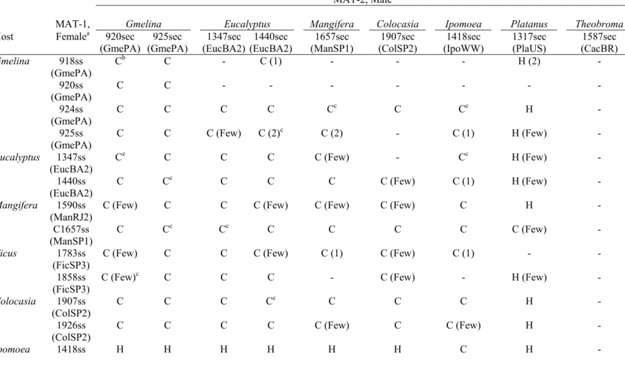
Mating studies
Testers from isolates of each of the five Brazilian host groups successfully crossed with the majority of the other Brazilian testers of opposite mating type (Table 6). In most of these crosses, there were many more than 25 perithecia with normal ascospore masses per plate. In successful crosses, perithecia usually developed within a week and produced thick, creamy ascospore masses at the tips of perithecial necks.
Analyses of single-ascospore progeny from thick, creamy ascospore masses demonstrated that the ascospore masses were not from an induced selfing. Single-ascospore progeny produced colonies of the mycelial morphology of the male and female parents in a roughly 1:1 ratio. Ten sets of progeny from ten crosses were analyzed for three microsatellite markers (Table 7). Eight to 24 progeny were tested for each set of progeny. All sets showed normal segregation (not deviating from 1:1:6 by the chi-square test) for male-parental:female-parental:non-parental types (Table 7), confirming that there had been a successful cross and meiotic segregation of three unlinked loci.
Some of the female testers performed poorly in crosses, perhaps through loss of femaleness (poor protoperithecia or perithecia development). Four female/MAT-1 testers (C925, C1590, C1783, and C1858) usually produced only a few perithecia per plate when paired with what appeared to be compatible MAT-2 testers (Table 6). Three of the four Gmelina female/MAT-1 testers (C918, C920, and C925) formed few or no perithecia with ascospores when spermatized with MAT-2 testers from eucalyptus, mango, or inhame. However, when MAT-2 Gmelina testers were used as males, they successfully crossed with all of the female/MAT-1 testers from Brazil.
recombinant progeny as determined by microsatellite markers (Table 7). In contrast, when the female/MAT-1 sweet potato tester was spermatized with male/MAT-2 testers of Brazilian isolates, few perithecia were produced, and the ascospore masses from these perithecia were watery, not creamy, and microscopic examination showed there to be misshapen ascospores and aborted asci in the perithecial centrum.
Discussion
Ceratocystis fimbriata populations on mango in São Paulo and Rio de Janeiro and on eucalyptus in Minas Gerais and Bahia have gene diversity (H) values ranging from 0.1889 to 0.3813, comparable to the values (using the same microsatellite markers) of what have been considered natural populations of other homothallic species from the C. fimbriata complex: H = 0.1979 for an Upper Amazon population of C. cacaofunesta (Engelbrecht et al., 2007b) and H = 0.2178 for the eastern USA population of C. platani (Engelbrecht et al., 2004). Using other neutral markers, similar values have been found for putatively natural populations of C. albifundus in South Africa (Roux et al., 2001; Barnes et al., 2005). These values of gene diversity are relatively low, but expected, for homothallic Ceratocystis species (Harrington et al., 1998). Selfing and asexual reproduction, as well as limited dispersal by insects or in insect frass should lead to isolated populations that are highly differentiated from each other and with relatively little gene flow between them (Harrington et al., 1998; Harrington, 2000; Baker et al., 2003). Aside from the eucalyptus populations in Minas Gerais and Bahia and the mango populations from Sao Paulo and southwestern Rio de Janeiro, which appeared to be closely related, most Brazilian populations of C. fimbriata were highly differentiated from each other and showed evidence of limited gene flow. Some of the Brazilian populations were of a single genotype or of only a few closely-related genotypes, and these appear to be the result of introductions by humans on plant propagative material.
crosses between different species of Ceratocystis produce no interaction when paired, or they form only a few perithecia with few ascospores that are misshapen, germinate poorly, and produce colonies of aberrant morphology (Engelbrecht & Harrington, 2005; Harrington & McNew, 1997; Harrington et al., 2001; Johnson et al., 2005). In our crosses, attempts were made to recover single-ascospore progeny and determine if the progeny segregated for colony morphology, which would indicate that the ascospore mass was the result of a crossing of two parents, or if the single ascospore progeny were uniform in appearance, an indication that the ascospore mass was the result of a selfing. Selected progeny were further studied for segregation of three microsatellite markers, and a 6:1:1 segregation ratio confirmed meiotic segregation. Fully interfertile crosses were between representatives of the Brazilian populations and also with C. fimbriata ss, but with one exception, the Brazilian testers were not interfertile with C. platani or C. cacaofunesta. Thus, the Brazilian populations should be considered a single biological species, C. fimbriata sensu stricto, and distinct from C. cacaofunesta and C. platani, which are host-specialized to the native American plant genera Theobroma and Platanus, respectively (Baker et al., 2003; Engelbrecht & Harrington, 2005).
The Brazilian populations most closely related to each other based on genetic distance analyses of populations and genotypes, as well as based on values of theta and estimates of gene flow, were the eucalyptus and mango populations from Minas Gerais, Bahia, São Paulo, and southwestern Rio de Janeiro. The analysis of molecular variance showed that most of the genetic variation among these populations was due to variation among regions and variation within populations, and little of the variation was due to variation among populations within regions. Thus, even in this limited part of Brazil, there are regional differences among populations.
soilborne inoculum was likely in the form of aleurioconidia that was originally formed in the wood of infected natural or cultivated trees, perhaps expelled from the trees by the tunneling of ambrosia beetles or other wood-boring insects (Carvalho, 1938; Viégas, 1960; Iton, 1960, 1961; Rossetto et al., 1997; Goitia & Rosales, 2001; Ocasio-Morales et al., 2007).
Surprisingly, few native hosts of C. fimbriata ss have been identified in Brazil, and then only on cultivated native hosts, such as Hevea brasiliensis (CABI, 2005). However, the high incidence of the disease and relatively high genetic diversity of the pathogen in eucalyptus plantations of Minas Gerais on sites that were recently natural Cerrado vegetation (Machado et al., 2004) suggest that C. fimbriata is native to this relatively dry forest type. At least one of the diverse Bahia populations (EucBA1) was on a site that was previously a farm, but the natural vegetation history of the Bahia populations and the sites of the mango isolates is not known. However, these regions of Bahia, Rio de Janeiro, and São Paulo would have been Mata Atlântica rainforest previous to agriculture or urban development. Thus, the region of the greatest population diversity of C. fimbriata in Brazil spans the very different Cerrado and Mata Atlântica forest types.
One of the mango populations, near the town of São Fidélis in northeastern Rio de Janeiro, differed substantially from the other mango and eucalyptus populations in UPGMA trees and in values of theta and Nm. The level of gene diversity for the São Fidélis population was somewhat lower than for most of the other mango and eucalyptus populations, and it is possible that the São Fidélis population is not natural and the fungus was brought into the area with the recent commercial plantings of mango. The isolates collected from the São Fidélis area were from scattered small farms, so more diversity had been expected for this population. However, much of this region was in pasture before cattle grazing, and the region may not have had much woody vegetation prior to establishment of these small farms. Aside from mango, cultivated annona or sugar-apple (Annona squamosa) is also a host of C. fimbriata in the São Fidélis area (Baker et al., 2003; Silveira et al., 2006), so the host range of this population may differ from the other eucalyptus and mango populations.
value for gene diversity (H = 0.1600) for this population was somewhat less that the values found for most of the mango and eucalyptus populations. Earlier genetic studies have suggested that strains of C. fimbriata and other Ceratocystis species have been readily moved on corms of inhame and related Araceae (Thorpe et al., 2005), and it was earlier speculated that much of the C. fimbriata on inhame in Brazil had originated from São Paulo (Harrington et al., 2005). Limited diversity in the inhame population that we sampled suggests that the genotypes of C. fimbriata found there may have been introduced from some other region on corms, which are the primary means of propagation. Alternatively, selection for aggressiveness (Pariaud et al., 2009) to inhame may have created a genetic bottleneck in an otherwise diverse local population of C. fimbriata in this part of São Paulo.
mango tree in São Paulo, where there are many nurseries producing mango seedlings, Ceratocystis wilt has been recognized for more than 70 years (Costa & Krug, 1935; Viégas, 1960), and the mango pathogen may be native.
Until recently, mango has been reported as a host of C. fimbriata only in Brazil (Viégas, 1960). Recently, serious mortality of mango in Oman and Pakistan has been reported, and anecdotal evidence suggests that the strains in Oman and Pakistan were introduced from Brazil on mango seedlings or grafted material (Al Adawi et al., 2006; Fateh et al., 2006). The DNA sequences of mango isolates from Oman and Pakistan have indicated that the C. fimbriata there is closely related to other South American isolates of C. fimbriata (van Wyk et al. 2007). Although detailed comparisons with Brazilian populations of C. fimbriata ss were not made, it was concluded by van Wyk et al. (2007) that the mango pathogen in Oman and Pakistan should be distinguished from C. fimbriata as a new species, C. manginecans, on the basis of the presence of barrel-shaped conidia. Isolates of the sweet potato strain of C. fimbriata do not produce barrel-shaped conidia (Engelbrecht & Harrington, 2005), but we have found the presence of barrel-shaped conidia to be a variable character in the Latin American clade of C. fimbriata, including isolates from Brazil, which we consider C. fimbriata ss. Thus, the history of Ceratocystis wilt on mango and DNA sequences suggest that C. manginecans is likely based on one or two strains of C. fimbriata from Brazil, perhaps from São Paulo, and until further study, C. manginecans should be considered a synonym of C. fimbriata.
more plantations to more accurately reflect the genetic diversity of the population there.
The fig population from São Paulo was collected from a small area where most of the commercial figs in Brazil are grown. Isolates were collected from various plantations, yet only two genotypes were identified among the 20 isolates studied. Two of the plantations were on sites previously in pasture for cattle, so it is likely that the fungus was introduced on vegetatively-propagated figs in at least these two plantations. The disease in these and the other fig plantations appeared in discrete, circular foci of dead and dying trees, suggesting that one plant had been infected initially and the fungus spread to adjacent plants through root systems.
Isolates of Ceratocystis fimbriata collected from exotic hosts in Brazil have shown substantial variation in rDNA sequences and microsatellite markers, and they have varied in aggressiveness to exotic hosts (Baker et al., 2003; Thorpe 2004; Thorpe et al., 2005). These isolates from Brazil do not appear to be highly host specialized, except that Gmelina isolates from Pará (population GemPA) are particularly aggressive on Gmelina seedlings, and two isolates from eucalyptus (populations EucBA2) were especially aggressive on eucalyptus (Baker et al., 2003; Thorpe, 2004). Zauza et al. (2004) tested 18 commercial clones of the hybrid Eucalyptus grandis×E. urophylla using the same two eucalyptus isolates, and there was significant clone × isolate interaction in the amount of discoloration found in the inoculated hosts. Baker et al. (2003) found that mango and eucalyptus isolates were pathogenic to both mango and eucalyptus, but there was substantial variation in aggressiveness. Thorpe et al. (2005) reported that inhame isolates from Brazil caused significantly greater discoloration than the control inoculations in inhame, whereas mango and fig isolates did not. Baker et al. (2003) reported that C. fimbriata ss from sweet potato was not pathogenic to mango or inhame, and Brazilian isolates from mango, eucalyptus, and Gmelina were not pathogenic to sweet potato or Platanusoccidentalis.
thus our samples are biased towards the portion of the population aggressive to these exotic hosts. These aggressive phenotypes may be further selected as they are spread and maintained on vegetatively propagated material of these same exotic hosts. In the most extreme example of such a genetic bottleneck, it is likely that isolates from sweet potato around the world originated from a single strain (Steimel et al., 2004) that was likely selected by humans on propagative material (storage roots), perhaps taken from northern South America, where the cultivated sweet potato may have originated (Engelbrecht & Harrington, 2005). Thus, the species C. fimbriata ss is based on a single, highly-selected strain of the fungus on sweet potato, taken from an unstudied population, probably a population of wound-colonizers (Johnson et al., 2005; Roux et al., 2007) that may have originally varied in aggressiveness to a wide variety of hosts.
the C. fimbriata complex, and perhaps more species will be recognized in Brazil. However, the evidence to date suggests that much of the variation in Brazil is found among geographically-isolated populations that vary in aggressiveness to certain exotic hosts. The level of aggressiveness to some of these cultivated hosts and the ease of spread of these strains to new regions in propagative or woody material is becoming an increasing concern.
Acknowledgements
This study was funded CNPq (Conselho Nacional de Desenvolvimento Científico e Tecnológico) and the National Science Foundation through grants DEB-987065 and DEB-0128104. We thank Christine Engelbrecht for providing testers and advice and Joe Steimel, Sujin Kim, and Doug McNew for technical assistance. Daniel Breda Binotti, Nilton Junqueira, Silvado Felipe da Silveira, José Pedro Pimentel, Paulo Sergio Brioso, Edson Furtado, and Edival Zauza provided invaluable assistance in collection of material and conducting isolations. We also thank the following Brazilian forest companies and their employees for their invaluable assistance: Bianca Vique Fernandes (V&M Florestal), José Urbano (RIMA Industrial S.A.), Raul Cesar Nogueira Melido (Votorantim Siderurgia), Reginaldo Gonçalves Mafia (Aracruz Celulose S.A.), Suzano Papel e Celulose, and Plantar S.A.
References
Al Adawi AO, Deadman ML, Rawahi AK, Maqbali YM, Al Jahwari A.A., Ak Saadi BA, Al Amri IS, Wingfield MJ, 2006. Aetiology and causal agents of mango sudden decline disease in Sultanate of Oman. European Journal of Plant Pathology 116, 247-54.
Alfenas AC, Zauza EAV, Mafia RG, Assis TF, 2004. Clonagem e doenças do eucalipto.Viçosa: Viçosa, MG: Universidade. Federal de Viçosa.
Barnes I, Gaur A, Burgess T, Roux J, Wingfield BD, Wingfield MJ, 2001. Microsatellite markers reflect intra-specific relationships between isolates of the vascular wilt pathogen Ceratocystis fimbriata. Molecular Plant Pathology 2, 31925.
Barnes J, Roux J, Wingfield BD, O'Neill M, Wingfield MJ, 2003. Ceratocystis fimbriata infecting Eucalyptus grandis in Uruguay. Australian Plant Pathology 32, 361-366.
Barnes I, Nakabonge G, Roux J, Wingfield BD, Wingfield MJ, 2005. Comparison of populations of the wilt pathogen Ceratocystis albifundus in South Africa and Uganda. Plant Pathology 54, 189-95.
Barton NH, Slatkin M, 1986. A quasi-equilibrium theory of the distribution of rare alleles in a subdivided population. Heredity 56, 40915.
CAB International, 2005. Ceratocystis fimbriata (original text prepared by T.C. Harrington). In: Crop Protection Compendium. CAB International, Wallingford, UK.
Castellani A, 1939. Viability of some pathogenic fungi in distilled water. Journal of Tropical Medicine and Hygiene 24, 270-76.
Carvalho MB, 1938. Sobre dois insetos nocivos à mangueira. Boletim da Secretaria da Agricultura. Indústria e Comércio do Estado de Pernambuco. Pernambuco. 3, 130-32.
Costa AS, Krug HP, 1935. Eine durch Ceratostomella hervorgerufene Welkekrankheit der Crotalaria juncea in Brasilien. Phytopathologische Zeitschrift Berlin. 8, 507-13.
DeScenzo RA, Harrington TC, 1994. Use of (CAT)5 as a DNA fingerprinting probe
for fungi. Phytopathology 84, 534-40.
Dettman JR, Taylor JW, 2004. Mutation and evolution of microsatellite loci in Neurospora. Genetics 168, 1231-48.
Engelbrecht CJB, Harrington TC, 2005. Intersterility, morphology, and taxonomy of Ceratocystis fimbriata on sweet potato, cacao, and sycamore. Mycologia 97, 57-69.
Engelbrecht CJ, Harrington TC, Alfenas AC, 2007a. Ceratocystis wilt of cacao - a disease of increasing importance. Phytopathology 97, 1648-49.
Engelbrecht CJ, Harrington TC, Alfenas AA, Suarez C, 2007b. Genetic variation of populations of the cacao wilt pathogen, Ceratocystis cacaofunesta. Plant Pathology 56, 923-33.
Excoffier L, Laval G, Schneider S, 2005. Arlequin ver. 3.0: An integrated software package for population genetics data analysis. Evolutionary Bioinformatics Online 1, 47-50.
Excoffier L, Smouse, PE, Quattro JM, 1992. Analysis of molecular variance inferred from metric distances among DNA haplotypes: application to human mitochondrial DNA restriction data. Genetics 131, 479-91.
Fateh FS, Kazmi MR, Ahmad I, Ashraf M, 2006. Ceratocystis fimbriata isolated from vascular bundles of declining mango tree in Sindh, Pakistan. Pakistan Journal of Botany 38, 1257-59.
Fearnside PM, 1988. Jari at age 19: Lessons for Brazil's silvicultural plans at Carajás. Interciencia 13, 12-24.
Felsenstein J, 1989. PHYLIPphylogeny inference package (Version 3.2). Cladistics 5, 164-6.
Felsenstein J, 1993. PHYLIP (phylogeny inference package) version 3.5c. Distributed by the author. Department of Genetics, University of Washington, Seattle, Washington.
Ferreira FA, Demuner AMM, Demuner NL, Pigato S, 1999. Murcha de Ceratocystis em eucalipto no Brasil. Fitopatologia Brasileira 24, 284.
Ferreira FA, Maffia LA, Barreto RW, Demuner NL, Pigatto S, 2006. Sintomatologia da murcha de Ceratocystis fimbriata em eucalipto. Revista Árvore 30, 155-62. Giraldo EA, 1957. La llaga macana del tronco del cacao. Acta Agronomica 7,
71-103.
Goita W, Rosales CJ, 2001. Relacion entre la incidencia de escolitidos y la necrosis del cacao em Aragua. Manejo Integrado de Plagas 62, 65-71.
Halsted BD, 1890. Some fungous diseases of the sweet potato. The black rot. New Jersey Agriculture Experiment Station Bulletin 76, 7-14.
Harrington TC, McNew DL, 1997. Self-fertility and uni-directional mating-type switching in Ceratocystis coerulescens, a filamentous ascomycete. Current Genetics 32, 52-9.
Harrington TC, McNew DL, 1998. Partial interfertility among the Ceratocystis species on conifers. Fungal Genetics and Biology 25, 44-53.
Harrington TC, Pashenova NV, McNew DL, Steimel J, Konstantinov MY, 2002. Species delimitation and host specialization of Ceratocystis laricicola and C. polonica to larch and spruce. Plant Disease 86, 418-22.
Harrington TC, Rizzo DM, 1999. Defining species in the fungi. In: Worrall JJ, ed. Structure and Dynamics of Fungal Populations. Dordrecht, Netherlands: Kluwer Press, 43-71.
Harrington TC, Steimel J, Kile G, 1998. Genetic variation in three Ceratocystis species with outcrossing, selfing and asexual reproductive strategies. European Journal of Forest Pathology 28, 217-26.
Harrington TC, Thorpe DJ, Marinho VLA, Furtado EL, 2005. First report of black rot of Colocasia esculenta caused by Ceratocystis fimbriata in Brazil. FitopatologiaBrasileira 30, 88-9.
Harrington TC, 2009. The genus Ceratocystis. Where does the oak wilt fungus fit? Proceedings of the 2nd National Oak Wilt Symposium, Appel, D.N. and R.F. Billings (eds.). Austin, TX. (in press).
Iton EF, 1960. Studies on a wilt disease of cacao at River Estate. II. Some aspects of wind transmission. In: Annual Report on Cacao Research, 1959-1960. St Augustine, Trinidad: Imperial College of Tropical Agriculture, University of the West Indies. 47-58.
Iton EF, Conway GR, 1961. Studies on a wilt disease of cacao at River Estate III. Some aspects of the biology and habits of Xyleborus spp. and their relation to disease transmission. In: Annual Report on Cacao Research, 1959-1960. St. Augustine, Trinidad: Imperial College of Tropical Agriculture. 59-65.
Johnson JA, Harrington TC, Engelbrecht CJB, 2005. Phylogeny and taxonomy of the North American clade of the Ceratocystis fimbriata complex. Mycologia 97, 1067-92.
Laia ML, Alfenas AC, Harrington TC, 2000. Isolation, detection in soil, and inoculation of Ceratocystis fimbriata, causal agent of wilting, die-back and canker in Eucalyptus. (Abstr.). Fitopatologia Brasileira 25, 384.
Machado RB, Ramos Neto MB, Pereira PGP Caldas EF, Gonçalves DA, Santos NS, Tabor K, Steininger M, 2004. Estimativas de perda da área do Cerrado brasileiro. Relatório técnico. Conservação Internacional, Brasília, DF. 26p.
Marin M, Castro B, Gaitan A, Preisig O, Wingfield BD, Wingfield MJ, 2003. Relationships of Ceratocystis fimbriata isolates from Colombian coffee-growing regions based on molecular data and pathogenicity. Journal of Phytopathology 151, 395405.
McDonald BA, 1997. The population genetics of fungi: tools and techniques. Phytopathology 87, 448-53.
Moller WJ, DeVay JE, 1968. Carrot as a species-selective isolation medium for Ceratocystis fimbriata. Phytopathology 58, 123-24.
Moller WJ, DeVay JE, Backman PA, 1969. Effect of some ecological factors on Ceratocystis canker in stone fruits. Phytopathology 59, 938-42.
Muchovej JJ, Albuquerque FC, Ribeiro GT, 1978. Gmelina arborea a new host of Ceratocystis fimbriata. Plant Disease Reporter 62, 717-719.
Murray MG, Thompson WF, 1980. Rapid isolation of high molecular weight DNA. Nucleic Acids Research 8, 4321-25.
Ocasio-Morales RG, Tsopelas P, Harrington TC, 2007. Origin of Ceratocystis platani on native Platanusorientalis in Greece and its impact on natural forests. PlantDisease 91, 901-4.
Pariaud B, Ravigné V, Halkett F, Goyeau H, Carlier J, Lannou C. 2009. Aggressiveness and its role in the adaptation of plant pathogens. Plant Pathology 58, 409-24.
Ribeiro IJA, Coral FJ, 1968. Estudo preliminary da ação do fungo Ceratocystis fimbriata Ell. and Halst., causador da seca da mangueira (Mangifera indica L.), sobre cacaueiros (Theobroma cacao L.). Bragantia 27, 87-9.