WILKA MESSNER DA SILVA BISPO
PHYSIOLOGICAL AND BIOCHEMICAL ASPECTS OF THE MANGO-Ceratocystis fimbriata INTERACTION
Tese apresentada à Universidade Federal de Viçosa, como parte das exigências do Programa de Pós-Graduação em Fisiologia Vegetal, para obtenção do título de Doctor Scientiae.
VIÇOSA
Fichi citilográfici prepiridi peli Biblioteci Centril di Universidide Federil de Viçosi - Câmpus Viçosi
T
Bispo, Wilka Messner da Silva, 1985-B662p
2014 Ceratocystis fimbriataPhysiological and biochemical aspects of the mango- interaction / Wilka Messner da Silva Bispo. - Viçosa, MG, 2014.
xi, 95 f. : il. ; 29 cm.
Orientador : Fabrício de Ávila Rodrigues.
Tese (doutorado) - Universidade Federal de Viçosa. Inclui bibliografia.
1. Manga - Doenças e pragas. 2. Fluorescência. 3. Clorofila. 4. Fotossíntese. 5. Seca-da-mangueira. I. Universidade Federal de Viçosa. Departamento de
Biologia Vegetal. Programa de Pós-Graduação em Fisiologia Vegetal. II. Título.
CDD 22. ed. 634.44
WILKA MESSNER DA SILVA BISPO
PHYSIOLOGICAL AND BIOCHEMICAL ASPECTS OF THE MANGO-Ceratocystis fimbriata INTERACTION
Tese apresentada à Universidade Federal de Viçosa, como parte das exigências do Programa de Pós-Graduação em Fisiologia Vegetal, para obtenção do título de Doctor Scientiae.
APROVADA: 19 de fevereiro de 2014.
________________________________ ________________________________ Prof. Fábio Murilo DaMatta Prof. Wagner Luiz Araújo
(Coorientador)
________________________________ ________________________________ Prof. Gleiber Quintão Furtado Profª. Diolina Moura Silva
________________________________ Prof. Fabrício Ávila Rodrigues
AGRADECIMENTOS
Aos meus pais e irmão, por todo o apoio, amor, paciência e incentivo;
À Universidade Federal de Viçosa, aos departamentos de Biologia Vegetal e Fitopatologia e, especialmente, ao Programa de Pós-graduação em Fisiologia Vegetal, pela oportunidade, amparo e auxílio na realização do curso;
À Coordenação de Aperfeiçoamento de Pessoal de Nível Superior e ao Conselho Nacional de Desenvolvimento Científico e Tecnológico, pelo apoio financeiro;
Ao meu orientador, Fabrício de Ávila Rodrigues, um exemplo de profissional e pessoa, pelos ensinamentos e pela amizade;
Aos professores do PPG-Fisiologia Vegetal pelos ensinamentos durante o curso, em especial aos Profs. Fábio Murilo DaMatta e Raimundo Santos Barros, pessoas muito queridas;
Aos integrantes do “Grupo Interação Planta-Patógeno”, pela ajuda e amizade;
Aos integrantes do “Grupo Café”, pela ajuda, amizade e pelas várias risadas ao longo do curso;
Ao sr. Mário e sra. Neuza, pela amizade e grande apoio durante os experimentos;
Aos técnicos Bruno, Camilo e Macabeu, pela ajuda e pelas histórias;
A todos os colegas de curso que tornaram essa jornada engrandecedora e que direta ou indiretamente tiveram papel fundamental na realização deste trabalho.
... e a Tabatã!
BIOGRAFIA
SUMÁRIO
RESUMO ... viii
ABSTRACT ... x
GENERAL INTRODUCTION... 1
LITERATURE CITED ... 3
CHAPTER 1 - Water relations and the photosynthetic performance of mango cultivars with different levels of resistance to Ceratocystis fimbriata infection ... 5
ABSTRACT ... 5
INTRODUCTION ... 7
MATERIALS AND METHODS ... 9
Plant material... 9
Inoculation procedure ... 9
Relative lesion indices. ... 9
Water potential and apparent hydraulic conductance ... 10
Leaf gas exchanges and chlorophyll a fluorescence measurements ... 10
Determination of photosynthetic pigments... 11
Total free amino acids (TFA) and proline (Pro) determinations ... 12
Lipid peroxidation assay ... 12
Vein density... 13
Experimental design and statistics ... 13
RESULTS ... 14
Analysis of variance ... 14
Disease assessments ... 14
Water relations variables ... 14
Gas exchanges and chlorophyl a fluorescence ... 14
Concentration of pigments, TFA, proline and lipid peroxidation ... 15
Veins density ... 15
DISCUSSION ... 16
ACKNOWLEDGMENTS ... 18
LITERATURE CITED ... 19
CHAPTER 2 – Pathogen-induced alterations in the antioxidative system of mango cultivars
with different levels of resistance to Ceratocystis fimbriata infection ... 33
ABSTRACT ... 33
INTRODUCTION ... 35
MATERIALS AND METHODS ... 37
Plant material... 37
Inoculation procedure ... 37
Relative lesion indices ... 37
Leaf gas exchanges ... 38
Biochemical assays ... 38
Lipid peroxidation assay ... 38
Determination of hydrogen peroxide (H2O2) concentration... 39
Determination of ascorbate (AsA) ... 39
Determination of total glutathione concentration (GSH+GSSG) ... 39
Determination of total phenolics (TP) concentration ... 40
Enzyme extractions and assays ... 40
Experimental design and statistics ... 43
RESULTS ... 44
Relative lesion indices ... 44
Leaf gas enchanges ... 44
Lipid peroxidation and H2O2 concentration ... 44
Ascorbate and total glutathione concentrations ... 44
Total phenolics concentration ... 44
Enzyme activities... 45
DISCUSSION ... 46
ACKNOWLEDGMENTS ... 49
LITERATUTE CITED ... 50
LIST OF TABLES AND FIGURES... 59
CHAPTER 3 – Photosynthetic performance and carbohydrate metabolism on leaves and stems of mango cultvars with different levels of basal resistance to Ceratocystis fimbriata infection ... 63
ABSTRACT ... 63
MATERIALS AND METHODS ... 68
Plant material... 68
Inoculation procedure ... 68
Relative lesion indices ... 69
Leaf gas exchanges and chlorophyll a fluorescence measurements ... 69
Biochemical assays ... 70
Extractions and determination of carbohydrates ... 70
Enzyme extractions and assays ... 71
Experimental design and statistics ... 72
RESULTS ... 73
Disease assesments ... 73
Gas exchanges and chlorophyll a fluorescence ... 73
Leaf starch concentration ... 73
Stem starch concentration ... 73
Leaf glucose, fructose and sucrose concentrations ... 74
Stem glucose, fructose and sucrose concentrations ... 74
Enzyme activity ... 74
DISCUSSION ... 76
ACKNOWLEDGMENTS ... 79
LITERATURE CITED ... 80
LIST OF TABLES AND FIGURES... 87
RESUMO
BISPO, Wilka Messner da Silva, D.Sc., Universidade Federal de Viçosa, Fevereiro de 2014. Aspectos fisiológicos e bioquímicos da interação mangueira-Ceratocystis fimbriata. Orientador: Fabrício Ávila Rodrigues. Coorientador: Fábio Murilo DaMatta.
A seca-da-mangueira, causada por Ceratocystis fimbriata Ellis & Halsted, tem grande impacto econômico, sendo uma das doenças que mais afeta a produção de manga no mundo. Observações empíricas têm mostrado grande variabilidade entre as cultivares em termos de seu nível basal de resistência à seca-da-mangueira, no entanto, poucos esforços tem sido realizados no sentido de elucidar os mecanismos fisiológicos envolvidos na resposta do hospedeiro ao processo de infecção. Considerando a lacuna de informações existente na literatura acerca da fisiologia da doença, este estudo teve como objetivo principal, investigar alterações fisiológicas nas plantas das cultivares de manga Ubá, Tommy Atkins e Palmer, as quais apresentam diferentes níveis de resistência basal à seca-da-mangueira. As relações entre a severidade da doença e as mudanças fisiológicas do hospedeiro foram avaliadas em três experimentos distintos, em que a cv. Ubá, considerada resistente, confrontou as cvs. Tommy Atkins (moderadamente resistente) e Palmer (suscetível). Os dois primeiros experimentos compararam o comportamento das cvs. Ubá e Tommy Atkins em relação às possíveis alterações no status hídrico das plantas e na ativação do sistema antioxidativo em resposta a infecção por C. fimbriata. Em suma, diminuições mais pronunciadas na condutância hidráulica aparente, na assimilação líquida de CO2 e na condutância estomática foram
ABSTRACT
BISPO, Wilka Messner da Silva, D.Sc., Universidade Federal de Viçosa, February, 2014. Physiological and biochemical aspects of the mango-Ceratocystis fimbriata interaction. Advisor: Fabrício Ávila Rodrigues. Co-advisor: Fábio Murilo DaMatta.
The mango (Mangifera indica L.) wilt, caused by Ceratocystis fimbriata Ellis & Halsted, has great economic impact, being one of the most important diseases affecting mango production worldwide. Empirical observations have shown great variability among mango cultivars in terms of their basal level of resistance to the mango wilt; nevertheless, to date, few attempts have been made to elucidate the physiological mechanisms underlying how mango respond to fungal infection. Considering the lack of information about the mango wilt physiology, this study aimed to investigate some physiological alterations in mango plants from cultivars Ubá, Tommy Atkins and Palmer, which present different levels of basal resistance to the mango wilt. The relationships between disease severity and the host physiological changes were assessed in three distinct experiments, in which cv. Ubá, considered resistant, confronted both cvs. Tommy Atkins (moderately resistant) and Palmer (susceptible). The first two experiments confronted cvs. Ubá and Tommy Atkins for the possible changes in plant water relations and the differences in the antioxidative responses upon fungal inoculation. The more pronounced decreases in apparent hydraulic conductance, net CO2 assimilation rate and
stomatal conductance were observed in cv. Tommy Atkins relative to cv. Ubá upon fungal inoculation, as well as decreases in total chlorophyll concentration and increases in total free amino acids, proline concentration and lipid peroxidation. Plants from cv. Tommy Atkins also presented more prominent increases in the activity of the superoxide dismutase (SOD), non-specific peroxidases (POX), ascorbate peroxidase (APX), glutathione peroxidase (GPX) and glutathione reductase (GR) enzymes and concentrations of metabolites (hydrogen peroxide and total phenolics) related to the oxidative stress responses. These modifications were minimal in cv. Ubá. Greater disease severity was also found for plants from cv. Palmer in the third experiment when compared to cv. Ubá. In addition to reduced photosynthetic performance, leaves from cv. Palmer showed decreased concentrations of starch and increases in hexoses concentrations as disease progressed, in accordance with the reduced activity of
ADP-glucose pyrophosphorylase (AGPase) and increased activity of both acid and alkaline
showed no changes in carbohydrates concentration and on the activity of enzymes, such as
those involved in the synthesis of starch (AGPase) and sucrose (sucrose phosphate synthase,
GENERAL INTRODUCTION
Due to favorable environmental conditions, Brazil is one of the largest producers of fruits worldwide, and the mango (Mangifera indica L.) stands out among the several cultivated and traded fruit species, receiving growing appreciation, due to the enhanced acceptance of internal and foreign markets (Oliveira et al, 2002; Poll et al, 2011). The Brazilian fruit production, as also observed in several countries, is intended, primarily, to the in natura market and there is great interest in producing and marketing exportable cultivars at the expense of those used for industrialization purposes. Among the different cultivars the cv. Tommy Atkins is the most demanded by the international market and also by the large consuming centers of the Centre-South (Souza et al., 2002), concentrating about 90% of the cropped area in Brazil. This cultivar presents great hardiness, attractive color, good conservation in the post-harvest period, in addition to production stability for consecutive years (Braz et al., 2008). Although not the main focus, great attention has been given to the use of the fruit in industrial processing and manufacturing of fruit pulp, being preferred for this destination, varieties which present high pulp yield, high soluble solids and absence of fibers (Braz et al., 2008). Among the Brazilian cultivars, the Ubá represents the most widespread as raw material for the processing industries (Benevides et al., 2008).
Despite the successful development of mango in several production centers for both fresh consumption and industry, the occurrence of diseases causes many losses in total yield. Additionally, diseases such as anthracnose and floral and vegetative malformation are becoming relevant in many producing regions (Araújo et al., 2004). The mango wilt, caused by Ceratocystis fimbriata Ellis and Halsted (Halsted, 1890), is among the most important mango tree diseases. This disease causes the death of plants in different growth stages from seedlings to mature trees, with the infection starting from the crown, with the aid of a beetle vector (Hypocryphalus mangiferae) or from the root system (Gallo et al., 2002; Masood et al., 2009). The gum extrusion from the points of damage in affected branches, necrosis and browning of vascular tissue are the initial symptoms of the disease (AL Adawi et al., 2006; Al-Sadi et al., 2010). The most characteristic symptom is the progressive drying of the plant, which initiates in branches of small caliber and progresses throughout the entire canopy, provoking the yellowing of leaves, wilting and drying of affected branches (Cunha et al., 2000; Batista et al., 2008).
LITERATURE CITED
Al-Adawi, A.O.; Deadman, M. L.; Al-Rawahi, A. K.; Al-Maqbali, Y. M.; Al-Jahwari, A. A.; Al-Saadi, B. A.; Al-Amri, I. S.; Wingfield, M. J. Aetiology and causal agents of mango sudden decline disease in the Sultanate of Oman. European Journal of Plant Pathology, v. 116, p. 247-254, 2006.
Al-Sadi, A. M.; Al-Ouweisi, F. A.; Al-Shariani, N. K.; Al-Adawi, A. O.; Kaplan, E. J.; Deadman, M. L. Histological changes in mango seedlings following infection with Ceratocystis manginecans, the cause of mango decline. Journal of Phytopathology, v. 158, p. 738–743, 2010.
Araújo, J. L. P.; Correia, R. C.; Guimarães, J.; Araújo, E. P. Production cost analysis and commercialization of mangos for exporting produced in the Sub-middle São Francisco Region, Brazil. Acta Horticulturae, v. 645, p. 379-381, 2004.
Batista, D. C.; Terao, D.; Barbosa, M. A. G.; Barbosa, F. R. Seca-da-mangueira: detecção, sintomatologia e controle. Petrolina: Embrapa Comunicado Técnico 138, 2008.
Benevides, S. D.; Ramos, A. M.; Stringheta, P. C.; Castro, V. C. Qualidade da manga e polpa da manga Ubá. Ciência e Tecnologia de Alimentos, v. 28, p. 571-578, 2008.
Braz, V. B.; Nunes, E. S.; Vieira, G.; Ribeiro Júnior, J. I.; Bertini, L. A.; Couto, F. L. D. Indução do amadurecimento de mangas cv. Tommy Atkins e cv. Ubá pela aplicação de ethephon pós-colheita. Bragantia, v.67, p.225-232, 2008.
Cunha, M. M. da; Santos Filho, H. P.; Nascimento, A. S. do. (Org.). Manga: fitossanidade. Brasília: Embrapa Comunicação para Transferência de Tecnologia, 2000. p. 25-47 (Frutas do Brasil; 6).
Halsted, B. D. Some fungous diseases of the sweet potato. New Jersey Agricultural College Experiment Station, Bulletin 76, pp. 25-27, 1890.
Masood, A.; Shafqat, S.; Asif, S.; Mudassar, A. Life cycle and biology of mango bark beetle, Hypocryphalus mangifera Stebbing: as a possible vector of sudden death disease of mango. Pakistan Journal of Zoology, v. 41, p. 281-288, 2009.
Mcelrone, A. J.; Sherald, J. L.; Forseth, I. N. Interactive effects of water stress and xylem-limited bacterial infection on the water relations of a host vine. Journal of Experimental Botany, v. 54, p. 419-430, 2003.
Nogués, S.; Cotxarrera, L.; Alegre, L.; Trillas, M. I. Limitations to photosynthesis in tomato leaves induced by Fusarium wilt. New Phytologist, v. 154, p. 461-470, 2002.
Oliveira, F. C.; Coelho, E. F.; Vasconcelos, L. F. L.; Araújo, E. C. E.Produção de manga sob diferentes regimes de irrigação, em condições subúmidas. Revista Brasileira de Engenharia Agrícola e Ambiental, v. 6, p. 390-396, 2002.
Poll, H; Vencato, A. Z.; Kist, B. B.; Santos, C.; Carvalho, C.; Reetz, E. R.; Beling, R. R. Anuário Brasileiro da Fruticultura. Santa Cruz do Sul: Editora Gazeta Santa Cruz, 2011, 128 p.
CHAPTER 1
Submitted as original paper to Phytopathology
Water relations and photosynthetic performance of mango cultivars with different levels of resistance to Ceratocystis fimbriata infection
Wilka M. S. Bispo, Leonardo Araújo, Isaías S. Cacique, Fábio M. DaMatta and Fabrício A. Rodrigues
First and fourth authors: Universidade Federal de Viçosa, Departamento de Biologia Vegetal, Viçosa, Minas Gerais, Zip Code 36570-000, Brazil; second, third and fifth authors: Universidade Federal de Viçosa, Departamento de Fitopatologia, Viçosa, Minas Gerais, Zip Code 36570-000, Brazil.
ABSTRACT
Bispo, W. M. S., Araújo, L., Cacique, I. S., DaMatta, F. M., and Rodrigues, F. A. Water relations and photosynthetic performance of mango cultivars with different levels of resistance to Ceratocystis fimbriata infection. Phytopathology 104:xx-xx
The mango (Mangifera indica L.) wilt is caused by Ceratocystis fimbriata Ellis & Halsted and it is of great economic importance in mango production. Considering that the physiological effects of this vascular disease are poorly characterized, this study aimed to investigate the physiological alterations in mango (Mangifera indica L.) leaves from cultivars Ubá and Tommy Atkins, which present different levels of resistance to the mango wilt. The relationships of disease severity to water relations, gas exchange and chlorophyll a fluorescence parameters were evaluated over 30 days. Leaf samples were collected to determine the lipid peroxidation, total pigments, total free amino acids and proline concentration. Briefly, more pronounced decreases in apparent hydraulic conductance as well as in the net CO2 assimilation rate and stomatal conductance were observed in cv. Tommy
and gas exchange coupled with symptoms of oxidative stress at advanced stages of fungal infection. In sharp contrast, cv. Ubá (resistant) was better able to postpone pathogen spread and xylem occlusion, and as a result, it suffered less from the dehydration imposed by fungal infection compared with the most susceptible cultivar.
Additional keywords: gas exchange; chlorophyll a fluorescence; mango wilt; osmotic adjustment, vascular pathogen; water potential.
INTRODUCTION
Due to the great variety of climatic conditions, Brazil is one of the largest producers of tropical fruits in the world, with a harvested area bigger than 2.5 million hectares (49). Among several cultivated and traded fruit species, mango (Mangifera indica L.) has experienced a growing domestic demand and increasing forecasts in activities related to production following the acceptance of the international market (48). The cultivar „Tommy Atkins‟ is the most in-demand by the international market and also by the high consumption centers of Mid-South Brazil (10,57), where approximately 80% of the cultivated area is concentrated. The remaining areas are used for the production of other varieties such as „Haden‟, „Palmer‟ and „Keitt‟. Some Brazilian cultivars are also cultivated such as Ubá (46), which is widely used as a raw material for processing industries because of its characteristics like high pulp yield, high soluble solid contents, and low fibers (7,10).
In addition to the successful development of various production hubs for both fresh consumption and industry, diseases cause significant yield losses and, consequently, reduced producer profit. Mango wilt is caused by the fungus Ceratocystis fimbriata Ellis & Halsted (27), and it is one of the most important diseases affecting mango trees worldwide (47). This disease causes death in plants at different development stages, from seedlings to mature trees (25). The initial disease symptoms are gummosis from the bark, bark splitting, streaking and necrosis of the vascular tissues beneath the gummosis (36,37). The most characteristic symptom of this disease is the progressive drying of the plant, which begins in small branches and progresses towards the trunk up to the canopy (6,17).
Tree death is associated with the blockage of vascular tissues by physical impairments such as gels, tyloses and fungal material, which may result in xylem embolism and a reduction in the useful conducting area of the xylem vessels (43,44,58). These dysfunctions usually lead to decreases in hydraulic conductivity and ultimately impair the flow of water and nutrients to the upper canopy (44,54). Usually, these alterations result in decreases in leaf water potential, thereby leading to internal water deficits (39,41). Water stress substantially impairs plant growth in addition to altering the photosynthetic activity and metabolism as a whole. Overall, decreases in photosynthetic performance under water deficit conditions have, to a large extent, been attributed to diffusion limitations through the stomata and mesophyll; however, only severe drought conditions leading to biochemical limitations in the CO2
MATERIALS AND METHODS
Plant material. Mango plants of approximately 1 year old from cultivars Ubá and Tommy Atkins, which are known to be resistant and moderately resistant, respectively, to C. fimbriata (14,50), were obtained from a commercial orchard (Dona Euzébia, Minas Gerais State, Brazil). Both cultivars were grafted onto plants from cv. Imbú, widely used as rootstock in the Zona da Mata region, Minas Gerais State, Brazil. The saplings were transplanted into plastic pots containing 8 kg of substrate consisting of a mixture of soil, sand and manure in a 2:1:1 proportion. The plants were kept in a greenhouse (temperature of 30 ± 2°C and relative humidity of 70 ± 5%) for two months before the beginning of the experiments. Plants were irrigated and fertilized as needed.
Inoculation procedure. The C. fimbriata isolate CEBS15, used to inoculate the plants, was obtained from symptomatic mango plants collected in the city of Brejo Santo (07° 29' 34" S, 38° 59' 06" W), Ceará State, Brazil. The isolate was preserved by Castellani's method (19). Plugs of malt extract agar medium containing fungal mycelia were transferred to Petri dishes containing potato dextrose agar (PDA). After three days, PDA plugs containing fungal mycelia were transferred to new Petri dishes containing the same culture medium and were maintained in an incubator chamber (at a temperature of 25°C with a 12 h photoperiod) for 14 days.
Plants were inoculated according to Al-Sadi et al. (3) with a few modifications. Stem disks (10 mm in diameter and approximately 2 mm in width) were removed from the stems with the aid of a punch at approximately 5 cm above the graft scar. A PDA plug (10 mm in diameter) obtained from a 14-day-old colony of each C. fimbriata isolate was carefully placed in the punch hole. Each hole containing a PDA plug with fungal mycelia was carefully covered with a piece of moistened cotton and then wrapped with parafilm to maintain adequate moisture for fungal infection. The disks used to inoculate each plant were taken from the middle portion of each fungal colony to make the inoculation as homogeneous as possible. Holes on the stems of plants receiving only PDA medium plugs served as the control treatment.
the length (in cm) of the internal necrotic tissue using digital calipers. The upward relative lesion length (URLL) and the downward relative lesion length (DRLL) were determined as the ratio between the length from the graft scar to the top of the stem (LGST) and the lesion length (LL) in the same interval (upward and downward) from the inoculation point according to the following formula: URLL or DRLL = LL × 100/LGST. The plants were standardized to a length of 20 cm (the distance from the graft scar to the top of the stem). The radial fungal colonization (RFC) was determined as the length of the necrotic tissue in relation to the total stem diameter × 100.
Water potential and apparent hydraulic conductance. The predawn leaf water potential (Ψpd) was determined at 2, 10, 20 and 30 days after inoculation (dai) with a Scholander-type
pressure chamber (model 1000, PMS Instruments, Albany, NY, USA). One single leaf from ten replications (plants) of each treatment was collected from the same plant portion for the measurements. Leaf samples were stuck through the petiole to the pressure chamber, which was slowly pressurized until a droplet of a translucent liquid appeared on the cut surface. The displayed pressure at the moment of liquid surfacing was recorded as a negative value of leaf water potential. As previously reported by Castro Neto et al. (15), the measurements of leaf water potential on mango leaves using a pressure chamber is problematic due to the presence of latex and this was especially observed for cv. Tommy Atkins. Thus, prior to the measurements, the latex drops were carefully removed with the aid of a piece of filter paper. The apparent hydraulic conductance (KL) was expressed as the ratio between the total plant
transpiration (gravimetrically estimated) between predawn and midday and the differences in water potential (∆Ψw) at the same interval (45,56). The soil surface in the plastic pots was
covered with a plastic film to minimize evaporation.
Leaf gas exchanges and chlorophyll a fluorescence measurements. The net carbon assimilation rate (A), stomatal conductance to water vapor (gs), internal-to-ambient CO2
concentration ratio (Ci/Ca) and transpiration rate (E) were measured in fully expanded leaves
with an infrared CO2/H2O gas analyzer (LI 6400, Li-Cor, Lincoln, NE) equipped with a
blue/red light source (Li-6400-02B). The measurements were conducted at ambient temperature and CO2 conditions under artificial light (1000 µmol photons m-2 s-1) from
replication, yielding similar values, but only one data set was recorded. The intrinsic (A/gs)
and instantaneous water use efficiency (A/E) at the aforementioned level of irradiance was also calculated.
Chlorophyll a fluorescence measurements were determined using a portable pulse amplitude modulation fluorometer (MINIPAM, Heinz Walz GmbH, Effeltrich, Germany) on the same leaves used for the gas exchange measurements. After 40 min of dark adaptation, the leaf tissues were exposed to a weak modulated measuring beam (0.03 µmol m-2 s-1) to determine the initial fluorescence (F0). Next, a saturating white light pulse of 6,000 µmol m-2
s-1 was applied for 0.8 s to ensure maximum fluorescence emission (Fm). From these initial
measurements, the maximum quantum efficiency of PSII photochemistry for dark-adapted leaves was calculated as follows: (Fv/Fm) [(Fm - F0)/Fm)]. The steady-state fluorescence yield
(Fs), the light-adapted maximum fluorescence (Fm‟), which was measured after 0.8 s of
saturating white light pulse (6,000 μmol m-2
s-1), and the light-adapted initial fluorescence (F0‟) estimated according to Oxborough and Baker (42) were determined in light-adapted
leaves. From these parameters, the efficiency of excitation energy capture by open PSII reaction centers (Fv‟/Fm‟) was calculated [(Fm‟ - F0‟/Fm‟)]. The estimated fraction of open
PSII centers (qL) was calculated as [(Fm‟ - Fs)*F0‟/ (Fm‟ - F0‟)*Fs] (30), and the
non-photochemical quenching coefficient (NPQ) was calculated as [(Fm/Fm‟) - 1)] (9). The actual
quantum yield of PSII electron transport (PSII) was computed as [(Fm‟ – Fs)/Fm‟], from
which the electron transport rate (ETR) was calculated as (PSII*PPFD*f*α), where f is a factor
that accounts for the partitioning of energy between PSII and PSI and is assumed to be 0.5, which indicates that the excitation energy is equally distributed between the two photosystems; α is the leaf absorbance by the photosynthetic tissues and is assumed to be 0.84 (38).
Determination of photosynthetic pigments. The concentrations of chlorophylls (Chl) a and b and carotenoids were determined using dimethyl sulfoxide (DMSO) as an extractor (59). Five leaf disks (10 mm in diameter) were punched from each leaf that had been previously used for gas exchange and Chl a fluorescence measurements at 2, 10, 20 and 30 dai. The collected disks were immersed in glass tubes containing 6 ml of a saturated DMSO solution and calcium carbonate (CaCO3) (5 g l-1) (53) and kept in the dark for 48 h. The
Total free amino acids (TFA) and proline (Pro) determinations. For each replication, four to five fully expanded leaves exposed to sunlight, including those used for gas exchange and Chl a fluorescence measurements, were collected from the same portion of the plant. The midrib region and the leaf ends were avoided. A composite sample was obtained and used for the biochemical analysis. From this sample, approximately 120 mg of leaf tissue were homogenized in 80% (v/v) ethanol as described by Robbins and Pharr (51). The total free amino acids concentration was determined by following the methodology of Moore and Stein (40). Aliquots of 100 µl from the ethanolic extract were diluted in water to 2 ml in glass tubes. A total of 1.5 ml of the ninhydrin reagent was added to the tubes, and the set was stirred [the ninhydrin reagent was prepared by the 1:1 (v/v) mixture (25°C) of a 1.6 mg/mL SnCl2.2H20/citrate buffer (pH 5.0) solution and a 40 mg/mL ninhydrin/ethylene glycol
monomethyl ether solution]. The mixture was incubated at 100°C for 20 min and then cooled in running water. Under agitation, 8 ml of 50% (v/v) ethanol were added to the tubes, and the readings were performed after 10 minutes at 570 nm in spectrophotometer (Thermo Scientific Multiskan GO UV/Vis). Amino acid concentrations were estimated using a standard curve with an equimolecular mixture of glycine, glutamic acid, phenylalanine and arginine in 50% (v/v) methanol.
The same ethanolic extract used to determine TFA concentration was used to quantify the proline concentration using the acid-ninhydrin method as described by Bates et al. (5) with a few modifications. A total of 150 µl of the ethanolic extract was diluted to 3 ml in water and mixed with a 4 ml solution of acid ninhydrin and acetic acid (1:1, v/v), and the mixture was incubated at 100°C for 50 min (the ninhydrin reagent was composed by a 1:25:17 (w/v/v) solution of ninhydrin/acetic acid/orthophosphoric acid). The reaction was stopped in an ice bath, and the chromophores were extracted with 4 ml of toluene. The resulting upper pink phase was collected, and the absorbance was read at 515 nm in spectrophotometer (Thermo Scientific Multiskan GO UV/Vis), using toluene as a blank. The proline concentration was determined from a standard curve of 50µM proline prepared in 70% (v/v) ethanol.
min in a boiling water bath. The reaction was stopped by immersion in an ice bath. The samples were centrifuged at 13,000 g for 4 min, and the absorbance of the supernatant was recorded at 532 nm in spectrophotometer (Thermo Scientific Multiskan GO UV/Vis). The nonspecific absorbance was estimated at 600 nm and subtracted from the specific absorbance values. An extinction coefficient of 155 mM-1 cm-1 was used to calculate the MDA concentration.
Vein density. Two leaves from six non-inoculated plants of each cultivar were collected at the end of the experiment. Fragments from the central portion of those leaves (400 mm2 in size) were collected and fixed in 70% ethanol for seven days. Leaf fragments were cleared and stained according to Berlyn and Miksche (8) with a few modifications. Initially, the fragments were cleared in 5% aqueous NaOH for 5 days, rinsed several times in distilled water and then exposed to an ethanolic series (30, 50, 70 and 100%) for 3 min per step. Leaf fragments were stained with safranin 1% (1 g safranin in 100 mL of 100% ethanol), submitted to a reverse ethanolic series and washed in distilled water to remove the excess stain. One stained fragment was mounted abaxial side up on glass slides containing 2-3 drops of 70% glycerol solution. The glass slides were then individually examined under a Carl Zeiss Axio Imager A1 microscope (50 × magnification) with phase contrast optics. The images were analyzed using Image-Pro Plus software (version 4.5, Media Cybernetics, Silver Spring, USA). The vein density (mm mm-2) was expressed as the sum of the length of all segments (mm) per unit area (mm2).
Experimental design and statistics. Data from all variables were subjected to analysis of variance (ANOVA). For ANOVA, the design was considered to be a 2 × 2 × 4 factorial experiment consisting of two cultivars, non-inoculated or inoculated plants and four sampling times (2, 10, 20 and 30 dai). The experiment was arranged in a completely randomized design. Each experimental unit consisted of one plastic pot with one mango sapling. At each evaluation time, ten plants from each treatment were used for the disease assessments and the Ψpd and KL measurements. From this initial group of plants, six plants were used for gas
exchange and Chl a fluorescence assessments, TFA, Pro, MDA and pigment concentrations. Means from non-inoculated and inoculated plants for each cultivar as well as between cultivars for non-inoculated and inoculated plants at each sampling time were compared by the t-test (P ≤ 0.05) using SAS (Release 8.02 Level 02M0 for Windows, SAS Institute, Inc.,
RESULTS
A preliminary experiment was carried out and, overall, yielded similar trends for the evaluated parameters in each treatment (data not shown) as discussed below.
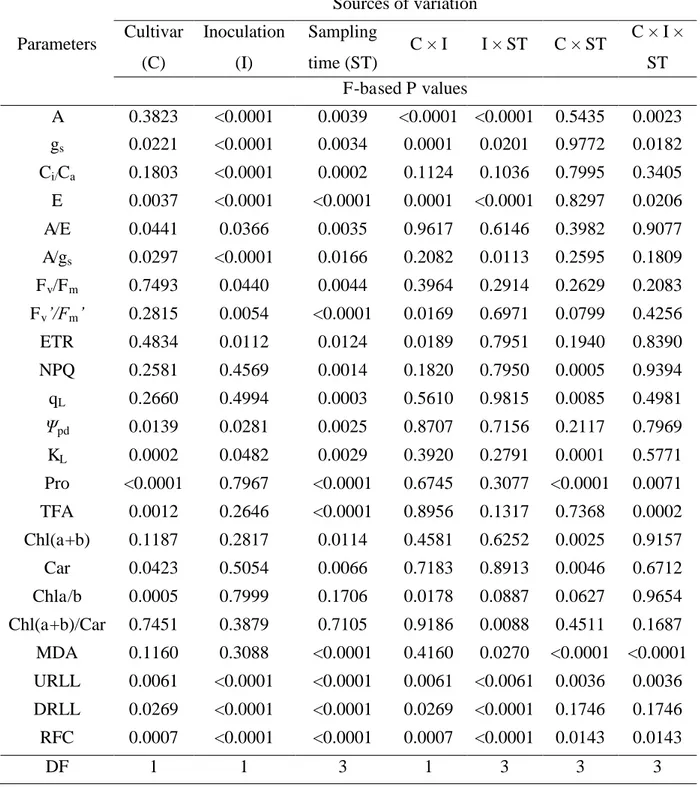
Analysis of variance. The factors cultivar, plant inoculation and sampling times were all significant for the disease indices DRLL, URLL and RFC. At least one of the factors and some of their interactions were significant for A, gs, Ci/Ca, E, A/E, A/gs, Ψpd, KL, Fv/Fm, Fv'/Fm', ETR, NPQ, qL, Chl(a+b), Chla/b, Chl(a+b)/car, MDA, TFA and Pro (Table 1).
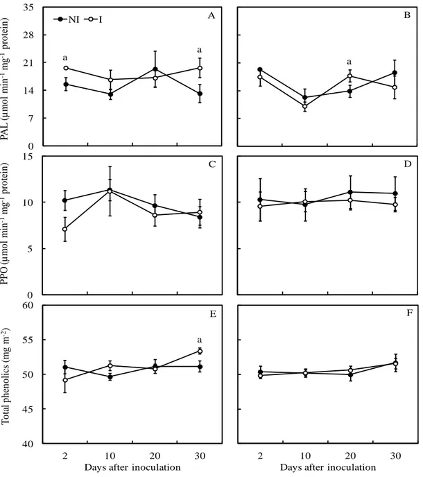
Disease assessments. The URLL, DRLL and RFC increased overtime in both Ubá and Tommy Atkins cultivars, but they reached the greatest values for cv. Tommy Atkins (Fig.
1A-C). Plants from cv. Tommy Atkins had higher values for all three disease indices than cv. Ubá
at 20 and 30 dai. DRLL, URLL and RFC were, respectively, 35, 51 and 35% higher at 20 dai.
At 30 dai, the differences were, respectively, of 25% for DRLL, 56% for URLL and 24% for
RFC between cultivars.
Water relations variables. There were no significant differences between non-inoculated and inoculated plants from both cultivars for Ψpd (Figs. 2A and B). In comparison to the
non-inoculated, inoculated plants from cv. Tommy Atkins presented a significant decrease around 26% for KL at 30 dai, while inoculated plants from cv. Ubá presented a decrease around 17%
at 30 dai (Figs. 2C and D). Overall, neither Ψpd nor KL differed significantly for
non-inoculated plants when comparing cultivars, except for the 64% lower KL for cv. Ubá at 2 dai
(Figs. 2A-D). Inoculated plants from cv. Tommy Atkins showed values for Ψpd about 25%
higher than those obtained for inoculated plants from cv. Ubá at 20 dai (Figs. 2A and B). Inoculated plants from cv. Tommy Atkins presented KL about 85% higher than those from
inoculated Ubá plants at 2 dai (Figs. 2C and D).
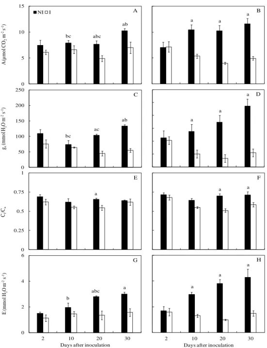
Gas exchanges and chlorophyll a fluorescence. Inoculated plants from cv. Tommy
Atkins presented average decreases of 55% for A, 73% for gs, 65% for E (considering the data
from 10, 20 and 30 dai) and a 17% average decrease for Ci/Ca (considering 20 and 30 dai) in
comparison to the non-inoculated plants. For the inoculated plants from cv. Ubá, average
decreases of 34% for A, 57% for gs, and 49% for E were found at 20 and 30 dai, while a
Average decreases of 19, 24 and 27% were found for A, gs and E, respectively, in inoculated
plants from cv. Tommy Atkins in relation to the inoculated plants from cv. Ubá. Overall,
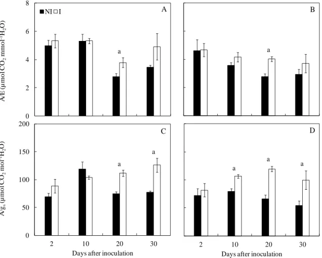
inoculated plants from both cultivars showed higher A/gs and A/E ratios, suggesting that water
use efficiency coupled to hydraulic impairments was improved. The differences between
non-inoculated and non-inoculated plants were more evident when considering A/gs in cv. Tommy
Atkins. Significant differences in Chl a fluorescence parameters were observed between non-inoculated and non-inoculated plants at 20 (for ETR and qL) and 30 dai (for Fv'/Fm', ETR and
NPQ) for Tommy Atkins but not for Ubá (Table 2). Decreases on the order of 17 and 22% were found for Fv'/Fm' and qL, and an average decrease of 28% in the ETR was found for the
inoculated plants, and the NPQ increased by 19% at 30 dai. Non-inoculated plants from cv. Tommy Atkins presented a higher Fv/Fm ratio along the evaluated time course when
compared to cv. Ubá.
Concentrations of pigments, TFA, proline and lipid peroxidation. Significant
differences between non-inoculated and inoculated cv. Tommy Atkins plants occurred in the concentrations of Chl (a+b), Chl (a+b)/Car, TFA, Pro and MDA (Table 3). There were significant decreases of 10 and 19%, respectively, for the Chl (a+b) and Chl (a+b)/Car
concentrations and increases of 24, 25 and 24% for the concentrations of TFA, Pro and MDA,
respectively, in inoculated cv. Tommy Atkins plants in relation to its non-inoculated
counterparts. Significant differences between cultivars occurred in inoculated plants (Table
3); relative to cv. Ubá, cv. Tommy Atkins displayed decreases (10%) in the Chl (a+b)
concentration and increases in TFA (18%), Pro (29%) and MDA (31%).
Vein density. The leaf vein density was significantly higher in cv. Ubá (13.6 mm mm-2)
DISCUSSION
The results of this study provided new information about the physiological traits of the mango wilt disease as mainly demonstrated by the alterations on water relations and photosynthetic performance arising from the colonization of mango plants by C. fimbriata. In agreement with the working hypothesis of the present study, the C. fimbriata infection of mango stem tissues appears to be associated with impaired plant water relations, as demonstrated by the lower KL values obtained for inoculated plants. These decreases are most
likely coupled with the intense stem tissue necrosis as the disease progressed, associated with a possible blockage of the colonized xylem vessels. In other interactions, such as bitternut hickory-Ceratocystis smalleyi and tanoak-Phytophthora ramorum, the rapid crown decline occurred in association with a reduced sap ascent, which was presumably caused by the obstruction provoked by tyloses or fungal material (43,44). Tyloses are usually formed in response to xylem cavitation and often induced by pathogens that physically block the vessels or secreted substances that alter the intrinsic properties of the sap (43,58).
Regardless of the cultivar, the radial stem tissues colonization (RFC) by C. fimbriata seemed to be of utmost importance for xylem vessel impairment. This finding could largely explain why the cv. Tommy Atkins plants, which displayed greater radial necrosis in the stem tissues compared to cv. Ubá, also experienced earlier and greater reductions in KL upon fungal
infection as can be seen by the noteworthy data tendency, especially from 20 dai onwards, which should ultimately have contributed to increased plant water shortages. The impairments in leaf hydration of cv. Tommy Atkins may have been further facilitated by its lower vein density, given that the water transport efficiency and the length of the hydraulic pathway through mesophyll are closely related to this anatomical variable (11,12). In any case, the occurrence of stem xylem cavitation, reductions in KL or even eventual changes in leaf or
stem water status can trigger hydraulic signals through pressure volume-changes in sensing cells or yet transient cavitation within leaf veins (16,28,52). These signals mediate various acclimation responses to water deficit and result in increased synthesis and concentration of abscisic acid (ABA), ultimately leading to reductions in gs and water loss by transpiration
(16). The increase in ABA concentrations may also be favored by the pathogen itself, since it has been shown that fungal species are capable of producing this hormone, including the ascomycete Ceratocystis fimbriata (21,55). Indeed, remarkable decreases in E and gs were
found for both cultivars. Given that these decreases were coupled with reductions in the Ci/Ca
(stomatal) constraints. In this regard, the disease-induced decrease in A is consistent with the effects of mild water deficits on photosynthesis, as noted in other studies where stomatal rather than biochemical limitations largely accounted for the observed decreases in the A of mango leaves under mild drought stress (18,34). Anyway, A decreased to a lesser extent than gs (and E) upon C.fimbriata infection, and thus, increases in both A/gs and A/E ratios were
found, which implies that the water use efficiency increased in the infected plants regardless of the cultivar, particularly after 20 dai.
Irrespective of the cultivar, decreases of A in inoculated plants occurred without any apparent alterations in the Fv/Fm, suggesting that primary photochemistry were unlikely to
have compromised the CO2 fixation. Furthermore, during most occasions, the ETR exceeded
90 µmol m-2 s-1, which is much higher than the photochemical requirement for the observed photosynthesis rates (35). A high ETR with a relatively low A usually leads to excess reducing power, which can be used to produce reactive oxygen species that can trigger a variety of photoinhibitory and photooxidative effects (32). Notwithstanding, the stronger decreases in A in cv. Tommy Atkins than in cv. Ubá were associated with adjustments at the photochemical level but only in the former cultivar, as demonstrated by the concomitant decreases in both the ETR and the efficiency of excitation energy captured by the open PSII reaction centers (estimated as Fv'/Fm') in parallel to increased NPQ at 30 dai. Collectively,
these changes suggest increases in thermal energy dissipation as a mean of protecting against increased excitation pressure preventing photodamage (2,4,31). Nevertheless, despite resorting to mechanisms involved in the dissipation of excess energy, it was observed an increase in lipid peroxidation in parallel to a slight decrease of the Chl pools in the plants selected for the analysis at later stages of disease development. As reported for the chestnut-Phytophthora cinnamomi (20) and tomato-Fusarium oxysporum f.sp. lycopersici (1,22) interactions, the decreases in Chl concentrations may be related not only to an oxidative stress response arising from the water shortage, but also to the release of non-selective toxins produced during the infection process.
In the present study, the declines in KL on inoculated plants from cv. Tommy Atkins
metabolism (26,29,33). In this regard, the higher levels of proline displayed by plants from cv. Tommy Atkins might be interpreted as an attempt to provide protection against water stress, which seems to be consistent with the higher level of disease severity in plants from this cultivar at advanced stages of fungal infection.
The results of the present study provide new insights into how mango plants respond to C. fimbriata infection at the physiological level. Irrespective of cultivar, the effects of fungal infection on stem tissues were probably associated with alterations in plant water relations. It was demonstrated that in the most susceptible Tommy Atkins cultivar, the disease symptoms developed faster and were associated with a more pronounced impairment of water relations and gas exchanges coupled with symptoms of oxidative stress at advanced stages of fungal infection. In contrast, although the most resistant cv. Ubá was better able to postpone disease development and, as such, suffered less from the stress imposition. In any case, the precise mechanisms underlying the responses to C. fimbriata infection may involve a myriad of physiological and biochemical processes and remain an important question that should be addressed in future studies. Considering that studies with contrasting results are often found for the involvement of water relation on vascular diseases development, it is suggested that this type of experiment must be carried out under natural conditions using direct measurements of the sap flow in order to favor the development of techniques suitable for early detection or control of the disease. Continued efforts in this direction should provide a better understanding of the alterations in plant water relations caused by mango wilt.
ACKNOWLEDGMENTS
LITERATURE CITED
1. Abdel-Fattah, G. M., and Al-Amri, S. M. 2012. Induced systemic resistance in tomato plants against Fusarium oxysporum f.sp. lycopersici by different kinds of compost. African J. Biotech. 11:12454-12463.
2. Adams, W. W. I., Zarter, C. R., Mueh, K. E., Amiard, V. S. E., and Demmig-Adams, B. 2005. Energy dissipation and photoinhibition: a continuum of photoprotection. In: Demmig-Adams, B., Demmig-Adams, W. W., and Mattoo, A. K. eds. Photoprotection, Photoinhibition, Gene Regulation and Environment. Springer-Verlag, Berlin, 49-64.
3. Al-Sadi, A. M., Al-Ouweisi, F. A., Al-Shariani, N. K., Al-Adawi, A. O., Kaplan, E. J., and Deadman, M. L. 2010. Histological changes in mango seedlings following infection with Ceratocystis manginecans, the cause of mango decline. J. Phytopathol. 158:738-743.
4. Baker, N. R. 2008. Chlorophyll Fluorescence : A Probe of Photosynthesis In Vivo. Annu. Rev. Plant Biol. 59:89-113.
5. Bates, L. S., Waldren, R. P., and Teare, D. 1973. Rapid determination of free proline for water stress studies. Plant Soil 39:205-207.
6. Batista, D. C., Terao, D., Barbosa, M. A. G., and Barbosa, F. R. 2008. Seca-da-mangueira Detecção, Sintomatologia e Controle. Comunicado Técnico 138, EMBRAPA, Petrolina, PE, Brazil.
7. Benevides, S. D., Ramos, A. M., Stringheta, P. C., and Castro, V. C. 2008. Qualidade da polpa da manga e polpa da manga Ubá. Ciênc. Tecnol. Alim. 28:571-578.
8. Berlyn, G. P., and Miksche, J. P. 1976. Botanical microtechnique and cytochemistry. Iowa State University Press, Ames, Iowa, USA.
10. Braz, V. B., Nunes, E. S., Vieira, G., Ribeiro Júnior, J. I., Bertini, L. A., and Couto, F. A. D. 2008. Indução do amadurecimento de mangas cv. Tommy Atkins e cv. Ubá pela aplicação de ethephon pós-colheita. Bragantia 67:225-232.
11. Brodribb, T. J., Feild, T. S., and Jordan, G. J. 2007 Leaf maximum photosynthetic rate and venation are linked by hydraulics. Plant Physiol. 144:1890-1898.
12. Brodribb, T. J., Field, T. S., and Sack, L. 2010. Viewing leaf structure and evolution from a hydraulic perspective. Funct. Plant Biol. 37:488-498.
13. Cakmak, I., and Horst, J. 1991. Effect of aluminium on lipid peroxidation, superoxide dismutase, catalase, and peroxidase activities in root tips of soybean (Glycine max). Physiol. Plantarum 83:463-468.
14. Carvalho, C. R. L., Rosseto, C. J., Mantovani, D. M. B., Morgano, M. A., Castro, J. V.,
and Bortoletto, N. 2004. Avaliação de cultivares de mangueira selecionadas pelo instituto agronômico de campinas comparadas a outras de importância comercial. Rev. Bras. Frutic.
26:264-271.
15. Castro Neto, M. T., Reinhardt, D. H., and da S Ledo, C. A. 2004. Determination of water potential on mango trees by pressure chamber. Acta Hort. 645:425-427.
16. Christmann, A., Grill, E., and Huang, J. 2013. Hydraulic signals in long-distance signaling. Curr. Opin. Plant Biol. 16:293-300.
17. Cunha, M. M., Santos Filho, H. P., and Nascimento, A. S. 2000. Manga: Fitossanidade. EMBRAPA, Brasília, DF, Brazil.
19. Dhingra, O. D., and Sinclair, J. B. 1995. Basic Plant Pathology Methods. Lewis Publisher, Boca Raton, FL, USA.
20. Dinis, L., Peixoto, F., Zhang, C., Martins, L., Costa, R., and Gomes-Laranjo, J. 2011. Physiological and biochemical changes in resistant and sensitive chestnut (Castanea) plantlets after inoculation with Phytophthora cinnamomi. Physiol. Mol. Plant Pathol. 75:146-156.
21. Dörffling, K., Petersen, W., Sprecher, E., Urbasch, I. and Hanssen, H.-P. 1984. Abscisic acid in phytopathogenic fungi of the genera Botrytis, Ceratocystis, Fusarium, and Rhizoctonia. Z. Naturforsch. 39:683-684.
22. El-Khallal, S. M. 2007. Induction and modulation of resistance in tomato plants against Fusarium Wilt disease by bioagent fungi (arbuscular mycorrhiza) and/or hormonal elicitors (jasmonic acid & salicylic acid): 1 - changes in growth, some metabolic activities and endogenous hormones related to defence mechanism. Aust. J. Basic Appl. Sci. 1:691-705.
23. Flexas, J., Bota, J., Cifre, J., Escalona, J. M., Galmés, J., Gulías, J., Lefi, E., Martínez-Cañellas, S. F., Moreno, M. T., Ribas-Carbó, M., Riera, D., Sampol, B., and Medrano, H. 2004. Understanding down-regulation of photosynthesis under water stress: future prospects and searching for physiological tools for irrigation management. Ann. Appl. Biol. 144:273-283.
24. Flexas, J., Diaz-Espejo, A., Galmés, J., Kaldenhoff, R., Medrano, H., and Ribas-Carbo, M. 2007. Rapid variations of mesophyll conductance in response to changes in CO2
concentration around leaves. Plant Cell Environ. 30:1284-1298.
25. Gallo, D., Nakano, O., Silveira Neto, S., Carvalho, R. P. L., Baptista, G. C., Berti Filho, E., Parra. J. R. P., Zucchi, R. A., Alves, S. B., Vendramin, J. D., Lopes, J. R. S., and Omoto, C. 2002. Entomologia Agrícola. 2nd Ed., FEALQ, Piracicaba, SP.
27. Halsted, B. D. 1890. Some fungous diseases of the sweet potato. New Jersey Agricultural College Experiment Station, Bulletin 76, pp. 25-27
28. Hubbard, R., Ryan, M., Stiller, V., and Sperry, J. 2001. Stomatal conductance and photosynthesis vary linearly with plant hydraulic conductance in ponderosa pine. Plant Cell Environ. 24:113–121.
29. Kaushal, N., Gupta, K., Bhandhari, K., Kumar, S., Thakur, P., and Nayyar, H. 2011. Proline induces heat tolerance in chickpea (Cicer arietinum L.) plants by protecting vital enzymes of carbon and antioxidative metabolism. Physiol. Mol. Biol. Plants 17:203-213.
30. Kramer, D. M., Johnson, G., Kiirats, O., and Edwards, G. E. 2004. New fluorescence parameters for the determination of QA redox state and excitation energy fluxes. Photosyn.
Res. 79:209-218.
31. Krause, G. H., and Weis, E. 1991. Chlorophyll fluorescence and photosynthesis: the basics. Annu. Rev. Plant Physiol. Plant Mol. Biol. 42:313-349.
32. Lima, A. L., DaMatta, F. M., Pinheiro, H. A., Totola, M. R., and Loureiro, M. E. 2002. Photochemical responses and oxidative stress in two clones of Coffea canephora under water deficit conditions. Environ. Exp. Bot. 47:239-247.
33. Liu, H., Yang, W., Liu, D., Han, Y., Zhang, A., and Li, S. 2011. Ectopic expression of a grapevine transcription factor VvWRKY11contributes to osmotic stress tolerance in Arabidopsis. Mol. Biol. Rep. 38:417-427.
34. Lu, P., Chacko, E. K., Bithell, S. L., Schaper, H., Wiebel, J., Cole, S., and Müller, W. J. 2012. Photosynthesis and stomatal conductance of five mango cultivars in the seasonally wet-dry tropics of northern Australia. Sci. Hort. 138:108-119.
36. Masood, A., Saeed, S., Malik, M. T., Iqbal, N., and Kazmi, M. R. 2010. Methodology for the evaluation of symptoms severity of Mango Sudden Death Syndrome in Pakistan. Pakistan J. Bot. 42:1289-1299.
37. Masood, A., Saeed, S., Silveira, S. F., Akem, C. N., Hussain, N., and Farooq, M. 2011. Quick decline of mango in Pakistan: survey and pathogenicity of fungi isolated from mango tree and bark beetle. Pakistan J. Bot. 43:1793-1798.
38. Maxwell, K., and Johnson, G. N. 2000. Chlorophyll fluorescence – a practical guide. J. Exp. Bot. 51:659-668.
39. McElrone, A. J., Sherald, J. L., and Forseth, I. N. 2003. Interactive effects of water stress and xylem-limited bacterial infection on the water relations of a host vine. Exp. Biol. 54:419-430.
40. Moore, S., and Stein, W. H. 1948. Photometric ninhydrin method for use in the chromatography of amino acids. J. Biol. Chem. 176:367-388.
41. Nogués, S., Cotxarrera, L., Alegre, L., and Trillas, M. I. 2002. Limitations to photosynthesis in tomato leaves induced by Fusarium wilt. New Phytol. 154:461-470.
42. Oxborough, K., and Baker, N. R. 1997. Resolving chlorophyll a fluorescence images of photosynthetic efficiency into photochemical and non-photochemical components: calculation of qP and Fv'/Fm' without measuring F0'. Photosyn. Res. 54:135-142.
43. Park, J. H., Juzwi, J., and Cavender-Bare, J. 2013. Multiple Ceratocystis smalleyi infections associated with reduced stem water transport in bitternut hickory. Phytopathology 103:565-574.
45. Pinheiro, H. A., DaMatta, F. M., Chaves, A. R. M., Loureiro, M. E., and Ducatti, C. 2005. Drought tolerance is associated with rooting depth and stomatal control of water use in clones of Coffea canephora. Ann. Bot. 96:101-108.
46. Pinto, A. C. Q., Andrade, S. E. M., Amaro, A. A., and Gomes, U. 2004. Mango industry in Brazil. Acta Hort. 645:37-50.
47. Ploetz, R. C. 2003. Diseases of mango. In: Ploetz, R. C., ed. Diseases of Tropical Fruit Crops. CABI Publishing, Wallingford, 327-363.
48. Poll, H., Vencato, A. Z., Kist, B. B., Santos, C., Carvalho, C., Reetz, E. R., and Beling, R. R. 2011. Anuário Brasileiro da Fruticultura. Editora Gazeta, Santa Cruz do Sul, RS, Brazil.
49. Pommer, C. V., and Barbosa, W. 2009. The impact of breeding on fruit production in warm climates of Brazil. Rev. Bras. Frutic. 31:612-634.
50. Ribeiro, I. J. A., Lourenção, A. L., Pilho, O. P., and Soares, N. B. 1984. Seca-da-mangueira. Vii. Resistência de cultivares de mangueira ao fungo Ceratocystis fimbriata Ell. & Halst. Bragantia 43:237-243.
51. Robbins, N. S., and Pharr, D. M. 1988. Effect of restricted root growth on carbohydrate metabolism and whole plant growth of Cucumis sativus L. Plant Physiol. 87:409-413.
52. Salleo, S., Nardini, A., Pitt, F., Lo Gullo, M. A. 2000. Xylem cavitation and hydraulic control of stomatal conductance in laurel (Laurus nobilis L.). Plant Cell Environ. 23:71-79.
53. Santos, R. P., Cruz, A. C. F., Iarema, L., Kuki, K. N., and Otoni, W. C. 2008. Protocolo para extração de pigmentos foliares em porta-enxertos de videira micropropagados. Ceres 55:356-364.
55. Siewers, V., Kokkelink, L., Smedsgaard, J., and Tudzynski, P. 2006. Identification of an abscisic acid gene cluster in the grey mold Botrytis cinerea. Appl. Environ. Microbiol. 72:4619-4626.
56. Silva, P. E. M., Cavatte, P. C., Morais, L. E., Medina, E. F., and DaMatta, F. M. 2013. The functional divergence of biomass partitioning, carbon gain and water use in Coffea canephora in response to the water supply: Implications for breeding aimed at improving drought tolerance. Environ. Exp. Bot. 87:49-57.
57. Souza, J. S., Almeida, C., Araujo, J. L. P., and Cardoso, C. E. L. 2002. Aspectos sócio-econômicos. In: Genú, P.J.C., and Pinto, A.C.Q. eds. A cultura da mangueira. Embrapa, Brasília, Brazil, 19-29.
58. Tyree, M. T., and Sperry, J. S. 1989. Vulnerability of xylem to cavitation and embolism. Annu. Rev. Plant Physiol. Mol. Biol. 40:l9-38.
LIST OF TABLES AND FIGURES
Table 1. Analysis of variance (ANOVA) of the effects of cultivar (C), plant inoculation (I), sampling time (ST) and the interactions between these factors on disease severity and some physiological and biochemical parameters.
Parameters
Sources of variation Cultivar
(C)
Inoculation (I)
Sampling time (ST)
C × I I × ST C × ST C × I × ST F-based P values
A 0.3823 <0.0001 0.0039 <0.0001 <0.0001 0.5435 0.0023 gs 0.0221 <0.0001 0.0034 0.0001 0.0201 0.9772 0.0182
Ci/Ca 0.1803 <0.0001 0.0002 0.1124 0.1036 0.7995 0.3405
E 0.0037 <0.0001 <0.0001 0.0001 <0.0001 0.8297 0.0206 A/E 0.0441 0.0366 0.0035 0.9617 0.6146 0.3982 0.9077 A/gs 0.0297 <0.0001 0.0166 0.2082 0.0113 0.2595 0.1809
Fv/Fm 0.7493 0.0440 0.0044 0.3964 0.2914 0.2629 0.2083
Fv’/Fm’ 0.2815 0.0054 <0.0001 0.0169 0.6971 0.0799 0.4256
ETR 0.4834 0.0112 0.0124 0.0189 0.7951 0.1940 0.8390 NPQ 0.2581 0.4569 0.0014 0.1820 0.7950 0.0005 0.9394 qL 0.2660 0.4994 0.0003 0.5610 0.9815 0.0085 0.4981
Ψpd 0.0139 0.0281 0.0025 0.8707 0.7156 0.2117 0.7969
KL 0.0002 0.0482 0.0029 0.3920 0.2791 0.0001 0.5771
Pro <0.0001 0.7967 <0.0001 0.6745 0.3077 <0.0001 0.0071 TFA 0.0012 0.2646 <0.0001 0.8956 0.1317 0.7368 0.0002 Chl(a+b) 0.1187 0.2817 0.0114 0.4581 0.6252 0.0025 0.9157 Car 0.0423 0.5054 0.0066 0.7183 0.8913 0.0046 0.6712 Chla/b 0.0005 0.7999 0.1706 0.0178 0.0887 0.0627 0.9654 Chl(a+b)/Car 0.7451 0.3879 0.7105 0.9186 0.0088 0.4511 0.1687 MDA 0.1160 0.3088 <0.0001 0.4160 0.0270 <0.0001 <0.0001 URLL 0.0061 <0.0001 <0.0001 0.0061 <0.0061 0.0036 0.0036 DRLL 0.0269 <0.0001 <0.0001 0.0269 <0.0001 0.1746 0.1746 RFC 0.0007 <0.0001 <0.0001 0.0007 <0.0001 0.0143 0.0143
Table 2. Maximum quantum efficiency of PSII photochemistry in dark-adapted leaves (Fv/Fm), the efficiency of excitation energy capture by
open PSII reaction centers (Fv'/Fm'), electron transport rate through PSII (ETR),non-photochemical quenching (NPQ) and fraction of open PSII
reaction centers (qL) determined in the leaves of plants from cultivars Ubá and Tommy Atkins non-inoculated (NI) or inoculated (I) with
Ceratocystis fimbriata.
Parameters
Ubá Tommy Atkins
20 dai 30 dai 20 dai 30 daí
NI I NI I NI I NI I
Fv/Fm 0.79 ± 0.002 0.79 ± 0.007 0.80 ± 0.005 0.80 ± 0.004 0.81 ± 0.003 b 0.80 ± 0.003 0.82 ± 0.003 b 0.79 ± 0.081
Fv'/Fm' 0.57 ± 0.02 0.55 ± 0.01 0.55 ± 0.02 0.56 ± 0.02 0.59 ± 0.01 0.53 ± 0.02 0.59 ± 0.01 0.46 ± 0.06 a
ETR 83.1 ± 11.2 79.6 ± 4.90 116 ± 6.40 112 ± 5.10 108 ± 8.30 78.9± 8.70 a 130 ± 5.00 91.2 ± 21.7 a
NPQ 1.94 ± 0.20 2.11 ± 0.17 2.29 ± 0.16 2.18 ± 0.21 1.92 ± 0.10 2.52 ± 0.29 2.08 ± 0.18 2.58 ± 0.27 a
qL 0.18 ± 0.03 0.19 ± 0.02 0.3 ± 0.02 0.28 ± 0.03 0.23 ± 0.02 0.19± 0.02 a 0.28 ± 0.02 0.27 ± 0.04
Table 3. Concentrations of total chlorophyll [Chl(a+b)] and carotenoids (Car), Chla/b ratio, Chl (a+b)/Car ratio and the concentrations of total free amino acids (TFA), proline (Pro) and malondialdehyde (MDA) equivalents determined in the leaves of plants from cultivars Ubá and Tommy Atkins non-inoculated (NI) or inoculated (I) with Ceratocystis fimbriata.
Parameters Ubá Tommy Atkins
NI I NI I
Chl(a+b) (g m-2) 0.67 ± 0.03 0.67 ± 0.03 0.67 ± 0.03 0.60 ± 0.02 ac
Car (g m-2) 0.11 ± 0.003 0.11 ± 0.005 0.10 ± 0.005 0.12 ± 0.007 c
Chla/b 3.08 ± 0.14 3.03 ± 0.04 3.05 ± 0.46 3.05 ± 0.31
Chl(a+b)/Car 6.04 ± 0.07 6.07 ± 0.06 6.7 ± 0.16 5.4 ± 0.15 a
TFA (mmol m-2) 11.26 ± 0.66 14.59 ± 2.95 13.53 ± 1.29 17.72 ± 0.99 ac
Pro (mmol m-2) 0.81 ± 0.09 0.75 ± 0.03 0.79 ± 0.05 1.06 ± 0.06 ac
MDA (µmol m-2) 26.12 ± 0.54 23.87 ± 1.22 26.47 ± 0.31 34.63 ± 0.84 ac
Figure 1. The upward relative lesion length (URLL), downward relative lesion length (DRLL) and the radial fungal colonization (RFC) on the stem tissues of mango plants from cultivars Ubá and Tommy Atkins inoculated with Ceratocystis fimbriata. Means followed by the letter a at each evaluation time are significantly different according to Student‟s t test (P ≤ 0.05). The error bars represent the standard error of the mean (n = 6).
0 10 20 30 D R L L ( % ) 0 20 40 60 U R L L ( % )
Ubá Tommy Atkins A
B C a a a a a a 0 30 60 90 120
2 10 20 30
R F C ( % )
Figure 2. Predawn leaf water potential (Ψpd) and apparent hydraulic conductance (KL)
on mango plants from cultivars Ubá (A and C) and Tommy Atkins (B and D) non-inoculated (NI) and non-inoculated (I) with Ceratocystis fimbriata. The means of NI and I treatments within each cultivar followed by the letter a at each evaluation time are significantly different according to Student‟s t test (P ≤ 0.05). The letter c indicates differences for I plants between the cultivars at each evaluation time according to Student‟s t test (P ≤ 0.05). The error bars represent the standard error of the mean (n = 6).
2 10 20 30
Days after inoculation 0
2 4 6
2 10 20 30
KL (m m o l H2 O M P a -1 m -2 s -1)
Figure 3. Net carbon assimilation rate (A) (A and B), stomatal conductance to water vapor (gs) (C and D), internal to ambient CO2 concentration ratio (Ci/Ca) (E and F) and transpiration
rate (E) (G and H) determined in the leaves of mango plants from cultivars Ubá (left column) and Tommy Atkins (right column) non-inoculated (NI) or inoculated (I) with Ceratocystis fimbriata. The means of NI and I treatments within each cultivar followed by the letter (a) at each evaluation time are significantly different according to Student‟s t test (P ≤ 0.05). The letters b and c indicate differences for, respectively, NI and I plants between cultivars at each evaluation time according to Student‟s t test (P ≤ 0.05). The error bars represent the standard error of the mean (n = 6).
0 50 100 150 200 250 gs (m m ol H2 O m -2s -1) 0 5 10 15 A (µ m o l C O2 m -2s -1) NI I 0 0.25 0.5 0.75 1 Ci /Ca 0 2 4 6
2 10 20 30
E (m m o l H 2 O m -2 s -1)
Days after inoculation
2 10 20 30
Days after inoculation abc
ab a a
Figure 4. Instantaneous water use efficiency (A/E) (A and B) and intrinsic water use efficiency (A/gs) (C and D) determined in the leaves of mango plants from cultivars Ubá (left
column) and Tommy Atkins (right column) non-inoculated (NI) or inoculated (I) with Ceratocystis fimbriata. The means of NI and I treatments within each cultivar followed by the letter a at each evaluation time are significantly different according to Student‟s t test (P ≤ 0.05). The error bars represent the standard error of the mean (n = 6).
0 2 4 6 8 A /E (µ m o l C O2 m m o l -1H 2 O) NI I 0 50 100 150 200
2 10 20 30
A /gs (µ m o l C O2 m o l -1H 2 O)
Days after inoculation
2 10 20 30
Days after inoculation
CHAPTER 2
Pathogen-induced alterations in the antioxidative system of mango cultivars with different levels of resistance to Ceratocystis fimbriata infection
Wilka M. S. Bispo, Leonardo Araújo, Maria B. Bermudez C., Isaías S. Cacique, Fábio M. DaMatta and Fabrício A. Rodrigues
First and fifth authors: Universidade Federal de Viçosa, Departamento de Biologia Vegetal, Viçosa, Minas Gerais, Zip Code 36570-000, Brazil; second, third, fourth and sixth authors: Universidade Federal de Viçosa, Departamento de Fitopatologia, Viçosa, Minas Gerais, Zip Code 36570-000, Brazil.
ABSTRACT
Bispo, W. M. S., Araújo, L., Bermudez C., M. B., Cacique, I. S., DaMatta, F. M., and Rodrigues, F. A. Pathogen-induced alterations in the antioxidative system of mango cultivars with different levels of resistance to Ceratocystis fimbriata infection.
infection, by less effectively restraining the spread of the pathogen in the vascular tissues, needed to trigger its antioxidant system at the leaf level to cope with the more rapid stress development when compared with the cv. Ubá.
Additional keywords: gas exchange; oxidative stress, phenolics, vascular pathogen.
INTRODUCTION
The mango (Mangifera indica L.) industry is one of the largest tropical fruit industries comprising approximately 100 countries in tropical and subtropical regions worldwide with a production of over 34.3 million tons (Bally, 2011). Brazil stands out as one of the chief producers of mango along with Mexico and Asian countries such as China and India, the major world producers (Bally, 2011). Among the mango cultivars commercially exploited in Brazil, the cv. Tommy Atkins concentrates around 90% of the cropped area mainly due to their hardiness, attractive color and good postharvest conditions besides the relative stability of production between consecutive years (Almeida et al., 2001). Some Brazilian cultivars are also cropped such as Ubá (Pinto et al., 2004), that is widely used as raw material for processing industries due to some characteristics of interest such as high pulp yield, high soluble solids content, and few fibers (Benevides et al., 2008; Braz et al., 2008). The greatest success in the development of various production hubs facing both industry and the fresh market is, however, confronted by a range of pests and diseases that can appreciably reduce fruit production and quality (Ploetz, 2003; Bally, 2011). The mango wilt, caused by the fungus Ceratocystis fimbriata Ell. & Halst. (Halsted, 1890), is one of the most important diseases affecting mango production worldwide because it causes the decline of entire orchards and thus ultimately cause losses in production and profits (Ploetz, 2003; Batista et al., 2008).
The genus Ceratocystis includes various plant pathogens affecting herbaceous and, especially, woody plants causing symptoms that include necrosis of the vascular tissues, cankers and leaf wilting (Upadhyay, 1993; van Wyk et al., 2007). Ceratocystis fimbriata is primarily considered a xylem pathogen (Harrington, 2000) causing the obstruction of the xylem vessels and dysfunctions on plant sap flow (Al-Sadi et al., 2010; Park et al., 2013). As a consequence of fungal infection, the whole-plant hydraulic conductivity is expected to decrease with the increasing tissue colonization by the pathogen (Fradin and Thomma, 2006; Parke et al., 2007, Park et al., 2013). The fungal structures such as mycelia and clamydospores and tissue necrosis, in addition to the promptly activation of defense structures (e.g., tyloses, gums and gels) by the hosts are closely related to the sealing of the vessels (Rahman et al., 1999; Parke et al., 2007; Al-Sadi et al., 2010; Yadeta and Thomma, 2013).
conditions of water deprivation, disproportionate increases in the production and accumulation of reactive oxygen species (ROS) as byproducts of various cellular processes may occur and may therefore lead to dramatic damages to cell structures and disruption of the metabolic activity including photosynthetic performance (Nogués et al., 2002; Chaves et al., 2003; Sharma et al., 2012). Fortunately, in order to restrain the deleterious action of the ROS, plant species are endowed with a complex antioxidant system which comprises metabolites and enzymes that can act over those oxidant compounds located in different organelles of the plant cells (Petridis et al., 2012). Some of the components involved in this cell scavenging process are the enzymes superoxide dismutases (SOD), that catalyze the dismutation of O2•- to
H2O2; catalases (CAT), responsible for the removal of H2O2, and the enzymes and metabolites
of the ascorbate/glutathione cycle, as well as phenolic compounds such as the flavonols (Chaves et al., 2003; Petridis et al., 2012).

![Table 3. Concentrations of t otal chlorophyll [Chl(a+b)] and carotenoids (Car), Chla/b ratio, Chl (a+b)/Car ratio and the concentrations of total free amino acids (TFA), proline (Pro) and malondialdehyde ( MDA) equivalents determined in the](https://thumb-eu.123doks.com/thumbv2/123dok_br/15367651.62963/41.1262.132.1175.312.580/concentrations-chlorophyll-carotenoids-concentrations-proline-malondialdehyde-equivalents-determined.webp)