1alpha (HIF-1
a
) Polymorphisms and Osteonecrosis
Georgia Chachami1,2, Alkmini Kalousi1,2, Loukia Papatheodorou3, Aggeliki Lyberopoulou1,2,
Vasileios Nasikas1, Keiji Tanimoto4, George Simos1,2, Konstantinos N. Malizos2,3, Eleni Georgatsou1,2*
1Laboratory of Biochemistry, Faculty of Medicine, School of Health Sciences, University of Thessaly, Biopolis, Larissa, Greece,2Institute of Biomedical Research and Technology (BIOMED/CERETETH), Larissa, Greece,3Department of Orthopaedic Surgery, Faculty of Medicine, School of Health Sciences, University of Thessaly, Biopolis, Larissa, Greece,4Department of Translational Cancer Research, Research Institute for Radiation Biology and Medicine, Hiroshima University, Hiroshima, Japan
Abstract
Bone hypoxia resulting from impaired blood flow is the final pathway for the development of osteonecrosis (ON). The aim of this study was to evaluate if HIF-1a, the major transcription factor triggered by hypoxia, is genetically implicated in susceptibility to ON. For this we analyzed frequencies of three known HIF-1apolymorphisms: one in exon 2 (C111A) and two in exon 12 (C1772T and G1790A) and their association with ON in a Greek population. Genotype analysis was performed using PCR-RFLP and rare alleles were further confirmed with sequencing. We found that genotype and allele frequency of C1772T and G1790A SNP ofHIF-1a(SNPs found in our cohort) were not significantly different in ON patients
compared to control patients. Furthermore these SNPs could not be associated with the different subgroups of ON. At the protein level we observed that the corresponding mutations (P582S and A588T, respectively) are not significant for protein function since the activity, expression and localization of the mutant proteins is practically indistinguishable from wt in HEK293 and Saos-2 cells. These results suggest that these missense mutations in theHIF-1agene are not important for the
risk of developing ON.
Citation:Chachami G, Kalousi A, Papatheodorou L, Lyberopoulou A, Nasikas V, et al. (2013) An Association Study between Hypoxia Inducible Factor-1alpha
(HIF-1a) Polymorphisms and Osteonecrosis. PLoS ONE 8(11): e79647. doi:10.1371/journal.pone.0079647
Editor:Ratna B. Ray, Saint Louis University, United States of America
ReceivedJuly 3, 2013;AcceptedOctober 2, 2013;PublishedNovember 18, 2013
Copyright:ß2013 Chachami et al. This is an open-access article distributed under the terms of the Creative Commons Attribution License, which permits
unrestricted use, distribution, and reproduction in any medium, provided the original author and source are credited.
Funding:The study was partially funded by a grant from the Hellenic Association of Orthopaedic Surgery and Trauma (http://www.eexot.gr/en/?about = 1) and
by the Musculoskeletal Research Unit of BIOMED/CERETETH (http://biomed.cereteth.gr/el/). The funders had no role in study design, data collection and analysis, decision to publish, or preparation of the manuscript.
Competing Interests:The authors have declared that no competing interests exist.
* E-mail: [email protected]
Introduction
Osteonecrosis (ON) is a disabling disorder mainly affecting the hips of young adults in the third and fourth decade of their life. ON has been associated with a variety of risk factors and can be characterized as idiopathic (of no apparent etiology) or secondary from several etiology-associated factors, such as hyperlipidaemia, hypercortisonism, dysbaric phenomena, autoimmune diseases, endotoxic reactions, alcoholism and smoking [1,2]. Thrombophil-ia and hypofibrinolysis have also been assocThrombophil-iated with the pathogenesis of ON which consequently results to bone ischemia, apoptosis of the osteocytes and the bone marrow and final establishment of a bone infarct. [3].
Low oxygen tension, also known as hypoxia, is one of the main pathophysiological characteristics of osteonecrosis [4]. Hypoxia can also occur in many other known diseases like ischemic conditions [5] (myocardial infarct, strokes), cancer [6] or even osteoarthritis [7]. HIF-1 (Hypoxia inducible factor-1) is the major transcriptional regulator triggered in hypoxia in order to promote adaptation to the new environment [8]. HIF-1 binds to regulatory DNA sequences called Hypoxia-Response Elements (HRE) thus controlling the expression of genes involved in cell metabolism, erythropoiesis, angiogenesis, invasion and metastasis. It is a
heterodimer that consists of HIF-1a, which is the oxygen regulated
subunit, and the constitutively expressed HIF-1b [9]. Under
normal oxygen conditions, HIF-1ais continuously produced and
destroyed in a process involving hydroxylation of two prolines within the oxygen-dependent degradation domain (ODD). Hydroxylation is mediated by conserved prolyl hydroxylases (PHDs), which act as oxygen sensors. Under hypoxic conditions,
hydroxylation is impaired and HIF-1ais stabilized, it translocates
to the nucleus where it dimerizes with HIF-1b, thus promoting the
transcription of its target genes [10].
The role of HIF-1ain osteonecrosis still remains elusive. It was
shown that HIF-1ais significantly increased in the ischemic side of
the epiphysial cartilage in the femoral head [11]. HIF-1awas also
detected at the reparative zone of collapsed femoral heads of ON patients [12]. Moreover, VEGF (Vascular Endothelial Growth
Factor), which is a target of HIF-1a, was also increased in ischemia
of femoral head, leading to revascularization of the cartilage thus promoting remodeling after the collapse [11,12]. The importance
of HIF-1a to the cartilage was known before, since it has been
reported that HIF-1a is essential not only for cell growth and
survival of growth plate chondrocytes in vivo [4,13] but also for extracellular matrix formation [14]. Latest studies suggest that
hypoxia promotes via HIF-1aa metabolic reprogramming in bone
marrow–derived angiogenic cells which leads to increased survival
in ischemic tissue [15]. For these reasons HIF-1a could be a
linked to the pathogenesis of ON [16]. For these reasons we investigated the genetic implication of three well-known and characterized polymorphisms (C111A in exon 2 and C1772T and
G1790A in exon 12) of HIF-1ain the appearance of ON.
Materials and Methods
Study subjects
The study population consisted of 134 ON patients (88 male, 46 female; mean age, 39.8 years ranging 18–69 years) and 63 controls (47 male, 16 female; mean age, 36.6 years ranging 17–59 years). ON diagnosis were established by evidence of osteonecrosis through magnetic resonance imaging (MRI) in stage 1 of the Association Research Circulation Osseous (ARCO) classification system and plain radiographs in Stages 2, 3, and 4.
According to etiological factors, patients were subgrouped into three groups of ON, group 1: idiopathic (32 cases), group 2: steroid-induced (78 cases), and group 3: other etiologies including alcohol-induced, anemia etc (24 cases). Patients with possible combined causes were excluded. Healthy control subjects were defined as individuals with no significant medical problems, no hip pain and their anteroposterior and frog leg lateral pelvic radiographs did not show any lesions with a sclerotic margin or subchondral collapse consistent with ON.
Idiopathic ON was defined by exclusion of cases with steroid-induced ON, alcohol-steroid-induced ON or possible combined cases. Steroid-induced ON was defined by a history of taking prednis-olone (1,800 mg) or an equivalent over 4 weeks [17]. Underlying diseases in steroid-induced ON were allergic respiratory or cutaneous diseases (21 cases), systemic lupus erythematosus (20 cases), organ transplantation (13 cases), idiopathic thrombocyto-penia purpura (11 cases), nephritic syndrome (8 cases), and others (5 cases). Alcohol-induced ON was diagnosed by the consumption of more than 400 ml of pure ethanol per week or alcohol-induced fatty liver and liver cirrhosis [18].
Clinical profiles of all patients used in this study are summarized in Table 1. There were no significant differences between patients and normal controls, in terms of age, sex, BMI and appearance of diabetes.
The study was approved by the Institutional Review Board (IRB) of the University Hospital of Larissa (Ref. N. 47714, 13/12/ 2012). Verbal informed consent was obtained from all participants in the study instead of written consent because the data were analyzed anonymously. The method of obtaining verbal consent
was approved by the IRB (for minors, consent was obtained from their parents). The procedures followed were in accordance with the ethical standards of the IRB at our institution (University Hospital of Thessaly, Larissa, Greece) on human experimentation and with the Helsinki Declaration of 1975, as revised in 2000.
DNA extraction and PCR
Genomic DNA was isolated from peripheral blood by using a Nucleospin Blood DNA extraction kit (Macherey-Nagel). The primers used for the exon 12 region were the following: forward
59-GCT GAA GAC ACA GAA GCA AAG AAC-39, reverse 59
-GGG TAG GAG ATG GAG ATG CAA TCA-39. The primers
for the C111A region were described in Konac et al [19]. The
PCR conditions used were: denaturation at 95uC for 4 min,
followed by 30 cycles of denaturation at 95uC for 30 s, annealing
and extension for exon 12: 57uC for 2 min and 72uC for 2 min, for
exon 2: 60uC for 30sec and 72uC for 30 sec, followed by a final
extension step at 72uC for 10 min. All PCR products were further
purified using the Nucleospin Gel and PCR clean up kit (Macherey-Nagel).
RFLP (Restriction Fragment Length Polymorphism) and Sequence analysis
The three polymorphisms were analyzed by digesting O/N at
37uC the PCR products with restriction endonuclease BglII for
C111A, HphI for C1772T and AciI (Fermentas, USA) for G1790A. The products were separated by a 2% agarose gel electrophoresis. Samples characterized by RFLP as heterozygous or homozygous for the rare allele were analysed further and verified by sequence analysis using the ABI PRISM BigDye Terminator v3.1 Ready Reaction Cycle Sequencing Kit in a ABI 3100/3130 Sequencer.
Statistics
Chi-square testing was used to determine the fit between observed genotype frequencies and those expected under Hardy– Weinberg equilibrium (HWE) using an online calculator tool http://www.tufts.edu/˜mcourt01/Documents/Court%20lab%20-%20HW%20calculator.xls. Frequencies of discrete variables between ON patients and controls were compared with the Chi-square test (fisher’s exact test) and continuous variables were compared by Student’s t test or Mann-Whitney test (non parametric variables). Allelic or genotype associations between
SNPs in control and ON patients, were compared using 262
Table 1.Clinical characteristics of controls and ON patients used in this study.
Control (N = 63) Patients (N = 134) Idiopathic (n = 32) Steroid-induced (n = 78) Other etiology (n = 24) P*
Age, mean (range) 36.6 (17–59) 39.8 (18–69) 46.6 (23–69) 37.3 (18–69) 38.5 (18–62) 0.086
Sex (male/female) 47/16 88/46 22/10 45/33 19/5 0.25
Concurrent or past medical history
Hypertension (%) 0 (0%) 21 (15.7%) 7 (21.8%) 9 (11.5%) 5 (20.8%) ,0.001
Hyperlipidemia (%) 0 (0%) 11 (8.2%) 5 (15.6%) 4 (5.1%) 2 (8.3%) 0.018
Diabetes mellitus (%) 0 (0%) 5 (3.7%) 2 (6.2%) 2 (2.5%) 1 (4.1%) 0.179
Renal diseases (%) 0 (0%) 13 (9.7%) 0 (0%) 13 (16.7%) 0 (0%) 0.01
SLE (%) 0 (0%) 20 (14.9) 0 (0%) 20 (25.6%) 0 (0%) ,0.001
BMI (kg/m2) 25.8 25.75 26.25 25.82 24.87 0.406
contingency tables and the 2-sided Fisher exact test. The strength of association was estimated by the odds ratio (OR), and their 95% confidence intervals (CI) were calculated. Statistical significance in luciferase assays was measured using the Student’s t-test. A P value
of less than 0.05 was considered to be statistically significant
(*P,0.05, ***P,0.001). All statistics were calculated through
Graph Pad Prism 5 software.
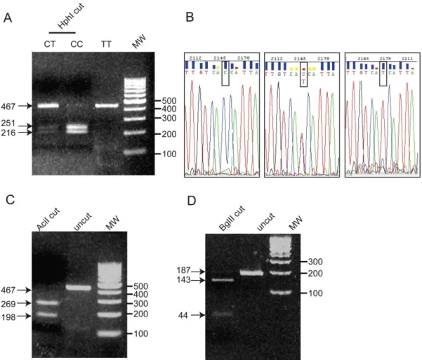
Figure 1. Analysis of HIF-1apolymorhisms using RFLP.(A) Analysis of HIF-1agene C1772T polymorphism with HphI enzyme, shown on 2% agarose electrophoresis. CT heterozygous genotype yielded three bands 467, 251 and 216 bp; C allele wt yielded two bands (251 and 216 bp). T allele remained uncut and yielded one fragment at 467 bp. Molecular weight standards are shown on the right. (B) Chromatograms of DNA sequence analysis of HIF-1aexon12 fragment showing the corresponding C, CT, T allelic variations at position 1772. (C) Analysis of HIF-1agene G1790A polymorphism with AciI enzyme, shown on 2% agarose electrophoresis. G allele wt yielded two bands (269 and 198 bp). A allele would remained uncut and yielded one fragment at 467 bp, as represented. Molecular weight standards are shown on the right. (D) Analysis of HIF-1agene C111A polymorphism with BglII enzyme, shown on 2% agarose electrophoresis. C allele wt yielded two bands (143 and 44 bp). A allele would remained uncut and yielded one fragment at 187 bp. Molecular weight standards are shown on the right.
doi:10.1371/journal.pone.0079647.g001
Table 2.Frequencies of HIF-1apolymorphisms between ON patients and controls.
SNPs POSITION GENOTYPE FREQUENCY* HWE**
control patients control patients control patients
C1772T exon 12 CC CT TT N CC CT TT N 0.111 0.126 0.777 0.367
50 12 1 63 101 32 1 134
G1790A exon 12 GG GA AA N GG GA AA N 0.024 0.011 0.846 ,0.0001
60 3 0 63 132 1 1 134
C111A exon 2 CC CA AA N CC CA AA N 0 0 nc nc
63 0 0 63 134 0 0 134
*Frequencies of rare alleles.
Cell cultures, plasmids and reporter assays
HEK293 and Saos-2 cells were maintained in Dulbecco’s medium supplemented with 10% fetal calf serum plus penicillin (50 IU/ml) and streptomycin (50 mg/ml).
pFLAG-CMV2-wild-type HIF-1a and mutant (P582S and A588T) constructs were
described in Tanimoto et al [20]. Transient co-transfection of
HIF-1aconstructs with a reporter gene pGL3–5HRE-VEGF was
carried out using the TransPassTM D2 Transfection Reagent (New England Biolabs Inc., Beverly, MA) and according to the manufacturer’s instructions.
The transcription activity of HIF-1aconstructs was analyzed in
a luciferase assay (described in [21]). Localization of HIF-1a
transfected constructs in Saos-2 and HEK293 cells was deter-mined using immunofluoresence (described in [21]) using an anti-FLAG antibody (Sigma). Protein expression of anti-FLAG constructs
was analyzed in Western blot using a homemade anti-HIF-1a
antibody [22] or a monoclonal anti-HIF-1a antibody (BD Biosciences) and anti-tubulin (Milipore, Billerica, MA,USA), as loading control.
Results
In order to examine the association of HIF-1agene
polymor-phisms with the susceptibility to osteonecrosis (ON) we analyzed the genotype and allele frequencies of three known SNPs located
in HIF-1atranslated regions (C111A in exon 2, and C1772T and
G1790A in exon 12 [23]) between ON patients of different etiologies and control subjects. Analysis of the genotypes was performed on PCR fragments amplified from DNA extracted from peripheral blood using specific restriction endonucleases (Fig 1, A, C, D). To confirm the polymorphisms found, sequencing of the PCR fragments of all subjects carrying the rare allele was performed (Fig 1B for C1772T SNP).
Concerning the C1772T SNP there was no significant deviation from the Hardy-Weinberg equilibrium in control (P = 0.777) and patient group (P = 0.367, Table 2). Out of the 63 controls, 50 cases were type CC, 12 cases were type CT and 1 case was type TT. Concerning patients diagnosed with ON, out of the 134 cases, 101 cases were type CC, 32 cases were type CT and 1 case was type TT. Statistics showed no significant differences in genotype (P = 0.592, OR (95% CI) = 1.257 (0.608 to 2.597)) and allele Table 3.HIF-1aSNP genotype and allele frequencies between ON patients and control subjects.
SNPs n (%) ON patients n (%) controls P-value* OR (95% CI)**
C1772T n = 134 n = 63
Genotypes
CC 101 (75.4) 50 (79.4)
CT 32 (23.9) 12 (19)
TT 1 (0.7) 1 (1.6)
CT+TT 33 (24.6) 13 (20.6) 0.5917 1.257 (0.6080 to 2.597)
Alleles
C 234 (87.3) 112 (88.9)
T 34 (12.7) 14 (11.1) 0.7425 1.162 (0.5996 to 2.254)
G1790A
Genotypes
GG 132 (98.5) 60 (95.2)
GA 1 (0.75) 3 (4.8)
AA 1 (0.75) 0 (0)
GA+AA 2 (1.5) 3 (4.8) 0.3297 0.303 (0.04932 to 1.862)
Alleles
G 265 (98.9) 123 (97.6)
A 3 (1.1) 3 (2.4) 0.3894 2.154 (0.4285 to 10.83)
C111A
Genotypes
CC 134 (100) 63 (100)
CA 0 (0) 0 (0)
AA 0 (0) 0 (0)
CA+AA 0 (0) 0 (0) nc nc
Alleles
C 268 (100) 126 (100)
A 0 (0) 0 (0) nc nc
*Fisher chi-square analysis, P values,0.05 are considered as statistically significant. **OR, Odds ratio; CI, confidence interval.
Calculations were performed for genotypes: CC vs CT+TT, GG vs GA+AA. nc: not calculated.
(P = 0.742, OR (95% CI) = 1.162 (0.600 to 2.254)) frequencies for the C1772T polymorphism, between ON patients and controls (Table 3). In accordance with a previous corresponding study in a Korean population, that similarly did not find association between the C1772T SNP and ON in the whole patient sample, but detected however an association within a subgroup [24], we discriminated subjects in males and females and in subgroups of ON, depending on the etiology of appearance. When we compared control cases versus different ON subgroups (1: idiopathic, 2: steroid induced and 3: other etiologies), we still could not find a significant difference in genotype (P vs idiop. = 1, OR (95% CI) = 0.887 (0.302 to 2.607), P vs steroid. = 0.433, OR (95% CI) = 1.417 (0.644 to 3.120), P vs other etiol. = 0.773, OR (95% CI) = 1.282 (0.436 to 3.881), Table 4) nor in allele frequencies (P vs idiop. = 0.807, OR (95% CI) = 0.827 (0.302 to 2.267), P vs steroid. = 0.590, OR (95% CI) = 1.244 (0.605 to 2.560), P vs other etiol. = 0.604, OR (95% CI) = 1.366 (0.515 to 3.623), Table 4). When we used the male population in our subgroups for the analysis, the difference still remained non significant (data not shown).
Regarding the G1790A SNP there was no deviation from the Hardy-Weinberg equilibrium for the control group (P = 0.846) but there was a significant deviation for the patient group, probably due to the low sample number and the rarity of our mutant allele
(P,0.0001, Table 2). Out of the 63 controls, 60 cases were type
GG, 3 cases were type GA and we found no case of type AA. Concerning total ON patients, out of the 134 cases, 132 cases were type GG, 1 case was type GA and 1 case was type AA. Statistics showed again no significant differences in genotype (P = 0.3297, OR (95% CI) = 0.303 (0.049 to 1.862)) and allele (P = 0.3894, OR (95% CI) = 2.154 (0.428 to 10.830)) frequencies for this polymor-phism between ON patients and controls (Table 3). Due to the small number of cases carrying the rare allele, we didn’t further compare controls with subgroups of ON patients.
As for C111A SNP, we did not find the rare A allele in controls nor in any of the subgroups of ON patients. Therefore, no association was attempted between controls and ON patients for this particular SNP (see Table 3).
Since the SNPs (C1772T and G1790A) that we tested were suggested not to be statistically significant concerning the appearance of ON, we further tried to validate our negative result by using a biological approach. For this we investigated the functional significance of the specific allelic variations in the
HIF-1alocus, at the protein level. At first we investigated the activity of
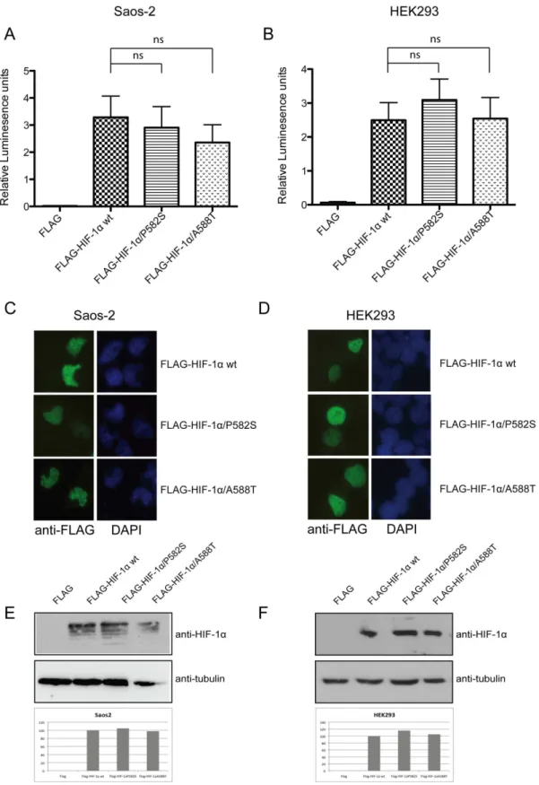
the variants using a transcription activity assay with a pGL3– 5HRE-VEGF luciferase reporter construct. We co-transfected osteosarcoma Saos-2 and HEK293 cells with the reporter plasmid
together with FLAG-HIF-1a wt or with the C1772T mutant
(corresponds to FLAG-HIF-1a/P582S) and the G1790A mutant
(corresponds to FLAG-HIF-1a/A588T, respectively [20]).
Trans-fection with HIF-1a wt led to a 2.5 to 3fold increase in
transcriptional activity both in Saos-2 and in HEK293 cells (Fig. 2, A and B). In Saos-2 cells the expression of both P582S and A588T variants originally led to a small decrease in activity compared to that observed in wt but this was proven not to be
statistically significant (Fig. 2A, Pvs P582S= 0.7338, Pvs
A588T= 0.3724). That was also the case for HEK293 cells, where the small differences observed between the activity of the variants
were also proven not significant (Fig. 2B, Pvs P582S= 0.4652, Pvs
A588T= 0.9537). Importantly enough, the variations observed between each mutant and the wt molecule showed different traits between the two cell lines tested. This fact implies their lack of significance as was already demonstrated by the statistic analysis. Since we excluded differences in transcriptional activity between
HIF-1avariants we next tested if localization or protein expression
of HIF-1ais affected by these two mutations. We showed that both
HIF-1a P582S and HIF-1a A588T remain nuclear when
transfected in Saos-2 and HEK293 cells, exactly like wt (Fig. 2C and D, respectively). Protein levels tested in both Saos-2 and HEK293 cells also remain unchanged between wt and mutants (Fig.2E and F).
In conclusion, in the present study we showed that C1772T and
G1790A SNPs found in translated loci of HIF-1a are not
associated with the occurrence of osteonecrosis. This could be because these specific polymorphisms seem to have no
conse-quence in the overall activity or protein expression of HIF-1a.
Discussion
Hypoxia is a main characteristic of several diseases like cancer, ischemic disorders and bone disorders like osteonecrosis and osteoarthritis, where it may also play a pivotal role in their pathogenesis [4,5,7,8]. Osteonecrosis (ON) is a pathological condition that has been attributed to the reduction or loss of vascular supply. This results in the lack of oxygen and
consequently to progressive collapse of the bone [3,25]. HIF-1a
is the key transcription factor activated upon lack of oxygen in order to promote the transcription of a great number of genes
needed for adaptation to hypoxic conditions [9]. HIF-1a
polymorphisms have been extensively studied in order to determine the association they may have in the appearance or progression of hypoxia related diseases. Among many (more than
30) polymorphisms, three are present in exons of HIF-1a
(C1772T, G1790A, C111A). Moreover, two of them (C1772T,
G1790A) are in the ODD domain of the HIF-1agene, where they
lead to ORF changes from Pro to Ser at codon 582 (C1772T) and from Ala to Thr at codon 588 (G1790A) [23], respectively. It has been shown before that C1772T and G1790A SNPs could be of importance in various types of cancer or specific disorders (like type II diabetes [26,27] and heart ischemic incidents [28,29]). However the genetic association or the level of importance they have in the etiology, progression or prognosis of these diseases is controversial among groups and remains subject of ongoing debate. Quite controversial are also the existing reports about the significance of these SNPs in the protein expression or function of
HIF-1a. There are reports [20,30,31] suggesting that P582S and
A588T HIF-1atranscriptional activities are higher than wild type.
Yamada et al [26] found that transcriptional activities of these
mutants differ significantly from wt HIF-1aonly under hypoxia. It
was also shown that P582S HIF-1a protein is expressed more
compared to the wt [31,32], maybe due to increased stability under normoxic conditions [31]. Furthermore, there is one study suggesting that the P582S mutation (or in combination with another mutation) may present decreased transcriptional activity compared to wt [29]. None of these studies has however addressed the mechanism underlying the alleles’ influence on trigger/ progression of the disease studied.
Concerning osteonecrosis, little is really known about HIF-1a
SNPs. On the contrary, VEGF SNPs’ role has been extensively studied, since VEGF is implicated in neovascularization of the necrotic bone, bone repair and formation [13,33]. It was shown that SNPs in the promoter region of VEGF are associated with ON of femoral head [34,35,36].
In the present study we have investigated the correlation of
C1772T, G1790A and C111A HIF-1a polymorphisms, with the
appearance of osteonecrosis in a Greek population. We found that the C111A allele was so rare that statistical analysis was not possible. For the other two SNPs (C1772T, G1790A) we could not detect a statistically significant association with the appearance of ON. This is in agreement with the only previous association study
about HIF-1a SNPs and ON (but of femoral head) in a Korean
population, where the authors have shown no significant association between four SNP genotype and allele frequencies of
HIF-1a(including C1772T in exon 12) and ON. Nevertheless in a
sub-analysis of the same study, the authors propose that C1772T SNP could be of importance only in the idiopathic ON male group [24]. To check this hypothesis in our own cohort we divided our ON patients into 3 etiological subgroups (idiopathic; steroid induced; of other etiologies) and discriminated between men and women. Our sample number was rather poor in some of our subcategories (especially the idiopathic cases) mostly due to the rarity of such cases in our population. Additionally, our control group was also rather small (but comparable to the different patient subcategories) due to the strict limitations we followed in order to exclude any ON indications and appropriately match with the patients’ clinical profile in respect to age, sex, BMI etc (see ‘Study Subjects’ under ‘Materials and Methods’). Despite the low number of patient samples in our subcategories we still did not observe a significant difference or at least a trend showing that
these specific polymorphisms of HIF-1a could be genetically
implicated with the appearance of any kind of ON.
Our biological approach in two different cell lines showed that
HIF-1amutants C1772T and G1790A do not differ from the wt at the protein level since they had no consequence on the
localization, expression and activity of HIF-1a protein. To our
knowledge this is the first study where the sub-cellular localization of the P582S and A588T mutants has been monitored and one of the very few where their protein levels are compared to wt. These findings concur with/and supplement our genetic results implying that the P582S and A588T mutations might not be genetically important for the appearance of ON because they do not have a profound impact on the overall function of the protein.
To summarize, we found no association of the two known SNPs
of HIF-1awith the risk of developing ON of any etiology. We thus
believe that these specific SNPs of HIF1ado not have a significant
genetic effect on ON diagnosis. Nevertheless a possible implication of other known SNPs of HIF-1a with ON cannot be excluded. Since hypoxia is a key feature of ON, we might expect that downstream
effectors of HIF-1a, like VEGF or other angiogenic or metabolic
agents could be of value in the diagnosis, progression and therapy of bone diseases like osteonecrosis. However, further biological and functional analysis would be needed to confirm that.
Acknowledgments
We acknowledge Dr. I. Mylonis for experimental help and support.
Author Contributions
Conceived and designed the experiments: EG KNM GS. Performed the experiments: GC AK AL VN EG. Analyzed the data: GC EG LP KNM GS. Contributed reagents/materials/analysis tools: KT KNM GS EG. Wrote the paper: GC EG.
software for image analysis ‘ImageJ’. FLAG-HIF-1aquantities were normalized against corresponding tubulin and expressed as fold increase against wt FLAG-HIF-1a(100%).
References
1. Assouline-Dayan Y, Chang C, Greenspan A, Shoenfeld Y, Gershwin ME (2002) Pathogenesis and natural history of osteonecrosis. Semin Arthritis Rheum 32: 94–124.
2. Malizos KN, Karantanas AH, Varitimidis SE, Dailiana ZH, Bargiotas K, et al. (2007) Osteonecrosis of the femoral head: etiology, imaging and treatment. Eur J Radiol 63: 16–28.
3. Childs SG (2005) Osteonecrosis: death of bone cells. Orthop Nurs 24: 295–301; quiz 302–293.
4. Maes C, Carmeliet G, Schipani E (2012) Hypoxia-driven pathways in bone development, regeneration and disease. Nat Rev Rheumatol 8: 358–366. 5. Goswami SK, Das DK (2010) Oxygen Sensing, Cardiac Ischemia, HIF-1alpha
and Some Emerging Concepts. Curr Cardiol Rev 6: 265–273.
6. Semenza GL (2012) Hypoxia-inducible factors: mediators of cancer progression and targets for cancer therapy. Trends Pharmacol Sci 33: 207–214. 7. Saito T, Kawaguchi H (2010) HIF-2alpha as a possible therapeutic target of
osteoarthritis. Osteoarthritis Cartilage 18: 1552–1556.
8. Semenza GL (2012) Hypoxia-inducible factors in physiology and medicine. Cell 148: 399–408.
9. Semenza GL (2009) Regulation of oxygen homeostasis by hypoxia-inducible factor 1. Physiology (Bethesda) 24: 97–106.
10. Schofield CJ, Ratcliffe PJ (2005) Signalling hypoxia by HIF hydroxylases. Biochem Biophys Res Commun 338: 617–626.
11. Kim HK, Bian H, Aya-ay J, Garces A, Morgan EF, et al. (2009) Hypoxia and HIF-1alpha expression in the epiphyseal cartilage following ischemic injury to the immature femoral head. Bone 45: 280–288.
12. Li W, Sakai T, Nishii T, Nakamura N, Takao M, et al. (2009) Distribution of TRAP-positive cells and expression of HIF-1alpha, VEGF, and FGF-2 in the reparative reaction in patients with osteonecrosis of the femoral head. J Orthop Res 27: 694–700.
13. Schipani E, Ryan HE, Didrickson S, Kobayashi T, Knight M, et al. (2001) Hypoxia in cartilage: HIF-1alpha is essential for chondrocyte growth arrest and survival. Genes Dev 15: 2865–2876.
14. Pfander D, Cramer T, Schipani E, Johnson RS (2003) HIF-1alpha controls extracellular matrix synthesis by epiphyseal chondrocytes. J Cell Sci 116: 1819– 1826.
15. Rey S, Luo W, Shimoda LA, Semenza GL (2011) Metabolic reprogramming by HIF-1 promotes the survival of bone marrow-derived angiogenic cells in ischemic tissue. Blood 117: 4988–4998.
16. Hadjigeorgiou G, Dardiotis E, Dardioti M, Karantanas A, Dimitroulias A, et al. (2008) Genetic association studies in osteonecrosis of the femoral head: mini review of the literature. Skeletal Radiol 37: 1–7.
17. Koo KH, Kim R, Kim YS, Ahn IO, Cho SH, et al. (2002) Risk period for developing osteonecrosis of the femoral head in patients on steroid treatment. Clin Rheumatol 21: 299–303.
18. Matsuo K, Hirohata T, Sugioka Y, Ikeda M, Fukuda A (1988) Influence of alcohol intake, cigarette smoking, and occupational status on idiopathic osteonecrosis of the femoral head. Clin Orthop Relat Res: 115–123. 19. Konac E, Onen HI, Metindir J, Alp E, Biri AA, et al. (2007) An investigation of
relationships between hypoxia-inducible factor-1 alpha gene polymorphisms and ovarian, cervical and endometrial cancers. Cancer Detect Prev 31: 102–109. 20. Tanimoto K, Yoshiga K, Eguchi H, Kaneyasu M, Ukon K, et al. (2003)
Hypoxia-inducible factor-1alpha polymorphisms associated with enhanced
transactivation capacity, implying clinical significance. Carcinogenesis 24: 1779–1783.
21. Lyberopoulou A, Venieris E, Mylonis I, Chachami G, Pappas I, et al. (2007) MgcRacGAP interacts with HIF-1alpha and regulates its transcriptional activity. Cell Physiol Biochem 20: 995–1006.
22. Mylonis I, Chachami G, Samiotaki M, Panayotou G, Paraskeva E, et al. (2006) Identification of MAPK phosphorylation sites and their role in the localization and activity of hypoxia-inducible factor-1alpha. J Biol Chem 281: 33095–33106. 23. Clifford SC, Astuti D, Hooper L, Maxwell PH, Ratcliffe PJ, et al. (2001) The pVHL-associated SCF ubiquitin ligase complex: molecular genetic analysis of elongin B and C, Rbx1 and HIF-1alpha in renal cell carcinoma. Oncogene 20: 5067–5074.
24. Hong JM, Kim TH, Chae SC, Koo KH, Lee YJ, et al. (2007) Association study of hypoxia inducible factor 1alpha (HIF1alpha) with osteonecrosis of femoral head in a Korean population. Osteoarthritis Cartilage 15: 688–694. 25. Lafforgue P (2006) Pathophysiology and natural history of avascular necrosis of
bone. Joint Bone Spine 73: 500–507.
26. Yamada N, Horikawa Y, Oda N, Iizuka K, Shihara N, et al. (2005) Genetic variation in the hypoxia-inducible factor-1alpha gene is associated with type 2 diabetes in Japanese. J Clin Endocrinol Metab 90: 5841–5847.
27. Nagy G, Kovacs-Nagy R, Kereszturi E, Somogyi A, Szekely A, et al. (2009) Association of hypoxia inducible factor-1 alpha gene polymorphism with both type 1 and type 2 diabetes in a Caucasian (Hungarian) sample. BMC Med Genet 10: 79.
28. Resar JR, Roguin A, Voner J, Nasir K, Hennebry TA, et al. (2005) Hypoxia-inducible factor 1alpha polymorphism and coronary collaterals in patients with ischemic heart disease. Chest 128: 787–791.
29. Hlatky MA, Quertermous T, Boothroyd DB, Priest JR, Glassford AJ, et al. (2007) Polymorphisms in hypoxia inducible factor 1 and the initial clinical presentation of coronary disease. Am Heart J 154: 1035–1042.
30. Putra AC, Tanimoto K, Arifin M, Hiyama K (2011) Hypoxia-inducible factor-1alpha polymorphisms are associated with genetic aberrations in lung cancer. Respirology 16: 796–802.
31. Fu XS, Choi E, Bubley GJ, Balk SP (2005) Identification of hypoxia-inducible factor-1alpha (HIF-1alpha) polymorphism as a mutation in prostate cancer that prevents normoxia-induced degradation. Prostate 63: 215–221.
32. Hebert C, Norris K, Parashar P, Ord RA, Nikitakis NG, et al. (2006) Hypoxia-inducible factor-1alpha polymorphisms and TSC1/2 mutations are comple-mentary in head and neck cancers. Mol Cancer 5: 3.
33. Komatsu DE, Hadjiargyrou M (2004) Activation of the transcription factor HIF-1 and its target genes, VEGF, HO-HIF-1, iNOS, during fracture repair. Bone 34: 680–688.
34. Liu B, Cao Y, Wang D, Yao G, Bi Z (2012) Vascular Endothelial Growth Factor -634G/C Polymorphism Associated with Osteonecrosis of the Femoral Head in a Chinese Population. Genet Test Mol Biomarkers 16: 739–743.
35. Lee YJ, Lee JS, Kang EH, Lee YK, Kim SY, et al. (2012) Vascular endothelial growth factor polymorphisms in patients with steroid-induced femoral head osteonecrosis. J Orthop Res 30: 21–27.