875 875 875 875 875 Mem Inst Oswaldo Cruz, Rio de Janeiro, Vol. 101(8): 875-880, December 2006
Production, characterization, and application of antibodies against
heat-labile type-I toxin for detection of enterotoxigenic
Escherichia coli
Caroline A Menezes, Sergio Y Imamura, Luiz R Trabulsi
†, Antônio Fernandes-Filho*,
Marina B Martinez*, Beatriz EC Guth**, Dennys M Girão***, Roxane MF Piazza/
+Laboratório de Bacteriologia, Instituto Butantan, Av. Vital Brazil 1500, 05503-900 São Paulo, SP, Brasil *Departamento de Análises Clínicas e Toxicológicas, Faculdade de Ciências Farmacêuticas, Universidade de São Paulo, São Paulo, SP, Brasil
**Departamento de Microbiologia, Imunologia e Parasitologia, Universidade Federal de São Paulo, São Paulo, SP, Brasil ***Departamento de Microbiologia Médica, Instituto Prof. Paulo de Góes, Universidade Federal do Rio de Janeiro,
Rio de Janeiro, RJ, Brasil
Strains of enterotoxigenic Escherichia coli (ETEC) are responsible for significant rates of morbidity and mortality among children, particularly in developing countries. The majority of clinical and public health laboratories are capable of isolating and identifying Salmonella, Shigella, Campylobacter, and Escherichia coli O157:H7 from stool samples, but ETEC cannot be identified by routine methods. The method most often used to identify ETEC is polymerase chain reaction for heat-stable and heat-labile enterotoxin genes, and subsequent serotyping, but most clinical and public health laboratories do not have the capacity or resources to perform these tests. In this study, polyclonal rabbit and monoclonal mouse IgG2b antibodies against ETEC heat-labile toxin-I (LT) were characterized and the potential applicability of a capture assay was analyzed. IgG-enriched fractions from rabbit polyclonal and the IgG2b monoclonal antibodies recognized LT in a conformational shape and they were excellent tools for detection of LT-producing strains. These findings indicate that the capture immunoassay could be used as a diagnostic assay of ETEC LT-producing strains in routine diagnosis and in epidemiological studies of diarrhea in developing countries as enzyme linked immunosorbent assay techniques remain as effective and economical choice for the detection of specific pathogen antigens in cultures.
Key words: detection - antibodies - heat-labile toxin - Escherichia coli
Diarrhea caused approximately two million deaths per year worldwide in the last decade (WHO-UNICEF 2002), and was responsible for an estimated 16-32% (median 21%) of mortality in children aged 0-4 years in devel-oping countries (Kosek et al. 2003). One of the major etiologic agents is enterotoxigenic Escherichia coli (ETEC), known to be endemic in essentially all devel-oping countries. Besides ETEC strains are also usually responsible for the acute diarrhea that affects visitors to the tropics (Ericsson 2003). In Brazil, these pathogens are responsible for up to 20% of cases of infantile diar-rhea, region and season dependent (Reis et al. 1982, Trabulsi et al. 1985, Gomes et al. 1991, Almeida et al. 1998, Souza et al. 2002, Fernandes-Filho et al. 2003, Franzolin et al. 2005, Barreto et al. 2006).
Specific virulence factors such as enterotoxins and colonization factors differentiate ETEC from other cat-egories of diarrheagenic E. coli. ETEC belongs to a
het-Financial support: Fapesp (grant 99/09458-0 to RMFP and fellowship to CAM), CNPq fellowship to SYI
+Corresponding author: [email protected] †In memoriam
Received 6 June 2006 Accepted 19 September 2006
erogeneous family of lactose-fermenting E. coli, belong-ing to a wide variety of O antigenic types. These strains produce enterotoxins (heat labile and/or heat stable), and colonization factors, which allow the organisms to readily colonize the small intestine and in this way cause diarrhea (Sack 1980, Wolf 1997, Nataro & Kaper 1998).
Since ETEC can be recognized by the enterotoxins it produces, diagnosis must depend upon identifying either heat-labile toxins (LT) and/or heat-stable toxin (ST). One or both toxins may be expressed by ETEC. LT belongs to a structurally and functionally related AB5 enterotoxin family, in which the A subunit has the toxic activity and B subunit is responsible for toxin binding to cell recep-tor.
con-876 876 876 876
876 Ab as tools for LT-I toxin detection • Caroline A Menezes et al.
ducting studies on diarrhea in developing countries do not include ETEC in their routine diagnosis (Qadri et al. 2005).
We have previously described the development of a capture immunoassay for detection of ETEC producing LT-I (Menezes et al. 2003). The estimated accuracy of this assay was 78% of sensitivity, 94% of specificity, and 92% of efficiency. In this analysis, we have em-ployed an IgG enriched fraction of a polyclonal rabbit and monoclonal mouse IgG2b anti-LT antibodies. In the present study, these antibodies were characterized im-munochemically, besides we better describe the capture assay and check its applicability to detec LT-I in super-natants of bacterial isolates.
MATERIALS AND METHODS
Polymyxin B sulphate, Freund complete adjuvant, orthophenylenediamine (OPD), 3,3-Diaminobenzidine tetrahydrochloride (DAB), Coomassie blue R-250, poly-ethylene glycol 4000 (PEG), HAT medium, HT medium, anti mouse IgG1, IgG2a, IgG2b, IgG3, IgA, and IgM, were obtained from (Sigma Aldrich Co, St Louis, MA, US). RPMI media and foetal calf serum from Cultilab (Campinas, SP, Brazil). Horseradish peroxidase-conju-gated anti-rabbit IgG, anti-mouse IgG, and anti-mouse IgG/A/M from Zymed Laboratories Inc (San Francisco, CA, US). Protein G – and protein A – Sepharose affin-ity chromatography column was purchased from GE Healthcare.
Bacterial strains - The bacterial isolates used in this study consisted in 44 strains previously defined as ETEC by genes presence: LT-I (30 isolates) and LT-I/ST-Ih (14 isolates) isolated from different geographic areas of Brazil (Nishimura et al. 1996, Girão et al. 2001, Souza et al. 2002, Fernandes-Filho et al. 2003). In addition, 298 E. coli strains isolated from stools of children, with and without diar-rhea, from sporadic cases in Brazil previously determined by molecular methods as LT/ST negative (Fernandes-Filho et al. 2003) was used, as well as 25 enteric Gram-negative strains, other than E. coli, including Vibrio cholerae, Salmonella spp., Shigella spp., Klebsilella spp., Edwardsiella, Citrobacter spp., Morganella morgani, Proteus spp., Enterobacter spp., and Providencia spp. isolates as part of our laboratory collection. E. coli H10407 (Evans et al. 1977) and 258909 (Qadri et al. 2000) were included as ETEC prototype strains.
Culture conditions - The strains were cultivated in 2% casamino acids, 0.15% yeast extract, and salts me-dium (CYE) (Evans et al. 1973) at 37ºC, 200 rpm for 18 h. Then each culture was incubated for 30 min at 37ºC, 200 rpm with polymyxin (1 mg/ml, final concentration) (Cerny & Tauber 1971, Strockbine et al. 1985) and cen-trifuged at 1600 x g for 20 min. The supernatants were kept at –20oC until use.
Purified LT toxin- LT toxin was purified as described by Bowman and Clements (2001) and was kindly donated by Dr John D Clements from Department of Microbiol-ogy and ImmunolMicrobiol-ogy, Tulane University Health Sciences Center, New Orleans, Louisiana, US.
Anti-LT-I polyclonal antibodies - New Zealand rab-bits were immunized subcutaneously with 100 µg of puri-fied LT in complete Freund’s adjuvant. Serum samples were obtained just before immunization by auricular-venom method to be used as negative control in specific anti-body evaluation. Serum samples were also obtained 30 days after antigen injection and subsequently analyzed by ELISA. The IgG-enriched fractions were obtained from rabbit antisera (Menezes et al. 2003) after being submitted to caprylic acid and ammonium sulphate precipitation as described by McKinney and Parkinson (1987), and its purity was observed by SDS/PAGE 15% (Laemmli 1970, Studier 1973) after Coomassie blue R-250 staining.
Monoclonal antibody (Mab) production - Four to six week-old female Balb/c mice were immunized with 2 µg of purified LT in complete Freund’s adjuvant. The immuniza-tion protocol consisted of three injecimmuniza-tions of 20-µl of the toxin in PBS at four-weeks interval. Serum samples were obtained just before the first immunization by retro-or-bital sinus method, to be used as negative control in spe-cific antibody evaluation. Serum samples were also ob-tained ten days after the last antigen injection and subse-quently analyzed by ELISA.The mouse with the highest antibody titer was boosted with 10 µg of purified LT with-out adjuvant four days prior to cell fusion, and then sac-rificed by cervical dislocation. Popliteal lymphnodes were removed aseptically and single cell suspensions were pre-pared by mechanically disrupting the tissues through a sterile nylon cloth sieve (100 µm pore size) into RPMI media containing penicillin/streptomycin. The popliteal lymphnode cells were fused with SP2/O-Ag14 mouse myeloma cells (2:1) using polyethylene glycol 4000. The fused cells were suspended and selected in HAT-RPMI 1640 medium containing 10% FCS into 96-well microplates (Techno Plastic Products AG, TPP, Switzerland). The microplates were incubated at 37°C in the presence of 5% CO2 for 12 days. In order to screen cultures for antibod-ies production, 100 µl of culture supernatant were added to wells of 96-well (Nunc-Immuno PolySorp) plate previ-ously coated with purified LT. The ELISA test was com-pleted as described above. Hybridoms from cultures show-ing significant antibody production were selected and cloned by limiting dilution culture.
Isotyping of monoclonal antibody - Nunc-Immuno PolySorp plate was coated overnight at 4°C with a solu-tion (100 µl/well) containing anti mouse IgG1 (10 µg/ml), IgG2a (10 µg/ml), IgG2b (10 µg/ml), IgG3 (10 µg/ml), IgA (100 µg/ml), and IgM (100 µg/ml) in 0.05 M sodium car-bonate-bicarbonate buffer (pH 9.6). The reaction was lowed by incubating supernatants of hybridoms and fol-lowed by a horseradish peroxidase-conjugated anti-mouse IgG/A/M antibody. The absence of non-specific IgM antibodies was assured by incubating the antibodies in wells coated with 0.05 M sodium carbonate-bicarbonate buffer (pH 9.6).
877 877 877 877 877 Mem Inst Oswaldo Cruz, Rio de Janeiro, Vol. 101(8), December 2006
0.1 M citric acid solution (pH 3.0) and harvested in 1 M Tris-solution (pH 9.8). The antibody solution was con-centrated by lyophilization and its purity was observed by SDS/PAGE 15% (Laemmli 1970, Studier 1973) after Coomassie blue R-250 staining.
Affinity of LT-antibodies- The dissociation constant (KD) of LT-antibodies was determined by their affinity to LT toxin as described by Friguet et al. (1985). Anti-bodies were incubated in solution with different antigen concentration until the equilibrium was reached, then a classical indirect ELISA measured the proportion of an-tibody that remained unsaturated at equilibrium. The dis-sociation constant was deduced by linear regression by Scatchard method.
LT-antibodies characterization by immunoblotting - To further access the reactivity of antibodies to puri-fied LT toxin, an immunoblotting analysis was performed using polyclonal and monoclonal LT-antibodies. Briefly, 10 µg per slot of purified LT toxin were applied to a 15% SDS-polyacrilamide gel (Laemmli 1970, Studier 1973). After electrophoresis, the separated proteins were trans-ferred to nitrocellulose membrane (Hybond C-Extra, Amersham Life Science) at 100 V for 18 h at 4oC. The
membrane was blocked with 3% BSA for 2 h and reacted with IgG-enriched fraction of polyclonal LT anti-body (5 µg/ml) and monoclonal LT-antibodies (18 µg/ml IgG1 or IgG2b) for 18 h at 4oC. The membrane was then
washed and incubated for 2 h with horseradish peroxi-dase-conjugated anti-mouse IgG diluted 1:10,000 or anti-rabbit IgG diluted 1/10,000. After washing, the sub-strate 3,3-Diaminobenzidine tetrahydrochloride plus hydrogen peroxide was added and the reaction was stopped by distilled water addition.
Sensitivity and specificity of capture assay in detect-ing LT-I - The sensitivity and specificity of the capture assay was determined as described by Menezes et al. (2003) in the standardization of the capture ELISA immu-noassay. Here we tested different concentrations of puri-fied LT (from 100 µg to 0.7 ng) or supernatants of bacterial isolates. ELISA optical densities (A492nm) data were ana-lyzed by mean and standard error using GraphPad Prism 3.00. Differences between ODs of LT producing- and non-producing strains were considered significant when the probability of equality was less than 0.05 (p < 0.05).
GM1 ELISA assay - Microtiter plates (Nunc Im-munoplate, Maxisorp) were incubated at 37oC for 16 h
with 1.25 µg/ml ganglioside GM1 (Sigma Aldrich Co.) in phosphate-buffered saline, pH 7.2 (PBS). And the reac-tion was developed according to Svennerholm and Wiklund (1983). Toxin bound to GM1 was then detected with 10 µg/ml of IgG2b monoclonal antibody, followed by anti-mouse IgG peroxidase diluted 1:10,000. The reaction was developed by adding freshly prepared solu-tion of OPD plus H2O2 recording of the A492 on a Multiskan EX ELISA reader.
RESULTS
Polyclonal and Mab characterization - The immuni-zation protocols used in both New Zealand rabbits and
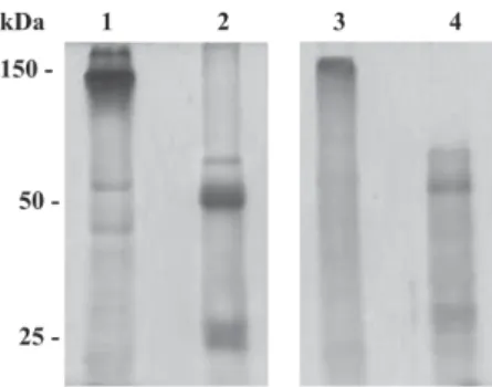
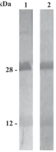
Balb/c mice generated high antibody titers against LT-I toxin. The mean of optical densities at 492 nm reached to 1.2 until 51,200 of serum dilutions in both rabbits and mice immunized with LT-I. In order to avoid cross-reaction of sera proteins other than IgG, polyclonal sera were submit-ted to caprylic acid and ammonium sulfate precipitations. The SDS/PAGE profile demonstrated that, despite the pres-ence of minor proteins, the major polypeptide of apparent molecular weight of 150 kDa under non-reducing condi-tions (Fig. 1, lane 1), and two components of molecular weigh of approximately 50 kDa and 25 kDa, under reduc-ing conditions, were observed in the IgG-enriched frac-tion (Fig. 1, lane 2). In addifrac-tion, this fracfrac-tion recognized both A and B subunits of LT-I by immunoblotting, with apparent molecular weights of 28 kDa and 12 kDa, respec-tively (Fig. 2, line 1), and presented a dissociation con-stant of 0.97 × 10⁻⁷ M.
After the fusion with popliteal lymphnode and mouse myeloma cells and several sub cloning by limiting dilu-tions, two Mab were obtained, one IgG1 and one IgG2b, both of them specific for LT toxin. The monoclonal an-tibodies were purified by affinity chromatography and a component of apparent molecular weight of 146 kDa was observed in SDS-PAGE under non-reducing conditions (Fig. 1, lane 3), and in a reducing condition two compo-nents of apparent molecular weight of 50 kDa and 25 kDa were observed (Fig. 1, lane 4). They recognized by immunoblotting the A and B subunits of LT-I (Fig. 2, line 2). The IgG2b showed a constant dissociation of 2.2 × 10⁻⁸ M. On the other hand, IgG1 Mab showed
dissocia-tion constant of 1.3 × 10⁻⁷ M.
Sensitivity and specificity of capture assay in de-tecting purified LT and LT-I in supernatant of bacte-rial isolates- Twenty-five µg/ml of anti-rabbit LT IgG enriched fraction and 10 µg/ml of IgG2b Mab allowed the capture of less then 10 ng of purified toxin (Fig. 3). In the same antibody concentrations, the reaction cut off was 0.173, defined by mean and standard error of opti-cal densities from supernatants of the 298 non-produc-ing LT-I E. coli strains and the 25 enteric Gram-negative strains. The 46 LT-I producing strains presented a range
878 878 878 878
878 Ab as tools for LT-I toxin detection • Caroline A Menezes et al.
of OD from 0.759 to 1.191 (median of 0.943), which allows a clear distinction between producing- and non-producing LT E. coli isolates (Fig. 4). Statistical analy-sis showed that the difference between LT producing-and non-producing strains by mean producing-and stproducing-andard devia-tion were considered significant (p < 0.0001) in the cap-ture assay.
DISCUSSION
Evidence has accumulated showing that both LT A-and B-subunits have distinct immunomodulatory activi-ties, which act synergistically, promoting a potent anti-toxin response (Pizza et al. 2001, Fraser et al. 2003). Thus, as expected, both New Zealand rabbits and Balb/c mice immunized with low concentration of LT-I toxin Fig. 2: 10 µg of purified heat-labile toxins (LT-I) were separated by 12% SDS/PAGE and transferred to nitrocellulose membrane. Nitro-cellulose membrane containing LT polypeptides was incubated in lane 1. With 5 µg/ml of IgG-enriched fraction of anti-LT-I polyclonal sera followed by horseradish peroxidase goat anti-rabbit IgG (1:10,000) and in lane 2. With 18 µg/ml of IgG2b of anti-LT-I monoclonal antibody followed by horseradish peroxidase goat anti-mouse IgG (1:10,000). Reaction was revealed with DAB + H2O2.
Fig. 3: titration curve of purified heat-labile toxins (LT-I) from 100 µg to 0.7 ng. Reaction was done using 25 µg/ml of IgG enriched fraction of rabbit polyclonal and 10 µg/ml of IgG2b monoclonal anti-LT. Goat anti-mouse IgG peroxidase labeled (1:10,000) and freshly prepared solution of OPD plus H2O2 recording of the A492 on a Multiskan EX ELISA reader.
Fig. 4:optical densities of enterotoxigenic Escherichia coli produc-ing heat-labile (LT) toxin, E. coli non-producing LT and entero-bacterial strains by capture assay. The reaction was detected with 25 µg/ ml of IgG enriched fraction of rabbit polyclonal and 10 µg/ml of IgG2b monoclonal anti-LT. Goat anti-mouse IgG peroxidase labeled (1:10,000) and freshly prepared solution of OPD plus H2O2 recording of the A492 on a Multiskan EX ELISA reader.
elicited high antibody titers. However, despite of LT high immunogenicity, seven fusions with popliteal lymphnode cells and mouse myeloma cells were necessary to get two stable and positives clones, classified as IgG1 and IgG2b, both of them specific for LT. In contrast, Lindholm et al. (1983) obtained 70 secreting Mab against cholera toxin, in which 14 clones showed full cross-re-activity with LT, and they presented different IgG isotypes after immunizing mice intraperitoneally or intravenously and fusing the spleen cells. In this way, the selection of few IgG-secreting clones is probably related to the fact that we used popliteal lymphnode cells of immunized mice for the hybridization experiments, which was re-sponsible for isotype selection and number of positive clones.
IgG-enriched fractions from rabbit polyclonal, and the IgG2b Mab recognized LT in a conformational shape (A and B subunit). Using these tools for detection of LT-producing strains the sensitivity and specificity of the capture immunoassay were 100 and 99%, respectively. The high sensitivity of the assay can be related to the use of CYE medium (Evans et al. 1973, Beutin et al. 1988) instead of Luria-Bertani (LB) broth medium, and also to the use of polymyxin (Cerny & Tauber 1971, Strockbine et al. 1985, Beutin et al. 1989, Bowman & Clements 2001). As LT is known to be peri-plasmatic in ETEC and not secreted into the medium, both conditions stimulate LT production.
879 879 879 879 879 Mem Inst Oswaldo Cruz, Rio de Janeiro, Vol. 101(8), December 2006
antibody dependent. The IgG2b Mab recognized with more intensity the A subunit. This fact might explain the low sensitivity (69%) of GM1-ELISA in detecting LT-I producing strains in supernatants. The efficiency of the capture immunoassay was due to IgG-enriched fraction of rabbit LT antisera and IgG2b Mab, since both of them recognized LT in a conformational shape (A and B sub-unit) thus allowing the capture of even low expressing-LT-I ETEC isolates.
It is important to mention that the results presented here are part of a project in which polyclonal and Mab against virulence factors of diarrheagenic E. coli patho-types have been raised. The development of immunoas-says for detecting these pathogens is the goal of our group (Menezes et al. 2003, Koga et al. 2003, Vilhena-Costa et al. 2006). These findings indicate that the capture immunoassay could be used as a diagnostic assay of ETEC LT-producing strains in routine diagnosis and in epidemiological studies of diarrhea in Brazil and other developing countries.
ACKNOWLEDGMENTS
To Dr John D Clements, Department of Microbiology and Immunology, Tulane University Health Sciences Center, New Orleans, US for kindly providing purified LT toxin; Vanessa Bueris, Dr Waldir P Elias, and Dr Martha H Sonobe for helpful suggestions and discussions.
REFERENCES
Almeida MTG, Silva RM, Donaire LM, Moreira LE, Martinez MB 1998. Enteropathogens associated with acute diarrheal disease in children. J Pediatr74: 291-298.
Barreto ML, Milroy CA, Strina A, Prado MS, Leite JP, Ramos EA, Ribeiro H, Alcântara-Neves NM, Teixeira M da G, Rodrigues LC, Ruf H, Guerreiro H, Trabulsi LR 2006. Com-munity-based monitoring of diarrhea in urban Brazilian chil-dren: incidence and associated pathogens. Trans R Soc Trop Med Hyg 100: 234-242.
Beutin L, Montenegro MA, Orskov I, Orskov F, Prada J, Zim-mermann S, Stephan R 1989. Close association of verotoxin (Shiga-like toxin) production with entero-hemol-ysin production in strains of Escherichia coli. J Clin Microbiol27: 2559-2564.
Bongaerts GP, Bruggeman-Ogle RM, Mouton RP 1985. Im-provements in the microtite GM1 ganglioside enzyme-linked immunosorbent assay for Escherichia coli heat-labile en-terotoxin. J Appl Bacteriol59: 443-449.
Bowman CC, Clements JD 2001. Differential biological and adjuvant activities of cholera toxin and Escherichia coli heat-labile enterotoxin hybrids. Infect Immun69: 1528-1535. Cerny G, Tauber M 1971. Differential release of periplasmic
ver-sus cytoplasmic enzymes from Escherichia coli B by poly-myxin B. Arch Mikrobiol 78: 166-179.
Ericsson CD 2003. Traveller’s diarrhoea. IntJ Antimicrob Agents 21: 116-124.
Evans DG, Evans Jr DJ, DuPont HL, 1977. Virulence factors of enterotoxigenic Escherichia coli. J Infect Dis136: S118-S123.
Evans DG, Evans Jr DJ, Pierce NF 1973. Differences in the
response of rabbit small intestine to heat-labile and heat-stable enterotoxins of Escherichia coli. Infect Immun 7: 873-880.
Fernandes-Filho A, Montemor LPG, Gomes TAT, Santos Filho L, Trabulsi LR, Martinez MB 2003. Enteropathogens associ-ated with acute diarrheal disease in urban infants in João Pessoa, Brazil. ASM 103 General Meeting, Washington, D.C., p. 139.
Franzolin MR, Alves RCB, Keller R, Gomes TAT, Beutin L, Barreto ML, Milroy C, Strina A, Ribeiro H, Trabulsi LR 2005. Prevalence of diarrheagenic Escherichia coli in children with diarrhea in Salvador, Bahia, Brazil. Mem Inst Oswaldo Cruz 100: 359-363.
Fraser SA, de Haan L, Hearn AR, Bone HK, Salmond RJ, Rivett AJ, Williams NA, Hirst TR 2003. Mutant Escherichia coli
heat labile toxin B subunit that separates toxoid-mediated sig-naling and immunomodulatory action from trafficking and delivery functions. Infect Immun71: 1527-1537.
Friguet B, Chaffotte AF, Djavadi-Ohaniance L, Goldberg ME 1985. Measurements of the true affinity constant in solution of antigen-antibody complexes by enzyme-linked immunosorbent assay. J Immun Meth 77: 305-319. Girão DM, Girão VBC, Gomes TAT, Trabulsi LR 2001. Etiologic
profile of infantile diarrhea in Rio de Janeiro. XXI Brazilian Microbiology Meeting, p. 123.
Gomes TAT, Rassi V, MacDonald KL, Ramos SRTS, Trabulsi LR, Vieira MAM, Guth BE, Candeias JA, Ivey C, Toledo MR 1991. Enteropathogens associated with acute diarrheal disease in urbans infants in São Paulo, Brazil. Infect Dis 164: 331-337.
Gustafsson B, Mollby R 1982. GM1 ganglioside enzyme-linked immunosorbent assay for detection of heat-labile enterotoxin produced by human and porcine Escherichia coli strains. J Clin Microbiol15: 298-301.
Koga PCM, Menezes CA, Lima FA, Nara JM, Magalhães CA, Cianciarulho AM, Ferreira-Júnior JMC, Trabulsi LR, Mendes-Ledesma MRB, Piazza RMF 2003. Polyclonal anti-intimin antibody: Immunological characterization and its use in EPEC and EHEC diagnosis. Braz J Microbiol 34: 5-7.
Kosek M, Bern C, Guerrant RL, 2003. The global burden of diarrhoeal disease, as estimated form studies published be-tween 1992 and 2000. Bull WHO81: 197-204.
Laemmli UK 1970. Cleavage of structural proteins during the assembly of the head of bacteriophage T4. Nature227: 680-685.
Lindholm L, Holmgren J, Wikstrom M, Karlsoon U, Andersson K, Lycke N 1983. Monoclonal antibodies to cholera toxin with special reference to cross-reactions with Escherichia coli heat-labile enterotoxin. Infect Immun40: 570-576. McKinney M, Parkinson A, 1987. A simple, non-chromatographic
procedure to purify immunoglobulins from serum and ascites fluid. JImmunol Meth96: 271-278.
880 880 880 880
880 Ab as tools for LT-I toxin detection • Caroline A Menezes et al.
Nataro JP, Kaper JB 1998. Diarrheagenic Escherichia coli. Clin Microbiol Rev 11: 142-201.
Nishimura LS, Ferreira LCS, Pacheco ABF, Guth BEC 1996. Relationship between outer membrane protein and li-popolysaccharide profiles and serotypes of enterotoxigenic
Escherichia coli isolated in Brazil. FEMS Microbiol Lett 143: 253-258.
Pizza M, Giuliani MM, Fontana MR, Monaci E, Douce G, Dougan G, Mills KH, Rappuoli R, Del Giudice G 2001. Mucosal vacines: non toxic derivatives of LT and CT as mucosal ad-juvants. Vaccine19: 2534-2541.
Qadri F, Das SK, Faruke ASG, Fuchs GJ, Albert MJ, Sack RB, Svennerholm AM 2000. Prevalence of toxin types and colo-nization factors in enterotoxigenic Escherichia coli isolated during a 2-year period from diarrheal patients in Bangladesh.
J Clin Microbiol 38: 27-31.
Qadri F, Svennerholm AM, Faruque ASG, Sack RB 2005. Enterotoxigenic Escherichia coli in developing countries: epidemiology, microbiology, clinical features, treatment, and prevention. Clin Microbiol Rev18: 465-483.
Reis MHL, Guth BEC, Gomes TAT, Murahovschi J, Trabulsi LR 1982. Frequency of Escherichia coli strains producing heat-labile toxin and heat-stable toxin or both in children with and without diarrhea in São Paulo. J Clin Microbiol15: 1062-1064.
Ristaino PA, Levine MM, Young CR 1983. Improved GM1-en-zyme-linked immunosorbent assay for detection of Escheri-chia coli heat-labile enterotoxin. J Clin Microbiol 18: 808-815.
Sack RB 1980. Enterotoxigenic Escherichia coli: identification and characterization. J Infect Dis142: 279-286.
Sen D, Saha MR, Pal SC 1984. Evaluation of three simple and rapid immunological tests for detection of heat-labile entero-toxin of enterotoxigenic Escherichia coli. J Clin Microbiol 19: 194-196.
Souza EC, Martinez MB, Taddei CR, Mukai L, Gilio AE, Racz ML, Silva L, Ejzenberg B, Okay Y 2002. Etiologic profile of acute diarrhea in children in São Paulo. J Pediatr78: 31-38. Speirs J, Stavric S, Buchanan B 1991. Assessment of two com-mercial agglutination kits for detecting Escherichia coli heat-labile enterotoxin. Can J Microbiol 37: 877-880.
Strockbine NA, Marques LRM, Randall KH, O’Brien AD 1985. Characterization of monoclonal antibodies against shiga-like toxin from Escherichia coli. Infect Immun50: 695-700. Studier FW 1973. Analysis of Bacteriophage T7 early RNAs
and proteins on slab gels. J Mol Biol79: 237-248. Svennerholm AM, Wiklund G 1983. Rapid GM1-enzyme-linked
immunosorbent assay with visual reading for identification of
Escherichia coli heat-labile enterotoxin. J Clin Microbiol 17: 596-600.
Trabulsi LR, Toledo MRF, Murahovschi J, Neto UF, Candeias AN 1985. Epidemiology of infatile bacterial diarrheal disease in Brazil, In Y Takeda, T Miwatani (eds), Bacterial Diar-rheal Diseases, KTK Scientific Pub, Tokyo, p. 25-36. Vilhena-Costa AB, Piazza RMF, Nara JM, Trabulsi LR,
Mar-tinez MB 2006. Slot Blot immunoassay as a tool for plasmid-encoded toxin detection in enteroaggregative Esche-richia coli culture supernatants. Diagn Microbiol Infect Dis 55: 101-106.
WHO-UNICEF 2002. Global water supply and sanitation assess-ment: 2000 report. WHO/UNICEF Joint monitoring programme for water supply and sanitation, Geneva/New York.