A Comparative Study on CT Image Segmentation
Using FCM-based Clustering Methods
Chih-Hung Wu, Xian-Ren Lo, and Chen-Sen Ouyang
Abstract—Identifying specific CT-image regions is an im-portant process in medical diagnosis. Clustering is a simple and useful means for automatic image segmentation. However, clustering results vary with the features of image pixels and the settings of parameters of the clustering methods. This study compares the results of CT image segmentation using FCM-based clustering algorithms running with intensity- and texture-based image features. Three types of image features, grayscale, LBP, and grayscale+LBP, are investigated. KM, FCM, and their medoid-variations are tested with various parameter settings. The results show that FCM and the grayscale+LBP feature can produce reasonable and satisfactory clustering results for CT-image segmentation.
Index Terms—image feature, image segmentation, local bi-nary pattern (LBP), FCM-based clustering.
I. INTRODUCTION
CT scan is an imaging modality which uses X-rays to obtain structural and functional information about the human body [1]. Because animal tissues have various degrees of X-ray absorption, they can be imaged in a CT scan as pixels with different intensity. For example, dense tissues such as bones are white in a CT image, soft tissues such as brain or liver are gray, tissues filled of air or cavity may be black, etc. With the help of the CT scan technology, medical diagnosis advances effectively and more accurately. The investigation of CT images usually relies on human medical doctors or experts, which is time-consuming and error-prone. Automated analysis of CT images can reduce human’s efforts and provide summarized information for fast diagnosis and has received increasing attention[2].
Automatically identifying specific image regions that may represent healthy tissues or suspicious nidus is an important process for CT image analysis. The technique of image segmentation is to partition a given image into homoge-neous and meaningful regions with specific features and is a useful tool for CT image analysis. Among various techniques that are developed for image segmentation, clus-tering is one of the commonly used methods. A clusclus-tering algorithm is an unsupervised learning process that collects data points with homogeneous features into the same cluster and discriminates clusters by data points with heterogeneous features [3]. For image segmentation, features associated with image pixels, such as color intensity, textures, pixel positions, are calculated for clustering validity. Usually, the
This work was supported in part by the Ministry of Science and Technology, Taiwan, under grant MOST 103-2221-E-390-016.
Chih-Hung Wu and Xian-Ren Lo are with the Department of Elec-trical Engineering, National University of Kaohsiung, No. 700, Kaohsi-ung University Rd., Nan-Tzu District, KaohsiKaohsi-ung 811, Taiwan. e-mail: (johnw@nuk.edu.tw)(my0936@gmail.com).
Chen-Sen Ouyang is with the Department of Information Engineer-ing, I-Shu University, Dashu District, Kaohsiung 840, Taiwan. e-mail: (ouyangcs@isu.edu.tw)
Euclidean-distance is used for discriminating homogene-ity/heterogeneity of clusters. Clustering results as well as the information obtained from clusters may vary with the use of different image features and parameters settings of clustering algorithms.
CT images are usually monochromatic grayscale so that image pixels form one-dimensional data for clustering. Clus-tering on one-dimensional data may not obtain correct image segments due to few discriminative information. Texture describes the variation among pixels and reserves structural information of image regions and serves as an effective feature for image clustering. Some texture-based clustering have been studied, such as [4], [5], [6]. The local binary patterns (LBP) is a widely used texture encoding for image clustering because of its robustness to illumination and pose variations and low computational complexity [7], [8], [9], [10].
This study compares the results of CT image segmenta-tion using intensity- and texture-based image features and clustering algorithms. Three features are used for cluster-ing: grayscale intensity values (one-dimensional), LBP tex-tures extracted from grayscale values (one-dimensional), and grayscale+LBP (two-dimensional). The clustering algorithms used in this study are the fuzzy c-means clustering (FCM) algorithm and its variations. There are some parameters effecting the performance of FCM, such as the selection of centroids, the stopping criteria, and the degree of fuzziness. This paper presents the clustering results of CT image segmentation using various settings of image features and FCM-based algorithms.
The remaining part of this paper is organized as follows. Section II reviews FCM and its variations. The extraction of features from CT images is discussed in Section III. The performance of CT clustering is presented and discussed in Section IV. Finally, conclusions are given in Section V.
II. FCM-BASED CLUSTERING
The following terms are used for describing the clustering algorithms.
• N: number of data points for clustering
• m: fuzzifier which determines the level of cluster
fuzzi-ness
• xi: thei-th, 1≤i≤N, data point
• K: number of clusters
• Ck: thek-th, 1≤k≤K, cluster
• ∥Ck∥: number of data points inCk
• vk: centroid of Ck
• ¯v: centroid of all data points, i.e.,¯v=N1
N
∑
i=1 xi
• |x−y|: distance between a pair of data points, a pair
y
• µik: membership degree ofxi “belonging-to”Ck
A. K-means Clustering
First of all, the K-means (KM) [11] is briefed. KM is a partition-based clustering method that clusters the data set ofN data points intokclusters withkknowna priori. KM performs an iterative process that assigns each data point to a cluster by considering their distance and minimizing the following objective function:
N ∑ i=1 K ∑ k=1
|xi−vk|2. (1)
Initially, KM starts with a given number K of clusters and randomly choosesKcentroids. The objective function shown in Eq.(1) is optimized by iteratively updatingvk as
vk=
1
|Ck| N
∑
i=1
xi. (2)
The iteration process stops when vk does not change. Oth-erwise, new centroids are calculated according to Eq.(2) and the iteration goes on.
B. Fuzzy C-means Clustering
The FCM algorithm, which is developed by Dunn [12], is a widely used clustering method. FCM can be considered an improved version of KM by considering the membership degree in terms of fuzziness. FCM also uses an iterative process similar to KM that assigns each data point to each cluster with a certain fuzzy membership degree through the minimization of the following objective function:
N ∑ i=1 K ∑ k=1
µmik|xi−vk|2, m≥1. (3)
As that in KM, FCM starts with a given numberKof clusters and randomly chooses K centroids. The objective function of Eq.(3) is optimized by iteratively updatingµik andvk as
µik=
1
K
∑
j=1
(|
xi−vk|
|xi−vj|
)m2−1
, (4)
vk = N
∑
i=1 µmikxi
N
∑
i=1 µmik
. (5)
The iteration stops when
∥Up+1−Up∥< ε, (6) whereUp=[µik] is the matrix composed of allµik’s,pis the number of iterations, and εis a threshold given by the user. Otherwise, new centroids are calculated according to Eq.(5) and the iteration goes on.
C. K-Medoids clustering
In both KM and FCM, the position of a centroid,vk, can be any location in the data space. By investigating Eq.(2) and Eq.(5), it is possible that there is no data point on the loca-tion calculated for vk. In some clustering applications, this may cause ridiculous explanation of data. The K-medoids (KMm) [13] clustering algorithm is designed for solving such a problem. KMm is similar to KM except the method of determining vk. For allocating vk, KMm first calculates vk’s location by .(2), then chooses the data point which is the nearest one to the position calculated by Eq.(2). That is, everyvk is allocated on the a data point, i.e.,∃i,vk=xi. Similarly, the same allocation policy can be applied to FCM. For convenience, we term the medoids-version of FCM as FCMm.
III. FEATURES OFCT IMAGES
As mentioned previously, CT-images are grayscale; they are described as a sequence of one-dimensional grayscale intensity values. In this paper, the texture encoded in LBP is also used for describing CT-images.
A. Grayscale Intensity
Ak×kgrayscale image can be viewed as a sequence of k2
integers,
⟨g1, g2, . . . , gk2⟩.
Each gi,1 ≤i≤ k2, represents the intensity of grayscale. For ab-bit grayscale image,0≤gi≤b2−1. For clustering, the distance between two pixelsgi andgj,1≤i, j≤k2, is the difference of their grayscale levels, i.e.,
√
(gi−gj)2. (7) B. LBP Texture
Given a circular neighborhood of radius R centered on pixelgc in a gray-scale intensity image, the LBP label ofgc is defined as below.
LBPP,R(gc) = P−1
∑
P=0
s(gp−gc)2P, (8)
s(x) = {
1, x≥0
0, x <0 (9)
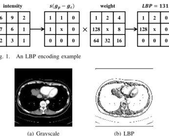
wheregpis the gray-scale intensity ofPpixels in the circular neighborhood ands(x)is the function which outputs 0 and 1 as the result of comparisons. Fig. 1 presents an LBP encoding example withP = 8and R= 1. A k×k grayscale image encoded by LBP labels can be viewed as a sequence ofk2 integers,
⟨p1, p2, . . . , pk2⟩.
Each pi, 1 ≤ i ≤ k2, represents the LBP label of gi, i.e., LBPP,R(gi). WhenP = 8 and R = 1, the range of LBP values in a b-bit grayscale CT image is defined, 0 ≤ pi ≤ b2−1
. For clustering, the distance between two pixelsgiand gj,1≤i, j≤k2, is the difference of their LBP values as
√
6 9 2
7 1
2 3 1 6 intensity
1 2 0
0
0 0 0 x 128 1 1 0
1 0
0 0 0 x
1 2 4
8
64 32 16 x 128
ൈ
࢙ሺࢍെࢍࢉሻ weight ࡸࡼൌ
Fig. 1. An LBP encoding example
(a) Grayscale (b) LBP
Fig. 2. A CT image in grayscale and LBP feautres
C. Grayscale + LBP
Combining grayscale and LBP labelling, ak×kgrayscale CT-image can be considered as a sequence of k2
integer-pairs,
⟨(g1, p1),(g2, p2), . . . ,(gk2, pk2)⟩.
For clustering, the distance between two pixels gi and gj,
1≤i, j≤k2
, is calculated by
√
(gi−gi)2+ (LBPP,R(gi)−LBPP,R(gj)2) (11) With both grayscale and LBP, a two-dimensional feature space is organized that may provide advanced discriminative information for cluster validity evaluation.
IV. EXPERIMENT
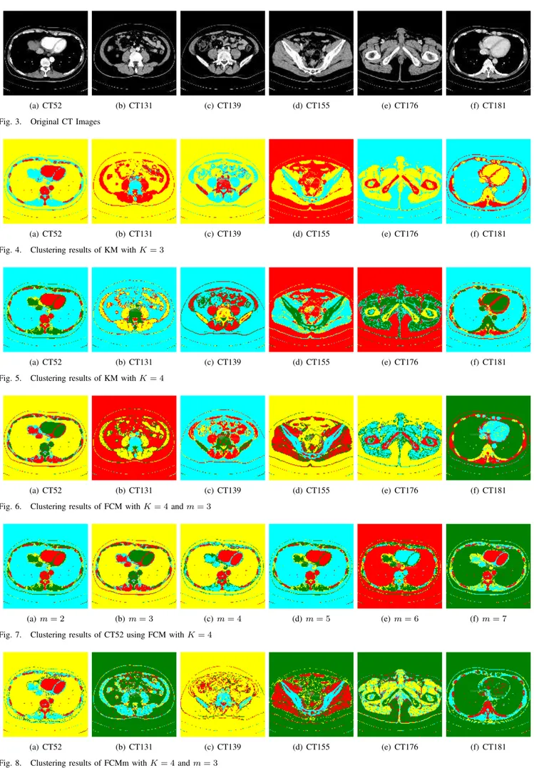
To evaluate the effectiveness of various features and clustering methods for CT-image clustering, the following experiments are performed. Six CT-images are chosen for test, as shown in Fig. 3. These images are in 8-bit grayscale and256×265in size. Data sets to be clustered are generated based on the three types of features, as mentioned in Sec-tion III. Four clustering algorithms are implemented; they are FCM, KM, FCMm, and KMm. These clustering algorithms are implemented in C++. All experiments are conducted in a personal computer with 8G RAM and a Core i3 CPU running on Windows 7. The clustering algorithms run with variousK=2–10 (number of clusters) and the best results are retained.
Fig. 4–Fig. 9 present some selected clustering results of CT-images. In these figures, pixels in the same color belong to the same cluster. It seems that clustering with grayscale+LBP as features can form better recognizable clusters; clustering with grayscale only may not always has satisfactory results. Table I–Table III presents the number of clusters obtained from the four clustering algorithms with fuzziness degree m=2.
V. CONCLUSION
Analysis of CT images by image segmentation is an import task in medical diagnosis. Clustering is a useful method for fast and effective image segmentation. This study compares the results of CT image segmentation using
TABLE I
FCMANDFCMM WITH GRAY, LBP,AND GRAY+LBPFEATURES
FCM FCM FCM FCMm FCMm FCMm
Image Gray LBP Gray+LBP Gray LBP Gray+LBP
CT52 3 2 3 3 2 3
CT131 3 2 4 4 2 3
CT139 3 2 3 3 2 3
CT155 3 2 4 4 2 3
CT176 3 2 3 3 2 3
CT181 3 2 3 3 2 3
TABLE II
RESULTS OFCTIMAGE CLUSTERING(m= 2)
Image FCM FCMm KM KMm
CT52 3 3 3 3
CT131 3 4 4 4
CT139 3 4 3 3
CT155 3 3 4 4
CT176 3 3 3 3
CT181 3 4 3 3
TABLE III
CLUSTERING RESULTS OFFCMWITH VARIOUSm
m= 1.5 2.0 3.0 4.0 5.0 6.0 7.0 8.0
CT52 3 3 3 3 3 4 4 4
CT131 4 3 3 3 3 3 4 4
CT139 3 3 3 3 3 3 3 3
CT155 3 3 4 3 3 4 3 3
CT176 4 3 3 3 3 3 3 3
CT181 3 3 3 3 3 3 4 4
TABLE IV
CLUSTERING RESULT OFFCMM WITH VARIOUSm
m= 1.5 2.0 3.0 4.0 5.0 6.0 7.0 8.0
CT52 3 3 3 3 3 4 4 4
CT131 4 3 3 3 3 3 4 4
CT139 3 3 4 3 3 3 3 3
CT155 3 3 4 3 3 4 3 3
CT176 4 3 3 3 3 3 3 3
CT181 3 3 4 3 3 3 4 4
intensity- and texture-based image features and clustering algorithms. The effectiveness of three features, grayscale, LBP, and grayscale+LBP, for clustering are investigated. The performance of four FCM-based clustering algorithms with various parameter settings are discussed. The results show that FCM and grayscale+LBP can produce reasonable and satisfactory clustering results for CT-image segmentation. Parameters associated with clustering algorithms and feature selection, such as the degree of fuzziness m in Eq.(3) and P and R in Eq.(8) should be investigated. These will be included in our future work.
REFERENCES
[1] N. Sharma and L. M. Aggarwal, “Automated medical image segmenta-tion techniques,”Journal of Medical Physics, vol. 35, pp. 3–14, 2010. [2] H. Ng, S. Ong, K. Foong, P. Goh, and W. Nowinski, “Medical image segmentation using k-means clustering and improved watershed algorithm,” in Proceedings of 7th IEEE Southwest Symposium on Image Analysis and Interpretation, 2006, pp. 61–65.
(a) CT52 (b) CT131 (c) CT139 (d) CT155 (e) CT176 (f) CT181
Fig. 3. Original CT Images
(a) CT52 (b) CT131 (c) CT139 (d) CT155 (e) CT176 (f) CT181
Fig. 4. Clustering results of KM withK= 3
(a) CT52 (b) CT131 (c) CT139 (d) CT155 (e) CT176 (f) CT181
Fig. 5. Clustering results of KM withK= 4
(a) CT52 (b) CT131 (c) CT139 (d) CT155 (e) CT176 (f) CT181
Fig. 6. Clustering results of FCM withK= 4andm= 3
(a)m= 2 (b)m= 3 (c)m= 4 (d)m= 5 (e)m= 6 (f)m= 7
Fig. 7. Clustering results of CT52 using FCM withK= 4
(a) CT52 (b) CT131 (c) CT139 (d) CT155 (e) CT176 (f) CT181
(a)m= 2 (b)m= 3 (c)m= 4 (d)m= 5 (e)m= 6 (f)m= 7
Fig. 9. Clustering results of CT52 using FCMm withK= 4
[4] A. Suruliandi and K. Ramar, “Local texture patterns - a univariate texture model for classification of images,” inProceedings of the In-ternational Conference on Advanced Computing and Communications, 2008, pp. 32–39.
[5] L. Zhou and H. Wang, “Open/closed eye recognition by local binary increasing intensity patterns,” inProceedings of the IEEE Conference on Robotics, Automation and Mechatronics, 2011, pp. 7–11. [6] A. R. Rivera, R. Castillo, and O. Chae, “Local directional number
pattern for face analysis: Face and expression recognition,” IEEE Transactions on Image Processing, vol. 22, no. 5, pp. 1740–1752, May 2013.
[7] T. Ojala, M. Pietikainen, and D. Harwood, “A comparative study of texture measures with classification based on featured distributions.”
Pattern Recognition, vol. 29, no. 1, pp. 51–59, 1996.
[8] T. Ojala, M. Pietikainen, and T. Maenpaa, “Multiresolution gray-scale and rotation invariant texture classification with local binary patterns,”
IEEE Transactions on Pattern Analysis and Machine Intelligence, vol. 24, no. 7, pp. 971–987, 2002.
[9] C.-H. Wu, L.-W. Lu, , and Y.-Y. Li, “A study on pattern encoding of local binary patterns for texture-based image segmentation,” inIn Proceedings of the 6th International Conference on Machine Learning and Cybernetics (ICMLC), 2014, pp. 13–16.
[10] D.-Q. Zhang and S.-C. Chen, “A novel kernelized fuzzy c-means algorithm with application in medical image segmentation,”Artificial Intelligence in Medicine, vol. 32, pp. 37 – 50, 1 2004.
[11] J. B. MacQueen, “Some methods for classification and analysis of mul-tivariate observations,” inProceedings of 5-th Berkeley Symposium on Mathematical Statistics and Probability, 1, Ed. Berkeley, University of California Press, 1967.
[12] J. C. Dunn, “A fuzzy relative of the isodata process and its use in detecting compact well-separated clusters,” Journal of Cybernetics, vol. 3, pp. 32 – 57, 1974.
[13] Y. Dodge, “An introduction to l1-norm based statistical data analysis,”

