Journal of HerbMed Pharmacology
Journal homepage: http://www.herbmedpharmacol.com J HerbMed Pharmacol. 2015; 4(4): 129-132.
The effect of gallic acid on Jurkat cell line
*Corresponding author: Batoul Pourgheysari, Medical Plants Research Center, Shahrekord University of Medical Sciences, Shahrekord, Iran. Email: [email protected]
Introduction
Acute lymphoblastic leukemia (ALL) is a malignancy of immature lymphoid cells. The over production of malig-nant cells in the bone marrow inhibits the production of other blood cells. ALL is the most prevalent malignancy in children, representing 25%-30% of all childhood malig-nancies (1). Many plants contain components against can-cer cells that can induce their effects in different stages of growth of cancer cells (2). A promising strategy for cancer prevention is introducing drugs that block the develop-ment of cancer (3). Food plays an important role in pre-venting the onset and progression of cancer. Nowadays, medicinal plants have attracted the attention of many cancer patients around the world. The scientists have fo-cused on the biological and therapeutic properties of these products (4). Induction of apoptosis in cells is one of the targets of anti-cancer therapy (5). Gallic acid (GA) is a polyhydroxyphenolic compound that is widely distributed
in the natural plants, fruits and food and has a wide range of biological functions (6). Many foods such as gallnuts, sumac, grape, green tea, oak bark, strawberry, lemon, ba-nana, pineapple, witch hazel, and apple peel are known to be rich in GA (7). GA and its analogs have been reported to have many biological activities, including antioxidant property (8), anti-tumor (7), mutagenic, and anti-carcinogenic properties (9). However, the major interest in GA and its analogs is related to its antitumor activity. It has been shown that propyl, lauryl, methyl gallate induce apoptosis and inhibit proliferation in tumor cell lines (10). GA has shown no cytotoxicity against normal fibroblast and endothelial cells (11). Some studies have shown that GA causes DNA fragmentation (10) and is also respon-sible for suppression of tumor angiogenesis, leading to in-hibition of tumor metastasis (12). In fact, anti-cancer ac-tivity of GA has been reported in various cancer cells, such as prostate (13), lung, gastric, colon, breast, cervical and
Zahra Sourani1, Batoul Pourgheysari2*, Mahmoud Raieian-Kopaei2, Hedayatollah Shirzad3, Moien Shirzad4
1Msc Student in Immunology, Shahrekord University of Medical Sciences, Shahrekord, Iran 2Medical Plants Research Center, Shahrekord University of Medical Sciences, Shahrekord, Iran 3Cellular & Molecular Research Center, Shahrekord University of Medical Sciences, Shahrekord, Iran 4Clinical Biochemistry Research Center, Shahrekord University of Medical Sciences, Shahrekord, Iran
Implication for health policy/practice/research/medical education:
Gallic Acid (GA) has anti-cancer property and this effect seems to be through apoptosis induction in lymphoblastic leukemia. These results can be a step toward targeted combination or alternative chemotherapy in cancer.
Please cite this paper as:Sourani Z, Pourgheysari B, Rafieian-Kopaei M, Shirzad H, Shirzad M. The effect of gallic acid on Jurkat cell line. J HerbMed Pharmacol. 2015;4(4):129-132.
Introduction: Acute lymphoblastic leukemia (ALL) is the most prevalent leukemia in children. Fruits and plants have a wide range of biological functions including anti-proliferative effect. Gallic acid (GA), is a polyhydroxyphenolic compound that is widely distributed in the natural plants. The aim of the present study was the evaluation of the effect of GA on proliferation inhibition of Jurkat cells, the lymphoblastic leukemia cell line.
Methods: Jurkat cell line was cultured in blood cells culture media in a standard conditions with different concentrations of GA (0-100 µm) for 24, 48 and 72 hours. The effect of GA on cell viability was measured using MTS assay.
Results: Decline of cell viability to less than 50% was observed at 60, 50 and 30 µm concentrations after 24, 48 and 72 hours incubation time, respectively.
Conclusion: The results demonstrated that the polyphenolic compound, GA with antioxidant capability is effective in proliferation inhibition in Jurkat lymphoblastic leukemia cell line with a time and dose dependent manner. Therefore, GA may be a potential compound for cancer prevention and treatment.
A R T I C L E I N F O
Keywords: Gallic acid Jurkat cell line
Acute lymphoblastic leukemia Proliferation
Article History: Received: 14 June 2015 Accepted: 5 September 2015 Article Type:
Original Article
Sourani Z et al
Journal of HerbMed Pharmacology, Volume 4, Number 4, October 2015 http://www.herbmedpharmacol.com 130
esophageal cancers (14). As there was no report of the ef-fect of GA on lymphoblastic leukemia cell line, we aimed to evaluate the effect of GA on proliferation inhibition on this cell line.
Materials and methods Cell culture
Lymphoblastic leukemia cell line (Jurkat cell) was cultured in RPMI 1640 supplemented with 10% heat-inactivated fetal bovine serum (FBS), 100IU/mL penicillin, 0.1 mg/ mL streptomycin (PS) and 0.3 mg/ml L-glutamine. The cells were incubated at 37°C in a humidified incubator with 5% carbon dioxide. The cells were maintained at the concentration of 2×10⁵/ml and then transferred to 96 and/ or 6-well plates for experiments. To determine the viable cells, the culture was harvested and the cells were counted by the trypan blue staining and standard hemocytometer.
Cell culture and treatment
Jurkat cells were cultured in RPMI-1640 medium contain-ing 10% FBS. The stock solution of GA was dissolved in RPMI-1640, and different concentrations (uM) were pre-pared in the RPMI-1640 medium.
Assessment of cell viability by MTS assay
This chromogenic assay involves the biological reduction by viable cells of the tetrazolium compound 3-(4, 5-di- methylthiazol-2-yl)-5-(3-carboxymethoxyphenyl)-2-(4-sulfophenyl)-2Htetrazolium (or MTS).
MTS assay reagent is composed of MTS and the elec-tron coupling agent phenazine methosulfate (PMS). The formazan product of MTS reduction is soluble in tissue culture medium. This reaction only takes place when mi-tochondrial reductase enzymes are active, and therefore the conversion can be directly related to the viability of cells in culture. The MTS reagent alone results in very low background absorbance values in the absence of cells. Jurkat cells were seeded in above complete media at the concentration of 10 × 103 cells per well in 96-well plates and allowed to grow overnight. The cells were treated with different concentrations (0-100 µM) of GA. After incuba-tion for 48 hours, 20 µL of MTS (5 mg/mL in phosphate buffered saline) was added to each well and incubated for 3 hours, in the dark. The absorbance was measured at 490-620 nm by an Elisa reader (stat fax-2100 awarenes).
Statistical analysis
Data were analyzed by SPSS using Kruskal-Wallis test and are presented as the means ± standard deviation from at least three independent experiments.
Results
Decrease cell viability by gallic acid
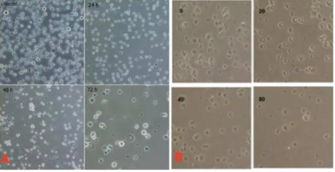
Jurkat cells were incubated with various concentrations (0, 10, 20, 30, 40, 50, 60, 70, 80, 90, 100µM) of GA for 24, 48 and 72 hours. Decrease of cell viability was observed after incubation time in treated cells in a time–dependent manner (Figure 1). The MTS assay was done to
investi-A B
Figure 1. A: Decrease of cell viability in the presence of GA
after 24, 48 and 72 hours (time-dependent). B: Decrease of cell viability in Jurkat cells incubated with various concentrations (0, 20, 40, 80) (dose-dependent) (Magnification 40x).
Figure 2. Jurkat cell viability by MTS assay. Cell viability was
determined by measuring the mitochondrial metabolism of MTS after 24, 48 and 72 hours.
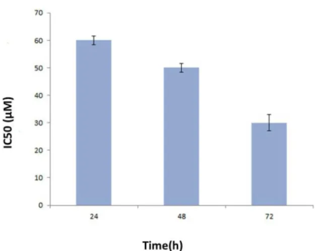
gate the cell viability. The data obtained from the absorp-tion at 490 nm was converted to a percentage. The results showed a dose- and time-dependent behavior (Figure 2). Cell viability was about 93%, 87%, 65%, at a concentration of 20μM after 24, 48 and 72 hours, respectively, however viability declined to 13%, 8% and 5% at a concentration of 100μM after above incubation periods. IC50 (inhibition concentration) was about 60μM in 24 hours, about 50μM in 48 hours and about 30μM in 72 hours (Figures 2 and 3).
Discussion
Efect of gallic acid on Jurkat cell line
Journal of HerbMed Pharmacology, Volume 4, Number 4, October 2015
http://www.herbmedpharmacol.com 131
cells (A549 and Calu-6). The inhibition of the growth of lung cancer cells and induction of cell death were related to GSH (glutathione) depletion as well as reactive oxygen species (ROS) level changes. The induction of apoptosis has been shown to be related to ROS in Larry et al study in prostate cancer cells. In this study, 80 μg/ml GA has the maximum inhibitory effect in 24 hours and caused activa-tion of apoptosis pathway (17). Chiaet al(18) studied the anticancer effects of GA in oral cancer cell lines including UM1, UM2, SCC-4 and SEC-9. They showed that the re-quired concentration of GA for the induction of apoptosis was different in various cell lines. Lu et al (12) have stud-ied GA-induced cell death and the proliferation, anti-invasion, and angiogenesis in human glioma cells. They showed that the inhibition of cell viability by GA was dose dependent. Madleneret al (19) studied the effect of GA in promyelocytic leukemia cells (HL-60) They showed that apoptosis induced by GA is dose dependent. Other studies have demonstrated the effect of GA on gastric cancer cells (20). Youet al (7) studied the effect of GA in Hela cervical cancer cells. They showed that GA was able to inhibit the growth of Hela cells and to induce apoptosis. GA-induced Hela cell death was also accompanied by ROS increase and GSH depletion. The effect of GA on Leukemia cells (K562) and L1210 (15,21) was also in consistent with our data. Our study showed that GA could inhibit cell pro-liferation in lymphoblastic leukemia cell line which was consistent with the studies of other cell lines.
Conclusion
It seems from the study that decrease of Jurkat cell num-bers in the presence of GA is dependent to the dose- and time which is a favorable effect of anti-cancer treatments. GA may help in cancer combination- chemotherapy but it needs to be investigated in other lymphoblastic cell lines, ALL lymphoblasts and then in vivo experimental models.
Acknowledgements
This paper was derived from MSc thesis of the first author which was supported by the Medical Plants Research
Cen-ter of Shahrekord University of Medical Sciences. The au-thors also wish to thank deputy of Research, Shahrekord University of Medical Sciences for financial support.
Authors’ contributions
ZS performed the experimental work and helped the writ-ing; BP led the design and writing the project; HS and MR helped with the design; MS helped in experimental design and analysis.
Conflict of interests
The authors declared no competing interests.
Ethical considerations
Ethical issues (including plagiarism, misconduct, data fabrication, falsification, double publication or submis-sion, redundancy) have been completely observed by the authors.
Funding/Support
This research was financially supported by Shahrekord University of Medical Sciences, Shahrekord, Iran.
References
1. Andrade AF, Borges KS, Silveira VS. Update on the use of l-asparaginase in infants and adolescent patients with acute lymphoblastic leukemia. Clin Med Insights Oncol. 2014;8:95-100.
2. Nejad Shahrokhabadi K, Tavakkol Afshari J, Rakhshandeh H, Barouk A. Study of cytotoxicity effect of total saffron extract on hepatocarcinoma cell line (HepG2. Medical Science Journal of Islamic Azad Univesity. 2009;19(3):154-159.
3. Gupta SK. Pharmacology and therapeutics in the new millennium. Springer Science & Business Media; 2001.
4. Abdullaev FI. Cancer chemopreventive and tumoricidal properties of saffron (Crocus sativus L.). Exp Biol Med (Maywood). 2002;227(1):20-25. 5. Denicourt C, Dowdy SF. Targeting apoptotic pathways
in cancer cells. Science. 2004; 305(5689):1411-1413. 6. Niemetz R, Gross GG. Enzymology of gallotannin
and ellagitannin biosynthesis. Phytochemistry. 2005; 66(17):2001-2011.
7. You BR, Moon HJ, Han YH, Park WH. Gallic acid inhibits the growth of HeLa cervical cancer cells via apoptosis and/or necrosis. Food Chem Toxicol. 2010;48(5):1334-40.
8. Locatelli C, Leal PC, Yunes RA, Nunes RJ, Creczynski-Pasa TB. Gallic acid ester derivatives induce apoptosis and cell adhesion inhibition in melanoma cells: the relationship between free radical generation, glutathione depletion and cell death. Chem Biol Interact. 2009;181(2):175-184.
9. Jang A, Lee NY, Lee BD, et al. Biological functions of a synthetic compound, octadeca-9, 12-dienyl-3, 4, 5-hydroxybenzoate, from gallic acid–linoleic acid ester. Food Chem. 2009;112(2):369-373.
Figure 3. Comparison of the IC50 (50% inhibition concentration) of
Sourani Z et al
Journal of HerbMed Pharmacology, Volume 4, Number 4, October 2015 http://www.herbmedpharmacol.com 132
10. Fiuza S, Gomes C, Teixeira L, et al. Phenolic acid derivatives with potential anticancer properties-a structure–activity relationship study. Part 1: Methyl, propyl and octyl esters of caffeic and gallic acids. Bioorg Med Chem. 2004;12(13):3581-3589. 11. Sohi KK, Mittal N, Hundal MK, Khanduja KL. Gallic
acid, an antioxidant, exhibits antiapoptotic potential in normal human lymphocytes: a Bcl-2 independent mechanism. J Nutr Sci Vitaminol (Tokyo). 2003; 49(4):221-227.
12. Lu Y, Jiang F, Jiang H, et al. Gallic acid suppresses cell viability, proliferation, invasion and angiogenesis in human glioma cells. Eur J Pharmacol. 2010;641(2):102-107.
13. Agarwal C, Tyagi A, Agarwal R. Gallic acid causes inactivating phosphorylation of cdc25A/cdc25C-cdc2 via ATM-Chk2 activation, leading to cell cycle arrest, and induces apoptosis in human prostate carcinoma DU145 cells. Mol Cancer Ther. 2006; 5(12):3294-2302.
14. Faried A, Kurnia D, Faried L, et al. Anticancer effects of gallic acid isolated from Indonesian herbal medicine, Phaleria macrocarpa (Scheff.) Boerl, on human cancer cell lines. Int J Oncol. 2007; 30(3):605-613.
15. Abdelwahed A, Bouhlel I, Skandrani I, et al. Study of antimutagenic and antioxidant activities of gallic acid and 1, 2, 3, 4, 6-pentagalloylglucose from Pistacia
lentiscus: Confirmation by microarray expression profiling. Chem Biol Interact. 2007;165(1):1-13. 16. You BR, Park WH. Gallic acid-induced lung cancer
cell death is related to glutathione depletion as well as reactive oxygen species increase. Toxicol In Vitro. 2010;24(5):1356-1362.
17. Russell LH, Mazzio E, Badisa RB, et al. Autoxidation of gallic acid induces ROS-dependent death in human prostate cancer LNCaP cells. Anticancer Res. 2012; 32(5):1595-1602.
18. Chia YC, Rajbanshi R, Calhoun C, Chiu RH. Anti-neoplastic effects of gallic acid, a major component of Toona sinensis leaf extract, on oral squamous carcinoma cells. Molecules. 2010;15(11):8377-8389. 19. Madlener S, Illmer C, Horvath Z, et al. Gallic acid
inhibits ribonucleotide reductase and cyclooxygenases in human HL-60 promyelocytic leukemia cells. Cancer Lett. 2007;245(1):156-162.
20. Ho HH, Chang CS, Ho WC, Liao SY, Wu CH, Wang CJ. Anti-metastasis effects of gallic acid on gastric cancer cells involves inhibition of NF-κB activity and downregulation of PI3K/AKT/small GTPase signals. Food Chem Toxicol. 2010;48(8):2508-2516.