Article
ISSN 0102-695X
http://dx.doi.org/10.1590/S0102-695X2011005000194 Received 25 May 2011 Accepted 8 Jul 2011 Available online 21 Oct 2011
antioxidant activity of melanoidin pigment
from the fermented leaves of
Orthosiphon
stamineus
Daniil N. Olennikov,
*Larisa M. Tankhaeva
Laboratory of Medical and Biological Research, Institute of General and Experimental Biology, Siberian Division, Russian Academy of Sciences, Russian Federation.
Abstract:The melanoidin pigment (OS-M) was isolated from fermented leaves of
Orthosiphon stamineus Benth. (Lamiaceae), with a 0.37% yield (from dry plant weight), and characterised. OS-M is a phenolic polymer with a molecular weight of 4.4 kDa. Determination of basic physicochemical parameters using elemental analysis, functional group analysis, UV-Vis- and FTIR-spectroscopy of OS-M indicated that the isolated polymer was similar to typical melanoidins. Experimental data show that aromatic fragments dominate the OS-M structure, which also contains a small amount of aliphatic fragments. Investigation into the antioxidant activity of OS-M under in vitro conditions demonstrated that O. stamineus melanoidin has a scavenging effect against free radicals (DPPH•, ABTS•+, O2•-) and NO molecules, inactivates molecules of H2O2, chelates Fe2+-ions and oxidises NADH.
Keywords: antioxidant activity fermented leaves Lamiaceae melanoidin
Orthosiphon stamineus
Introduction
Orthosiphon stamineus Benth. [O. aristatus
(Blume) Miq., O. spicatus (Thunb.) Bak., Ocymum
grandiflorum Bl.; Lamiaceae fam.] is a popular
traditional medicinal plant from South Asia that is used to treat a wide range of diseases. In Indonesia, O. stamineus, or kumis kucing, is used to treat rheumatism, diabetes, hypertension, tonsillitis, epilepsy, menstrual disorders, and sexually transmitted diseases; in Vietnam, for kidney and liver stones, hepatitis, colds, and inflammatory diseases; in Myanmar as a secho or myit-shwe for disorders of the genitourinary system and in the traditional medicine of Java for hypertension (Ohashi et al., 2000). In Japan, O. stamineus is used as a Java tea or neko no hige for cleansing (detoxification) of the organism (Awale et al., 2004). Because of its popularity and demonstrated effectiveness, phytochemical and pharmacological research on O. stamineus has been carried out since the 1930s.
Chemical studies of O. stamineus have isolated di- and triterpenes (Awale et al., 2004; Tezuka et al., 2000), carotenoids (Kudritskaya et al., 1987) and essential oils (Hossain et al., 2008). Phenolic compounds of O. stamineus were detected, including phenylpropanoids, flavones, flavonols, coumarins and chromens (Akowuah et al., 2005; Matsubara et al.,
1999; Sumaryono et al., 1991).
Pharmacological studies of extracts and individual compounds from O. stamineus have shown biological activity including antimicrobial, antifungal (Hossain et al., 2008), hypoglycaemic, diuretic, litolytic, saluretic (Dharmaraj et al., 2006), anti-inflammatory (Awale et al., 2003), antioxidant (Akowuah et al., 2005), cytotoxic (Tezuka et al., 2000) and hypotensive (Matsubara et al., 1999) properties.
Commercial raw material of O. stamineus
consists of dried leaves and hops of shoots, which were pre-fermented within 24-36 hours and then quickly dried. This treatment increased the extractability of this plant material. During fermentation, the raw material turns from dark green to dark brown. The nature of the dark pigment of fermented O. stamineus leaves has not previously been investigated, but it is probably similar to the melanoidin from fermented Camelia sinensis
leaves (black tea) (Sava et al., 2001).
In this study, we present the results of a physicochemical investigation of melanoidin pigment from fermented O. stamineus leaves and an examination of its antioxidant properties using in vitro methods.
Materials and Methods
Elemental analysis (CHNS/O) was performed in a 2400 Series II elemental analyser (Perkin Elmer, USA). Potentiometric titration was performed on a Metron automatic titrator (Germany). Spectrophotometric analysis was performed on SF-2000 UV-Vis spectrophotometer (Lomo, Russian Federation). Size-exclusion chromatography (SEC) was performed on a Sephadex G-150 column (120 × 1.5 cm, Pharmacia, Sweden) coupled to an SF-2000 UV-Vis flow-detector (Lomo, Russian Federation) at 280 nm. 0.3% NaCl was used as an eluent. Preliminary calibration of the column was conducted using standard dextrans of different molecular weights (Mw 2000, 100, 10 kDa) and raffinose (Mw 0.5 kDa). The molecular weights were calculated using a calibration curve. FTIR spectra were determined using a Fourier-transform infrared spectrometer FT-801 (Simex, Russian Federation) using KBr pellets in the frequency range 4000-600 cm-1. The substance was ground with
spectroscopic grade KBr powder and pressed into 1-mm pellets for FT-IR measurement.
Plant material
Green and fermented aerial parts of
Orthosiphon stamineus Benth. (Lamiaceae family)
were obtained from Trisko Co., Indonesia
(production-run No. В105346). The identity of the plant was kindly
identified by Prof. Dr. Tamara A. Aseeva (Laboratory of Botany, Institute of General and Experimental Biology, Russia). Voucher specimens are deposited in the herbarium of Department of Biologically Active Substances, Institute of General and Experimental Biology, Russia.
Analytical methods
Physicochemical characteristics of melanoidin were determined according to the following methods: total sugar content - anthrone-sulphuric method with D-glucose as standard (Olennikov et al., 2006) and protein content using the Bradford method with bovine serum albumin as a standard (Bradford, 1976). The carboxyl group content and total phenol group content were determined using potentiometric titration according to the method of Koroleva et al. (2007). Carbonyl group content was determined using the 2,4-dinitrophenylhydrazine method with acetophenone as a standard (Lappin & Clark, 1951), and pyrocatechol group content was determined using the FeSO4/tartrate method with pyrocatechol as a standard (Falkehag et al., 1966). Total melanoidin content was determined using a spectrophotometry, according to the method of Santos & Stephanopoulos (2008), and rosmarinic acid content was determined using the HPTLC-densitometric
method (Fecka et al., 2007).
Isolation procedures
For isolation of O. stamineus melanoidin pigment (OS-M), powdered fermented leaves (700 g) were first successively extracted with n-hexane and chloroform to remove lipids. The defatted powder was extracted four times with distilled water at 80 °C using a solid:liquid ratio of 1:20 (w/v). The mixture was filtered and the combined water extract was concentrated under vacuum at 40 °C to a final volume of 2 L. The concentrated extract was centrifuged (6000 x g, 20 min). The supernatant was treated with Sevag reagent (mixture of chloroform and n-butanol 4:1) to remove proteins, dialysed (72 h), and concentrated under vacuum at 40 °C to a final volume of 500 mL. The resulting solution was acidified with concentrated H2SO4 to a final concentration of 1% and stored for 6 h at 10 °C. The brown precipitate was centrifuged (10000 x g, 30 min), washed with 1% H2SO4, dissolved in 100 mL of 0.1% NaHCO3 and dialysed (48 h). The undialysed fraction was dried by lyophilisation, and the powder obtained was suspended in 20% HCl and stored for five days at 5 oC. The mixture of powder and
HCl was centrifuged (6000 x g, 30 min) and washed with 1% HCl; the precipitate was dissolved in 100 mL of 0.1% NaHCO3 and dialysed (48 h). The undialysed part was dried by lyophilisation and dissolved in 50 mL of 0.1 M Na2CO3, and the solution was transferred to a Sephadex G-150 column (110 × 4.5 cm) coupled to an SF-2000 UV-Vis flow detector (Lomo, Russian Federation); 6-mL fractions were collected using 0.1 M Na2CO3 as an eluent. The fractions No. 98-118 were combined and dialysed (48 h), and the undialysed part was dried by lyophilisation, giving fractions containing OS-M. The yield of OS-M was 2.62 g.
UV-Vis spectroscopic analysis
For determination of UV-Vis and differential spectra ten milligrams of OS-M was transferred to a volumetric flask (25 mL) and dissolved in 25 mL of a 1:1 dioxane:EtOH mixture (v/v). Then, 2 mL of the obtained solution was transferred into volumetric flasks (25 mL), one filled with pH 6.0 buffer (495 mL of 0.2 M KH2PO4 + 113 mL 0.1 M NaOH + 1392 mL of distilled water; solution A) and the other with pH 12.0 buffer (400 mL 0.1 M Na2B4O7 + 600 mL 0.1 M NaOH; solution B). The UV-Vis spectra were determined in a 1-cm quartz cell in the 190-800 nm range. Distilled water was used as a blank. To determine differential spectra, solution B was used as a sample and solution A was used as a blank.
determined using the solutions of OS-M prepared in
final concentrations of 8, 16, 32 and 64 μg/mL using
pH 12.0 buffer as a solvent. The UV-Vis spectra were determined in the 190-800 nm range. Distilled water was used as blank. For optical density data, a common logarithm was used to build a graph of optical density logarithm against wavelength. Linear regression was used to determine the regression equation, and the value of the slope was used as a logarithmic slope of absorbance. All measurements were carried out in triplicate.
The chromaticity coefficient E465/E665 was determined as a ratio of optical densities at 465 and 665 nm for the substance in pH 12.0 buffer solutions.
For determination colour value E1%1 cm of
OS-M solutions were prepared in final concentrations of
8, 16, 32, 64, 128 and 256 μg/mL using pH 12.0 buffer
as a solvent. The optical densities of the solutions were determined at 195 nm using distilled water as a blank. The data were used to construct a graph of optical density versus concentration. After linear regression was determined, the regression equation and the colour value (E1%1 cm) were calculated as the optical density
of an OS-M solution with concentration 10 mg/mL (=1%). All measurements were carried out three times.
Melanoidin-Fe-salt preparation (OS-M-Fe)
One hundred milligrams of OS-M was dissolved in 2 mL of DMSO, and 48 mL of distilled water was added. Twenty milliliters of a 1% solution of FeSO4 in distilled water was added and stirred for 2 h at 30 °C. The reaction mixture was centrifuged (12000 x g, 30 min), and the precipitate was washed with distilled water and dried under vacuum at 20 °C. The yield of melanoidin-Fe-salt (OS-M-Fe) was 124 mg.
Aqueous extract of O. stamineus leaves (AEOS)
preparation
Powdered fermented O. stamineus leaves (200 g) were extracted three times with water at 80 °C using a solid:liquid ratio of 1:25 (w/v). The mixture was filtered, and the combined water extracts were concentrated under vacuum at 40 °C to a final volume of 1 L. The concentrated extract was centrifuged (6000 x g, 20 min), and the supernatant was dried in a ShSV-45k vacuum-drying box (KZMA, Inc., Kazan’, Russia) to give dried water extract of O. stamineus
leaves (AEOS), which was then powdered in a VSI-05 crushing machine (KZMA, Inc., Kazan’, Russia). The yield of AEOS was 30.74 g.
Antioxidant activity assays
The ability to scavenge DPPH• free radicals was assessed as described by Asker & Shawky (2010); the radical-scavenging activity against ABTS•+ radical cation was measured using the method of Ding et al. (2010); the determination of superoxide anion scavenging activity was measured in phenazine methosulphate-nicotinamide adenine dinucleotide-nitroblue tetrazolium systems using the method of Ozen et al. (2011); the NO scavenging activity was measured using the sodium nitroprusside method (Kumar et al., 2008); the H2O2 inactivating activity was measured using the method of Badami & Channabasavaraj (2007); the chelating activity for Fe2+-ions was measured by the o-phenanthroline method (Olennikov et al.,
2011a); β-carotene bleaching assay was performed in β-carotene-oleic acid-DMSO-H2O2-system (Olennikov
et al., 2011b); the oxidation of NADH was determined by the method of Mosca et al. (1998).
Statistical analysis
All measurements were carried out in triplicate. Statistical analyses were performed using a one-way
analysis of variance (ANOVA), and the signiicance
of the mean difference was determined by Duncan’s multiple range test. Differences at p<0.05 were considered
statistically signiicant. The results were presented as mean
values±SD (standard deviations).
Results and Discussion
The melanoidin pigment OS-M was isolated from fermented O. stamineus leaves using water extraction, acid hydrolysis, repeated precipitation, gel-permeation chromatography on a Sephadex G-150 column and dialysis. The physical and chemical properties of the purified melanoidin OS-M were studied. The average yield of OS-M was 0.37% (dry basis). The O. stamineus melanoidin, an amorphous dark-brown substance that was precipitated in alkaline and neutral solutions of FeSO4 and FeCl3 and at pH below 2.5-3.0 and was bleached by H2O2, KMnO4 and NaOCl. The OS-M does not contain sugars or proteins.
The elemental composition of OS-M was as follows: C=57.14±1.14%, H=3.92±0.07%, N=2.02±0.04%, O=36.92±0.73%; notably, the presence of sulphur was not detected. The C/H ratio of 1.21 indicates an aromatic nature (Koroleva et al., 2007). Comparative analysis of these results with the same data for black tea melanoidin (Sava et al., 2001) shows that OS-M was characterised by higher carbon content and a higher C/H value, indicating a higher level of aromaticity for O. stamineus melanoidin.
OS-M contained 9.31±0.32% carboxylic groups, 7.07±0.24% carbonyl groups, 8.67±0.28% phenolic hydroxyl groups and 3.52±0.11% pyrocatechol groups. The SEC of OS-M on Sephadex G-150 was visible as a single peak, which corresponded to a molecular weight of 4.4 kDa.
The absorption spectrum of OS-M was outwardly simple and contained a single strong band at 228 nm and two weak shoulders at 267 and 337 nm. The band at 228 and the shoulder at 267 nm were the primary and secondary B-band resulting from A1g' → B1u' and A1g' → B2u' transition types, respectively. The appearance of the shoulder at 337 nm (K-band) was the
result of a lengthening of the π-conjugation chain. The
absorption spectra of OS-M in alkaline media displayed a slight reduction in the intensity of the primary B-band and its weak hypsochromic shift to 224 nm. The secondary B-band (267 nm) and K-band (337 nm) were marked by an increase in intensity and a bathochromic shift to 271 and 365 nm, respectively. These changes were caused by the ionisation of phenolic hydroxyl groups in alkaline medium. The differential spectrum of OS-M (alkaline solution vs. neutral solution) displayed three bands at 261, 301 and 378 nm and a number of very weak bands and shoulders in the visible region (487, 587, 654, 702, 766 nm). The origin of these bands was the result of bathochromic shifts of the primary and secondary B-bands and K-bands in the initial spectra. In the case of OS-M, the intensity of the band was maximal at 378 nm as a result of the presence of a
sufficient quantity of α-carbonyl containing groups in
the melanoidin structure.
A plot of the logarithm of absorbance versus wavelength for an alkaline solution of OS-M was described by linear dependence. The dependence was linear regardless of the concentration of the melanoidin solution. However, it should be noted that the linearity of this dependence increased as the concentration of melanoidin increased. For example, for OS-M solutions with concentrations of 8, 16, 32
and 64 μg/mL, the coefficients of determination (r2)
were 0.9952, 0.9981, 0.9985 and 0.9992, respectively. The linearity of this dependence was observed in the
wavelength range from 230 to 700 nm. The values of the logarithmic slope of absorbance for solutions with different concentrations were similar (-0.0034 to -0.0038). Therefore, to determine this characteristic, we recommend an arithmetic mean value using a series of solutions with different concentrations of melanoidin. The value of the logarithmic slope of absorbance for OS-M defined in this way was -0.00365, similar to those previously calculated for other melanoidins (Ellis & Griffits, 1974).
The value of the chromaticity coefficient (E465/ E665) of OS-M was 4.47±0.11, indicating a small amount of aliphatic fragments and high content of aromatic components. The colour value of the OS-M (E1%1 cm)
was 81.25±2.59, higher than the colour value for the general pigment (45.0) and Osmanthus fragrans’ seed melanoidin (60.24) (Wang et al., 2006).
The FTIR spectrum of OS-M indicates the presence of bands similar to those of other melanoidins
(Bilińska, 1996). A broad band at 3400 cm-1 can be
attributed to stretching vibrations of OH and NH2 groups, and weak vibrations at 2929 and 2858 cm-1 are assigned
to stretching vibrations of aliphatic C=H, CH2 and CH3 groups. The strong absorbances at 1720 and 1650 cm-1 were recognised as vibrations of free carboxylic groups and of aromatic C=C and/or C=O groups, respectively. A small amount of aliphatic fragments and the high degree of
aromaticity in the structure of OS-M was conirmed by the
elemental analysis, C/H ratio and the value of chromaticity
coeficient. The absence of bands in the 1610-1625 and
960-970 cm-1 region indicated a low level of double bonds
in the structure of melanoidin. The FTIR spectrum of OS-M also contained bands at 1515 cm-1 (aromatic rings
C-C stretching, NH deformation in amide II), 1443 cm-1
(deformation of aliphatic C-H and a stretching of phenolic OH and symmetric stretching of COO-), 1230 cm-1 (C-H
deformations and C-O stretching in phenolic groups), 1207 cm-1 (stretching of ester C-O-C and valence vibrations of
phenolic C-O groups), 1125 cm-1 (ring breathing and C-O
groups stretching), 1056 cm-1 (aromatic esters), 880, 861,
834, 778 cm-1 (vibrations of H-atoms in aromatic rings),
673, and 595 cm-1 (Bilińska, 1996).
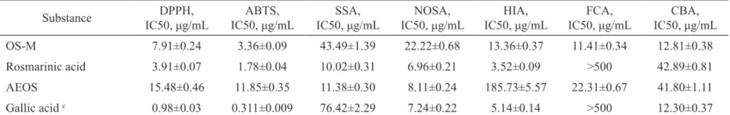
Table 1. Antioxidant activity of OS-M, rosmarinic acid and aqueous extract of Orthosiphon stamineus leaves (AEOS).a, b
Substance DPPH,
IC50, μg/mL IC50, μg/mLABTS, IC50, μg/mLSSA, IC50, μg/mLNOSA, IC50, μg/mLHIA, IC50, μg/mLFCA, IC50, μg/mLCBA,
OS-M 7.91±0.24 3.36±0.09 43.49±1.39 22.22±0.68 13.36±0.37 11.41±0.34 12.81±0.38
Rosmarinic acid 3.91±0.07 1.78±0.04 10.02±0.31 6.96±0.21 3.52±0.09 >500 42.89±0.81
AEOS 15.48±0.46 11.85±0.35 11.38±0.30 8.11±0.24 185.73±5.57 22.31±0.67 41.80±1.11
Gallic acid c 0.98±0.03 0.311±0.009 76.42±2.29 7.24±0.22 5.14±0.14 >500 12.30±0.37 aDPPH• radical-scavenging assay (DPPH), ABTS•+ radical cation-scavenging assay (ABTS), superoxide-anion scavenging assay (SSA), NO scavenging assay (NOSA), H2O2 inactivating assay (HIA), Fe
2+-chelating activity (FCA), β-carotene bleaching assay (CBA); bValues are means±SD of triplicate
The effect of OS-M on hydrogen peroxide (H2O2) inactivation was higher than for WEOS; the IC50
values of OS-M and AEOS were 13.36 and 185.73 μg/
mL, respectively. However, rosmarinic acid provoked more effective inactivation of H2O2 molecules (3.52
μg/mL). The reduced activity of AEOS was probably
caused by the presence of other accompanying components of the O. stamineus extracts. Analogous parameters determined previously for synthetic melanoidin and melanoidin from Aspergillus nidulans
were significantly lower - 57.91 and 186.17 μg/mL,
respectively (Goncalves & Pombeiro-Sponchiado, 2005). OS-M can therefore be considered a good H2O2 inactivator.
Reports of melanoidins as good chelators of metal ions (Fogarty & Tobin, 1996) led to our examination of the Fe2+-chelating activity of OS-M. Experimental data showed that O. stamineus melanoidin expressed chelating action on Fe2+-ions (IC50 11.41 μg/ mL) and was more effective than low-molecular-weight compounds. For example, the chelating activity of
rosmarinic and gallic acids as an IC50 was >500 μg/mL.
The presence of this kind of activity for AEOS (IC50
22.31 μg/mL) was partially caused by the presence of
OS-M. Fe2+ ions react with melanoidin to form Fe2+ -melanoidin complexes that were confirmed by FTIR-spectroscopy. Some changes in the FTIR spectra of Fe2+ -melanoidin complex (OS-M-Fe) were observed. The band at 1714 cm-1 disappeared, indicating the formation
of chemical bonds between free carboxylic groups and Fe2+-ions. The FTIR spectrum of OS-M-Fe was also characterised by the presence of two intense bands at 1392 and 1274 cm-1, which were initially absent in the
spectra of melanoidin. All detected changes indicated that O. stamineus melanoidin reacted with Fe2+-ions to form a Fe2+-melanoidin complex, removing Fe2+-ions and limiting the possibility of their participation in the lipid peroxidation processes.
The examination of the influence of OS-M
on the oxidative destruction of β-carotene in the oleic
acid-DMSO-H2O2 system demonstrated a high value of
antioxidant activity, with IC50 12.81 μg/mL, similar to the 12.30 μg/mL value of parameter for gallic
acid. The efficiency of rosmarinic acid in this assay
was slightly lower (42.89 μg/mL). One feature of the β-carotene bleaching assay is the ability to investigate
the influence of OS-M on the presence of a complex of damaging factors, including H2O2, O2•-, OH•-, and alkyl-radicals that form in this in vitro system. Our results were shown that O. stamineus melanoidin is a highly effective antioxidant.
It is known that DOPA-melanoidin can oxidise NADH through the action of free radicals in the polymer structure (Gan et al., 1974). We assayed the oxidising properties of O. stamineus melanoidin and On the basis of these studies, we concluded
that the biopolymer isolated from the fermented leaves of O. stamineus was melanoidin.
Comparative analysis of the melanoidin content in native and fermented leaves of O. stamineus showed that native (green) leaves do not contain melanoidin, with the exception of stems, which contain 0.124 mg/g (from dry weight) of melanoidin. The formation of melanoidin in the leaves of O. stamineus is therefore due to the fermentation process. The highest content of melanoidin was characteristic of old leaves (4.37 mg/g); melanoidin content in young leaves and stems was 3.21 and 1.43 mg/g, respectively.
Experimental investigations of the antioxidant activity of O. stamineus melanoidin (OS-M) were conducted using the traditional assays: DPPH• radical-scavenging assay (DPPH), ABTS•+ radical cation-scavenging assay (ABTS), superoxide-anion scavenging assay (SSA), NO scavenging assay (NOSA), H2O2 inactivating assay (HIA), Fe2+-chelating
activity (FCA), and β-carotene bleaching assay (CBA)
(Table 1). All experiments included determination and comparative estimation of the same antioxidant factors for rosmarinic acid, the predominant component of O. stamineus with known antioxidant activity (Akowuah et al., 2005), aqueous extract of O. stamineus leaves (AEOS) characterised by co-presence of rosmarinic acid (56.34±1.12 mg/g from dry AEOS weight) and melanoidin (12.16±0.14 mg/g from dry AEOS weight) and gallic acid as an antioxidant reference compound. The radical-scavenging activity of OS-M against DPPH• and ABTS•+ radicals was very high, with IC50=7.91
and 3.36 μg/mL, respectively. The same parameters for rosmarinic acid were 3.91 μg/mL (DPPH•) and 1.78
μg/mL (ABTS•+); for gallic acid the values were 0.98
μg/mL (DPPH•) and 0.311 μg/mL (ABTS•+). This data
classifies OS-M as a radical scavenger. The high radical-scavenging activity of melanoidins has previously been shown for other representative melanoidins (Tu et al., 2009).
The superoxide-anion scavenging activity of
OS-M was IC50 43.49 μg/mL, higher than the activity of gallic acid (76.42 μg/mL) but lower than the activity of rosmarinic acid (10.02 μg/mL) and AEOS (11.38 μg/
mL). This kind of antioxidant action was previously determined for synthetic (DOPA-, TPT-, Leuenk- and Tyr-Gly-melanoidins) (Mosca et al., 1998) and natural melanoidins (Sava et al., 2001). The superoxide anion-scavenging activity of OS-M is enabled by its stable free radical (Mosca et al., 1998).
The activity of OS-M in the NO scavenging
assay was characterised as medium (IC50 22.22 μg/
mL) because the activity of rosmarinic and gallic acids
and AEOS were quite high (6.96, 7.24, 8.11 μg/mL,
established that OS-M influenced NADH in a dose-dependent manner (Figure 1). In the study, the degree of autoxidation of NADH was 3.52%. The introduction of various concentrations of melanoidin increased this value to 4.10, 10.02 and 29.61% for the melanoidin
concentrations of 33, 83 and 167 μg/mL, respectively.
Figure 1. NADH oxidation by OS-M. (1) NADH, (2) NADH
+ OS-M (33 μg/mL), (3) NADH + OS-M (83 μg/mL), (4) NADH + OS-M (167 μg/mL).
Conclusions
We investigated the pigment OS-M, isolated from fermented O. stamineus leaves, by a physical-chemical analysis. Elemental analysis, functional group analysis, UV-Vis- and FTIR-spectroscopy indicated the aromatic nature of OS-M and confirmed its membership in the class of melanoidin pigments.
The study of the antioxidant activity of OS-M using traditional in vitro methods indicated that OS-M strongly scavenges free radicals (DPPH•, ABTS•+, O2•-) and NO molecules, inactivates hydrogen peroxide, chelates Fe2+-ions and oxidises NADH. Comparative analysis of the data indicates that in some cases, the antioxidant activity of melanoidin met or exceeded the activity of rosmarinic acid, the known antioxidant compound from O. stamineus leaves. For example, the Fe2+-chelating activity OS-M and the antioxidant
activity in the β-carotene bleaching assay were
significantly higher than that of rosmarinic acid. Our data add to the information about compounds from O. stamineus leaf extracts that exhibit antioxidant activity. We hypothesise that the presence of melanoidin in fermented O. stamineus leaves also contributes to the inhibitory effect of its extracts on lipid peroxidation processes.
Acknowledgment
The authors thank the Lavrent’ev’s Foundation for financial support (grant No 6.22).
References
Akowuah GA, Ismail Z, Norhayati I, Sadikun A 2005. The effects of different extraction solvents of varying polarities on polyphenols of Orthosiphon stamineus
and evaluation of the free radical-scavenging activity.
Food Chem 93: 311-317.
Asker MMS, Shawky BT 2010. Structural characterization and antioxidant activity of an extracellular polysaccharide isolated from Brevibacterium otitidis BTS 44. Food Chem 123: 315-320.
Awale S, Tezuka Y, Banskota AH, Adnyana IK, Kadota S 2003. Highly-oxygenated isopimaran-type diterpenes from Orthosiphon stamineus of Indonesia and their nitric oxide inhibitory activity. Chem Pharm Bull 51: 268-275.
Awale S, Tezuka Y, Kobayashi M, Ueda J, Kadota S 2004. Neoorthosiphonone A; a nitric oxide (NO) inhibitory diterpene with new carbon skeleton from Orthosiphon stamineus. Tetrahedr Lett 45: 1359-1362.
Badami S, Channabasavaraj KP 2007. In vitro antioxidant activity of thirteen medicinal plants of India’s Western Ghats. Pharm Biol 45: 392-396.
Bilińska B 1996. Progress in infrared investigations of melanin
structures. Spectrochim Acta A 52: 1157-1162.
Bradford MM 1976. A rapid and sensitive method for the quantification of microgram quantities of protein utilizing the principle of protein-dye binding. Anal Biochem 72: 248-254.
Dharmaraj S, Hossain MA, Zhari S, Harn GL, Ismail Z 2006. The use of principal component analysis and self-organizing map to monitor inhibition of calcium oxalate crystal growth by Orthosiphon stamineus
extract. Chemometr Intell Lab Syst 81: 21-28. Ding H, Chou T, Liang C 2010. Antioxidant and
antimelanogenic properties of rosmarinic acid methyl ester from Origanum vulgare. Food Chem 123: 254-262.
Ellis DH, Griffiths DA 1974. The location and the analysis of melanins in the cell walls of some soil fungi. Can J Microbiol 20: 1379-1386.
Falkehag SI, Marton J, Adler E 1966. Chromophores in craft lignin. In Marton J (ed.) Lignin structure and reactions. Advances in Chemistry. Ser. 59. Washington: American Chemical Society, p. 75-89.
Fecka I, Raj D, Krauze-Baranowska M 2007. Quantitative determination of four water-soluble compounds in herbal drugs from Lamiaceae using different chromatographic techniques. Chromatographia 66: 87-93.
Fogarty RV, Tobin JM 1996. Fungal melanins and their interaction with metals. Enzyme Microb Technol 19: 311-317.
Acta Enzymol 370: 62-69.
Goncalves RCR, Pombeiro-Sponchiado SR 2005. Antioxidant activity of the melanin pigment extracted from
Aspergillus nidulans. Biol Pharm Bull 28: 1129-1131.
Hossain MA, Ismail Z, Rahman A, Kang SC 2008. Chemical composition and anti-fungal properties of the essential oils and crude extracts of Orthosiphon stamineus
Benth. Ind Crop Prod 27: 328-334.
Koroleva OV, Kulikova NA, Alekseeva TN, Stepanova EV, Davidchuk VN, Belyaeva EY, Tsvetkova EA 2007. A comparative characterization of fungal melanin and the humin-like substances synthesized by Cerrena maxima 0275. Appl Biochem Microbiol 43: 61-67. Kudritskaya SE, Fishman GM, Zagorodskaya LM, Chikovani
DM 1987. Carotenoids of Orthosiphon stamineus. Chem Nat Comp 23: 767-768.
Kumar S, Kumar D, Manjusha, Saroha K, Singif N, Vashishta B 2008. Antioxidant and free radical scavenging potential of Citrullus colocynthis (L.) Schrad. methanolic fruit extract. Acta Pharm 58: 215-220. Lappin GR, Clark LC 1951. Colorimetric method for
determination of traces of carbonyl compounds. Anal Chem 23: 541-542.
Matsubara T, Bohgaki T, Watarai M, Suzuki H, Ohashi K, Shibuya H 1999. Antihypertensive action of methylripariochromene A from Orthosiphon aristatus, an Indonesian traditional medicinal plant. Biol Pharm Bull 22: 1083-1088.
Mosca L, Blarzino C, Coccia R, Foppoli C, Rosei AM 1998. Melanins from tetrahydroisoquinolines: Spectroscopic characteristics, scavenging activity and redox transfer properties. Free Rad Biol Med 24: 161-167.
Ohashi K, Bohgaki T, Shibuya H 2000. Antihypertensive substance in the leaves of Kumis kucing (Orthosiphon aristatus) in Java island. Yakugaku Zasshi 120: 474-482. Olennikov DN, Agafonova SV, Stolbikova AV, Rokhin AV
2011a. Melanin of Laetiporus sulphureus (Bull.: Fr.) Murr sterile form. Appl Biochem Microbiol 47: 298-303.
Olennikov DN, Tankhaeva LM, Agafonova SV 2011b. Antioxidant components of Laetiporus sulphureus
(Bull.: Fr.) Murr. fruit bodies. Appl Biochem Microbiol
47: 419-425.
Olennikov DN, Tankhaeva LM, Samuelsen AB 2006. Quantitative analysis of polysaccharides from Plantago major leaves using the Dreywood method.
Chem Nat Comp 42: 265-268.
Ozen T, Demirtas I, Aksit H 2011. Determination of antioxidant activities of various extracts and essential oil compositions of Thymus praecox subsp. skorpilii
var. skorpilii. Food Chem 124: 58-64.
Santos CNS, Stephanopoulos G 2008. Melanin-based high-throughput screen for L-tyrosine production in
Escherichia coli. Appl Environ Microbiol 74: 1190-1197.
Sava VM, Yang S, Hong M, Yang P, Huang GS 2001. Isolation and characterization of melanic pigments derived from tea and tea polyphenols. Food Chem 73: 177-184. Sumaryono W, Proksch P, Wray V, Witte L, Hartmann T 1991.
Qualitative and quantitative analysis of the phenolic constituents from Orthosiphon aristatus. Planta Med 57: 176-180.
Tezuka Y, Stampoulis P, Banskota AH, Awale S, Tran KQ, Saiki I, Kadota S 2000. Constituents of the Vietnamese medicinal plant Orthosiphon stamineus. Chem Pharm Bull 48: 1711-1719.
Tu Y, Sun Y, Tian Y, Xie M, Chen J 2009. Physicochemical characterization and antioxidant activity of melanin from the muscles of Taihe Black-bone silky fowl (Gallus gallus domesticus Brisson). Food Chem 114: 1345-1350.
Wang H, Pan Y, Tang X, Huang Z 2006. Isolation and characterization of melanin from Osmanthus fragrans’ seeds. LWT 39: 496-502.
*Correspondence
Daniil N. Olennikov
Laboratory of Medical and Biological Research, Institute of General and Experimental Biology, Siberian Division, Russian Academy of Sciences
Sakh’yanovoy str., 6, Ulan-Ude, Russian Federation, 670047 [email protected]